Embed Size (px)
Citation preview
Anthonio Research Center © 2013 55 Ogba and Abia-Bassey, IJBAIR; 2(3): 55-60
International Journal of Basic, Applied and Innovative ResearchIJBAIR, 2013, 2(3): 55 - 60
www.arpjournals.com; www.antrescentpub.com
CASE STUDY
THE INCIDENCE OF UTERINE FIBROID AMONG REPRODUCTIVE AGE WOMEN: A FIVE YEAR REVIEW OF CASES AT ISTH, IRRUA, EDO, NIGERIA
1Elugwaraonu O., *1Okojie A.I.O, 2Okhia O., 3Oyadoghan G.P.1Post Basic School of Nursing, Irrua Specialist Teaching Hospital (ISTH), Irrua, Edo, Nigeria. 2Department of
Nursing Sciences, Ambrose Alli University, Ekpoma, Edo State, Nigeria. 3 Department of Anatomy, Abia State University, Uturu, Abia State, Nigeria.
*Corresponding author: [email protected]
Received: 7th July, 2013 Accepted: 29th September, 2013 Published: 30th September, 2013
ABSTRACT
This study is a 5 year retrospective analysis on the incidence and age distributions of uterine fibroid among reproductive age women presenting at the gynecology department of Irrua Specialist Teaching Hospital (ISTH), Irrua, Edo, Nigeria, from January 2008 to December 2012. Using judgmental probability technique to select units in the wards, records were reviewed and the data subjected to statistical analysis. Overall, 4536 case files were reviewed among which 896 were positive for uterine fibroid; giving a prevalence of 19.75. Specifically, the year 2012 recorded the highest incidence of uterine fibroid (23.59), followed by the year 2008 (23.36), 2009 (19.78), 2011 (18.91) and 2010 (14.56) respectively. Women within 26 – 35 years were significantly affected in all the years under study, with an incident rate of 66.96, while older women (>35) and those younger (<26), presented an incident rate of 29.58 and 3.46 respectively. Judging by these findings, it is obvious that uterine fibroid remains an issue that needs attention in Edo state and its environs. It is our opinion therefore, that an effective awareness/screening programme strategy be adopted in line with efforts to meet the millennium development goals on maternal mortality and morbidity.
Keywords: Uterine fibroid, Reproductive age, Irrua,
______________________________________________
INTRODUCTION
Globally, gynecological cancers have remained an important health concern (Nkyekyer, 2000), especially uterine fibroid, which has been reported to be the commonest benign-type tumor that develops in the muscular wall of the uterus (Lowe, 1999; Newbold et al., 2000; Vollenhoven, 1998; Ross et al., 1986), with an estimated incidence rate of 20% - 45% among women above the age of 30 years (Akinyemi et al., 2004).
Although its aetiology is unclear, epidemiologic studies suggests however, that it is a hormone-dependent benign tumor that follows the reproductive life cycle of a woman, increasing in risk with age up until the fifth decade, and then declines precipitously at menopause (Schwartz et al., 2000a,b; Cramer et al., 1995). In fact, its growth is said to be regulated by ovarian steroids and growth factors, with nulliparity, polycystic ovary syndrome, hypertension and diabetes mellitus as associated increased risk factors (Okolo, 2008). Other risk factors noted by several studies include age (Marshall et al., 1997; Wilcox et al., 1994; Ross et al., 1986), African-American ethnicity (Day Baird et al., 2003; Faerstein et al., 2001; Kjerulff et al., 1996, Marshall et al., 1997), obesity (Okoronkwo, 1999; Marshall et al., 1998; Sato et al., 1998; Lumbiganon et al., 1996), diet (Chiaffarino et al., 1999), excessive radiation (Kawamura et al., 1997; Wong et al., 1993), family history (Luoto et al., 2000; Van Voorhis et al., 2002; Sato et al., 2002; Gross
ASN-PH-020919
ISSN: 2315-5388
Anthonio Research Center © 2013 56 Ogba and Abia-Bassey, IJBAIR; 2(3): 55-60
and Morton, 2001; Schwartz et al., 2000a), age at menarche and infertility (Sato et al., 2000; Faerstein et al., 2001; Cramer et al., 1995, Marshall et al., 1998).
Of greater concern however, is the fact that fibroids are common among Black women than Caucasians (Oguniyi and Fasuba, 1990). For example, the incidence rate of fibroids in Nigeria ranges from 17.9- 26% (Sagay et al., 1998; Vollenhoven et al., 1990) as against 11% in Europe and United States (Ogedengbe, 2003). In fact, uterine fibroids account for 3.2 – 7.8% of new gynaecological cases and 68.1% of hysterectomy cases in Nigeria (Aboyeji and Ijaiya, 2002; Otolorin et al., 1987).
Although uterine fibroids are sometimes asymptomatic, the common symptoms in symptomatic cases includes:menstrual dysfunction, pains, pressure related symptoms, sub-fertility, and pregnancy related problems (Mcllveen and Li, 2005; Ande et al., 2004).
There is no doubt that uterine fibroid represents a significant public health concern, considering its poorly understood aetiology, the symptoms in symptomatic cases, and the nature of its African dominance. However, the aim of this study is to evaluate the incidence and age distribution of uterine fibroids among child bearing women presenting at the Gyneacology department of ISTH from January, 2008 to December, 2012.
MATERIALS AND METHODS
Study area: The Irrua specialist teaching hospital is located in Irrua, the administrative head quarters of Esan Central Local Government Area of Edo State, Nigeria. The hospital was established and commissioned in 1993 as a 230 bedded hospital but later upgraded to a specialist teaching Hospital. Apart from patients resident in Edo state, ISTH receives patients from neighboring states like Delta, Kogi and Ondo states. ISTH serves also as a teaching hospital for medical interns and residents, as well as students of the School of Post Basic Nursing, Irrua, Zuma Memorial School of Midwifery, Irrua; Saint Camilus School of Midwiffery, Uromi; and Students of the College of Medicine, Ambrose Alli University, Ekpoma.
Study design: This is a retrospective study of child bearing women presenting at the gyneacological unit of ISTH Irrua, from January, 2008 to December, 2012.
Ethical Consideration: Permission to carry out this retrospective study was sort for and obtained from the Head, Department of Gyneacology, ISTH Irrua.
Sampling techniques: Judgmental probability technique was used to select the units in the department of Gyneacology, ISTH Irrua, which included the antenatal clinic, post natal unit, gynaecology ward, female surgical ward, and the Medical laboratory unit.
Data analysis: Data obtained from this study were collated and analyzed using SPSS version 16 (Inc. Chicago Illinois, USA), and the results presented with suitable tables.
RESULTS
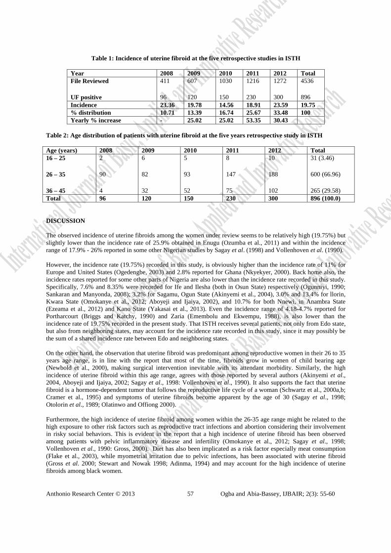
Overall, there were 4536 reproductive age women that presented at gynaecology department of ISTH during the period under study. The files reviewed showed that 896 of the women were positive for uterine fibroid giving an incidence of 19.75%. The breakdown of this figure for the five-year period is as follows: 2008 (96 cases; 23.36%); 2009 (120 cases; 19.78%); 2010 (150 cases; 14.56%); 2011 (230 cases; 18.91%); and 2012 (300 cases; 23.59%) (Seetable 1). It was observed that the number of uterine fibroid cases increased as years progressed, with the year 2012 having an overall highest percentage increase of 33.48%. Also, the comparative percentage increase between adjoining years was highest between 2010 and 2011 (53.35%), followed by that between 2011 and 2012 (30.43%).
The age distribution of patients with uterine fibroids during the five-year period is shown in table 2, and it indicates that majority of the women were between the ages of 26 and 35 (66.96%), followed by women between the ages of 36 and 45 (29.58%), and those between the ages of 16 and 25 (with the least percentage incidence of 3.46%) respectively. It was also observed that in each of the year under review, women within the ages of 26 – 35 were considerably represented compared to women above or below this age range.
Anthonio Research Center © 2013 57 Ogba and Abia-Bassey, IJBAIR; 2(3): 55-60
Table 1: Incidence of uterine fibroid at the five retrospective studies in ISTH
Year 2008 2009 2010 2011 2012 TotalFile Reviewed
UF positive
411
96
607
120
1030
150
1216
230
1272
300
4536
896Incidence 23.36 19.78 14.56 18.91 23.59 19.75% distribution 10.71 13.39 16.74 25.67 33.48 100Yearly % increase - 25.02 25.02 53.35 30.43
Table 2: Age distribution of patients with uterine fibroid at the five years retrospective study in ISTH
Age (years) 2008 2009 2010 2011 2012 Total16 – 25
26 – 35
36 – 45
2
90
4
6
82
32
5
93
52
8
147
75
10
188
102
31 (3.46)
600 (66.96)
265 (29.58)Total 96 120 150 230 300 896 (100.0)
DISCUSSION
The observed incidence of uterine fibroids among the women under review seems to be relatively high (19.75%) but slightly lower than the incidence rate of 25.9% obtained in Enugu (Ozumba et al., 2011) and within the incidence range of 17.9% - 26% reported in some other Nigerian studies by Sagay et al. (1998) and Vollenhoven et al. (1990).
However, the incidence rate (19.75%) recorded in this study, is obviously higher than the incidence rate of 11% for Europe and United States (Ogedengbe, 2003) and 2.8% reported for Ghana (Nkyekyer, 2000). Back home also, the incidence rates reported for some other parts of Nigeria are also lower than the incidence rate recorded in this study. Specifically, 7.6% and 8.35% were recorded for Ife and Ilesha (both in Osun State) respectively (Ogunniyi, 1990; Sankaran and Manyonda, 2008); 3.2% for Sagamu, Ogun State (Akinyemi et al., 2004), 3.0% and 13.4% for llorin, Kwara State (Omokanye et al., 2012; Aboyeji and Ijaiya, 2002), and 10.7% for both Nnewi, in Anambra State (Ezeama et al., 2012) and Kano State (Yakasai et al., 2013). Even the incidence range of 4.18-4.7% reported for Portharcourt (Briggs and Katchy, 1990) and Zaria (Emembolu and Ekwempu, 1988), is also lower than the incidence rate of 19.75% recorded in the present study. That ISTH receives several patients, not only from Edo state,but also from neighboring states, may account for the incidence rate recorded in this study, since it may possibly be the sum of a shared incidence rate between Edo and neighboring states.
On the other hand, the observation that uterine fibroid was predominant among reproductive women in their 26 to 35 years age range, is in line with the report that most of the time, fibroids grow in women of child bearing age (Newbold et al., 2000), making surgical intervention inevitable with its attendant morbidity. Similarly, the high incidence of uterine fibroid within this age range, agrees with those reported by several authors (Akinyemi et al., 2004, Aboyeji and Ijaiya, 2002; Sagay et al., 1998: Vollenhoven et al., 1990). It also supports the fact that uterine fibroid is a hormone-dependent tumor that follows the reproductive life cycle of a woman (Schwartz et al., 2000a,b; Cramer et al., 1995) and symptoms of uterine fibroids become apparent by the age of 30 (Sagay et al., 1998; Otolorin et al., 1989; Olatinwo and Offiong 2000).
Furthermore, the high incidence of uterine fibroid among women within the 26-35 age range might be related to the high exposure to other risk factors such as reproductive tract infections and abortion considering their involvement in risky social behaviors. This is evident in the report that a high incidence of uterine fibroid has been observed among patients with pelvic inflammatory disease and infertility (Omokanye et al., 2012; Sagay et al., 1998;Vollenhoven et al., 1990: Gross, 2000). Diet has also been implicated as a risk factor especially meat consumption(Flake et al., 2003), while myometrial irritation due to pelvic infections, has been associated with uterine fibroid (Gross et al. 2000; Stewart and Nowak 1998; Adinma, 1994) and may account for the high incidence of uterine fibroids among black women.
Anthonio Research Center © 2013 58 Ogba and Abia-Bassey, IJBAIR; 2(3): 55-60
Judging by the findings of this study, it is obvious that uterine fibroid remains a public health concern indicating that if Nigeria must achieve her vision 2020 and the millennium development goal on maternal and child mortality and morbidity, uterine fibroids must be given the attention it deserves. It is our opinion therefore, that an effective awareness/screening strategy be adopted in line with efforts to meet the millennium development goals on maternal mortality and morbidity.
ACKNOWLEDGMENT
Our special thanks go to the Head, Department of Gynecology, ISTH Irrua, and all Head Doctors and Nurses of the units where data were collected for this study. We also thank the Post Basic School of Nursing, for approving and supporting this study through Okojie A.I.O.
REFERENCES
Aboyeji, A.P. and Ijaiya, M.A. (2002). Uterine fibroids. A ten year clinical review at University of Ilorin Teaching Hospital, Ilorin, Nigeria. Nig. J. Med.; 11 (1): 16 – 19.
Adinma, J.I.B. (1994). Uterine fibroid and fertility in Enugu. Nig. Med. J.; 5: 3-5.
Akinyemi, B.O., Adewoye, B.R. and Fakoya, T.A. (2004). Uterine fibroid: A review. Nig. J. Med.; 13(4): 318 –329.
Ande, A.B.A, Ehigiegba, A.E. and Umeora, O.U.J. (2004). Repeat myomectomy at caesarean section. Arch.Gynaecol. Obstet.; 270: 296-298
Briggs, N.D. and Katchy, K.C. (1990). Pattern of primary gynecological malignancies as seen in a tertiary hospital situated in the Rivers State of Nigeria. Int. J. Gynecol. Obstet.; 31:157-61.
Chiaffarino, F., Parazzini, F., La Vecchia, C., Chatenoud, L., Di Cintio, E., Marsico, S. (1999). Diet and uterine myomas. Obstet. Gynecol.;94:395–398.
Cramer, S.F., Horiszny, J.A. and Leppert, P. (1995). Epidemiology of uterine leiomyomas. With an etiologic hypothesis. J. Reprod. Med.; 40:595–600.
DayBaird, D., Dunson, D.B., Hill, M.C., Cousins, D. and Schectman, J.M. (2003). High cumulative incidence of uterine leiomyoma in black and white women: ultrasound evidence. Am. J. Obstet. Gynecol.; 188:100–7.
Emembolu, J.O. and Ekwempu, C.C. (1988). Carcinoma of the cervix uteri in Zaria: Etiological factors. Int. J.Gynecol. Obstet.; 26:265-9.
Ezeama, C.O., Ikechebelu, J.I., Obiechina, N.J. and Ezeama, N.N. (2012). Clinical presentation of uterine fibroids inNnewi, Nigeria: A 5-year review. Ann. Med. Health Sci. Res.; 2 (2): 114-118.
Faerstein, E., Szklo, M. and Rosenshein, N. (2001). Risk factors for uterine leiomyoma: a practice-based case-control study. I. African-American heritage, reproductive history, body size, and smoking. Am. J. Epidemiol.;153:1–10.
Flake, G.P., Andersen, J. and Dixon, D. (2003). Etiology and pathogenesis of uterine leiomyomas: A review. Environ. Health. Perspect.; 111:1037-1049.
Gross K, Morton, C. and Stewart, E. (2000): Finding genes for uterine fibroids. Obstet. Gynaeco.; 95 (4 suppl): 560 – 561.
Gross, K. and Morton, C.C. (2001). Genetics and the development of fibroids. Clin. Obstet. Gynecol.; 44:335–349.
Kawamura, S., Kasagi, F., Kodama, K., Fujiwara, S., Yamada, M., Ohama, K. and Oto, K. (1997). Prevalence of uterine myoma detected by ultrasound examination in the atomic bomb survivors. Radiat. Res.; 147:753–758.
Anthonio Research Center © 2013 59 Ogba and Abia-Bassey, IJBAIR; 2(3): 55-60
Kjerulff, K.H., Langenberg, P., Seidman, J.D., Stolley, P.D. and Guzinski, G.M. (1996). Uterine leiomyomas. Racial differences in severity, symptoms and age at diagnosis. J. Reprod. Med.; 41:483–90.
Lowe, D.G. (1999). Benign. Tumors of the uterus ln: Dewhurst’s Textbook of Obstetrics and Gynaecology for Postgraduate. 6th edition Edmonds DK (Ed) Blackwell Science Publication London; Pp. 552 – 559.
Lumbiganon, P., Rugpao, S., Phandhu-fung, S., Laopaiboon, M. and Vudhikamraksa, N. (1996). Protective effect of depot-medroxyprogesterone acetate on surgically treated uterine leiomyomas: a multicentre case-control study. Br.J. Obstet. Gynaecol.; 103:909–14.
Luoto, R., Kaprio, J., Rutanen, E.M., Taipale, P., Perola, M. and Koskenvuo, M. (2000). Heritability and risk factors of uterine fibroids—the Finnish Twin Cohort Study. Maturitas.; 37:15–26.
Marshall, L., Spiegelman, D., Barbieri, R., Goldman, M.B., Manson, J.E., Colditz, G.A., Willett, W.C. and Hunter,D.J. (1997). Variation in the incidence of uterine leiomyoma among premenopausal women by age and race. Obstet.Gynecol.; 90:967–73.
Marshall, L., Spiegelman, D., Manson, J., Goldman, M.B., Barbieri, R.L., Stampfer, M.J., Willett, W.C. and Hunter, D.J. (1998). Risk of uterine leiomyomata among premenopausal women in relation to body size and cigarette smoking. Epidemiology.;9:511–17.
Mcllveen, M. and Li, T.C. (2005). Myomectomy: a review of surgical technique. Human. Fertil.; 8(1): 27-33.
Newbold, R.R., DiAugustine, R.P., Risinger, J.I., Everitt, J.I., Walmer, D.K., Parrott, E.C. and Dixon, D. (2000). Advances in uterine leiomyoma research: conference overview, summary, and future research recommendations. Environ. Health Perspect.; 108:769–73.
Nkyekyer, K. (2000). Pattern of gynecological cancers in Ghana. East. Afr. Med. J.; 77:534-8.
Ogedengbe, O.K. (2003). Uterine Fibroids In: Contemporary obstetrics and gynaecology for developing countries. Okonofua .F and Odunsi K (Ed) Intec printers limited Ibadan. Pg 202 – 213.
Oguniyi, S.O. and Fasuba, O. (1990). Uterine fibromata in Ilesha, Nigeria. Nig. Med. Practitioner; 191: 93 – 95
Okolo, S. (2008). Incidence, aetiology and epidemiology of uterine fibroids. Best Pract. Res. Clin. Obstet.Gynaecol.; 22(4): 571-88.
Okoronkwo, M.O. (1999). Body weight and uterine leiomyomas among women in Nigeria. West Afr. J. Med.;18:52–4.
Olatinwo, A.W.O. and Offiong, R.A. (2000). An analysis of surgically treated cases of uterine fibroid at the University of Ilorin Teaching Hospital, Ilorin, Nigeria. Nig. J. Surgical Res.; 92: 6 – 11.
Omokanye, L.O., Salaudeen, G.A., Saidu, R., Jimoh, A.A.G. and Balogun, O.R. (2012). Surgical management of uterine fibroids at the University of Ilorin Teaching Hospital: A 5 year review. Global Res. J. Med. Sci.; Vol.2(2) pp.018 – 022.
Otolorin, E.O., Ojengbede, O. and Falase, A.O. (1987): Laparoscopic evaluation of the tubo peritoneal factor in infertile Nigerian; women. Gynaecol. Obstetric.; 25: 42 – 52.
Otolorin, E.O., Owude, L.J. and Ladipo, O.A. (1989). Results of myomectomy for infertility in Ibadan Nigeria. Nig.Med. J.; 19(14): 173 – 175.
Ozumba, B.C., Nzegwu, M.A. and Nyikam, A.A. (2011). histological patterns of gynecological lesions in Enugu, Nigeria. A five-year review from January 1, 2000 to December 31st 2004. Advances in Bioresearch; Volume 2, Issue 2, December 2011: 132 – 136

Anthonio Research Center © 2013 60 Ogba and Abia-Bassey, IJBAIR; 2(3): 55-60
Ross, R., Pike, M.C., Vessey, M.P., Bull, D., Yeates, D. and Casagrande, J.T. (1986). Risk factors for uterine fibroids: reduced risk associated with oral contraceptives. Br. Med. J.; 293:359–62.
Sagay, S., Udoeyop, E.U., Pam, C., Karshina, J.A., Daru, P.H. and Otubu, J.A.M. (1998): Laparoscopic evaluation of 1000 consecutive infertile women in Jos Nigeria. Trop.J. Obstet.Gynaecol.; 15(1): 30 – 35.
Sankaran, S. and Manyonda, I.T. (2008). Medical management of fibroids. Best Pract. Res. Clin. Obstet. Gynaecol.; 22(4): 655-676.
Sato, F., Miyake, H., Nishi, M., Mori, M. and Kudo, R. (2000). Early normal menstrual cycle pattern and the development of uterine leiomyomas. J. Womens Health Gend. Based. Med.; 9:299–302.
Sato, F., Mori, M., Nishi, M., Kudo, R. and Miyake, H. (2002). Familial aggregation of uterine myomas in Japanese women. J. Epidemiol.; 12:249–253.
Sato, F., Nishi, M., Kudo, R. and Miyake, H. (1998). Body fat distribution and uterine leiomyomas. J. Epidemiol.;8:176–80.
Schwartz, S., Voigt, L., Tickman, E., Yarbro, P., Daling, J. and Scholes, D. (2000a). Familial aggregation of uterine leiomyomata. (Abstract). Am J Epidemiol.;151(suppl):S10.
Schwartz, S.M., Marshall, L.M. and Baird, D.D. (2000b). Epidemiologic contributions to understanding the etiology of uterine leiomyomata. Environ Health Perspect.; 108:821–7.
Stewart, E.A. and Nowak, R.A. (1998). New concepts in the treatment of uterine leiomyoma. Obstet Gynecol.;92:624-7.
Van Voorhis, B.J., Romitti, P.A. and Jones, M.P. (2002). Family history as a risk factor for development of uterine leiomyomas. Results of a pilot study. J Reprod Med.; 47: 663–9.
Vollenhoven, B. (1998). Introduction: the epidemiology of uterine leiomyomas. Baillieres Clin Obstet Gynaecol.;12:169–76.
Vollenhoven, B.J., Lawrence, A.S. and Healey, D.C. (1990). Uterine fibroid: a clinical review. British J. Obstet. Gynaecol.., 1990: 97: 285 – 298.
Wilcox, L., Koonin, L.M., Pokras, R., Strauss, L.T., Xia, Z. and Peterson ,H.B. (1994). Hysterectomy in the United States, 1988–1990. Obstet Gynecol.; 83:549–55.
Wong, L.F., Yamada, M., Sasaki H., Kodama, K., Akiba, S., Shimaoka, K. and Hosoda, Y. (1993). Non cancer disease incidence in the atomic bomb survivors: 1958–1986. Radiat Res.; 135(3):418–30.
Yakasai, I.A., Ugwa, E.A. and Otubu, J. (2013). Gynecological malignancies in Aminu Kano Teaching Hospital Kano: A 3 year review. Niger. J. Clin. Pract.; 16:63-66.
AUTHOR(S) CONTRIBUTION
Elugwaraonu, O., was involved in the collection of data with assistant and supervision from Okojie A.I.O., and Okhia, O. All authors including Oyadoghan G.P. were involved in the presentation, criticism and approval of the final submitted draft.


![The Incidence of Postpartum Hemorrhage in Pregnant … · between endometrial damage and uterine scarring and subsequent placenta previa [4]. Mean-while, the condition is frequently](https://img.dokumen.tips/doc/110x75/5ba8e78509d3f2f51d8b4ab9/the-incidence-of-postpartum-hemorrhage-in-pregnant-between-endometrial-damage.jpg)


![Uterine Cancer Incidence and Mortality — United … [myometrium], C54.3 [fundus uteri], C54.8 [overlapping lesion of corpus uteri], C54.9 [corpus uteri]) and uterus, not otherwise](https://img.dokumen.tips/doc/110x75/5cc84ff988c993f00b8d5f2d/uterine-cancer-incidence-and-mortality-united-myometrium-c543-fundus-uteri.jpg)