Embed Size (px)
Citation preview

neurospora ms
1
The Genome Sequence of the Filamentous Fungus Neurospora crassa James E. Galagan1, Sarah E. Calvo1, Katherine A. Borkovich2, Eric U. Selker3, Nick D. Read4, David Jaffe1, William FitzHugh5, Li-Jun Ma1, Serge Smirnov1, Seth Purcell1, Bushra Rehman1, Timothy Elkins1, Reinhard Engels1, Shunguang Wang1, Cydney B. Nielsen1, Jonathan Butler1, Matthew Endrizzi1, Dayong Qui1, Peter Ianakiev1, Deborah Bell-Pedersen6, Mary Anne Nelson7, Margaret Werner-Washburne7, Claude P. Selitrennikoff8, John A. Kinsey10, Edward L. Braun11, Alex Zelter4,26, Ulrich Schulte12, Gregory O. Kothe3, Gregory Jedd13, Werner Mewes9,17, Chuck Staben14, Edward Marcotte15, David Greenberg16, Alice Roy1, Karen Foley1, Jerome Naylor1, Nicole Stange-Thomann1, Robert Barrett1, Sante Gnerre1, Michael Kamal1, Manolis Kamvysselis1, Cord Bielke9, Stephen Rudd17, Dmitrij Frishman17, Svetlana Krystofova2, Carolyn Rasmussen18, Robert L. Metzenberg19, David D. Perkins19, Scott Kroken18, David Catcheside21, Weixi Li 14, Robert J. Pratt6, Stephen A. Osmani23, Colin P.C. DeSouza24, Louise Glass18, Marc J. Orbach25, J. Andrew Berglund3, Rodger Voelker3, Oded Yarden26, Mike Plamann27, Stephan Seiler27, Jay Dunlap22, Alan Radford28, Rodolfo Aramayo6, Donald O. Natvig7, Lisa A. Alex29, Gertrud Mannhaupt9, Daniel J. Ebbole30, Michael Freitag3, Ian Paulsen16, Matthew S. Sachs31, Eric S. Lander1,32, Chad Nusbaum1 & Bruce Birren1
1Whitehead Institute Center for Genome Research, 320 Charles Street, Cambridge, MA 02141, USA 2Department of Plant Pathology, University of California, Riverside, CA 92521 USA 3Institute of Molecular Biology, University of Oregon, Eugene, OR 97403 USA 4Institute of Cell and Molecular Biology, University of Edinburgh, Edinburgh EH9 3JH UK 5Celera Genomics, Rockville, MD 20850 USA 6Department of Biology, Texas A & M University, College Station, TX 77843, USA 7Department of Biology, University of New Mexico, Albuquerque, NM 87131 USA 8Department of Cellular and Structural Biology, University of Colorado Health Sciences Center, Denver, CO 80262, USA 9Department of Genome Oriented Bioinformatics, Technical University of Munich, Wissenschaftzentrum Weihenstephan, 85350 Freising-Weihenstephan, Germany 10Department of Microbiology, University of Kansas Medical School, Kansas City, KS 66160 USA 11Department of Zoology, University of Florida, Gainesville, FL 32611-8525, USA 12Institute of Biochemistry, Heinrich Heine University, 40225 Duesseldorf Germany 13Lab of Plant Molecular Biology, Rockefeller University, New York, NY 10021 US 14T.H. Morgan School of Biological Sciences, University of Kentucky, Lexington, KY 40506 USA 15The Institute for Cellular and Molecular Biology, The University of Texas at Austin, Austin, TX 78712 USA 16The Institute for Genomic Research, 9712 Medical Center Drive, Rockville, Maryland 20878, USA 17Institute for Bioinformatics (MIPS), GSF-National Research Center for Environment and Health, Ingolstaedter Landstrasse 1, 85764 Neuherberg, Germany 18Department of Plant and Microbial Biology, University of California, Berkeley, Berkeley, CA 94720 USA 19Department of Biological Sciences, Stanford University, Stanford, CA 94305 21School of Biological Sciences, Flinders University, P.O. Box 2100, Adelaide, South Australia 5001
neurospora ms
2
22Department of Genetics, Dartmouth Medical School, Hanover, NH 03755 USA 23Department of Molecular Genetics, The Ohio State University, Columbus, OH 43210 24Department of Molecular Genetics, The Ohio State University, Columbus, OH 43211 25Department of Plant Pathology, University of Arizona, Tucson, AZ 85721 USA 26Dept. of Plant Pathology and Microbiology, Faculty of Agricultural, Food and Environmental Quality Sciences, The Hebrew University of Jerusalem, Rehovot 76100 Israel 27School of Biological Sciences, University of Missouri-Kansas City, Kansas City, MO 64110 USA 28School of Biology, Leeds University, Leeds LS2 9JT UK 29Department of Chemistry, California State Polytechnic University Pomona, Pomona, CA 91768 30Department of Plant Pathology and Microbiology, Texas A & M University, College Station, TX 77843, USA 31Department of Biochemistry and Molecular Biology, OGI School of Science and Engineering, Oregon Health & Science University, Beaverton, OR 97006 USA 32Department of Biology, MIT, Cambridge, Massachusetts 02139, USA

neurospora ms
3
Neurospora crassa is a central organism in the history of 20th century genetics, biochemistry, and molecular biology. Here, we report a high-quality draft sequence of the Neurospora genome. The ~40 Mb genome encodes ~10,000 protein-coding genes —more than twice as many as the fission yeast S. pombe and only ~25% fewer than the fruit fly D. melanogaster. Analysis of the gene set yields insights into unexpected aspects of Neurospora biology including the identification of genes potentially associated with red light photobiology, genes implicated in secondary metabolism, and important differences in Ca2+ signalling as compared to plants and animals. Neurospora possesses the widest array of genome defence mechanisms known for any organism, including a process unique to fungi called Repeat Induced Point Mutation (RIP). Genome analysis suggests that RIP has had a profound impact on genome evolution, greatly slowing the creation of new genes through genomic duplication and resulting in a genome with an unusually low proportion of closely related genes.
Research on Neurospora in the early part of the 20th century paved the way for modern genetics and molecular biology. First documented in 1843 as a contaminant of bakeries in Paris1, Neurospora was developed as an experimental organism in the 1920s2,3. Subsequent work on Neurospora by Beadle and Tatum4 in the 1940s established the relationship between genes and proteins, summarized in the �one-gene-one-enzyme� hypothesis. In the latter half of the century, Neurospora played a key role as a model organism, contributing to the fundamental understanding of genome defence systems, DNA methylation, mitochondrial protein import, circadian rhythms, post-transcriptional gene silencing, and DNA repair5. Because Neurospora is a multicellular filamentous fungus, it has also provided a system to study cellular differentiation and development as well as other aspects of eukaryotic biology6.
The legacy of over 70 years of research7, coupled with the availability of molecular and genetic tools, offers enormous potential for continued discovery. The sequencing of the Neurospora crassa genome was undertaken to maximize this potential. Here, we report an initial sequence and analysis of the Neurospora genome.
Neurospora Genome Sequence
The Neurospora genome is much larger (>40 Mb) than that of S. pombe and S. cerevisiae (both ~12 Mb). Accordingly, we sought to first produce and analyse a high-quality draft sequence, en route to a finished sequence.
The genome sequence was assembled from deep whole-genome shotgun (WGS) coverage obtained by sequencing both ends of a variety of clone types, including plasmids, Fosmids, bacterial artificial chromosomes and jumping clones (Table 1 and Methods). In all, the data provided an average of >20-fold sequence coverage and >98-fold physical coverage of the genome. The Arachne package8 was used to assemble the draft genome sequence. The resulting assembly consists of 958 sequence contigs with a total length of 38.6 Mb (Table 2) and an N50 length of 114.5 kb (that is, 50% of all bases are contained in contigs of at least 114.5 kb). The sequence contigs are assembled into scaffolds (or supercontigs) when they are joined by at least two forward-reverse links arising from the paired-end reads. The

neurospora ms
4
assembly consists of 163 scaffolds with a total length of 39.9 Mb (including gaps between contigs) and an N50 length of 1.56 Mb.
The vast majority of the assembly (97%) is contained in the 44 largest scaffolds. At the opposite end of the spectrum, there are 38 tiny scaffolds with lengths <4 kb. Forty-two of these large scaffolds (and one of the smaller ones) could be readily anchored to the Neurospora genetic map7 by virtue of their containing known genetic markers with sequence, allowing the assembly to be positioned along the seven Neurospora chromosomes.
The genome assembly is of very high quality in terms of continuity and representation. The assembly has long-range continuity, with the N50 scaffold size being nearly 1000-fold larger than the average gene size (see below). The assembly represents the vast majority of the genome, as assessed by comparison with available finished sequence and genetic markers. It contains 99.13% of available finished sequence (17 Mb from linkage groups II and V9) and all of the 252 genetic markers with sequence. This estimate, however, does not account for unusual genomic regions such as the ribosomal DNA repeats; such regions may contain ~1.7 Mb of additional sequence10 corresponding to 2-3% of the genome that cannot be readily assembled with available techniques.
The assembly also has high accuracy, with 99.5% of the sequence having Arachne quality scores ≥30. Comparison with finished sequence confirms the sequence accuracy, with the discrepancy rate for this subset being less than 10-5. The comparison also largely confirms the assembly. Twelve minor discrepancies were identified in 17 Mb. Five lie at the end of contigs and are most likely caused by a misaligned or low-quality terminal read. Four are short insertions or deletions, ranging from 9-559 bp. The remaining three discrepancies appear to be instances in which the finished sequence does not correctly represent the genome owing to chimerism in the BACs and in which the draft sequence is correct.
Efforts are currently underway to produce a complete sequence of the genome (apart from problematic regions such as the rDNA repeats). The existing high quality draft sequence, however, provides the substrate for genomic analysis.
Genes
Gene Count and Basic Characteristics. A total of 10,082 protein-coding genes (9,200 longer than 100 amino acids) were predicted (Table 2). This constitutes nearly twice as many genes as in S. pombe (~4800) and S. cerevisiae (~6300) and nearly as many as in [is 4300 fewer really �nearly as many�D. melanogaster (~14,300). Genes span at least 44% of the genome sequence (38% in coding exons and an additional 6% in introns between such exons), with an average gene density of one gene per 3.7 kb. The average gene length of 1.67 kb, is slightly longer than the 1.4 kb average gene length for both S. cerevisiae and S. pombe. The difference in gene length is due to the greater number of introns in Neurospora genes -- an average of 1.7 introns per gene with an average intron size of 134 nucleotides.

neurospora ms
5
Interestingly, most predicted Neurospora introns lack a polypyrimidine (PY) tract, which are common in other eukaryotic introns, but do contain a strong branchpoint (BP) sequence. The position of the BP sequence in Neurospora introns is unusual, in comparison to well-characterized human and S. cerevisiae introns, in being close to the 3� acceptor site. The distance to the acceptor site in Neurospora averages 14 nt, in contrast to an average of 31-40 nucleotides in yeast11 and 24 nucleotides in humans12. The strong BP sequence and its proximity to the 3� splice site (consensus of AYAG) suggest that in Neurospora these elements play an important role in defining the 3� end of the intron in the absence of a PY tract.
Comparative Analysis. Comparison of the Neurospora predicted protein set to those of selected sequenced eukaryotes (see Methods) highlights the biological complexity of the filamentous fungi. Fully 4,140 (41%) Neurospora proteins lack significant matches to known proteins from public databases (Table 2), reflecting the early stage of fungal genome exploration and the diversity of fungal genes remaining to be described. Furthermore, 5,805 (57%) Neurospora proteins do not have significant matches to genes in either of the sequenced yeasts (Figure 1a), underscoring the greater complexity of this filamentous fungus as compared to the yeast models.
When compared to sequenced eukaryotes, a total of 1,421 (14%) Neurospora genes
display best BLASTP matches to proteins in either plants or animals (Figure 1b). Of these, 584 lack high scoring hits to either sequenced yeast. These data reflect the biology shared between filamentous fungi and other higher eukaryotes, which in a number of cases is absent in the yeasts. These results, and others discussed below, emphasize the importance of Neurospora as a model organism for the study of biology in eukaryotic organisms.
Protein Domain Architectures. To characterize the functional organization of the proteome, we analysed the domain structure of the Neurospora protein set. Predicted proteins were searched against the Interpro database of protein families and domains using InterproScan13. The domain architecture for each protein was then defined as the ordered list (from N terminus to C terminus) of corresponding Interpro domains. An identical analysis was performed on selected sequenced eukaryotes for comparison.
As shown in Table 3, larger genome size is generally correlated with a greater number
of distinct domains. Consistent with this, Neurospora possesses a number of protein domain families that display expansions in number relative to yeast (Table 4). However, the relative increase in domains between organisms is modest, with Neurospora displaying an 8% increase in the number of domains compared with the yeasts. In contrast, the number of unique permutations of domains increases substantially with genome size. In particular, Neurospora has 26% more identifiable architectures than the yeasts, with a 19% increase in the average number of architectures per domain. Finally, organisms with larger genomes also possess a larger proportion of architectures represented by more than one gene, with the single notable exception of Neurospora (Table 3). In contrast to the other eukaryotes considered, Neurospora possesses a smaller proportion of duplicated architectures than expected given its genome size. This difference may be due to the phenomenon of RIP (Repeat Induced Point Mutation, see below).

neurospora ms
6
Epigenetics, Genome Defence, and Genome Evolution
Epigenetic phenomena, heritable changes in gene expression that do not involve changes to the DNA code, are widespread among organisms. Neurospora is an important model for the study of epigenetic phenomena, possessing a wide variety of epigenetic mechanisms and related genome defence mechanisms. The most remarkable of these phenomena is Repeat Induced Point Mutation (RIP), a process unique to fungi. The availability of a Neurospora genome sequence allows an analysis of RIP and its dramatic impact on genome evolution.
Repeat Induced Point Mutation. First discovered in Neurospora14,15, RIP is a genome defence system that efficiently detects and mutates both copies of a sequence duplication. RIP acts during the haploid dikaryotic stage of the Neurospora sexual reproductive cycle, causing numerous C:G to T:A mutations within duplicated sequences. In a single passage through the sexual cycle up to 30% of the C:G pairs in duplicated sequences can be mutated, with a strong preference for C to T mutations occurring at CpA dinucleotides16. The pattern of mutations produces a characteristic skewing of dinucleotide frequencies that allows RIP-mutated sequences to be accurately detected17. RIP requires a minimal duplicated sequence length of about 400bp18 and greater than roughly 80% sequence identity between duplicates19, consistent with the proposal that RIP involves homologous pairing20,21. In addition to suffering mutations, RIP-mutated sequences are frequent targets for DNA methylation. As with mammals, DNA methylation has been shown to cause gene silencing in Neurospora22. RIP thus mutates and epigenetically silences repetitive DNA.
RIP has been proposed to act as a defence against selfish or mobile DNA15,20.
Consistent with this hypothesis, examples of mobile elements inactivated by RIP have been reported for Neurospora crassa and related species17,23,24. However, because RIP mutation and methylation can extend beyond the bounds of duplicated sequences25, RIP can have both mutational and epigenetic effects on neighbouring unique sequences. Furthermore, RIP acts on all duplicated sequences, including those arising from large-scale chromosomal duplications as well as gene duplications26. The presence of RIP thus has profound consequences for the evolution of the Neurospora genome. Indeed, it has been proposed that RIP might prevent gene innovation through gene duplication15. With the availability of the Neurospora genome sequence, we were able to address this hypothesis.
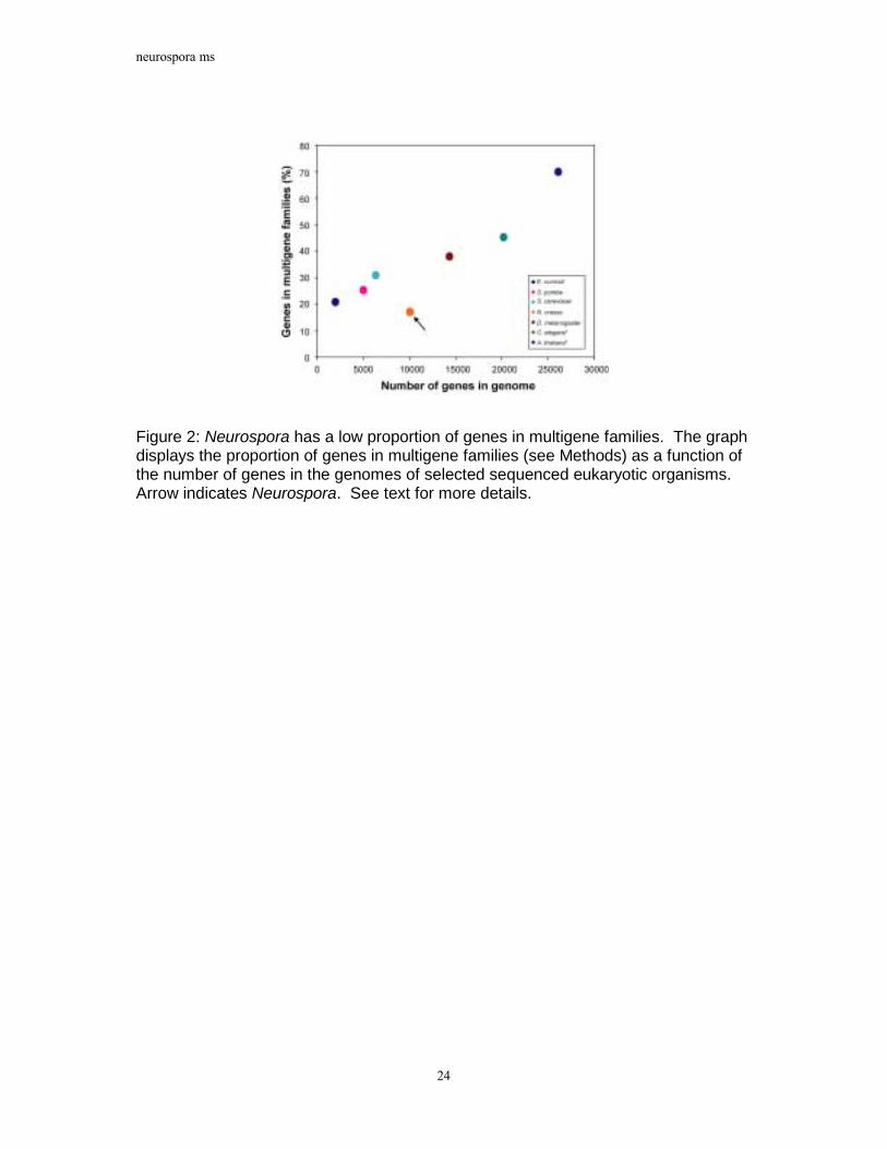
Multigene Families. To investigate the impact of RIP on protein families in Neurospora, genes were clustered into �multigene families� based on an all-vs.-all comparison of protein sequences (see Methods). As shown in Figure 2, the percentage of genes in multigene families in selected sequenced eukaryotes is correlated with genome size. However, in striking contrast to the other analysed organisms, Neurospora possesses many fewer genes in multigene families than expected given its size. When the analysis is expanded to include an additional 17 sequenced prokaryotes, only Mycoplasma genitalium, Mycoplasma pulminus, Ureaplasma urealyticum and Vibrio cholerae display a correspondingly small proportion of genes in families. This is noteworthy considering that the Mycoplasmas are thought to have undergone reductive evolution and represent minimal life forms27.

neurospora ms
7
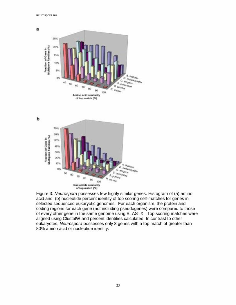
The analysis reveals another characteristic of Neurospora gene families. Unlike other
sequenced eukaryotes, Neurospora possesses only a handful of highly similar gene pairs. Figure 3 displays histograms of amino acid and nucleotide similarities between each gene in the six organisms analysed and the best matching gene in that organism. A significant proportion of genes have best matches with greater than 80% amino acid and nucleotide identity in all the organisms considered except Neurospora. Neurospora contains only 8 genes with top matches of greater than 80% amino acid or coding sequence identity. This value is significant because, as described above, RIP mutates duplicated sequences that display greater than 80% nucleotide similarity. Thus, the small proportion of genes in multigene families and the near absence of highly similar genes are consistent with the actions of RIP.
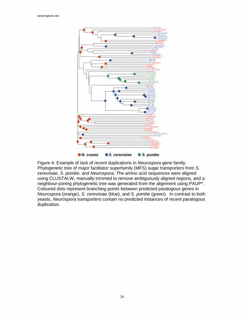
A striking example of the lack of highly similar genes in multigene families is revealed
in an analysis of predicted major facilitator superfamily (MFS) sugar transporters (Figure 4). Neurospora has a similar number of predicted MFS sugar transporters as S. cerevisiae. However, a phylogenetic analysis of fungal sugar transporters indicates that the Neurospora proteins are substantially more divergent than those of S. cerevisiae as well as those of S. pombe. Furthermore, the Neurospora transporters contain no apparent instances of recent gene duplication. In contrast, the majority of S. cerevisiae HXT hexose and S. pombe GHT transporters represent two relatively recent and independent expansions and include very recently duplicated genes such as HXT15 and HXT16. Thus, despite a diversity of MFS sugar transporters, Neurospora appears to lack close paralogs in this gene family, consistent with the results of the genome-wide multigene family analysis. Analyses of other gene families yielded similar results (data not shown).
The paucity of closely related sequences is evident not only at the level of complete
genes, but even at the level of individual exons and protein domains. Analysis of the 27,200 predicted exons in Neurospora revealed only 36 pairs of exons (0.1%) with greater than 80% nucleotide identity. Furthermore, an analysis of 4,047 PFAM protein domains predicted by the HMMER program28 revealed only 37 pairs (0.9% of predicted domains) with greater than 80% amino acid identity over more than 50% the length of the predicted domain. This compares with 16% of predicted domains in S. cerevisiae and 10% in S. pombe.
Gene Evolution through Gene Duplication. The above results suggest that RIP has had a powerful impact on suppressing the creation of new genes or partial genes through genomic duplication. This is consistent with the large number of mutations induced in duplicated sequences by RIP. Indeed, computer simulation (see Methods) indicates that, following a gene duplication, each copy has an 80% probability of acquiring an in-frame stop codon after only a single round of RIP and a 99.5% probability by the point that RIP has mutated the copies to less than 85% nucleotide similarity. The high frequency of stop codons reflects the preference of RIP for mutating CpA to TpA, increasing the prevalence of the stop codons TAA and TAG.
neurospora ms
8
These results raise the critical question of whether any significant gene duplication has occurred in Neurospora subsequent to the acquisition of RIP. We searched for empirical evidence of duplicated genes that have survived RIP by analysing the set of Neurospora coding sequences using two different previously developed measures17 for detecting RIP-mutated sequences (see Methods). These measures utilize the characteristic skewing of dinucleotides produced by RIP to detect mutated sequences. According to these measures, only 59 of the 9,200 predicted genes encoding proteins ≥ 100 amino acids show evidence of having been mutated by RIP. Of these, only 8 consist of pairs of predicted duplicated genes (genes in the same multigene family) in which both copies are predicted to be RIP-mutated. Thus, few pairs of duplicated genes display evidence of having both survived RIP. The remaining genes predicted to be RIP-mutated include 3 genes that are in multigene families but lack a matching RIP-mutated gene, and 48 single-copy genes. Of the latter, 18 are situated in or adjacent to RIP-mutated relics of mobile elements and were thus potentially mutated as a consequence of this association.
Gene duplication is thought to play a primary role in the innovation of new genes29-31.
However, taken together, these data support the conclusion that most, if not all, paralogous genes in Neurospora duplicated and diverged prior to the emergence of RIP, and since that point the evolution of new genes through gene duplication has been virtually arrested. This remarkable conclusion raises the question of whether and how Neurospora is able to evolve new genes. A number of mechanisms not involving gene duplication are conceivable, including gene sharing and lateral transfer. Ultimately, a conclusive analysis of these and other potential mechanisms of evolution may only be possible by comparing Neurospora to the genomes of closely related species to illuminate recent evolutionary history. Nonetheless, our results make clear that the cost to Neurospora for increased genome security through RIP is a significant impact on the evolution of new gene functions through gene duplication.
Repetitive DNA. The lack of genic redundancy in the Neurospora predicted proteome is mirrored by a relative paucity of repetitive DNA in the genome. An analysis of repeat sequences longer than 200 bp and with greater than 65% similarity (see Methods), revealed that only 10% of the Neurospora assembly consists of repeat sequences, consistent with previously reported estimates32,33.
The repeat content of Neurospora provides a testament to the efficiency of RIP.
Applying the measures of RIP mentioned above to the Neurospora genome revealed that the vast majority of repetitive sequences (81%) in Neurospora have been mutated by RIP. Conversely, only 18% of predicted RIP-mutated sequence is non-repetitive, likely reflecting the loss of the corresponding duplicated sequence. As described above, duplications greater than about 400 bp are susceptible to RIP18. In keeping with this, we observe that over 97% of genomic repeats greater than 400 bp in length are RIP-mutated. Moreover, repeats longer than 400 bp clustered based on sequence similarity display an average sequence identity within clusters of 78%, with 93% of repeat clusters displaying an average identity of less than 85%. This corresponds to previous estimates indicating that RIP requires greater than approximately 80% sequence identity to detect duplicated sequences.

neurospora ms
9
Consistent with the hypothesis that RIP acts as a defence mechanism against selfish DNA34, no intact mobile elements were identified. Furthermore, a significant proportion (46% of repetitive nucleotides) of the Neurospora RIP-mutated repetitive sequence can be identified as relics of mobile elements (Figure 5). The largest proportion of identified repeat elements consists of Tad relics, followed by relics of Punt, Pogo, gypsy, and copia-like elements, all previously described for Neurospora17,23,24,35-37. Strikingly, nearly half of all identified elements appear to be relics of mobile element families not previously known for Neurospora. Over 25 copies of a Tc1/mariner superfamily member were identified, similar to members of the fungal impala family that have also been characterized in Magnaporthe grisea and Fusarium oxysporum38-40. Twenty-five RIP-mutated copies of the Pot2 transposon, previously characterized in M. grisea41, and 37 retroposon relics with similarity to the gypsy-like MAGGY elements in M. grisea42 were also found.
Ribosomal RNA. The only repetitive sequences known to have survived RIP in Neurospora are the roughly 175-200 copies10 of the large rDNA tandem repeat containing the 17S, 5.8S, and 25S rRNA genes. As in higher eukaryotes, these tandem repeats occur within the nucleolar organizer region (NOR), and their resistance to RIP appears to stem from this localization15. Within the genome sequence we found several copies of the rDNA repeat in regions outside the NOR. In every case they display evidence of mutation by RIP, consistent with previous observations15. Thus, the sequence of the rDNA repeat does not in itself appear to confer resistance to RIP.
The 5S rRNA genes in Neurospora have survived RIP in a different manner. In
contrast to most higher eukaryotes in which the 5S rRNA genes form tandem repeats43-47, the 5S genes are dispersed through the genome in Neurospora48. A total of 74 copies comprising several different subtypes of 5S rDNA are dispersed through all seven chromosomes. This dispersed distribution coupled with the small size (~120 nt) of the 5S rRNA genes ensures they are not recognized by RIP.
DNA Methylation. Neurospora has been extensively used as a model for studying DNA methylation in eukaryotes. As with animals and plants, methylation has been shown to cause epigenetic silencing in Neurospora22,49. In contrast to mammals, methylation in Neurospora is not restricted to symmetric CpG sites50,51. In Neurospora, an estimated 1.5% of cytosines are methylated52,53, and it has been suggested that nearly all DNA methylation in the Neurospora genome is a result of RIP51,54-56. The Neurospora genome includes two potential cytosine DNA methyltransferase genes. One, called DIM-2, is required for all known DNA methylation57. The other, called RID, is required for RIP and is a member of a family found thus far only in filamentous fungi58. Importantly, the genome sequence confirms that Neurospora does not have proteins bearing canonical methyl-binding domains (MBD), suggesting the existence of alternative motifs involved in recognition and maintenance of methylated DNA59.
Plasmid reads for Neurospora were sequenced from libraries cloned separately in
methylation-tolerant and methylation-intolerant strains of E. coli (Table 1). Although not intended for this purpose, the two libraries provided a basis for predicting regions of methylation in the genome by comparing the representation of regions in sequence obtained
neurospora ms
10
from each library (see Methods). Testing the accuracy of such predictions, we found that 8 of 10 regions predicted to be methylated were experimentally confirmed as such. The predictions thus have good specificity. (The data does not provide high sensitivity, however. The predictions include only a minority of actually methylated regions, probably because the methylation intolerance is incomplete (see Methods). This could be overcome by sequencing of bisulfite-treated genomic DNA60.)
The specificity of the predictions provides valuable insight into the pattern of
methylation in the Neurospora genome. Regions predicted to be methylated show a remarkable correspondence to regions predicted to be repetitive and RIP-mutated (Figure 6). Fully 85% correspond to predicted RIP-mutated sequences. However, a small proportion (10%) of predicted methylated regions correspond to predicted non-repetitive and non-RIP-mutated sequence. In two out of ten such cases, both the methylation and the non-repetitive nature of these sequences were experimentally verified. This raises the possibility that methylation in Neurospora may also play non-defence roles, as has been proposed for methylation in higher organisms.
RNA Silencing. Post-transcriptional gene silencing (PTGS), or RNA silencing, is widespread among organisms61 and is increasingly being recognized as a key switch for controlling eukaryotic gene expression62. For example, co-suppression in plants63 and RNA interference (RNAi) in animals64-66 both control gene expression through the specific degradation of homologous RNAs. RNA silencing pathways are thought to be derived from ancestral natural defence systems directed against invading nucleic acids67. Consistent with this, all known PTGS mechanisms share similar components including RNA-dependent RNA polymerases (RdRPs), Argonautes (related to translation initiation factors), Dicers (double stranded-specific endonucleases), as well as many other ATP-dependent DNA and ATP-dependent RNA helicases and dsRNA-binding proteins68.
Neurospora possesses two RNA silencing pathways. The first, called quelling, silences
expression of transgenes during vegetative growth. This pathway was uncovered through the study of three genes, qde-1, qde-2 and qde-3, coding respectively for an RdRP, an Argonaute, and a RecQ helicase69-74. The second pathway, called meiotic silencing by unpaired DNA, acts during sexual reproduction75,76. A gene called sad-1, encoding an RdRP, has been identified for this pathway76,77.
Our analysis of the Neurospora genome sequence uncovered several additional genes
implicated in RNA silencing (Table 5). These include one RdRP, one Argonaute-like protein, and one RecQ-like helicase, as well as two Dicer-like ribonucleases (also known as CAF proteins). A phylogenetic analysis of the predicted RdRPs, Argonaute-like proteins, and Dicer-like proteins (see Methods) indicates that the Neurospora genes comprise two paralogous sets. One set includes the three qde genes and is thus predicted to correspond to the quelling pathway. The other set includes sad-1, and in phylogenetic trees these genes branch consistently with those of the single pathway observed in S. pombe76,78. Based on this analysis, we predict that one of the identified Dicers, Sms-3, is part of the meiotic silencing pathway, while the other, dcl-2, is part of the quelling pathway (Table 5). In addition, we

neurospora ms
11
predict that the identified Argonaute, Sms-2, is also part of the meiotic silencing pathway. Experimental work has confirmed the roles for Sms-279 and Sms-380. Taken together, these results support the conclusion that meiotic silencing and quelling represent two separate RNA-dependent silencing pathways. We further hypothesize that both pathways might have evolved from a single ancestral RNA silencing pathway.
Fungal Biology and Evolution
The availability of the Neurospora genome sequence provides an opportunity to study the genetic basis underlying the extraordinary biochemical and metabolic diversity exhibited by a filamentous fungus. Our analysis of the genome sequence has resulted in a number of surprising insights into the biology and evolution of Neurospora and other filamentous fungi.
Cell Signalling and Environmental Responses
Unexpected discovery of putative red light sensing genes. Blue light is an important regulator of Neurospora growth and development, impacting the circadian rhythm of conidiation, carotenogenesis of hyphae, and numerous facets of sexual development81. Although Neurospora photobiology has been intensively studied for more than two decades, the genome sequence has nonetheless revealed a number of previously uncharacterized sequences with similarity to blue light sensing genes, including both a cryptochrome homolog and a gene whose product contains a single PAS/LOV-type domain associated with light sensing.
In addition, Neurospora possesses two putative homologs of phytochromes, genes
known for their role in red light sensing in other organisms, and a putative homolog of the Aspergillus nidulans velvet gene implicated in the regulation of both red and blue light responses. The presence of these genes is unexpected given that no red light photobiology has been described for Neurospora to date. It has been recently shown that in addition to red light sensing, some Arabidopsis phytochromes associate with cryptochromes to play a role in blue light sensing and signaling82. Therefore, the two phytochromes and the velvet homolog may also regulate this aspect of Neurospora photobiology.
Expansion of histidine kinases reflects the importance of two-component signalling in filamentous fungi. Mitogen-activated protein kinase (MAPK) pathways83 integrate signals from multiple receptor pathways including two-component signalling systems84. The basic two-component system consists of a histidine kinase and a cognate response regulator protein. The nine MAPK pathway proteins identified in the Neurospora genome sequence (Figure 7) correspond to those found in S. pombe and S. cerevisiae, indicating that the basic MAPK machinery is conserved between these species. In contrast, Neurospora has a significantly expanded complement of histidine kinases as compared to both sequenced yeasts. Neurospora possesses 11 histidine kinases, compared with one in S. cerevisiae and three in S. pombe. Two of the 11 genes have been previously characterized in Neurospora85, while a third is similar to proteins in A. fumigatus and A. nidulans that affect conidiation85,86.

neurospora ms
12
Functions for the remaining genes are unknown, although 7 (including the two phytochromes discussed above) contain PAS/PAC domains, implicating them in oxygen and light responses. The expansion of histidine kinases in Neurospora relative to yeasts, coupled with the conservation of the core MAPK pathway, suggests a larger role than previously expected for two-component signalling in filamentous fungi. The large number of histidine kinases reveals filamentous fungi to be more similar in this regard to plants, where two component systems are abundant, than to animals, where these systems are absent.
A novel family G protein coupled receptors. Eukaryotic cells sense many environmental stimuli via seven-transmembrane helix, G protein-coupled receptors (GPCRs)87. Our analysis indicates that Neurospora possesses a total of 10 predicted seven-transmembrane helix proteins (Figure 7) three of which belong to a novel class not previously identified in any fungus. These genes encode proteins similar to cAMP GPCRs from the protists D. discoideum88 and Polysphodylium pallidum, and also to predicted proteins from A. thaliana89 and C. elegans. The D. discoideum proteins have been shown to sense cAMP levels during chemotaxis and multicellular development90. This suggests a possible analogous function in Neurospora. The existence of an extracellular cAMP signalling pathway has never been previously demonstrated in any fungal system.
In support of this hypothesis, along with the presence of putative cAMP receptors,
Neurospora was found to possess the full complement of proteins required for the synthesis and degradation of cAMP. For example, one adenylyl cyclase91, one cyclase-associated protein, and four genes encoding putative cAMP phosphodiesterases are present. Furthermore, Neurospora wild-type strains accumulate cAMP in the extracellular medium92, although a role in extracellular signalling has not been established. These data, coupled with the presence of GPCRs similar to slime mould cAMP receptors, suggest the possibility that cAMP or a related molecule may serve as an extracellular signal in Neurospora.
Numerous calcium signalling proteins support the importance of Ca2+ sensory transduction in filamentous fungi. A considerable body of evidence, primarily from pharmacological studies, indicates that Ca2+-signaling regulates numerous processes in filamentous fungi including secretion, cytoskeletal organization, hyphal tip growth, hyphal branching, sporulation, and circadian rhythmicity93. However, the identification of the main components of even one Ca2+-mediated response pathway in filamentous fungi has remained elusive. The genome sequence of Neurospora has provided for the first time over 25 of the proteins likely to be necessary for Ca2+-signaling in filamentous fungi (Figure 7). The presence of the genes encoding these proteins underscores the importance of Ca2+-signalling for filamentous fungi.
A surprising difference between Ca2+-signalling in Neurospora as compared with plants
and animals was also revealed by the genome sequence. An important aspect of Ca2+ -signalling in plant and animal cells involves Ca2+ release from internal stores. This is commonly mediated by the second messengers inositol 1,4,5 trisphosphate (InsP3) and cADP ribose, or by Ca2+-induced Ca2+ release (although other second messenger systems also exist)94. InsP3 is present within Neurospora hyphae95 and physiological evidence, including intracellular membrane associated InsP3-activated Ca2+ channel activity, supports a role in

neurospora ms
13
Ca2+-signaling96,97. In spite of this, Neurospora (and S. cerevisiae) lacks recognizable InsP3 receptors. In addition, neither ADP ribosyl cyclase (which synthesizes cADP ribose) nor ryanodine receptor proteins, key components of Ca2+ release mechanisms in plant and animal cells, are found in Neurospora. These observations raise the question of whether other, perhaps novel, second messenger systems responsible for Ca2+ release from internal stores remain to be discovered in filamentous fungi.
Growth and Development
Hyphal growth. True hyphae produced by filamentous fungi are tubular structures consisting of cellular compartments usually delineated by incomplete septa5. In contrast, the pseudohyphae produced by yeasts such as S. cerevisiae consist of chains of uninucleate elongated cells98 with no apparent cytoplasmic continuity between cellular compartments. The molecular mechanisms underlying these two modes of growth are not well understood.
The two parallel signalling pathways that regulate pseudohyphal growth in S. cerevisiae
- the MAPK and cAMP modules - are both conserved in the Neurospora genome. In C. albicans, which is capable of pseudohyphal, true hyphal, and budding growth, both pathways are required for true hyphal production, suggesting a similar role in Neurospora99. However, at least three transcription factors, Tec1p, Flo8p and Sfl1, specifically required for regulating pseudohyphal development in S. cerevisiae99 were not found in Neurospora. Conversely, Neurospora possesses a gene with similarity to a transcription factor shown to be critical for hyphal growth in C. albicans75 (Efg1). This transcription factor is not required for pseudohyphal growth in C. albicans, nor is the homologous gene in S. cerevisiae (Phd1p) required for pseudohyphal growth99. More study of the complex pathways underlying these modes of growth is required. Nonetheless, these data clarify in part the distinctions and similarities between the signalling pathways and regulatory components of hyphal and pseudohyphal growth.
Macroconidiation pathway differs from that in A. nidulans. Macroconidia are asexual spores common to filamentous fungi, but absent from yeast. Macroconidiation, the development of macroconidia, begins with the differentiation of specialized aerial hyphae that rise perpendicular to the substrate5,100. The tips of aerial hyphae switch in Neurospora from an elongation to an apical budding program to form chains of proconidia in a structure termed the conidiophore. Maturation of the conidiophore culminates in the release of multinucleated conidia.
Components of the macroconidiation pathway have been identified in both Neurospora
and the filamentous fungus A. nidulans, and known upstream signalling proteins appear to be conserved in both species101. In contrast, there is little conservation of downstream components between the two fungi. In Neurospora, the acon-2, acon-3, fld, and fl genes are essential for conidiation5, whereas in A. nidulans, the FlbC, FlbD, BrlA, AbaA and WetA gene products are required. Our analysis of the genome sequence revealed that Neurospora possesses no FlbC, BrlA or AbaA homologues, and a protein with only very weak similarity to approximately 100 amino acids at the C-terminus of WetA. These data make clear that

neurospora ms
14
the molecular machinery underlying macroconidiation in Neurospora differs significantly from that in A. nidulans. This may have important implications for the evolution of macroconidiation.
Secondary Metabolism
The fungal kingdom produces a spectacular array of small, bioactive compounds termed secondary metabolites best known for their roles as pigments, antibiotics, and mycotoxins. With the exception of the synthesis of carotenoid and melanin pigments, Neurospora has not been demonstrated to possess secondary metabolism. It was thus surprising that the Neurospora genome sequence revealed a number of putative genes for secondary metabolite production.
Non-ribosomal Peptide Synthetases. Three genes resembling non-ribosomal peptide synthetase (NRPS) genes and one NRPS-related gene were identified in the Neurospora genome sequence (Figure 8). One gene is orthologous to an Aureobasidium pullulans NRPS. The most closely related NRPS of known function is sid2 of Ustilago maydis that is responsible for production of hydroxamate siderophores102. The remaining two NRPS genes are of unknown function, although one is orthologous to an NRPS in the genome of Magnaporthe grisea (Whitehead, Dean et al., unpublished data), and the other is orthologous to an NRPS found in the genomes of all filamentous ascomycetes sequenced to date, including four (Cochliobolus heterostrophus, Botryotinia fuckeliana, Gibberella verticillioides and G. zeae) sequenced by the Torrey Mesa Research Institute/Syngenta. The product of this gene is required for virulence of C. heterostrophus (B.-N. Lee et al, Torrey Mesa Research Institute, unpublished data) The NRPS-related gene is orthologous to the C. heterostrophus CPS1 gene , a representative of a novel group of adenylate forming enzymes; the CPS1 product contributes to virulence of C. heterostrophus on maize, C. victoriae on oats and G zeae on wheat (S.-W. Lu et al).
Polyketide Synthases. Seven polyketide synthase (PKS) genes were identified in the Neurospora genome, which could be classified into three groups based on domain structure (Figure 8). The first class contains genes similar to DHN-melanin PKS genes of Exophila dermatitidis103, Colletotrichum lagenarium104, and Alternaria alternata105. Sequence identity to a large number of EST sequences from sexual and perthecial libraries suggest a role for this PKS in melanin pigment synthesis during sexual development106. The genes in the second class of PKSs are similar in structure to several fungal genes including the Aspergillus terreus lovF gene required for lovastatin synthesis. The genes in the third class resemble other fungal genes including the Aspergillus terreus lovB gene that is also required for lovastatin synthesis.
Diterpene metabolism. Diterpenes comprise a diverse group of compounds, primarily in plants and fungi, with roles in defence, pathogenicity and regulation of plant growth. The genome sequence revealed several genes that are associated with diterpene biosynthesis in other organisms, including terpene synthase, several genes related to gibberellin oxidases, and a member of the cytochrome P450 monooxygenase gene family. These genes include at

neurospora ms
15
least one member of each of the three enzyme classes required for the biosynthesis of gibberellic acid (GA). GA, a normal growth regulator in plants, was first identified as a metabolic product of the plant pathogen Gibberella (Fusarium) fujikuroi, a relative of Neurospora that causes "foolish seedling" disease in rice107. The presence of these genes in Neurospora suggests that many components necessary for GA production, an important factor in pathogenicity, were present in the ancestors of Neurospora and G. fujikuroi.
The discovery of genes associated with secondary metabolism provides new insight
into the biology and evolution of Neurospora. We speculate that these genes may play roles in morphogenesis and chemotropism108, interspecies communication, and possibly even chemical defence. Moreover, the identification of these genes in Neurospora suggests that apparent major differences in lifestyles among related fungi, such as pathogenicity, may derive in part from minor modifications of gene function and expression.
Plant Pathogenicity and Neurospora
The ability to parasitise living plants is widespread throughout the fungal kingdom. Although Neurospora is a saprotroph (that is, feeds on dead or decaying matter), the genome sequence contains numerous genes similar to those required for plant pathogenesis identified in fungal pathogens. A number of these genes have no other known function in other organisms except pathogenesis (Table 6). Neurospora possesses a wide range of extracellular enzymes capable of digesting plant cell wall polymers, although there is no clear cutinase homolog. Cutin is one of the main layers protecting the epidermis of the leaves of plants and many, but not all, plant pathogens have cutinase activity. Neurospora has a wide range of cytochrome P450 enzymes that are important in some host-pathogen systems for detoxification of plant anti-fungal compounds. In addition, a large number of identified ABC and MFS drug efflux systems could play a role in combating toxic plant compounds. The capability to form secondary metabolite members of the PKS, NRPS and terpenoid families, as described above, is present. Also, Neurospora contains all signal transduction components implicated in ascomycete pathogenesis that have been described to date (G protein, MAPK , calcium, and cAMP signalling). Finally, putative homologs were identified to several genes whose only defined roles are in infection efficiency and pathogen aggressiveness (Table 6). In sum, although Neurospora is not known to be a pathogen, the genome sequence has revealed many genes with similarity to those required for pathogenesis.
Conclusion
Although Neurospora has been a subject of intense study for over 70 years, the analysis of the genome sequence has provided many new insights into a variety of cellular processes including cell signalling, growth and differentiation, secondary metabolism, and genome defence. The analysis has also uncovered surprising similarities between the saprotrophic Neurospora and pathogenic fungi, providing a new perspective on the molecular underpinnings of these two lifestyles. Finally, the genome sequence has revealed the remarkable impact of RIP on the evolution of genes in Neurospora. Recent reports indicating the apparent presence of RIP in other fungi109,110 broaden the implications of our findings.
neurospora ms
16
The apparent lack of functional gene duplication in Neurospora provides a unique opportunity to study other modes of evolution in this experimentally tractable organism.
The genome sequence of Neurospora provides only a first glimpse into the genomic
basis for the biological diversity of the filamentous fungi. Fungal genome sequences from the many ongoing111 and planned112 projects will expand this view as well as provide extraordinary opportunities for comparative analyses. This resulting revolution in fungal biology promises to yield insight into this important group of organisms, as well as to provide a deeper understanding of the fundamental cellular processes common to all eukaryotes.

neurospora ms
17
Tables
Table 1 Sequencing Coverage by Clone Type
Clone Type Methylation Tolerant Host
Average Insert Size (kb)
Assembled Reads
Sequence Coverage1
Physical Coverage1
Plasmid No 4.6 424,269 6.0x 20.6x
Plasmid Yes 4.0 921,589 13.2x 38.2x
Fosmid Yes 41.2 30,234 0.33x 11.7x
Cosmid Yes 40.3 12,958 0.18x 4.6x
Cosmid Yes 38.5 6,938 0.10x 2.2x
BAC Yes 66.9 16,873 0.17x 8.8x
Jumping Clones Yes 47.0 18,442 0.26x 9.6x
Jumping Clones Yes 28.7 892 0.01x 0.3x 1Based on estimated genome size of 41 Mb

neurospora ms
18
Table 2 Neurospora crassa Genome Features
General Size (Assembly 5) 38,639,769 bp Chromosomes 7 %G+C 50% Protein-coding genes 10,082 Protein-coding genes > 100aa 9200 tRNA genes 424 5S rRNA genes 74 Percent coding 44% Average gene size 1673 bp (481aa) Average intergenic distance 1953 bp
Predicted protein-coding sequences Identified by similarity to known sequences 1336 (13%) Conserved hypothetical proteins 4606 (46%) Predicted proteins (no similarity to known sequences) 4140 (41%)

neurospora ms
19
Table 3 Summary of Interpro Domain and Domain Architecture Analysis
Organism Total Genes Domains
%Genes w/Domain
Hits Distinct
ArchitecturesAve #Genes/ Architecture
Ave # Architectures per Domain
Ave # Domains per Architecture
Percent Duplicated
Architectures
A. thaliana 26147 539 38% 1579 5.4 5.4 3.2 46%
C. elegans 20206 586 37% 1959 6.9 6.9 4.4 36%
D. melanogaster 14335 580 35% 1696 5.9 5.9 4.3 33%
N. crassa 92001 477 31% 965 3.5 3.5 3.1 28%
S. cerevisiae 6306 443 34% 786 3.0 3.0 3.0 34%
S. pombe 4824 438 38% 744 2.9 2.9 3.0 31%
E. cuniculi 1996 209 35% 296 2.2 2.2 2.7 23% 1Only genes coding for proteins longer than 100 aa were included in the analysis
neurospora ms
20
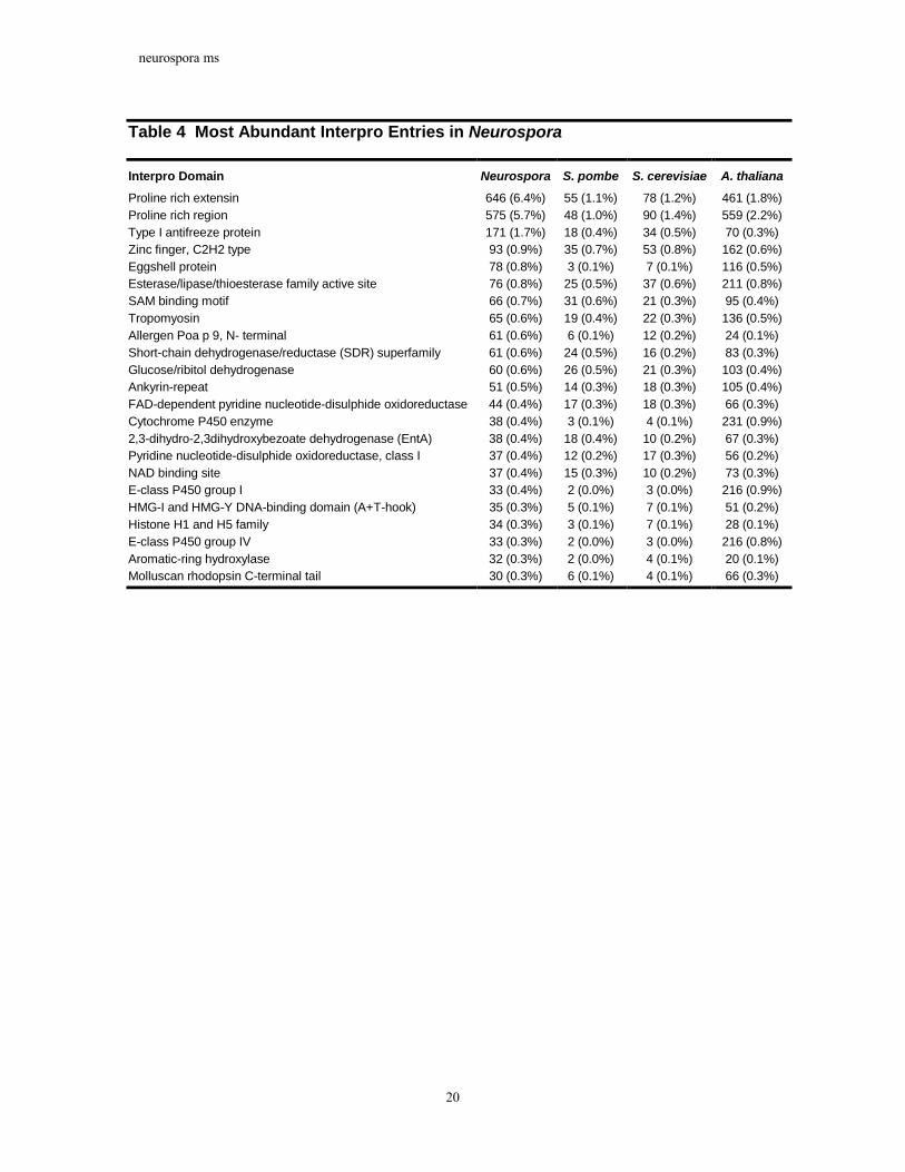
Table 4 Most Abundant Interpro Entries in Neurospora
Interpro Domain Neurospora S. pombe S. cerevisiae A. thaliana Proline rich extensin 646 (6.4%) 55 (1.1%) 78 (1.2%) 461 (1.8%) Proline rich region 575 (5.7%) 48 (1.0%) 90 (1.4%) 559 (2.2%) Type I antifreeze protein 171 (1.7%) 18 (0.4%) 34 (0.5%) 70 (0.3%) Zinc finger, C2H2 type 93 (0.9%) 35 (0.7%) 53 (0.8%) 162 (0.6%) Eggshell protein 78 (0.8%) 3 (0.1%) 7 (0.1%) 116 (0.5%) Esterase/lipase/thioesterase family active site 76 (0.8%) 25 (0.5%) 37 (0.6%) 211 (0.8%) SAM binding motif 66 (0.7%) 31 (0.6%) 21 (0.3%) 95 (0.4%) Tropomyosin 65 (0.6%) 19 (0.4%) 22 (0.3%) 136 (0.5%) Allergen Poa p 9, N- terminal 61 (0.6%) 6 (0.1%) 12 (0.2%) 24 (0.1%) Short-chain dehydrogenase/reductase (SDR) superfamily 61 (0.6%) 24 (0.5%) 16 (0.2%) 83 (0.3%) Glucose/ribitol dehydrogenase 60 (0.6%) 26 (0.5%) 21 (0.3%) 103 (0.4%) Ankyrin-repeat 51 (0.5%) 14 (0.3%) 18 (0.3%) 105 (0.4%) FAD-dependent pyridine nucleotide-disulphide oxidoreductase 44 (0.4%) 17 (0.3%) 18 (0.3%) 66 (0.3%) Cytochrome P450 enzyme 38 (0.4%) 3 (0.1%) 4 (0.1%) 231 (0.9%) 2,3-dihydro-2,3dihydroxybezoate dehydrogenase (EntA) 38 (0.4%) 18 (0.4%) 10 (0.2%) 67 (0.3%) Pyridine nucleotide-disulphide oxidoreductase, class I 37 (0.4%) 12 (0.2%) 17 (0.3%) 56 (0.2%) NAD binding site 37 (0.4%) 15 (0.3%) 10 (0.2%) 73 (0.3%) E-class P450 group I 33 (0.4%) 2 (0.0%) 3 (0.0%) 216 (0.9%) HMG-I and HMG-Y DNA-binding domain (A+T-hook) 35 (0.3%) 5 (0.1%) 7 (0.1%) 51 (0.2%) Histone H1 and H5 family 34 (0.3%) 3 (0.1%) 7 (0.1%) 28 (0.1%) E-class P450 group IV 33 (0.3%) 2 (0.0%) 3 (0.0%) 216 (0.8%) Aromatic-ring hydroxylase 32 (0.3%) 2 (0.0%) 4 (0.1%) 20 (0.1%) Molluscan rhodopsin C-terminal tail 30 (0.3%) 6 (0.1%) 4 (0.1%) 66 (0.3%)
neurospora ms
21
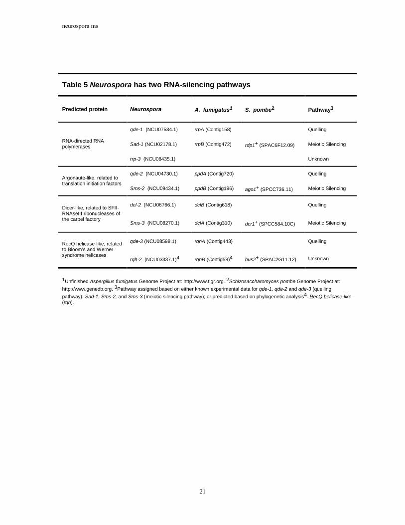
Table 5 Neurospora has two RNA-silencing pathways
Predicted protein Neurospora A. fumigatus1 S. pombe2 Pathway3
qde-1 (NCU07534.1) rrpA (Contig158) Quelling
Sad-1 (NCU02178.1) rrpB (Contig472) rdp1+ (SPAC6F12.09) Meiotic Silencing RNA-directed RNA polymerases
rrp-3 (NCU08435.1) Unknown
qde-2 (NCU04730.1) ppdA (Contig720) Quelling Argonaute-like, related to translation initiation factors
Sms-2 (NCU09434.1) ppdB (Contig196) ago1+ (SPCC736.11) Meiotic Silencing
dcl-2 (NCU06766.1) dclB (Contig618) Quelling Dicer-like, related to SFII-RNAseIII ribonucleases of the carpel factory
Sms-3 (NCU08270.1) dclA (Contig310) dcr1+ (SPCC584.10C) Meiotic Silencing
qde-3 (NCU08598.1) rqhA (Contig443) Quelling RecQ helicase-like, related to Bloom’s and Werner syndrome helicases
rqh-2 (NCU03337.1)4 rqhB (Contig58)4 hus2+ (SPAC2G11.12) Unknown
1Unfinished Aspergillus fumigatus Genome Project at: http://www.tigr.org. 2Schizosaccharomyces pombe Genome Project at: http://www.genedb.org. 3Pathway assigned based on either known experimental data for qde-1, qde-2 and qde-3 (quelling pathway); Sad-1, Sms-2, and Sms-3 (meiotic silencing pathway); or predicted based on phylogenetic analysis4. RecQ helicase-like (rqh).

neurospora ms
22
Table 6 Pathogenesis associated genes in Neurospora
Gene Homologa Organism Pathogenesis Function
NCU08038 GAS1 M. grisea Infection efficiency
NCU06170 GAS2 M. grisea Infection efficiency
NCU02903 PTH11 M. grisea Putative membrane receptor for host sensing
NCU07432 PLS1 M. grisea Tetraspanin, req. for pathogenic development
NCU05521 PATH531 M. grisea Aggressiveness factor
NCU05730 PEP2 N. haematococcab Aggressiveness factor
NCU03370 CAP20 C. gloeosporioidesc Aggressiveness factor
NCU06937 NCU06938 NCU02021 NCU04482 NCU09660
ECP2 C. fulvumd Aggressiveness factor, avirulence factor. Gene family in Neurospora
aOnly genes with no known role other than pathogenesis. bNectria haematococca. cColletotrichum gloeosporioides. dCladosporium fulvum.

neurospora ms
23
Figures
Figure 1: Summary of BLASTP Analysis of Neurospora proteins. (a) Proportion of Neurospora proteins with BLASTP hits (p<1e-5) to S. cerevisiae and S. pombe. (b) Proportion of Neurospora proteins with top BLASTP hits to selected sequenced eukaryotic organisms.
neurospora ms
24
Figure 2: Neurospora has a low proportion of genes in multigene families. The graph displays the proportion of genes in multigene families (see Methods) as a function of the number of genes in the genomes of selected sequenced eukaryotic organisms. Arrow indicates Neurospora. See text for more details.
neurospora ms
25
Figure 3: Neurospora possesses few highly similar genes. Histogram of (a) amino acid and (b) nucleotide percent identity of top scoring self-matches for genes in selected sequenced eukaryotic genomes. For each organism, the protein and coding regions for each gene (not including pseudogenes) were compared to those of every other gene in the same genome using BLASTX. Top scoring matches were aligned using ClustalW and percent identities calculated. In contrast to other eukaryotes, Neurospora possesses only 8 genes with a top match of greater than 80% amino acid or nucleotide identity.
neurospora ms
26
Figure 4: Example of lack of recent duplications in Neurospora gene family. Phylogenetic tree of major facilitator superfamily (MFS) sugar transporters from S. cerevisiae, S. pombe, and Neurospora. The amino acid sequences were aligned using CLUSTALW, manually trimmed to remove ambiguously aligned regions, and a neighbour-joining phylogenetic tree was generated from the alignment using PAUP*. Coloured dots represent branching points between predicted paralogous genes in Neurospora (orange), S. cerevisiae (blue), and S. pombe (green). In contrast to both yeasts, Neurospora transporters contain no predicted instances of recent paralogous duplication.

neurospora ms
27
Figure 5: Distribution of identified RIP-mutated repeat elements. A total of 46% of repetitive sequence in the Neurospora assembly could be identified as mobile element relics. No intact mobile elements were identified.
38%
15%3%
2%1%
0%6%
10%
3%
2%
1%
0%
1%
11%
6% 1%
class I LINE (Tad)class I LTR-gypsyclass I LTR-copiaclass I LTR (Tto-1)class I LTR (Tf)class I LTR (yoyo)undefined class Iclass II-Marinersclass II-Pot2class II-Pogoclass II-Punt class II-En/Spmundefined class IIrDNA repeats Centromere-proximal flanking Repeats with inverted repeats at both ends

neurospora ms
28
Figure 6: Correspondence between predicted RIP, methylation, and repetitive DNA. Prediction of RIP, methylation, and repeat sequence in 1kb windows for selected contigs. Red lines plot the TpA/ApT RIP Index (see Methods). Red dots indicate windows predicted to be RIP-mutated (TpA/ApT>1.2). Blue lines plot proportion of reads from methyl-tolerant library. Blue dots indicate windows predicted to be methylated based on >70% methyl-tolerant reads (see Methods). Black lines plot repeat content as fraction of nucleotides in each window that is in repetitive sequence. Black dots indicate windows with >50% repeat sequence. Contigs were selected to illustrate regions predicted as methylated.

neurospora ms
29
Figure 7: Overview of major intracellular signalling pathways in Neurospora. Abbreviations: AC, adenylyl cyclase; C, Ca2+-channel protein; CaM calmodulin; Ca2+/CaM-Reg, calcium and calmodulin regulated protein; CAP, cyclase-associated protein; GPCR, G-protein coupled receptor; Gα, G-protein α subunit; Gβ, G-protein β subunit; Gγ, G-protein γ subunit; HPT, histidine-containing phosphotransfer domain protein; MAPK, MAP kinase; MAPKK, MAPK kinase; MAPKKK, MAPKK kinase; PKA-R, protein kinase A regulatory subunit; PKA-C, protein kinase A catalytic subunit; PLC, phospholipase C; PKC, protein kinase C; T, Ca2+-transport protein (P-type Ca2+-ATPase, H+/Ca2+-exchanger, or Na+/Ca2+-exchanger);. *Location in plasma membrane and/or organelle membranes not determined.

neurospora ms
30
Figure 8: Domain structures of predicted Neurospora non-ribosomal peptide synthase (NRPS) and polyketide synthase (PKS) genes. Domains were predicted using a combination of PFAM searches using HMMER, protein alignments, and manual inspection.

neurospora ms
31
Methods Strain and Growth Conditions. Twenty 5 ml cultures of N. crassa wild-type strain N150 ("74-OR23-1VA"; FGSC#2489) were grown on a shaker in Vogel's minimal medium5 for 3 days at 32o C. Tissues were harvested, freeze-dried overnight and DNA extracted as previously described113. DNA from the twenty samples was mixed and used for library construction.
Sequencing and assembly. The genome was sequenced by the whole-genome shotgun method. Plasmid (4-kb inserts) and Fosmid (40-kb inserts) libraries were generated as described at http://www-genome.wi.mit.edu/. Jumping clone (subclone) libraries with 50-kb inserts were generated as described elsewhere114. Neurospora cosmid and BAC clones were obtained from previously constructed libraries115,116. Sequencing methods for all clone types are described at http://www-genome.wi.mit.edu/. All inserts were sequenced from both ends to generate paired-reads. The sequence coverage generated is shown in Table 1. The sequence was assembled using Arachne8. Finished sequence from linkage groups II and V was provided by MIPS and is available at http://mips.gsf.de/proj/Neurospora/.
Annotation and Analysis. The Neurospora genome was annotated using the Calhoun annotation system. The genome sequence was searched against the public protein databases using BLASTX with threshold E<1e-5. Genes were predicted using a combination of FGENESH, FGENESH+, and GENEWISE. The gene calling programs were validated against a test set of 191 previously characterized Neurospora proteins. Predicted genes were validated against ESTs aligned to the genome using SIM4. All predicted genes were searched against the PFAM set of hidden Markov models using the HMMER program and the public protein databases using BLASTP. Transfer RNAs were identified using the tRNAScan-SE program (version X). Multigene families were constructed by searching each annotated gene against every other gene using BLASTP, requiring matches with E < 1e-5 over 60% of the longer gene length, and clustering genes based on single linkage transitive closure. Repeat sequences were detected by searching the genome sequence against itself using CrossMatch, filtering for alignments longer than 200bp in length, and clustering pairs based on region overlap.
RIP-mutated regions were detected by calculating one or both of two different dinucleotide ratios for sequence regions17. Regions with TpA/ApT>2 or (CpA+TpG)/(ApC+GpT)<0.7 were predicted as RIP-mutated. Prediction of RIP sequence across the genome utilized only the TpA/ApT ratio, while the analysis of coding sequences used both (with a positive prediction by either measure taken as a prediction as RIP). RIP simulations were based on parameters derived from Watters et al. (1999). DNA methylation was predicted by calculating the proportion of plasmid reads overlapping 1 kb windows from both the methylation-tolerant and methylation-intolerant libraries. Regions with greater than 70% reads derived from the methylation-tolerant library were predicted methylated. Specificity was estimated as described in the text. Methylation was experimentally assessed using Southern analyses as described elsewhere51. Sensitivity was estimated by testing 19 repetitive and RIP mutated 1 kb regions that were not predicted to be methylated. Of the 19 regions, 14 were in fact methylated.
Predicted RNA silencing genes were aligned with homologs from plants, animals and other fungi using T-Coffee v1.37. C-terminal and N-terminal regions of low homology were removed and the sequences realigned until alignments started and stopped at regions of

neurospora ms
32
unambiguous similarity. Both joining-joining trees, using ClustalX, and maximum posterior probability trees, using MrBayes 2.01, were generated and analysed.
Additional details, analysis results, and the genome sequence are available at http://www-genome.wi.mit.edu/.

neurospora ms
33
References
1. Payen, A. (rapporteur) Extrait d'un rapport adressé à M. Le Maréchal Duc de Dalmatie, Ministre de la Guerre, Président du Conseil, sur une altération extraordinaire du pain de munition. Ann. Chim. Phys. 3e Ser. 9, 5-21 (+one plate) (1843).
2. Shear, C. L. & Dodge, B. O. Life histories and heterothallism of the red bread-mold fungi of the Monilia sitophila group. J. Agr. Res. 34, 1019-1042 (1927).
3. Lindegren, C. C. A six-point map of the sex chromosome of Neurospora crassa. J. Genet. 32, 243-256 (1936).
4. Beadle, G. W. & Tatum, E. L. Genetic control of biochemical reactions in Neurospora. Proc Natl Acad Sci U S A 27, 499-506 (1941).
5. Davis, R. H. Neurospora : contributions of a model organism (Oxford University Press, New York, 2000).
6. Davis, R. H. & Perkins, D. D. Timeline: Neurospora: a model of model microbes. Nat Rev Genet 3, 397-403 (2002).
7. Perkins, D. D., Radford, A. & Sachs, M. S. The Neurospora Compendium: Chromosomal loci (Academic Press, San Diego, CA, 2001).
8. Batzoglou, S. et al. ARACHNE: a whole-genome shotgun assembler. Genome Res 12, 177-89 (2002).
9. Schulte, U., Becker, I., Mewes, H. W. & Mannhaupt, G. Large scale analysis of sequences from Neurospora crassa. J Biotechnol 94, 3-13 (2002).
10. Free, S. J., Rice, P. W. & Metzenberg, R. L. Arrangement of the genes coding for ribosomal ribonucleic acids in Neurospora crassa. J Bacteriol 137, 1219-26. (1979).
11. Spingola, M., Grate, L., Haussler, D. & Ares, M., Jr. Genome-wide bioinformatic and molecular analysis of introns in Saccharomyces cerevisiae. Rna 5, 221-34 (1999).
12. Voelker, R. & Berglund, J. A. (Unpublished). 13. Zdobnov, E. M. & Apweiler, R. InterProScan--an integration platform for the
signature-recognition methods in InterPro. Bioinformatics 17, 847-8. (2001). 14. Selker, E. U., Cambareri, E. B., Jensen, B. C. & Haack, K. R. Rearrangement of
duplicated DNA in specialized cells of Neurospora. Cell 51, 741-52. (1987). 15. Selker, E. U. Premeiotic instability of repeated sequences in Neurospora crassa. Annu
Rev Genet 24, 579-613 (1990). 16. Cambareri, E. B., Jensen, B. C., Schabtach, E. & Selker, E. U. Repeat-induced G-C to
A-T mutations in Neurospora. Science 244, 1571-5. (1989). 17. Margolin, B. S. et al. A methylated Neurospora 5S rRNA pseudogene contains a
transposable element inactivated by repeat-induced point mutation. Genetics 149, 1787-97. (1998).
18. Watters, M. K., Randall, T. A., Margolin, B. S., Selker, E. U. & Stadler, D. R. Action of repeat-induced point mutation on both strands of a duplex and on tandem duplications of various sizes in Neurospora. Genetics 153, 705-14. (1999).
19. Cambareri, E. B., Singer, M. J. & Selker, E. U. Recurrence of repeat-induced point mutation (RIP) in Neurospora crassa. Genetics 127, 699-710. (1991).
20. Selker, E. U. Repeat-induced gene silencing in fungi. Adv Genet 46, 439-50 (2002). 21. Selker, E. U. & Garrett, P. W. DNA sequence duplications trigger gene inactivation in
Neurospora crassa. Proc Natl Acad Sci U S A 85, 6870-4. (1988). 22. Rountree, M. R. & Selker, E. U. DNA methylation inhibits elongation but not
initiation of transcription in Neurospora crassa. Genes Dev 11, 2383-95. (1997).

neurospora ms
34
23. Cambareri, E. B., Aisner, R. & Carbon, J. Structure of the chromosome VII centromere region in Neurospora crassa: degenerate transposons and simple repeats. Mol Cell Biol 18, 5465-77. (1998).
24. Kinsey, J. A., Garrett-Engele, P. W., Cambareri, E. B. & Selker, E. U. The Neurospora transposon Tad is sensitive to repeat-induced point mutation (RIP). Genetics 138, 657-64. (1994).
25. Irelan, J. T., Hagemann, A. T. & Selker, E. U. High frequency repeat-induced point mutation (RIP) is not associated with efficient recombination in Neurospora. Genetics 138, 1093-103. (1994).
26. Perkins, D. D., Margolin, B. S., Selker, E. U. & Haedo, S. D. Occurrence of repeat induced point mutation in long segmental duplications of Neurospora. Genetics 147, 125-36. (1997).
27. Razin, S., Yogev, D. & Naot, Y. Molecular biology and pathogenicity of mycoplasmas. Microbiol Mol Biol Rev 62, 1094-156 (1998).
28. Apweiler, R. et al. InterPro--an integrated documentation resource for protein families, domains and functional sites. Bioinformatics 16, 1145-50. (2000).
29. Ohno, S. Evolution by Gene Duplication (Springer-Verlag, 1970). 30. Walsh, J. B. How Often Do Duplicated Genes Evolve New Functions? Genetics 139,
421-428 (1995). 31. Lynch, M. & Conery, J. S. The evolutionary fate and consequences of duplicate
genes. Science 290, 1151-1155 (2000). 32. Krumlauf, R. & Marzluf, G. A. Genome organization and characterization of the
repetitive and inverted repeat DNA sequences in Neurospora crassa. J Biol Chem 255, 1138-45. (1980).
33. Krumlauf, R. & Marzluf, G. A. Characterization of the sequence complexity and organization of the Neurospora crassa genome. Biochemistry 18, 3705-13. (1979).
34. Selker, E. U. Epigenetic phenomena in filamentous fungi: useful paradigms or repeat-induced confusion. Trends Genet 13, 296-301 (1997).
35. Centola, M. & Carbon, J. Cloning and characterization of centromeric DNA from Neurospora crassa. Mol Cell Biol 14, 1510-9. (1994).
36. Bibbins, M., Cummings, N. J. & Connerton, I. F. DAB1: a degenerate retrotransposon-like element from Neurospora crassa. Mol Gen Genet 258, 431-6. (1998).
37. Schechtman, M. G. Characterization of telomere DNA from Neurospora crassa. Gene 88, 159-65. (1990).
38. Langin, T., Capy, P. & Daboussi, M. J. The transposable element impala, a fungal member of the Tc1-mariner superfamily. Mol Gen Genet 246, 19-28. (1995).
39. Hua-Van, A., Hericourt, F., Capy, P., Daboussi, M. J. & Langin, T. Three highly divergent subfamilies of the impala transposable element coexist in the genome of the fungus Fusarium oxysporum. Mol Gen Genet 259, 354-62. (1998).
40. Villalba, F., Lebrun, M. H., Hua-Van, A., Daboussi, M. J. & Grosjean-Cournoyer, M. C. Transposon impala, a novel tool for gene tagging in the rice blast fungus Magnaporthe grisea. Mol Plant Microbe Interact 14, 308-15. (2001).
41. Kachroo, P., Leong, S. A. & Chattoo, B. B. Pot2, an inverted repeat transposon from the rice blast fungus Magnaporthe grisea. Mol Gen Genet 245, 339-48. (1994).
42. Nakayashiki, H., Nishimoto, N., Ikeda, K., Tosa, Y. & Mayama, S. Degenerate MAGGY elements in a subgroup of Pyricularia grisea: a possible example of successful capture of a genetic invader by a fungal genome. Mol Gen Genet 261, 958-66. (1999).
neurospora ms
35
43. Campell, B. R., Song, Y., Posch, T. E., Cullis, C. A. & Town, C. D. Sequence and organization of 5S ribosomal RNA-encoding genes of Arabidopsis thaliana. Gene 112, 225-8 (1992).
44. Timofeeva, M. et al. [Organization of a 5S ribosomal RNA gene cluster in the human genome]. Mol Biol (Mosk) 27, 861-8 (1993).
45. Belkhiri, A., Intengan, H. & Klassen, G. R. A tandem array of 5S ribosomal RNA genes in Pythium irregulare. Gene 186, 155-9 (1997).
46. Martins, C. & Galetti, P. M., Jr. Two 5S rDNA arrays in neotropical fish species: is it a general rule for fishes? Genetica 111, 439-46 (2001).
47. Valarik, M. et al. Isolation, characterization and chromosome localization of repetitive DNA sequences in bananas (Musa spp.). Chromosome Res 10, 89-100 (2002).
48. Selker, E. U. et al. Dispersed 5S RNA genes in N. crassa: structure, expression and evolution. Cell 24, 819-28. (1981).
49. Irelan, J. T. & Selker, E. U. Cytosine methylation associated with repeat-induced point mutation causes epigenetic gene silencing in Neurospora crassa. Genetics 146, 509-23. (1997).
50. Selker, E. U. & Stevens, J. N. DNA methylation at asymmetric sites is associated with numerous transition mutations. Proc Natl Acad Sci U S A 82, 8114-8. (1985).
51. Selker, E. U., Fritz, D. Y. & Singer, M. J. Dense nonsymmetrical DNA methylation resulting from repeat-induced point mutation in Neurospora. Science 262, 1724-8. (1993).
52. Russell, P. J., Rodland, K. D., Rachlin, E. M. & McCloskey, J. A. Differential DNA methylation during the vegetative life cycle of Neurospora crassa. J Bacteriol 169, 2902-5. (1987).
53. Foss, H. M., Roberts, C. J., Claeys, K. M. & Selker, E. U. Abnormal chromosome behavior in Neurospora mutants defective in DNA methylation. Science 262, 1737-41. (1993).
54. Selker, E. U., Jensen, B. C. & Richardson, G. A. A portable signal causing faithful DNA methylation de novo in Neurospora crassa. Science 238, 48-53. (1987).
55. Singer, M. J., Marcotte, B. A. & Selker, E. U. DNA methylation associated with repeat-induced point mutation in Neurospora crassa. Mol Cell Biol 15, 5586-97. (1995).
56. Miao, V. P., Freitag, M. & Selker, E. U. Short TpA-rich segments of the zeta-eta region induce DNA methylation in Neurospora crassa. J Mol Biol 300, 249-73. (2000).
57. Kouzminova, E. & Selker, E. U. dim-2 encodes a DNA methyltransferase responsible for all known cytosine methylation in Neurospora. Embo J 20, 4309-23. (2001).
58. Freitag, M., Williams, R. L., Kothe, G. O. & Selker, E. U. A cytosine methyltransferase homologue is essential for repeat-induced point mutation in Neurospora crassa. Proc Natl Acad Sci U S A 99, 8802-7. (2002).
59. Selker, E. U. et al. Induction and maintenance of nonsymmetrical DNA methylation in Neurospora. Proc Natl Acad Sci U S A (2002).
60. Frommer, M. et al. A genomic sequencing protocol that yields a positive display of 5-methylcytosine residues in individual DNA strands. Proc Natl Acad Sci U S A 89, 1827-31 (1992).
61. Grant, S. R. Dissecting the mechanisms of posttranscriptional gene silencing: divide and conquer. Cell 96, 303-6 (1999).
62. Zamore, P. D. Ancient pathways programmed by small RNAs. Science 296, 1265-9 (2002).

neurospora ms
36
63. Vance, V. & Vaucheret, H. RNA silencing in plants--defense and counterdefense. Science 292, 2277-80 (2001).
64. Hammond, S. M., Caudy, A. A. & Hannon, G. J. Post-transcriptional gene silencing by double-stranded RNA. Nat Rev Genet 2, 110-9 (2001).
65. Rougvie, A. E. Control of developmental timing in animals. Nat Rev Genet 2, 690-701 (2001).
66. Hannon, G. J. RNA interference. Nature 418, 244-51 (2002). 67. Wolffe, A. P. & Matzke, M. A. Epigenetics: regulation through repression. Science
286, 481-6 (1999). 68. Hutvagner, G. & Zamore, P. D. RNAi: nature abhors a double-strand. Curr Opin
Genet Dev 12, 225-32 (2002). 69. Cogoni, C. & Macino, G. Gene silencing in Neurospora crassa requires a protein
homologous to RNA-dependent RNA polymerase. Nature 399, 166-9 (1999). 70. Catalanotto, C., Azzalin, G., Macino, G. & Cogoni, C. Involvement of small RNAs
and role of the qde genes in the gene silencing pathway in Neurospora. Genes Dev 16, 790-5 (2002).
71. Catalanotto, C., Azzalin, G., Macino, G. & Cogoni, C. Gene silencing in worms and fungi. Nature 404, 245 (2000).
72. Cogoni, C. et al. Transgene silencing of the al-1 gene in vegetative cells of Neurospora is mediated by a cytoplasmic effector and does not depend on DNA-DNA interactions or DNA methylation. Embo J 15, 3153-63 (1996).
73. Cogoni, C. & Macino, G. Isolation of quelling-defective (qde) mutants impaired in posttranscriptional transgene-induced gene silencing in Neurospora crassa. Proc Natl Acad Sci U S A 94, 10233-8 (1997).
74. Cogoni, C. & Macino, G. Posttranscriptional gene silencing in Neurospora by a RecQ DNA helicase. Science 286, 2342-4 (1999).
75. Aramayo, R. & Metzenberg, R. L. Meiotic transvection in fungi. Cell 86, 103-13 (1996).
76. Shiu, P. K., Raju, N. B., Zickler, D. & Metzenberg, R. L. Meiotic silencing by unpaired DNA. Cell 107, 905-16 (2001).
77. Shiu, P. K. & Metzenberg, R. L. Meiotic Silencing by Unpaired DNA. Properties, regulation and suppression. Genetics 161, 1483-95 (2002).
78. Volpe, T. A. et al. Regulation of Heterochromatic Silencing and Histone H3 Lysine-9 Methylation by RNAi. Science 297, 1833-7 (2002).
79. Lee, D., Pratt, R. J., McLaughlin, M. & Aramayo, R. Genetics (In Press). 80. McLaughlin, M., Lee, D., Pratt, R. J., Dado, J. & Aramayo, R. (In Preparation). 81. Linden, H., Ballario, P., Arpaia, G. & Macino, G. Seeing the light: news in
Neurospora blue light signal transduction. Adv Genet 41, 35-54 (1999). 82. Devlin, P. F. & Kay, S. A. Circadian photoperception. Annu Rev Physiol 63, 677-94
(2001). 83. Chang, L. & Karin, M. Mammalian MAP kinase signalling cascades. Nature 410, 37-
40 (2001). 84. West, A. H. & Stock, A. M. Histidine kinases and response regulator proteins in two-
component signaling systems. Trends Biochem Sci 26, 369-76 (2001). 85. Alex, L. A. & Simon, M. I. (Unpublished data). 86. Pott, G. B., Miller, T. K., Bartlett, J. A., Palas, J. S. & Selitrennikoff, C. P. The
isolation of FOS-1, a gene encoding a putative two-component histidine kinase from Aspergillus fumigatus. Fungal Genet Biol 31, 55-67. (2000).

neurospora ms
37
87. Dohlman, H. G., Thorner, J., Caron, M. G. & Lefkowitz, R. J. Model systems for the study of seven-transmembrane-segment receptors. Annu Rev Biochem 60, 653-88 (1991).
88. Klein, P. S. et al. A chemoattractant receptor controls development in Dictyostelium discoideum. Science 241, 1467-72 (1988).
89. Josefsson, L. G. & Rask, L. Cloning of a putative G-protein-coupled receptor from Arabidopsis thaliana. Eur J Biochem 249, 415-20 (1997).
90. Aubry, L. & Firtel, R. Integration of signaling networks that regulate Dictyostelium differentiation. Annu Rev Cell Dev Biol 15, 469-517 (1999).
91. Kore-eda, S., Murayama, T. & Uno, I. Isolation and characterization of the adenylate cyclase structural gene of Neurospora crassa. Jpn J Genet 66, 317-34 (1991).
92. Ivey, F. D., Yang, Q. & Borkovich, K. A. Positive regulation of adenylyl cyclase activity by a galphai homolog in Neurospora crassa. Fungal Genet Biol 26, 48-61 (1999).
93. Gadd, G. M. in The Growing Fungus (eds. Gow, N. A. R. & Gadd, G. M.) 183-210 (Chapman & Hall, London, 1994).
94. Bootman, M. D. et al. Calcium signalling--an overview. Semin Cell Dev Biol 12, 3-10 (2001).
95. Lakin-Thomas, P. L. Effects of inositol starvation on the levels of inositol phosphates and inositol lipids in Neurospora crassa. Biochem J 292 ( Pt 3), 805-11 (1993).
96. Silverman-Gavrila, L. B. & Lew, R. R. Regulation of the tip-high [Ca2+] gradient in growing hyphae of the fungus Neurospora crassa. Eur J Cell Biol 80, 379-90 (2001).
97. Cornelius, G., Gebauer, G. & Techel, D. Inositol trisphosphate induces calcium release from Neurospora crassa vacuoles. Biochem Biophys Res Commun 162, 852-6 (1989).
98. Gimeno, C. J., Ljungdahl, P. O., Styles, C. A. & Fink, G. R. Unipolar cell divisions in the yeast S. cerevisiae lead to filamentous growth: regulation by starvation and RAS. Cell 68, 1077-90 (1992).
99. Lengeler, K. B. et al. Signal transduction cascades regulating fungal development and virulence. Microbiol Mol Biol Rev 64, 746-85 (2000).
100. Griffin, D. H. Fungal Physiology (Wiley-Liss, New York, 1994). 101. Ivey, F. D., Kays, A. M. & Borkovich, K. A. Shared and independent roles for a
Galpha(i) protein and adenylyl cyclase in regulating development and stress responses in Neurospora crassa. Eukaryot Cell 1, 634-42 (2002).
102. Yuan, W., Gentil, G., Budde, A. & Leong, S. Characterization of the Ustilago maydis sid2 gene encoding a multidomain peptide synthetase in the ferrichrome biosynthetic gene cluster. J. Bacteriol. 183, 4040-4051 (2001).
103. Feng, B. et al. Molecular cloning and characterization of WdPKS1, a gene involved in dihydroxynaphthalene melanin biosynthesis and virulence in Wangiella (Exophiala) dermatitidis. Infect. Immun. 69, 1781-1794 (2001).
104. Takano, Y. et al. Structural analysis of PKS1, a polyketide synthase gene involved in melanin biosynthesis in Colletotrichum lagenarium. Mol. Gen. Genet. 249, 162-167 (1995).
105. Takano, Y., Kubo, Y., Kawamura, C., Tsuge, T. & Furusawa, I. The Alternaria alternata melanin biosynthesis gene restores appressorial melanization and penetration of cellulose membranes in the melanin-deficient albino mutant of Colletotrichum lagenarium. Fungal Genet. and Biol., 131-140 (1997).
106. Nelson, M. A. et al. Expressed sequences from conidial, mycelial, and sexual stages of Neurospora crassa. Fungal Genet Biol 21, 348-63. (1997).
neurospora ms
38
107. Kurosawa, E. Experimental studies on the nature of the substance excreted by the "bakanae" fungus (in Japanese). Trans. Nat. His. Soc. Formosa 16, 213-227 (1926).
108. Vigfusson, N. & Cano, R. Artificial induction of the sexual cycle of Neurospora crassa. Nature 249, 383-385 (1974).
109. Graia, F. et al. Genome quality control: RIP (repeat-induced point mutation) comes to Podospora. Mol Microbiol 40, 586-95. (2001).
110. Ikeda, K. et al. Repeat-induced point mutation (RIP) in Magnaporthe grisea: implications for its sexual cycle in the natural field context. Mol Microbiol 45, 1355-64 (2002).
111. TIGR Microbial Database, http://www.tigr.org/tdb/mdb/mdbinprogress.html. 112. Fungal Genome Initiative, http://www-genome.wi.mit.edu/seq/fgi/. (2002). 113. Luo, Z., Freitag, M. & Sachs, M. S. Translational regulation in response to changes in
amino acid availability in Neurospora crassa. Mol Cell Biol 15, 5235-45 (1995). 114. Venter, J. C. et al. The sequence of the human genome. Science 291, 1304-51 (2001). 115. Kelkar, H. S. et al. The Neurospora crassa genome: cosmid libraries sorted by
chromosome. Genetics 157, 979-90 (2001). 116. Aign, V., Schulte, U. & Hoheisel, J. D. Hybridization-based mapping of Neurospora
crassa linkage groups II and V. Genetics 157, 1015-20 (2001).


![The History and Scope of Microbiology · Some organisms studies by microbiologists CAN be visualized without the aid of amplification [bread molds (fungus) and filamentous algae]](https://img.dokumen.tips/doc/110x75/5e7873555e72f05112460566/the-history-and-scope-of-some-organisms-studies-by-microbiologists-can-be-visualized.jpg)