Embed Size (px)
Citation preview
REVIEW
The general surgeon’s perspective of rectus diastasis. A systematicreview of treatment options
Elwin H. H. Mommers1• Jeroen E. H. Ponten2
• Aminah K. Al Omar1•
Tammo S. de Vries Reilingh3• Nicole D. Bouvy1
• Simon W. Nienhuijs2
Received: 9 February 2017 / Accepted: 16 May 2017 / Published online: 8 June 2017
� The Author(s) 2017. This article is an open access publication
Abstract
Background Diastasis of the rectus abdominis muscles
(DRAM) is characterised by thinning and widening of the
linea alba, combined with laxity of the ventral abdominal
musculature. This causes the midline to ‘‘bulge’’ when intra-
abdominal pressure is increased. Plastic surgery treatment
for DRAM has been thoroughly evaluated, though general
surgical treatments and the efficacy of physiotherapy remain
elusive. The aim of this systematic literature review is to
evaluate both general surgical and physiotherapeutic treat-
ment options for restoring DRAM in terms of postoperative
complications, patient satisfaction, and recurrence rates.
Method MEDLINE�, Embase, PubMed, PubMed Cen-
tral�, The cochrane central registry of controlled trials
(CENTRAL), Google Scholar, and the Physiotherapy
Evidence Database (PEDro) were searched using the fol-
lowing terms: ‘rectus diastasis’, ‘diastasis recti’, ‘midline’,
and ‘abdominal wall’. All clinical studies concerning
general surgical or physiotherapeutic treatment of DRAM
were eligible for inclusion.
Result Twenty articles describing 1.691 patients (1.591
surgery/100 physiotherapy) were included. Surgical inter-
ventions were classified as plication techniques (313 patients;
254 open/59 laparoscopic), modified hernia repair techniques
(68 patients, all open), and combined hernia & DRAM
techniques (1.210 patients; 1.149 open/40 hybrid). The
overall methodological quality was low. Plication techniques
with interrupted sutures and mesh reinforcement were
applied most frequently for DRAM repair. Open repairs were
performed in 85% of patients. There was no difference in
postoperative complications or recurrence rate after laparo-
scopic or open procedures, or between plication and modified
hernia repair techniques. Physiotherapy programmes were
unable to reduce IRD in a relaxed state. Though reduction of
IRD during muscle contraction was described.
Conclusion Both plication-based methods and hernia
repair methods are used for DRAM repair. Based on the
current literature, no clear distinction in recurrence rate,
postoperative complications, or patient reported outcomes
can be made. Complete resolution of DRAM, measured in
a relaxed state, following a physiotherapy training pro-
gramme is not described in current literature. Physiother-
apy can achieve a limited reduction in IRD during muscle
contraction, though the impact of this finding on patient
satisfaction, cosmesis, or function outcome is unclear.
Keywords Diastasis of the rectus abdominis muscles
(DRAM) � Diastasis repair � Surgical treatment �Physiotherapy
Diastasis of the rectus abdominis muscles (DRAM) is
characterised by a protruding midline following an increase
in intra-abdominal pressure. The condition is characterised
by a gradual thinning and widening of the linea alba,
combined with a general laxity of the ventral abdominal
wall muscles [1]. DRAM is frequently misclassified as a
primary ventral hernia, though the musculofascial
Meeting presentation information The results published in thismanuscript have not been published elsewhere.
& Elwin H. H. Mommers
1 Department of Surgery, Maastricht University Medical
Center, Maastricht, The Netherlands
2 Department of Surgery, Catharina Hospital, Eindhoven, The
Netherlands
3 Department of Surgery, Elkerliek Hospital, Helmond, The
Netherlands
123
Surg Endosc (2017) 31:4934–4949
DOI 10.1007/s00464-017-5607-9
and Other Interventional Techniques
continuity of the midline and subsequent absence of a true
hernia sac is what sets DRAM apart from a ventral hernia.
DRAM is defined according to the Beer classification as an
inter-rectus distance (IRD) of 22 mm, three centimetres
above the umbilicus measured in a relaxed state [2].
DRAM occurs most frequently during pregnancy and
regresses spontaneously after childbirth in most women.
However, 12 months postpartum, 33% of women still
experience DRAM [3].
Patients with DRAM can experience similar complaints
as patients with ventral hernias, such as lower back pain,
functional, and cosmetic impairment, although DRAM
does not pose any threat of strangulation [4–6]. DRAM
repair is challenging for most general surgeons since
guidelines on indication and methods for repair do not
exist. The similarity to primary ventral hernias causes
frequent misclassification of the disease, and potential
mistreatment of DRAM. In recent years, the overall com-
plexity of evidence concerning DRAM treatment has
increased. This is due to the development and implemen-
tation of several new reconstructive techniques, combined
with heterogenous outcome measurements, heterogenous
definitions for DRAM, and the lack of high quality data.
DRAM is mostly treated conservatively, with or without
the help of a physiotherapist. If conservative therapy is
preferred, patients can be referred to a physiotherapist for
training programmes that specifically target DRAM, with
the aim of reducing IRD and improvement of quality-of-
life (QoL). Benjamin et al. evaluated the efficacy of these
training programmes in 2014, though due to the low quality
of the included studies, no conclusions could be drawn [7].
In case of severe functional or cosmetic impairment, the
patient can be referred to a plastic or general surgeon.
Patients that suffer from excess skin or want to tailor their
waistline simultaneously with DRAM repair should be
referred to a surgeon in the field of plastic and recon-
structive surgery for an abdominoplasty. Publications
describing DRAM repair in combination with abdomino-
plasty, liposuction, or other strictly plastic surgical tech-
niques are numerous. A recent review of Akram et al. in
2014 concerning abdominoplasty repairs in combination
with plication of the linea alba concluded that most evi-
dence is of low quality and RCTs are required to gain more
insight in the short- and long-term effects of these com-
bined procedures [8].
Patients with the sole diagnosis of DRAM are frequently
referred to the general surgeon. If surgical treatment is
considered, several techniques ranging from laparoscopic,
endoscopic, hybrid, and open repairs are available. Cur-
rently, there is no consensus on the preferred surgical
management of DRAM. In contrast to evidence from the
reconstructive field, a thorough literature review comparing
different surgical techniques for DRAM is noticeably
absent in general surgery. The aim of this systematic lit-
erature review is to provide insight in the general surgical
treatment options for DRAM in terms of postoperative
complications, patient satisfaction, and recurrence rates,
and to evaluate if physiotherapy is an alternative for sur-
gical intervention.
Methods
This review was registered on PROSPERO [No.:
CRD42016048176], and conducted according to the PRISMA
statement [9]. Before the start of the review process, the
review protocol was evaluated and approved by an indepen-
dent, external expert in the field of ventral hernia repair.
Search strategy
A structured literature search of MEDLINE�, Embase,
PubMed, PubMed Central� (PMC), The cochrane central
registry of controlled trials (CENTRAL), Google Scholar,
and the Physiotherapy Evidence Database (PEDro) was
performed by two independent reviewers (E.M. and
A.A.O.) using the following terms:
‘Diastasis recti’ OR ‘rectus diastasis’ OR ‘diastasis of
the rectus abdominis’ OR ‘diastasis of the recti’ OR ‘ab-
dominal diastasis’ OR ‘abdominal separation’ OR ‘diasta-
sis recti abdominis’ OR ‘separation of the recti’ OR
‘separation of the rectus abdominis’ OR ‘divarication of
the recti’ OR ‘divarication the rectus abdominis’ (See
Table 1 for PubMed search algorithm).
The last search was performed on 8 September 2016.
The search was performed using validated methods of the
Cochrane collaboration [10]. Both medical subject heading
(MeSH) terms and free-text terms were used to construct
the search algorithm. To create a sensitive algorithm, only
one domain of search terms (DRAM population) was used.
In addition to the above-mentioned database searches, all
reference lists of included studies were cross-referenced to
retrieve additional articles eligible for inclusion. In case of
disagreement between the reviewers regarding the eligi-
bility for inclusion of an article, the study quality, or the
data abstraction, a third reviewer (NB) was consulted for
arbitration.
Outcome definition and study selection criteria
The primary outcome for surgical studies was recurrence
rate, secondary outcomes were complication rate within
30 days, and patient satisfaction.
The primary outcome for physiotherapy studies was the
effect of the treatment on IRD, secondary outcomes were
were patient satisfaction, and recurrence rate.
Surg Endosc (2017) 31:4934–4949 4935
123
Physiotherapy studies reporting the effect of a single
exercise, performed only once, on IRD were considered as
functional anatomy studies; these studies were not included
in this systematic review. Physiotherapy studies focussing
on rectus diastasis during pregnancy or during the imme-
diate postpartum period (B24 h after childbirth) are not
included in this review since results obtained during this
period cannot be translated to a later time point, and
therefore, do not provide an answer to the secondary end-
point of this review.
Any study (comparative, non-comparative, randomised,
or observational studies) describing the effect of a surgical
or physiotherapy intervention for DRAM in at least one
patient C18 years old and reporting on the primary and/or
secondary outcome of this systematic literature review was
eligible for inclusion, with the exclusion of plastic surgery
interventions such as abdominoplasty or liposuction. No
limitations to subjects, type of article, or language were
applied, though articles published before 1975 which
described techniques that have not been applied or
described since 1975 were excluded. Articles reporting on
the treatment of DRAM combined with primary ventral
midline hernias were included, yet analysed separately.
Quality assessment
The quality of randomised controlled trials (RCTs) was
evaluated using a 14-item modified Jadad score and the
Cochrane risk of bias tool [11, 12]. The quality of non-
randomised clinical studies was evaluated using the
methodological index for non-randomised studies (MIN-
ORS) criteria [13]. In case of missing data for the quality
assessment tool or data abstraction, the first and corre-
sponding author of the study was contacted via email for
completion of the missing data. If the author did not
respond, a reminder was sent after two weeks. Studies
which scored below three on the MINORS checklist were
excluded due to lack of critical data needed for correct
interpretation of the results, combined with an
unacceptable risk of bias, as at least six of eight domains of
the MINORS checklist are either not reported, or partially
reported in those publications.
Data abstraction
Data abstraction was performed in duplicate by two inde-
pendent reviewers (E.M. and A.A.O.) with a standardised
electronic data extraction form including but not limited to
the following study variables: title, source, year of publi-
cation, study design incl. retrospective or prospective
design, demographics of study population, sample size,
type of rectus diastasis according to Nahas et al., descrip-
tion of intervention, type of analysis (intention to treat vs
per protocol), complications within and after 30 days
postoperative, follow-up period, follow-up assessment tool,
risk factors for recurrence, and recurrence rate [14]. The
Nahas classification defines four different aesthetic types of
DRAM: Type A, DRAM secondary to pregnancy; Type B,
DRAM and laxity of the lateral and infra-umbilical
aponeurosis; Type C, congenital lateral insertion of the
rectus abdominis muscles; and Type D, DRAM combined
with poor waistline.
Results
After screening 3.689 citations and removal of 20 duplicate
manuscripts, 37 articles were selected for full-text review
(Fig. 1). Seventeen articles did not meet the inclusion cri-
teria. Twenty studies describing a total of 1.691 patients,
1.591 (94 males, 1497 females) in general surgical tech-
niques and 100 (all female) in physiotherapy training
programmes, were included in this systematic review.
Fourteen articles described surgical techniques, and six
described physiotherapy interventions (Table 1). Five
articles were written in Russian, and one in Italian. The
included surgical techniques were divided into three cate-
gories: (1) plication techniques for DRAM repair, (2)
Table 1 Search algorithm for PubMed search
((((((((((((diastasis[All Fields] AND recti[All Fields]) OR (rectus[All Fields] AND diastasis[All Fields])) OR (diastasis[All Fields] AND (‘‘rectus
abdominis’’[MeSH Terms] OR (‘‘rectus’’[All Fields] AND ‘‘abdominis’’[All Fields]) OR ‘‘rectus abdominis’’[All Fields]))) OR (diastasis[All
Fields] AND recti[All Fields])) OR ((‘‘abdomen’’[MeSH Terms] OR ‘‘abdomen’’[All Fields] OR ‘‘abdominal’’[All Fields]) AND diastasis[All
Fields])) OR ((‘‘abdomen’’[MeSH Terms] OR ‘‘abdomen’’[All Fields] OR ‘‘abdominal’’[All Fields]) AND (‘‘divorce’’[MeSH Terms] OR
‘‘divorce’’[All Fields] OR ‘‘separation’’[All Fields]))) OR (diastasis[All Fields] AND recti[All Fields] AND abdominis[All Fields])) OR
((‘‘divorce’’[MeSH Terms] OR ‘‘divorce’’[All Fields] OR ‘‘separation’’[All Fields]) AND recti[All Fields])) OR ((‘‘divorce’’[MeSH Terms] OR
‘‘divorce’’[All Fields] OR ‘‘separation’’[All Fields]) AND (‘‘rectus abdominis’’[MeSH Terms] OR (‘‘rectus’’[All Fields] AND
‘‘abdominis’’[All Fields]) OR ‘‘rectus abdominis’’[All Fields]))) OR (diastasis[All Fields] AND rectus[All Fields] AND abdominus[All
Fields])) OR (divarication[All Fields] AND recti[All Fields])) OR (divarication[All Fields] AND rectus[All Fields] AND abdominus[All
Fields])) OR (divarication[All Fields] AND (‘‘rectus abdominis’’[MeSH Terms] OR (‘‘rectus’’[All Fields] AND ‘‘abdominis’’[All Fields]) OR
‘‘rectus abdominis’’[All Fields])))
Search algorithm for PudMed database search, performed on 8th of September 2016
4936 Surg Endosc (2017) 31:4934–4949
123
hernia repair techniques modified for DRAM repair, and
(3) techniques for DRAM associated with small midline
hernias.
Surgery
Plication techniques
Six retrospective studies described a surgical technique that
included plication of the midline, anterior, or posterior
rectus fascia, whilst maintaining the myofascial continuity
of the ventral abdominal wall [15–20]. A total of 313
patients were included in this section. Four studies were
case series, and two were case reports (Table 2). Quality of
the included studies was low–to-moderate with MINORS
scores ranging from 4 to 7.
Techniques
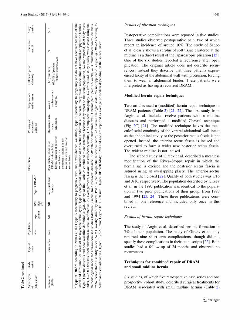
Four studies used a laparoscopic plication technique
(Fig. 2A). All laparoscopic studies used mesh reinforce-
ment, of which three used interrupted sutures, and one used
a continuous suture. The study of Palanivelu et al.
describes a technique in which they place interrupted
sutures which run in and out of the widened linea alba
several times, causing the midline to fold like a ‘Venetian
blind’ when the sutures are tied [19]. The difference in
suture technique did not lead to a difference in recurrence
rate, which were 0% in all the laparoscopic repair groups
after a follow-up ranging from 6 to 24 months.
Three studies used an open technique, and all plicated a
different layer of the ventral abdominal wall (Fig. 2B)
[16–18]. The study of Nahas et al. included two female
patients with a recurrent DRAM, and is the only study in
this section that did not use mesh reinforcement. They
describe two type C (congenital lateral implantation of the
rectus abdominis muscle) DRAM patients that had under-
gone plication of the anterior rectus fascia during
abdominoplasty surgery and were operated using an open
plication technique of the posterior rectus fascia [18]. The
anterior rectus fascia was then sutured to the midline, to
mimic the anatomic situation. The other two ‘open’ studies
described either a plication technique of the anterior rectus
fascia or solely the widened linea alba, combined with a
sublay mesh.
Fig. 1 Flow of trials through
review. PRISMA flowchart of
study selection
Surg Endosc (2017) 31:4934–4949 4937
123
Ta
ble
2E
evid
ence
tab
lesu
rger
y
Au
tho
r
(yea
ro
f
pu
bli
cati
on
)
Stu
dy
per
iod
Ty
pe
of
stu
dy
Po
pu
lati
on
Inte
rven
tio
nP
rim
ary
and
seco
nd
ary
ou
tco
me
Co
mp
lica
tio
ns
and
/or
resu
lts
Fo
llo
w-u
pp
erio
d
(Met
ho
d)
Rec
urr
ence
rate
%
Stu
dy
qu
alit
yN=
…A
ge
(yea
rs)
BM
I
(Kg
/
m2)
Ty
pe
of
DR
AM
*
Pli
cati
on
Shir
ah,
B.H
.
(2016)
2004–2013
Cas
ese
ries
216
40.9
26.4
NR
(mea
nIR
D
10
cm)
Open
repai
rw
ith
pli
cati
on
of
post
erio
rre
ctus
fasc
iaan
d
subla
ypoly
pro
pyle
ne
mes
h
(n=
179).
Lap
arosc
opic
repai
rw
ith
mid
line
pli
cati
on
wit
h
conti
nuous
sutu
rean
din
tra-
abdom
inal
poly
pro
pyle
ne
mes
h(n
=37).
Rec
urr
ence
rate
,
post
op.
com
pli
cati
ons,
abdom
inal
wal
l
funct
ion,
cosm
etic
outc
om
e
Open
:
Wound
infe
ctio
n
(n=
11)
6.1
%
Ser
om
a(n
=9)
5%
Hem
atom
a
(n=
5)
2.8
%
Cosm
etic
(exc/
good
95,6
%,
unsa
tis.
4,4
%)
Lap
arosc
opic
:
Pai
n(n
=4)
10.8
%
Cosm
etic
(exc/
good
91,2
%,
unsa
tis.
8,1
%))
24
month
s(C
T?
clin
ical
exam
inat
ion)
0%
7/1
6
Sah
oo,
M.R
.
(2014)
NR
Cas
ese
ries
335–45
NR
A,
BL
apar
osc
opic
mid
line
pli
cati
on
wit
hin
terr
upte
d
sutu
res
and
intr
a-ab
dom
inal
mes
hre
info
rcem
ent.
Rec
urr
ence
rate
,
post
op.
com
pli
cati
ons
Pai
nan
dti
ghtn
ess
of
abdom
en
obse
rved
(no
spec
ifica
tion),
whic
hdec
reas
ed
duri
ng
foll
ow
-
up.
12
month
s(N
R_
0%
5/1
6
Sid
dik
y,
A.H
.
(2010)
NR
Cas
ere
port
138
NR
BL
apar
osc
opic
mid
line
pli
cati
on
wit
hin
terr
upte
d
mat
tres
ssu
ture
sw
ithout
mes
hre
info
rcem
ent.
Rec
urr
ence
rate
,
post
op.
Com
pli
cati
ons
Post
oper
ativ
epai
n
and
ileu
s
des
crib
ed,
del
ayed
dis
char
ge
afte
r
5day
s.
8w
eeks
(NR
)0%
4/1
6
Pal
aniv
elu,
C.
(2009)
1998–2007
Cas
ese
ries
18
42
28.2
A,
B,
DL
apar
osc
opic
mid
line
pli
cati
on
wit
hin
terr
upte
d
ven
etia
nbli
nds
sutu
res
and
mes
hre
info
rcem
ent.
Rec
urr
ence
rate
,
post
op.
com
pli
cati
ons
Pai
n(n
=2)
11.1
%
Pneu
monia
(n=
1)
5.5
%
Chro
nic
pai
n(n
=2)
11.1
%
6–48
month
s(C
T)
0%
7/1
6
Nah
as,
F.X
.
(2004)
NR
Cas
ere
port
238
&59
NR
CO
pen
mid
line
pli
cati
on
of
the
post
erio
rre
ctus
shea
thw
ith
inte
rrupte
dsu
ture
s,an
d
anch
ori
ng
of
the
ante
rior
rect
us
fasc
iato
the
post
erio
r
rect
us
fasc
iain
the
mid
line.
Rec
urr
ence
rate
,
post
op.
com
pli
cati
ons
Unev
entf
ul
reco
ver
yan
d
sati
sfac
tory
cosm
etic
resu
lts.
Cas
e1:
30
month
s(C
T)
Cas
e2:
6m
onth
s(C
T)
0%
4/1
6
4938 Surg Endosc (2017) 31:4934–4949
123
Ta
ble
2co
nti
nu
ed
Auth
or
(yea
r
of
publi
cati
on)
Stu
dy
per
iod
Type
of
study
Popula
tion
Inte
rven
tion
Pri
mar
yan
d
seco
ndar
y
outc
om
e
Com
pli
cati
ons
and/o
rre
sult
s
Foll
ow
-up
per
iod
(Met
hod)
Rec
urr
ence
rate
%
Stu
dy
qual
ity
N=
…A
ge
(yea
rs)
BM
I
(Kg/
m2)
Type
of
DR
AM
*
Der
yugin
a,
M.S
.(2
001)
NR
Cas
ese
ries
73
45.9
NR
NR
Open
repai
rw
ith
mid
line
pli
cati
on
of
the
linea
alba
usi
ng
inte
rrupte
dsu
ture
s
mes
hsu
bla
yre
info
rcem
ent
fine-
pore
dw
oven
Lav
san
tape.
Rec
urr
ence
rate
Mis
sing
1–11
yea
rs(N
R)
4%
(n=
3)
4/1
6
Modifi
ed
her
nia
repai
r
met
hod
Angio
,L
.G.
(2007)
NR
Cas
ese
ries
12
43
NR
NR
Open
modifi
edC
hev
rel
tech
niq
ue
wit
hout
ente
ring
the
abdom
inal
cavit
y.
Mid
line
pla
sty
wit
honla
y
mes
hre
info
rcem
ent.
Rec
urr
ence
rate
,
cosm
etic
resu
lt,
post
op.
com
pli
cati
ons
Ser
om
a(n
=3)
7.0
%
24
month
s
(12
month
s=
CT
,18
and
24
month
s=
clin
ical
exam
inat
ion)
0%
8/1
6
Gir
eev,
G.I
.
(1983,
1994,
1997)!
1980–1989
Cas
ese
ries
56
NR
NR
NR
Open
modifi
edR
ives
–S
toppa
repai
rw
ithout
mes
h
rein
forc
emen
t.
Rec
urr
ence
rate
,
work
impai
rmen
t,
pai
n
9sh
ort
-ter
m
com
pli
cati
ons,
no
work
impai
rmen
t
71.4
%,
moder
ate
impai
rmen
t
16%
,an
dse
ver
e
impai
rmen
t0%
.
24
month
s(N
R)
0%
3/1
6
Com
bin
ed(h
ernia
and
DR
AM
)
Pri
vet
t,B
.J.
(2016)
2013-2
015
Cas
ese
ries
58
NR
NR
NR (h
ernia\
4cm
)
Open
repai
rof
DR
AM
wit
h
smal
lum
bil
ical
her
nia
.
Sm
all
um
bil
ical
inci
sion
and
pre
per
itonea
lpla
cem
ent
of
self
-adhes
ive
mes
h.
Rec
urr
ence
rate
,
post
op.
com
pli
cati
ons
No
post
op.
com
pli
cati
ons
NR
(NR
)1.7
%(n
=1)
3/1
6
Ko
cker
ling,
F.
(2016)
2015-2
016
Cas
ese
ries
40
53.6
32.6
NR
EL
AR
plu
sfo
rD
RA
Mw
ith
um
bil
ical
or
epig
astr
ic
her
nia
.E
ndosc
opic
-ass
iste
d
ante
rior
rect
us
fasc
iatu
rn
over
wit
hm
esh
augm
enta
tion
rese
mble
sa
hybri
dver
sion
of
the
modifi
edC
hev
rel
tech
niq
ue
wit
honly
asm
all
um
bil
ical
inci
sion.
Post
op.
com
pli
cati
ons
Um
bil
ical
nec
rosi
s(n
=1)
2.5
%
Impai
red
wound
hea
ling
(n=
1)
2.5
%
Ser
om
a(n
=1)
2.5
%
Inte
rmit
tent
pai
n
on
exer
tion
(n=
3)
7.5
%
NA
NA
4/1
6
Surg Endosc (2017) 31:4934–4949 4939
123

Ta
ble
2co
nti
nu
ed
Auth
or
(yea
r
of
publi
cati
on)
Stu
dy
per
iod
Type
of
study
Popula
tion
Inte
rven
tion
Pri
mar
yan
d
seco
ndar
y
outc
om
e
Com
pli
cati
ons
and/o
rre
sult
s
Foll
ow
-up
per
iod
(Met
hod)
Rec
urr
ence
rate
%
Stu
dy
qual
ity
N=
…A
ge
(yea
rs)
BM
I
(Kg/
m2)
Type
of
DR
AM
*
Bel
lido
L.A
.
(2015)
2011-2
012
Pro
spec
tive
cohort
study
21
37.6
27.4
A,
B,
C,
D
(her
nia
C2
cm)
DR
AM
wit
hum
bil
ical
or
epig
astr
icher
nia
C2
cm.
Endosc
opic
,su
bcu
taneo
us,
mid
line
pli
cati
on
wit
h
V-l
ock
sutu
re,
and
onla
y
mes
hre
info
rcem
ent.
Rec
urr
ence
rate
,
post
op.
com
pli
cati
ons,
cosm
etic
appea
rance
,
pai
n(V
AS
)
Ser
om
ain
supra
pubic
area
(n=
5)
23%
Subcu
taneo
us
emphyse
ma
(n=
2)
9%
20
month
s
(US?
clin
ical
exam
inat
ion)
0%
11/1
6
Mat
ei,
O.A
.
(2014)
2010–2012
Cas
ese
ries
44
60.2
31.2
NR
Open
repai
rw
ith
smal
l
um
bil
ical
her
nia
wit
h
Riv
es–S
toppa
repai
r
com
bin
edw
ith
subla
ym
esh
pla
cem
ent.
Post
op.
com
pli
cati
ons
Min
imal
um
bil
ical
nec
rosi
s(n
=1)
NR
NA
3/1
6
Yura
sov,
A.V
.
(2014)
2006–2013
Cas
ese
ries
374
deg
ree
I:
n=
174
deg
ree
II:
n=
162
deg
ree
III:
n=
38
NR
NR
NR
Open
Riv
es–S
toppa
like
repai
r
for
DR
AM
wit
hum
bil
ical
her
nia
wit
hsu
bla
ym
esh
pla
cem
ent
conti
nuous
sutu
ring
of
the
post
erio
ran
d
ante
rior
rect
us
fasc
ia.
The
abdom
inal
cavit
yis
not
open
ed.
Post
op.
com
pli
cati
ons
gro
up
I:
com
pli
cati
on
rate
2.5
%
(Hem
atom
aof
subcu
taneo
us
fatn=
1(1
.2%
)
Ileu
sn=
1
(1.2
%))
gro
up
II:
com
pli
cati
on
rate
5.4
%(W
ound
infe
ctio
nn=
1
(1.3
%)
Hem
atom
aof
subcu
taneo
us
fat
n=
2
(2.7
%)
Ileu
sn=
1
(1.3
%))
gro
up
III:
com
pli
cati
on
rate
5%
(Hem
atom
aof
subcu
taneo
us
fat
n=
1(5
%))
NR
NA
5/1
6
4940 Surg Endosc (2017) 31:4934–4949
123
Results of plication techniques
Postoperative complications were reported in five studies.
Three studies observed postoperative pain, two of which
report an incidence of around 10%. The study of Sahoo
et al. clearly shows a surplus of soft tissue clustered at the
midline as a direct result of the laparoscopic plication [15].
One of the six studies reported a recurrence after open
plication. The original article does not describe recur-
rences, instead they describe that three patients experi-
enced laxity of the abdominal wall with protrusion, forcing
them to wear an abdominal binder. These patients were
interpreted as having a recurrent DRAM.
Modified hernia repair techniques
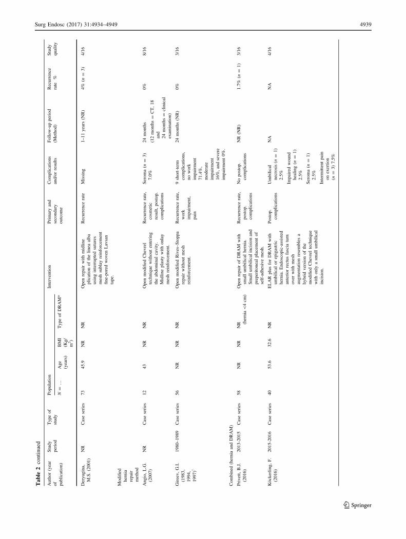
Two articles used a (modified) hernia repair technique in
DRAM patients (Table 2) [21, 22]. The first study from
Angio et al. included twelve patients with a midline
diastasis and performed a modified Chevrel technique
(Fig. 2C) [21]. The modified technique leaves the mus-
culofascial continuity of the ventral abdominal wall intact
as the abdominal cavity or the posterior rectus fascia is not
opened. Instead, the anterior rectus fascia is incised and
overturned to form a wider new posterior rectus fascia.
The wident midline is not incised.
The second study of Gireev et al. described a meshless
modification of the Rives–Stoppa repair in which the
hernia sac is excised and the posterior rectus fascia is
sutured using an overlapping plasty. The anterior rectus
fascia is then closed [22]. Quality of both studies was 8/16
and 3/16, respectively. The population described by Gireev
et al. in the 1997 publication was identical to the popula-
tion in two prior publications of their group, from 1983
and 1994 [23, 24]. These three publications were com-
bined in one reference and included only once in this
review.
Results of hernia repair techniques
The study of Angio et al. described seroma formation in
7% of their population. The study of Gireev et al. only
reported nine short-term complications, though did not
specify these complications in their manuscripts [22]. Both
studies had a follow-up of 24 months and observed no
recurrences.
Techniques for combined repair of DRAM
and small midline hernia
Six studies, of which five retrospective case series and one
prospective cohort study, described surgical treatments for
DRAM associated with small midline hernias (Table 2)Ta
ble
2co
nti
nu
ed
Auth
or
(yea
r
of
publi
cati
on)
Stu
dy
per
iod
Type
of
study
Popula
tion
Inte
rven
tion
Pri
mar
yan
d
seco
ndar
y
outc
om
e
Com
pli
cati
ons
and/o
rre
sult
s
Foll
ow
-up
per
iod
(Met
hod)
Rec
urr
ence
rate
%
Stu
dy
qual
ity
N=
…A
ge
(yea
rs)
BM
I
(Kg/
m2)
Type
of
DR
AM
*
Ran
ney
,B
.
(1990)
NR
Cas
ese
ries
673
NR
NR
A,
B(u
mbil
ical
her
nia
)
Open
mid
line
pli
cati
on
of
DR
AM
wit
hum
bil
ical
her
nia
.P
lica
tion
of
post
erio
r
rect
us
fasc
ia,
and
conti
nuous
sutu
ring
of
the
rect
us
musc
les
and
ante
rior
rect
us
fasc
ia.
Rec
urr
ence
rate
,
wound
deh
isce
nce
Wound
deh
isce
nce
not
obse
rved
.
14.8
yea
rs(a
v)
67.2
8%
of
pat
ients
(cli
nic
alex
amin
atio
n)
0%
5/1
6
*T
yp
eo
fD
RA
Mac
cord
ing
toN
ahas
etal
.;(T
yp
eA
(sec
on
dar
yto
pre
gn
ancy
wit
han
da
wel
l-d
efin
edw
aist
lin
e),
Ty
pe
B(s
eco
nd
ary
top
reg
nan
cyan
dd
on
ot
hav
ead
equ
ate
ten
sio
no
fth
e
late
ral
and
infr
a-u
mb
ilic
alar
eas
of
the
my
oap
neu
roti
cla
yer
),T
yp
eC
(co
ng
enit
alla
tera
lin
sert
ion
of
the
rect
us
abd
om
inis
atth
eco
stal
mar
gin
san
das
soci
atio
no
fu
mb
ilic
alo
rep
igas
tric
her
nia
),
Ty
pe
D(r
ectu
sd
iast
asis
and
po
or
wai
stli
ne
defi
nit
ion
));QoL
Qu
alit
y-o
f-li
fe,
!ar
ticl
esco
mb
ined
too
ne
refe
ren
ceb
ecau
seth
eyre
po
rto
nth
esa
me
po
pu
lati
on
,NR
no
tre
po
rted
,BMI
bo
dy
mas
s
ind
ex,DRAM
Dia
stas
isR
ecti
abd
om
inis
mu
scle
,Med
med
ian
;Av
aver
age/
mea
n,Unsatis.
un
sati
sfac
tory
resu
lts,CT
com
pu
ted
tom
og
rap
hy
.USultrasound,
stu
dy
qu
alit
yw
asas
sess
edu
sin
gth
e
met
ho
do
log
ical
ind
exfo
rn
on
-ran
do
mis
edst
ud
ies
(MIN
OR
S)
sco
re,IRD
inte
r-re
cti
dis
tan
ce,AAW
ante
rio
rab
do
min
alw
all,
Ch
ron
icp
ain
:p
ain[
6w
eek
s,RCT
ran
do
mis
edco
ntr
oll
edtr
ials
,
PFDI
pel
vic
flo
or
dis
abil
ity
ind
ex,ODI
Osw
estr
yd
isab
ilit
yin
dex
,P
SF
S=
pat
ien
t-sp
ecifi
cfu
nct
ion
alsc
ore
,VAS
vis
ual
anal
og
ue
scal
e,NA
no
tap
pli
cab
le.DegreeofDRAM
acco
rdin
gto
Ask
erh
ano
vcl
assi
fica
tio
n(D
egre
eI:
22
–5
0m
m;
Deg
ree
II:
51
–8
0m
m;
Deg
ree
III:[
80
MM
);B
MI
and
age
are
rep
ort
edas
aver
age
or
med
ian
dep
end
ing
on
the
sou
rce
arti
cle
Surg Endosc (2017) 31:4934–4949 4941
123
[6, 25–29]. The total number of patients in this section was
1.210. Quality of the included studies was low-to-moderate
as one studies scored 11/16 and the remaining MINORS
scores ranged from 3 to 5.
Two studies included both umbilical and epigastric
hernias. Four studies included small umbilical hernias.
Hernia repair in five out of six studies consisted of suture
closure of the hernia sac combined with mesh reinforce-
ment. The oldest study (1990), of Ranney et al., describes a
modified Rives–Stoppa repair without mesh reinforcement.
Four studies described open techniques, mostly resembling
either modifed Chevrel or Rives–Stoppa procedures
(Fig. 2C, D respectively). One described an endoscopic
procedure in the anatomical plane between the subcuta-
neous fat and the anterior rectus fascia, with plication of
the midline and onlay mesh reinforcement. One described a
hybrid version in the same plane, which resembled a
modified Chevrel repair, performed partially endoscopic,
with onlay mesh reinforcement.
Results of combined repair DRAM and midline hernia
Five of the six studies reported on postoperative compli-
cations [6, 25–28]. Two of these studies encountered no
postoperative complications, and the remaining three
encountered only minor (Clavien-Dindo I–II) postoperative
complications. The majority of minor complications were
seromas, with an incidence ranging from 2.5%, reported by
Kockerling et al. to 23%, reported by Bellido et al. [6, 26].
Follow-up was only reported in two studies and ranged
from 20 months to 14.8 years. Only one recurrence was
observed during the follow-up in the study of Privett et al.
in a patient with a combined DRAM and umbilical hernia
[28]. They mentioned that open repair with preperitoneal
placement of mesh without approximation of the rectus
fascia (resembling a bridged repair) leads to fluctuating
cosmetic results, since protrusion of DRAM may still be
present after mesh placement.
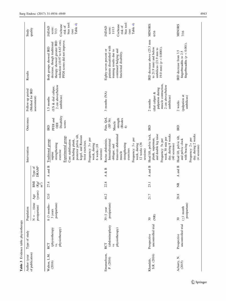
Physiotherapy
Six studies evaluated the effect of a physiotherapy inter-
vention on IRD in a total of one hundred postpartum
women. Two RCTs, two prospective uncontrolled trials,
and two case reports were included in this section (Table 3)
[5, 30–34]. All studies focussed on females at different
postpartum intervals, ranging from one month to three
years. Scientific quality of the included studies was mod-
erate with MINORS scores ranging from six to nine for the
non-randomised studies, and a Jadad score of 7–11 for the
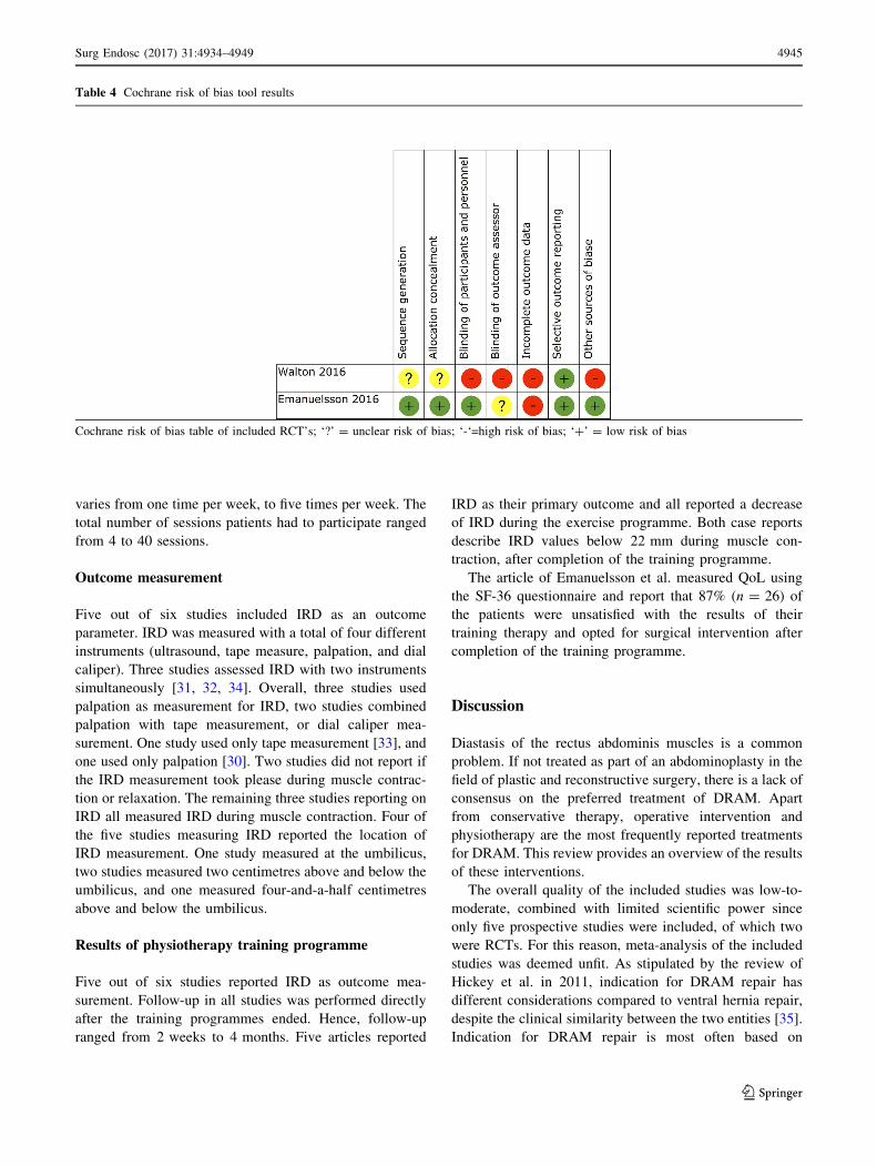
included RCTs. See Table 4 for results of The Cochrane
risk of bias tool.
Training programme
One case study used a single exercise (prone kneeling) to
train the patients [33]. Two studies used general (not fur-
ther specified) strengthening exercises for the abdominal
wall, hip, and trunk muscles [5, 32]. The remaining three
studies used the head lift exercise, combined with pelvic
lock or pelvic tilt exercises [30, 31, 34].
Frequency of the training programme varied between
the studies. One study let patients train on their own and
the frequency of the exercise during the training pro-
gramme is not reported [33]. The remaining studies used
counselling of a physiotherapist to train patients in allo-
cated training sessions. Frequency of the programmes
Fig. 2 Illustrations of surgical
interventions. Four main
surgical interventions for
treating DRAM. A laparoscopic
plication of the entire midline
with mesh reinforcement,
performed in 59 patients;
B open plication of the posterior
rectus fascia, performed in 254
patients; C modified Chevrel
repair, performed in 52 patients;
D Rives-Stoppa like repair with
or without mesh reinforcement,
performed in 948 patients
4942 Surg Endosc (2017) 31:4934–4949
123
Ta
ble
3E
vid
ence
tab
lep
hy
sio
ther
apy
Au
tho
r(y
ear
of
pu
bli
cati
on
)
Ty
pe
of
stu
dy
Po
pu
lati
on
Inte
rven
tio
nO
utc
om
esF
oll
ow
-up
per
iod
(Met
ho
dfo
rIR
D
asse
ssm
ent)
Res
ult
sS
tud
y
qu
alit
yN
=…
(tim
e
po
stp
artu
m)
Ag
e
(yea
rs)
BM
I
(Kg
/
m2)
Ty
pe
of
DR
AM
*
Wal
ton
,L
.M.
(20
16
)
RC
T
(ph
ysi
oth
erap
y
vs.
ph
ysi
oth
erap
y)
8(3
mo
nth
s–
3y
ears
po
stp
artu
m)
32
.02
7.4
Aan
dB
Tra
dit
ion
alg
rou
p:
sup
ine
stre
ng
then
ing
exer
cise
Ex
per
imen
tal
gro
up
:
Co
rest
abil
isat
ion
incl
ud
ing
pla
nk
,
po
ster
ior
pel
vic
tilt
,
keg
el,
and
Ru
ssia
n
twis
tex
erci
ses.
IRD
PF
DI
and
OD
I
Dis
abil
ity
sco
res
1.5
mo
nth
s
(US
&d
ial
cali
per
,
2cm
abo
ve/
bel
ow
um
bil
icu
s)
Bo
thg
rou
ps
sho
wed
IRD
dec
reas
e,th
ou
gh
trad
itio
nal
ther
apy
sho
wed
gre
ater
IRD
red
uct
ion
(10
.97
to6
.63
cm),
PF
DI
sco
res
did
no
tim
pro
ve.
JAD
AD
sco
re:
7/1
3
Co
chra
ne
risk
of
bia
sto
ol:
(see
Tab
le4
)
Fre
qu
ency
:39
per
wee
k,
du
rin
g
6w
eek
s(1
8
sess
ion
s)
Em
anu
elss
on
,
P.
(20
16
)
RC
T
(ab
do
min
op
last
y
vs
ph
ysi
oth
erap
y)
30
(1y
ear
po
stp
artu
m)
44
.22
2.8
A&
BR
ectu
sab
do
min
is,
Inte
rnal
/ex
tern
al
ob
liq
ue,
and
tran
sver
sab
do
min
al
mu
scle
stre
ng
then
ing
exer
cise
s
Pai
n,
Qo
L
(SF
-36
)
Mu
scle
stre
ng
th
(Bio
dex
4)
3m
on
ths
(NA
)E
igh
ty-s
even
per
cen
to
r
pat
ien
tsd
issa
tisfi
edw
ith
trai
nin
gre
sult
sd
ue
to
con
tin
ued
bu
lgin
gan
d
fun
ctio
nal
dis
abil
ity
.
JAD
AD
sco
re:
11
/13
Co
chra
ne
risk
of
bia
sto
ol:
(see
Tab
le4
)F
req
uen
cy:
39
per
wee
k,
du
rin
g
12
wee
ks
(36
sess
ion
s)
Kh
and
ale,
S.R
.(2
01
6)
Pro
spec
tiv
e
un
con
tro
lled
tria
l
30
(NR
)
21
.72
3.1
Aan
dB
Hea
dli
ft,
pel
vic
lock
,
pla
nk
,su
per
man
,
and
do
ub
lele
gra
ise
IRD
2m
on
ths
(dia
lca
lip
er&
pal
pat
ion
du
rin
g
mu
scle
con
trac
tio
n,
2cm
abo
ve/
bel
ow
um
bil
icu
s)
IRD
dec
reas
eab
ov
e(2
5.3
mm
to2
1.9
mm
)an
db
elo
w
um
bil
icu
s(2
1.9
mm
to
19
.0m
m)
(p\
0.0
00
1).
MIN
OR
S:
6/1
6
Fre
qu
ency
:5
xp
er
wee
k,
30
min
per
day
,d
uri
ng
8w
eek
s
(40
sess
ion
s)
Ach
arry
,N
.
(20
15
)
Pro
spec
tiv
e
un
con
tro
lled
tria
l
30
(B1
mo
nth
po
stp
artu
m)
28
.8N
RA
and
BH
ead
lift
,p
elv
icti
lt,
and
pel
vic
clo
ck
wit
hb
raci
ng
IRD
2w
eek
s
(pal
pat
ion
at
um
bil
icu
s)
IRD
dec
reas
efr
om
3.5
fin
ger
bre
adth
sto
2.5
fin
ger
bre
adth
s(p\
0.0
01
).
MIN
OR
S
7/1
6
Fre
qu
ency
:29
per
day
,d
uri
ng
2w
eek
s
(4se
ssio
ns)
Surg Endosc (2017) 31:4934–4949 4943
123
Ta
ble
3co
nti
nu
ed
Au
tho
r(y
ear
of
pu
bli
cati
on
)
Ty
pe
of
stu
dy
Po
pu
lati
on
Inte
rven
tio
nO
utc
om
esF
oll
ow
-up
per
iod
(Met
ho
dfo
rIR
D
asse
ssm
ent)
Res
ult
sS
tud
y
qu
alit
yN
=…
(tim
e
po
stp
artu
m)
Ag
e
(yea
rs)
BM
I
(Kg
/
m2)
Ty
pe
of
DR
AM
*
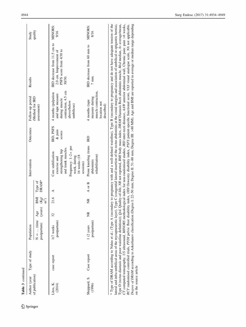
Lit
os,
K.
(20
14
)
case
rep
ort
1(7
wee
ks
po
stp
artu
m)
32
21
.6A
Co
rest
abil
isat
ion
exer
cise
and
stre
ng
then
ing
hip
and
tru
nk
mu
scle
s.
IRD
,P
SF
S
&p
ain
sco
res
4m
on
ths
(pal
pat
ion
and
tap
em
easu
re
du
rin
gm
usc
le
con
trac
tio
n,
4.5
cm
abo
ve/
bel
ow
um
bil
icu
s)
IRD
dec
reas
efr
om
11
.5cm
to
2.0
cm.
Imp
rov
emen
to
f
PS
FS
sco
refr
om
4/3
0to
30
/30
.
MIN
OR
S:
9/1
6
Fre
qu
ency
:1
–29
per
wee
k,
du
rin
g
16
wee
ks
(18
sess
ion
s)
Sh
epp
ard
,S
.
(19
96
)
Cas
ere
po
rt1
(2y
ears
po
stp
artu
m)
NR
NR
Ao
rB
Pro
ne
kn
eeli
ng
(tra
ns
abd
om
inis
reh
abil
itat
ion
)
IRD
4m
on
ths
(tap
e
mea
sure
du
rin
g
mu
scle
con
trac
tio
n,
loca
tio
nn
ot
des
crib
ed)
IRD
dec
reas
efr
om
60
mm
to
7m
m.
MIN
OR
S:
8/1
6
*T
yp
eo
fD
RA
Mac
cord
ing
toN
ahas
etal
.;(T
yp
eA
(sec
on
dar
yto
pre
gn
ancy
wit
han
da
wel
l-d
efin
edw
aist
lin
e),
Ty
pe
B(s
eco
nd
ary
top
reg
nan
cyan
dd
on
ot
hav
ead
equ
ate
ten
sio
no
fth
e
late
ral
and
infr
a-u
mb
ilic
alar
eas
of
the
my
oap
neu
roti
cla
yer
),T
yp
eC
(co
ng
enit
alla
tera
lin
sert
ion
of
the
rect
us
abd
om
inis
atth
eco
stal
mar
gin
san
das
soci
atio
no
fu
mb
ilic
alo
rep
igas
tric
her
nia
),
Ty
pe
D(r
ectu
sd
iast
asis
and
po
or
wai
stli
ne
defi
nit
ion
));QoL
Qu
alit
y-o
f-li
fe,NR
no
tre
po
rted
,BMI
bo
dy
mas
sin
dex
,DRAM
Dia
stas
isR
ecti
abd
om
inis
mu
scle
,Med
med
ian
,Av
aver
age/
mea
n,
CT
com
pu
ted
tom
og
rap
hy
.US
ult
raso
un
d,MINORS
met
ho
do
log
ical
ind
exfo
rn
on
-ran
do
mis
edst
ud
ies,IRD
inte
r-re
cti
dis
tan
ce,AAW
ante
rio
rab
do
min
alw
all,
Ch
ron
icp
ain
:p
ain[
6w
eek
s,
RCT
ran
do
mis
edco
ntr
oll
edtr
ials
,PFDI
pel
vic
flo
or
dis
abil
ity
ind
ex,ODI
Osw
estr
yd
isab
ilit
yin
dex
,PSFS
pat
ien
t-sp
ecifi
cfu
nct
ion
alsc
ore
,VAS
vis
ual
anal
og
ue
scal
e,NA
no
tap
pli
cab
le.
DegreeofDRAM
acco
rdin
gto
Ask
erh
ano
vcl
assi
fica
tio
n(D
egre
eI:
22
–5
0m
m;
Deg
ree
II:
51
–8
0m
m;
Deg
ree
III:[
80
MM
).A
ge
and
BM
Iar
ere
po
rted
inav
erag
eo
rm
edia
nra
ng
ed
epen
din
g
on
the
sou
rce
arti
cle
4944 Surg Endosc (2017) 31:4934–4949
123
varies from one time per week, to five times per week. The
total number of sessions patients had to participate ranged
from 4 to 40 sessions.
Outcome measurement
Five out of six studies included IRD as an outcome
parameter. IRD was measured with a total of four different
instruments (ultrasound, tape measure, palpation, and dial
caliper). Three studies assessed IRD with two instruments
simultaneously [31, 32, 34]. Overall, three studies used
palpation as measurement for IRD, two studies combined
palpation with tape measurement, or dial caliper mea-
surement. One study used only tape measurement [33], and
one used only palpation [30]. Two studies did not report if
the IRD measurement took please during muscle contrac-
tion or relaxation. The remaining three studies reporting on
IRD all measured IRD during muscle contraction. Four of
the five studies measuring IRD reported the location of
IRD measurement. One study measured at the umbilicus,
two studies measured two centimetres above and below the
umbilicus, and one measured four-and-a-half centimetres
above and below the umbilicus.
Results of physiotherapy training programme
Five out of six studies reported IRD as outcome mea-
surement. Follow-up in all studies was performed directly
after the training programmes ended. Hence, follow-up
ranged from 2 weeks to 4 months. Five articles reported
IRD as their primary outcome and all reported a decrease
of IRD during the exercise programme. Both case reports
describe IRD values below 22 mm during muscle con-
traction, after completion of the training programme.
The article of Emanuelsson et al. measured QoL using
the SF-36 questionnaire and report that 87% (n = 26) of
the patients were unsatisfied with the results of their
training therapy and opted for surgical intervention after
completion of the training programme.
Discussion
Diastasis of the rectus abdominis muscles is a common
problem. If not treated as part of an abdominoplasty in the
field of plastic and reconstructive surgery, there is a lack of
consensus on the preferred treatment of DRAM. Apart
from conservative therapy, operative intervention and
physiotherapy are the most frequently reported treatments
for DRAM. This review provides an overview of the results
of these interventions.
The overall quality of the included studies was low-to-
moderate, combined with limited scientific power since
only five prospective studies were included, of which two
were RCTs. For this reason, meta-analysis of the included
studies was deemed unfit. As stipulated by the review of
Hickey et al. in 2011, indication for DRAM repair has
different considerations compared to ventral hernia repair,
despite the clinical similarity between the two entities [35].
Indication for DRAM repair is most often based on
Table 4 Cochrane risk of bias tool results
Cochrane risk of bias table of included RCT’s; ‘?’ = unclear risk of bias; ‘-‘=high risk of bias; ‘?’ = low risk of bias
Surg Endosc (2017) 31:4934–4949 4945
123
cosmetic or functional impairment, as DRAM poses no risk
of strangulation. Therefore, the cosmetic results of a sur-
gical technique or physiotherapy training programme,
along with other patient reported outcomes (PROs), should
be an important outcome of scientific studies. Remarkably,
cosmetic outcome was only included in one study, and
measured subjectively with an instrument that was not
validated. Other PROs were not measured in surgical
publications and only reported twice in physiotherapy
studies.
Surgical technique
Based on the published literature, the surgical techniques
available for DRAM repair are either plication-based or
hernia repair-based. The plication-based techniques include
open plication, laparoscopic plication, or hybrid plication
of either the anterior or posterior rectus fascia. Based on
the results of this review, there is no clear difference in
postoperative complications between these methods.
Nearly all studies that described a plication technique used
interrupted sutures and mesh reinforcement, which could
account for the low recurrence rates, though comparative
data are not available. The plication techniques can leave a
surplus of skin directly after surgery, as described in the
study of Sahoo et al., though due to the lack of cosmetic
outcome measurement no evidence-based statements
regarding the cosmetic postoperative appearance can be
made [15].
Hernia repair techniques can be used for DRAM treat-
ment. The musculofascial continuity of the ventral
abdominal wall is an important anatomical structure to be
considered during DRAM repair. If the midline is incised
and the continuity is disturbed, the risk of incisional hernia
formation and subsequent risk of strangulation will become
larger, though alignment of the rectus muscles could be
easier. The current evidence is of insufficient quality to
detect differences between techniques that preserve the
musculofascial continuity versus techniques that incise the
midline. Hernia-based techniques for DRAM repair are
often modifications of the original Chevrel or Rives–
Stoppa techniques [21, 36, 37].
An important reason for DRAM patients to seek medical
attention is the cosmetic impairment they experience.
Despite the importance of cosmetic results, the majority
(85% of patients) of published literature in general surgery
for DRAM concerns open procedures. Recently developed
hybrid techniques such as the ELAR plus described by
Kockerling et al., the endoscopic midline plication by
Bellido et al., or the eMILOS by Reinpold et al. could
increase the number of minimally invasive procedures in
the DRAM population [6, 26, 38]. These procedures are
promising variations of classic (open) hernia repair
techniques that respect the anatomical myofascial conti-
nuity of the ventral abdominal wall, and leave only mini-
mal scarring, without risk of incisional hernia formation
because the abdominal cavity is not opened. Given the
recent invention of these techniques, the amount of pub-
lished data is limited and with short follow-up. The study
of Reinpold et al. unfortunately had to be excluded since
the article did not describe critical data about the study
methodology or the population [38]. Nevertheless, the
technique seems to be promising for DRAM treatment.
It was not possible to isolate any difference in outcome
between male and female patients of the included studies
due to quality and reporting limitations of the included
studies. It is the author’s opinion that the pathophysiology
between males and females, or between type A/B and type
C/D DRAM is different. The A/B type DRAM may be
based on a physiological response during pregnancy, when
collagen is remodelled under the influence of the hormone
‘relaxin’ to allow widening or strentching of the midline
that is not corrected properly after pregnancy [39, 40]. In
males, or type C/D DRAM, genetic predisposition or
altered collagen 1:3 ratio’s may have a more pronounced
role.
Physiotherapy
The literature regarding physiotherapy interventions is
heterogeneous in nature and of low quality. The type of
exercises used to reduce IRD, the frequency of the exer-
cises, the total number of sessions within a training pro-
gramme, and the instruments used to asses IRD vary
greatly amongst the included studies. For instance, the case
study of Litos et al. informed their patient to avoid
abdominal exercises that could increase IRD by recruit-
ment of the transverse abdominal muscles, such as sit-ups,
crunches, and rotational trunk exercises, whilst other
studies from Ramesh et al. and Walton et al. target
specifically the transverse abdominal muscles with these
exercises to reduce IRD [31, 32, 34]. The included studies
only report on postpartum women (type A, B), making
translation of the results to men and type C and D DRAM
difficult, if not impossible.
Brauman et al. has investigated the clinical anatomy of
DRAM and reported that DRAM is not only associated
with a gradual thinning and stretching of the linea alba, but
also by a laxity of the ventral abdominal musculature [1].
Considering Brauman’s findings, physiotherapy could play
a role in treating the laxity of the ventral abdominal
musculature.
Despite the potential benefits of physiotherapy, current
literature does not describe the successful treatment of
rectus diastasis nor a reduction of IRD measured in a
relaxed state. Since diastasis rectus is defined as a
4946 Surg Endosc (2017) 31:4934–4949
123
separation of the rectus abdominis muscles as measured in
a relaxed state, we must conclude that the currently
available evidence does not describe the successful treat-
ment of rectus diastasis after a physiotherapy training
programme. Physiotherapy was able to moderately reduce
IRD during muscle contraction. The impact of these results
on quality-of-life or functional outcome is currently
unknown, as is the sustainability of these results after a
follow-up exceeding four months. Based on the study of
Emanuelsson et al., physiotherapy alone is unlikely to lead
to satisfying functional and cosmetic results [5]. Ema-
nuelsson et al. compared DRAM patients treated with a
physiotherapy training programme with patients whom
received abdominoplasty for DRAM in a randomised
controlled trial. Eighty-one percent of the patients in the
training group were unsatisfied with their functional and
cosmetic appearance at the end of the programme and
opted for surgical intervention after the trial ended.
Physiotherapy could be an alternative to surgery for
patients who are unable or reluctant to undergo surgical
intervention. Surgical treatment only corrects the widening
of the linea alba and will not influence the general laxity of
the ventral abdominal wall. Therefore, physiotherapy could
be a useful addition to surgical intervention for DRAM, to
achieve satisfying functional outcome.
limitations
Most of the included studies were performed in postpartum
women (rectus diastasis type A and B), reducing the trans-
latability to men and rectus diastasis type C and D. Most of
the included studies were retrospective and non-comparative
in nature, reducing the scientific power of the review. The
quality of the included studies according to the MINORS
criteria was low; this is in part due to the ‘how-to-do-it’ type
of publication which describes surgical techniques in sci-
entific journals. These articles only include a small popula-
tion, limiting the description of the randomisation, inclusion,
end point, and blinding methods. Moreover, the MINORS
score is sensitive for retrospective studies, as a retrospective
study will automatically lose four out of sixteen points. Due
to the low quality of the included studies, gender differences
could not be isolated from the included population. Despite
the above-mentioned limitations some general recommen-
dations and conclusions can be drawn from this review.
Considerations for DRAM treatment
DRAM is not a hernia
The continuity of the myofascial anatomy in the ventral
abdominal wall is what sets DRAM apart from an
abdominal wall hernia. There are endoscopic, hybrid, and
open techniques available that leave the anatomical
myofascial continuity intact, and could potentially protect
the DRAM patient from the risk of incisional hernia for-
mation in case of a failed repair. Whether these techniques
have any cosmetic or functional advantage over traditional
hernia repair techniques, and if there is indeed no risk of
incisional hernia formation, is currently unclear.
Align and use mesh
For the repair of DRAM (without midline hernia), plication
techniques with mesh reinforcement and interrupted
sutures are most frequently used to reconstruct the ventral
abdominal wall. Only using mesh reinforcement without an
approximation of the rectus fascia of some sort, may lead
to unsatisfying cosmetic results. Other minimally invasive,
hybrid, or open techniques are promising and can be used,
though long-term results and comparative controlled data
are not available.
Evaluate what is important for the patient
The current body of evidence focuses primarily on recur-
rence rates. It is well known that the risk of recurrence is
not the most important variable for the patient. Instead,
PROs such as postoperative pain, cosmetic outcome, and
functional result are variables that directly concern the
patients’ wellbeing. The use of PROs is low in both hernia-
and DRAM-related research, and should be increased
during the coming years [41].
Cosmetic outcome is important
Cosmetic impairment is an important factor for DRAM
patients to seek medical attention. Therefore, cosmetic
result of a surgical intervention is of high importance in the
DRAM population. Which surgical procedure (endoscopic,
hybrid, laparoscopic, or open) has the most satisfying
cosmetic outcome is not yet evaluated.
Consider physiotherapy
Physiotherapy is unlikely to completely treat DRAM, since
cases in which IRD was reduced to normal during a relaxed
state are currently not described in literature. However, a
moderate reduction in IRD during muscle contraction is
reported. Whether this has an influence on functional out-
comes or quality-of-life is not described. Physiotherapy
combined with surgery could potentially have favourable
results over surgery alone, this combination of treatments is
currently not investigated. Moreover, patients that are
reluctant or unable to undergo surgery may be referred to a
Surg Endosc (2017) 31:4934–4949 4947
123
physiotherapist for strengthening exercises of the abdomi-
nal wall.
Recommendations for future studies
The authors would like to recommend future studies
reporting on the efficacy of any DRAM repair to include
PROs such as cosmetic outcome, quality-of-life, and work
impairment, measured with a validated questionnaire (Eu-
raHS QoL, COMI-Hernia) as their primary outcome, and
IRD or recurrence rate as a secondary outcome. Both
should be measured using an objective tool (dial caliper or
ultrasound), as finger palpation should not be used for
scientific outcome reporting [42]. The location of mea-
surement and the cut-of value should be standardised for
patients’ age, location of measurement, and pre- or post-
partum status according to previously published classifi-
cations [2, 42–44]. IRD should be measured whilst the
rectus abdominis muscles are relaxed, as references values
of normal midline width and DRAM classification
are measured/based on measurements during muscle
relaxation. Based on the pathophysiology described by
Brauman, the combination of physiotherapy and surgical
repair has great theoretical potential to solve both the
anatomical divarication and the laxity of the ventral
abdominal muscles [1]. The authors recommend that future
RCTs focus on the combination of surgery and physio-
therapy for the repair of DRAM.
Conclusion
Published literature on surgical treatments for rectus dias-
tasis is of low scientific and methodological quality. Both
plication-based methods and hernia repair methods are
used for DRAM repair. Based on the current literature, no
clear distinction in recurrence rate, postoperative compli-
cations, or patient reported outcomes can be made. DRAM
is most frequently repaired using plication techniques
combined with mesh reinforcement. Current literature does
not describe the successful treatment of DRAM or a
reduction of IRD in a relaxed state following physiother-
apy. Physiotherapy can achieve a moderate reduction in
IRD during muscle contraction, though it is currently
unclear if this has any positive effect on quality-of-life or
functional outcomes.
Acknowledgements The authors would like to thank Prof. Dr.
G. Beets of the Antoni van Leeuwenhoek Hospital, Amsterdam, The
Netherlands, for his time and effort associated with reviewing the
study protocol in his role as an independent, external, expert reviewer.
Funding Not applicable. No research funder or external grant was
obtained for this manuscript.
Disclosures Drs. E.H.H. Mommers, Drs. J.E.H. Ponten, Ms. A.K. Al-
Omar, Dr. T.S. de Vries Reilingh, Prof. Dr. N.D. Bouvy, and Dr. S.W.
Nienhuijs have no conflict of interest or financial ties to disclose.
Open Access This article is distributed under the terms of the
Creative Commons Attribution 4.0 International License (http://crea
tivecommons.org/licenses/by/4.0/), which permits unrestricted use,
distribution, and reproduction in any medium, provided you give
appropriate credit to the original author(s) and the source, provide a
link to the Creative Commons license, and indicate if changes were
made.
References
1. Brauman D (2008) Diastasis recti: clinical anatomy. Plast
Reconstr Surg 122(5):1564–1569
2. Beer GM et al (2009) The normal width of the linea alba in
nulliparous women. Clin Anat 22(6):706–711
3. Sperstad JB et al (2016) Diastasis recti abdominis during preg-
nancy and 12 months after childbirth: prevalence, risk factors and
report of lumbopelvic pain. Br J Sports Med 50(17):1092–1096
4. Parker MA, Millar LA, Dugan SA (2009) Diastasis rectus
abdominis and lumbo-pelvic pain and dysfunction-are they rela-
ted? J Women’s Health Phys Ther 33(2):15–22
5. Emanuelsson P et al (2016) Operative correction of abdominal
rectus diastasis (ARD) reduces pain and improves abdominal wall
muscle strength: A randomized, prospective trial comparing
retromuscular mesh repair to double-row, self-retaining sutures.
Surgery 160(5):1367–1375
6. Bellido Luque J et al (2015) Totally endoscopic surgery on
diastasis recti associated with midline hernias. The advantages of
a minimally invasive approach. Prospective cohort study. Hernia
19(3):493–501
7. Benjamin DR, van de Water AT, Peiris CL (2014) Effects of
exercise on diastasis of the rectus abdominis muscle in the
antenatal and postnatal periods: a systematic review. Physio-
therapy 100(1):1–8
8. Akram J, Matzen SH (2014) Rectus abdominis diastasis. J Plast
Surg Hand Surg 48(3):163–169
9. Moher D et al (2009) Preferred reporting items for systematic
reviews and meta-analyses: the PRISMA statement. PLoS Med
6(7):e1000097
10. Higgins JPT, Green S (2011) Cochrane Handbook for Systematic
Reviews of Interventions. Green S (ed). Vol. Version 5.1.0.: The
Cochrane Collaboration.
11. Jadad AR et al (1996) Assessing the quality of reports of ran-
domized clinical trials: is blinding necessary? Control Clin Trials
17(1):1–12
12. Higgins JP et al (2011) The Cochrane Collaboration’s tool for
assessing risk of bias in randomised trials. BMJ 343:d5928
13. Slim K et al (2003) Methodological index for non-randomized
studies (minors): development and validation of a new instru-
ment. ANZ J Surg 73(9):712–716
14. Nahas FX (2001) An aesthetic classification of the abdomen
based on the myoaponeurotic layer. Plast Reconstr Surg
108(6):1787–1795; discussion 1796–1797
15. Kumar AT, Sahoo MR (2014) Laparoscopic plication and mesh
repair for diastasis recti: a case series. Int J Case Rep
Images 5(9):610
16. Deriugina MS (2001) A method of surgical treatment of diastasis
recti abdominis. Vestn Khir Im I I Grek 160(3):77–78
17. Shirah BH, Shirah HA (2016) The effectiveness of polypropylene
mesh in the open and laparoscopic repair of divarication of the
recti. Med Imp Surg. doi:10.4172/jims.1000105
4948 Surg Endosc (2017) 31:4934–4949
123

18. Nahas FX, Ferreira LM, Mendes A (2004) Jde, An efficient way
to correct recurrent rectus diastasis. Aesthetic Plast Surg
28(4):189–196
19. Palanivelu C et al (2009) Laparoscopic repair of diastasis recti
using the ‘Venetian blinds’ technique of plication with prosthetic
reinforcement: a retrospective study. Hernia 13(3):287–292
20. Siddiky AH, Kapadia CR (2010) Laparoscopic plication of the
linea alba as a repair for diastasis recti—a mesh free approach.
J Surg Case Rep 2010(5):3
21. Angio LG et al (2007) The surgical treatment of the diastasis recti
abdominis: an original technique of prosthesis repair of the
abdominal wall. G Chir 28(5):187–198
22. Gireev GI, Zagirov UZ, Shakhnazarov AM (1997) Treatment of
linea alba hernia and diastasis of rectus abdominis. Khirurgiia
(Mosk) 7:58–61
23. Gireev GIG (1992) The pathogenesis and surgical treatment of
diastasis recti abdominis. Vestnik khirurgii imeni II. Grekova
148(3):372–375
24. Zagirov UZ, Gireev GI, Shakhnazarov AM (1994) Results of
surgical treatment of diastasis of the rectus abdominis muscles.
Khirurgiia (Mosk) 3:24–27
25. Aiekseev AVYLAADNKMVlAK (2014) Short-term surgical
treatment outcomes of patients with combined umbilical hernia
and diastasis recti. Moscal Surg J 5(39):4
26. Kockerling F et al (2016) Endoscopic-assisted linea alba recon-
struction plus mesh augmentation for treatment of umbilical and/
or epigastric hernias and rectus abdominis diastasis - early results.
Front Surg 3:27
27. Matei OA, Runkel N (2014) A novel technique of midline mesh
repair for umbilical hernia associated with diastasis recti. Surg
Technol Int 24:183–187
28. Privett BJ, Ghusn M (2016) Proposed technique for open repair of
a small umbilical hernia and rectus divarication with self-grip-
ping mesh. Hernia 20(4):527–530
29. Ranney B (1990) Diastasis recti and umbilical hernia causes,
recognition and repair. S D J Med 43(10):5–8
30. Acharry N, Kutty RK (2015) Abdominal exercise with bracing, a
therapeutic efficacy in reducing diastasis-recti among postpartal
females. Int J Physiother Res 3(2):999–1005
31. Khandale SR, Hande D (2016) Effects of abdominal exercises on
reduction of diastasis recti in postnatal women. IJHSR
6(6):182–191
32. Litos K (2014) Progressive therapeutic exercise program for
successful treatment of a postpartum woman with a severe
diastasis recti abdominis. J Women&s Health Phys Ther
38(2):58–73
33. Sheppard S (1996) The role of transversus abdominus in post
partum correction of gross divarication recti. Manual Ther
1(4):214–216
34. Walton LM et al (2016) The effects of a 6 week dynamic core
stability plank exercise program compared to a traditional supine
core stability strengthening program on diastasis recti abdominis
closure, pain, oswestry disability index (ODI) and pelvic floor
disability index scores (PFDI). Phys Ther Rehabil 3(1):3
35. Hickey F, Finch JG, Khanna A (2011) A systematic review on the
outcomes of correction of diastasis of the recti. Hernia
15(6):607–614
36. Stoppa RE (1989) The treatment of complicated groin and inci-
sional hernias. World J Surg 13(5):545–554
37. Chevrel JP (1979) The treatment of large midline incisional
hernias by ‘‘overcoat’’ plasty and prothesis (author’s transl).
Nouv Presse Med 8(9):695–696
38. Reinpold, Endoscopic assisted mini-open transhernial sublay
repair (eMILOS) of symptomatic diastasis recti. Hernia, 2016.
20(1)
39. Samuel CS, Lekgabe ED, Mookerjee I (2007) The effects of
relaxin on extracellular matrix remodeling in health and fibrotic
disease. Adv Exp Med Biol 612:88–103
40. MacLennan AH (1981) Relaxin–a review. Aust N Z J Obstet
Gynaecol 21(4):195–202
41. Antonescu I et al (2014) Outcomes reported in high-impact sur-
gical journals. Br J Surg 101(5):582–589
42. van de Water AT, Benjamin DR (2016) Measurement methods to
assess diastasis of the rectus abdominis muscle (DRAM): a sys-
tematic review of their measurement properties and meta-analytic
reliability generalisation. Man Ther 21:41–53
43. Coldron Y et al (2008) Postpartum characteristics of rectus
abdominis on ultrasound imaging. Manual Ther 13(2):112–121
44. van de Water ATM, Benjamin DR (2016) Measurement methods
to assess diastasis of the rectus abdominis muscle (DRAM): a
systematic review of their measurement properties and meta-an-
alytic reliability generalisation. Manual Ther 21:41–53
Surg Endosc (2017) 31:4934–4949 4949
123