Embed Size (px)
DESCRIPTION
The Eyelids. DR. NAILA ALI ASSISTANT PROFESSOR OPHTHALMOLOGY. The eyelids are movable folds which act as a shutter protecting eye from injury or excessive light. Both the upper and lower eyelids meet at medial and lateral canthi with the opening the papebral fissure between them. - PowerPoint PPT Presentation
Citation preview

The EyelidsThe Eyelids
DR. NAILA ALIDR. NAILA ALIASSISTANT PROFESSORASSISTANT PROFESSOR
OPHTHALMOLOGYOPHTHALMOLOGY
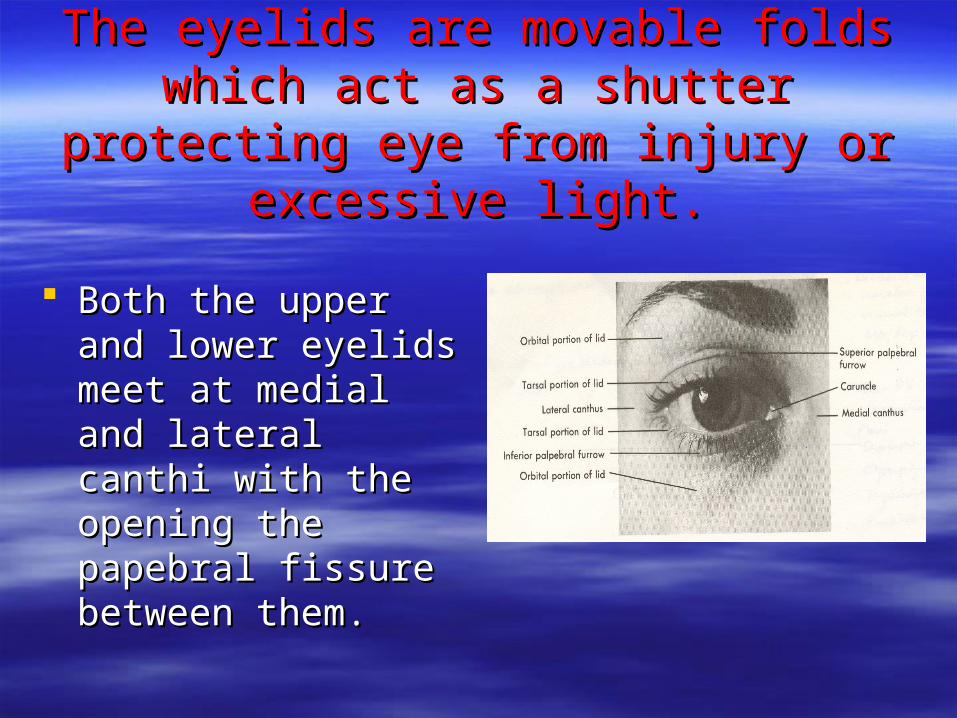
The eyelids are movable folds which act The eyelids are movable folds which act as a shutter protecting eye from injury or as a shutter protecting eye from injury or
excessive light.excessive light.
Both the upper and Both the upper and lower eyelids meet at lower eyelids meet at medial and lateral medial and lateral canthi with the opening canthi with the opening the papebral fissure the papebral fissure between them.between them.
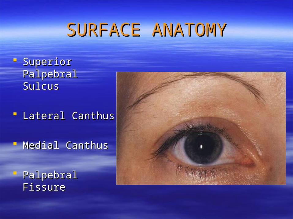
SURFACE ANATOMYSURFACE ANATOMY
Superior Superior Palpebral SulcusPalpebral Sulcus
Lateral CanthusLateral Canthus
Medial CanthusMedial Canthus
Palpebral FissurePalpebral Fissure
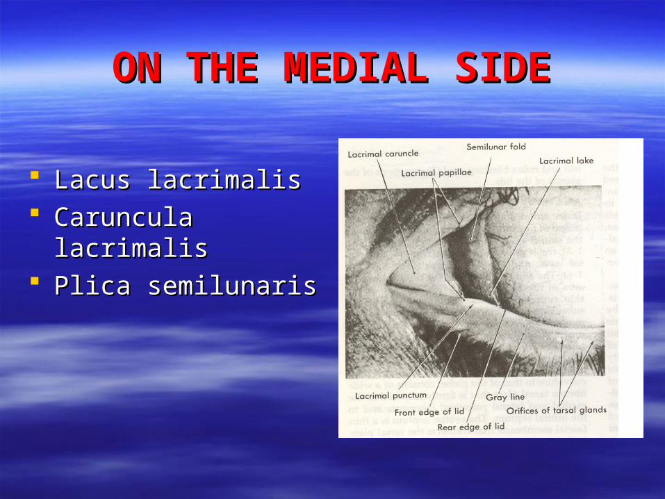
ON THE MEDIAL SIDEON THE MEDIAL SIDE
Lacus lacrimalisLacus lacrimalis Caruncula lacrimalisCaruncula lacrimalis Plica semilunaris Plica semilunaris
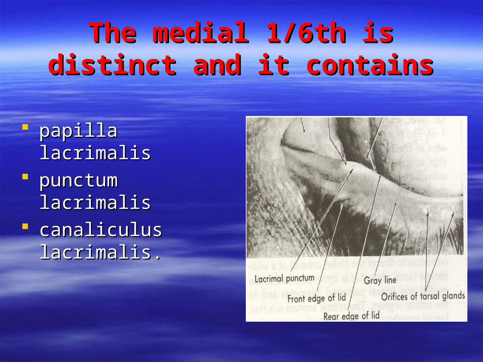
The medial 1/6th is distinct and it The medial 1/6th is distinct and it containscontains
papilla lacrimalispapilla lacrimalis punctum lacrimalis punctum lacrimalis canaliculus lacrimalis.canaliculus lacrimalis.
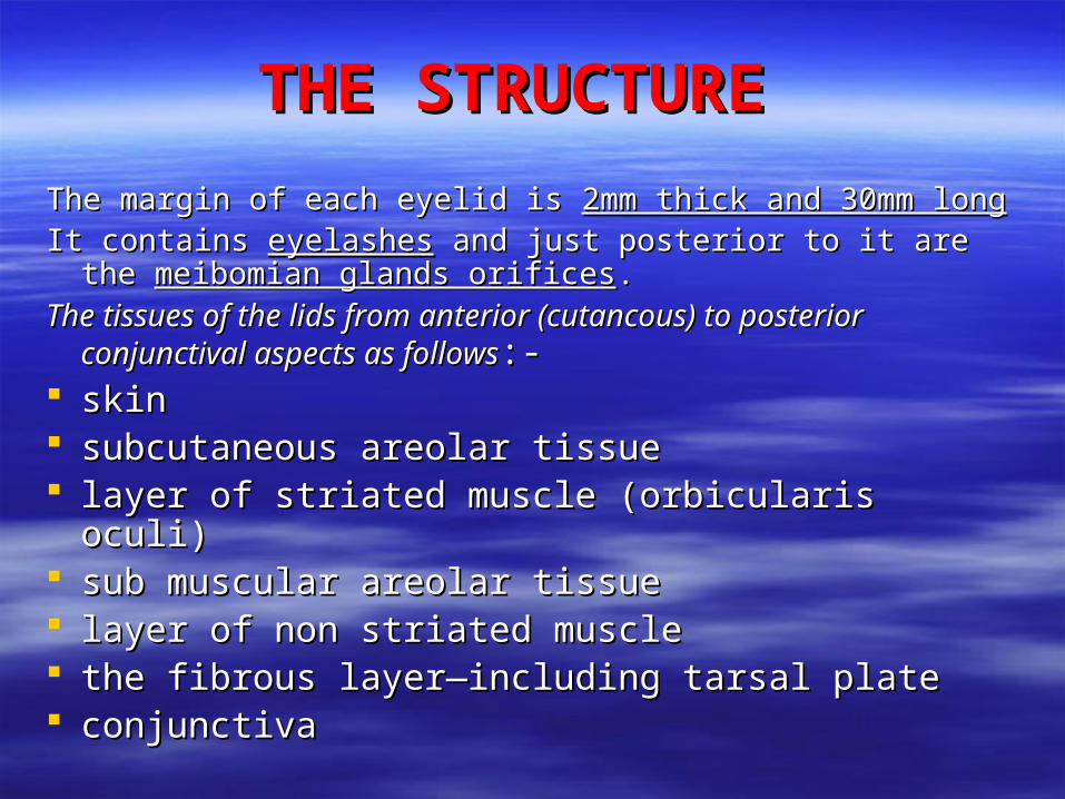
THE STRUCTURE THE STRUCTURE
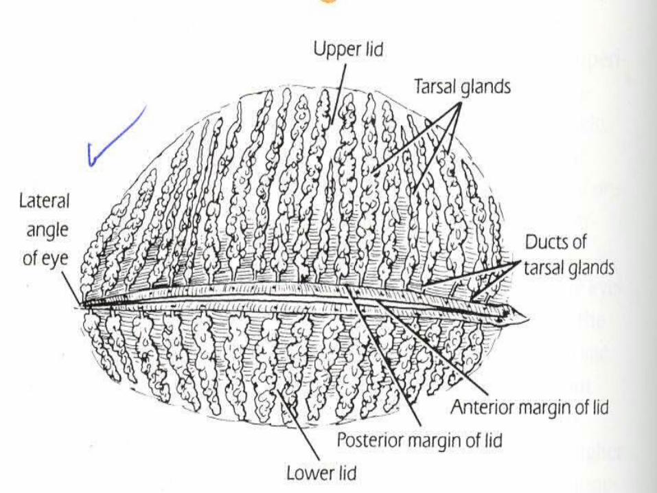
The margin of each eyelid is The margin of each eyelid is 2mm thick and 30mm long2mm thick and 30mm longIt contains It contains eyelasheseyelashes and just posterior to it are the and just posterior to it are the
meibomian glands orificesmeibomian glands orifices..
The tissues of the lids from anterior (cutancous) to posterior The tissues of the lids from anterior (cutancous) to posterior conjunctival aspects as followsconjunctival aspects as follows:-:-
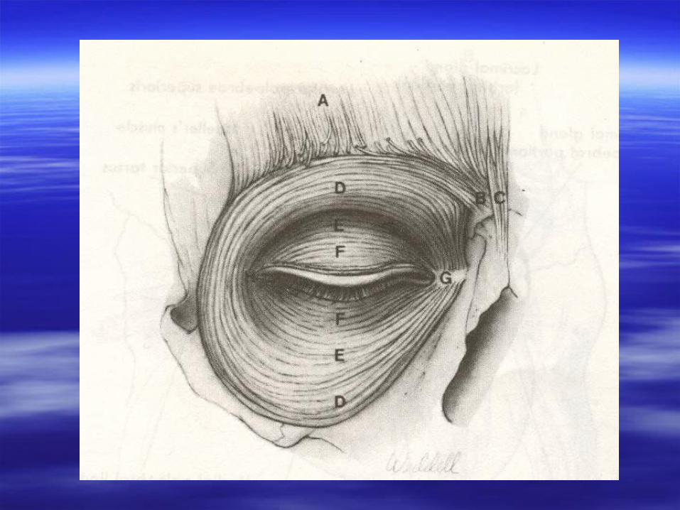
skin skin subcutaneous areolar tissuesubcutaneous areolar tissue layer of striated muscle (orbicularis oculi)layer of striated muscle (orbicularis oculi) sub muscular areolar tissuesub muscular areolar tissue layer of non striated musclelayer of non striated muscle the fibrous layer—including tarsal platethe fibrous layer—including tarsal plate conjunctivaconjunctiva
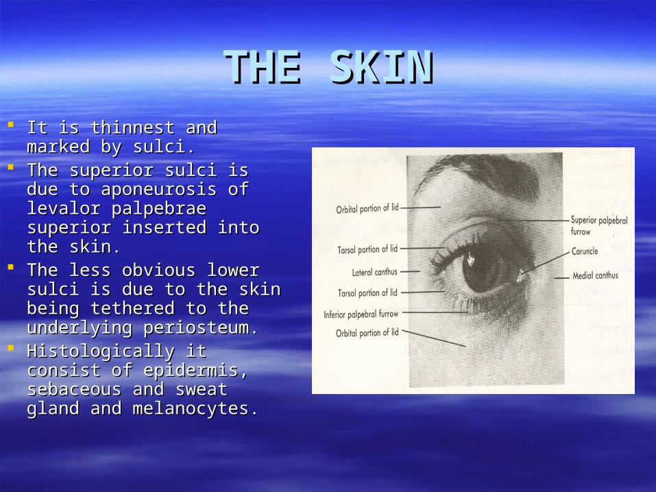
THE SKINTHE SKIN It is thinnest and marked by It is thinnest and marked by
sulci.sulci. The superior sulci is due to The superior sulci is due to
aponeurosis of levalor aponeurosis of levalor palpebrae superior inserted palpebrae superior inserted into the skin. into the skin.
The less obvious lower The less obvious lower sulci is due to the skin sulci is due to the skin being tethered to the being tethered to the underlying periosteum.underlying periosteum.
Histologically it consist of Histologically it consist of epidermis, sebaceous and epidermis, sebaceous and sweat gland and sweat gland and melanocytes.melanocytes.
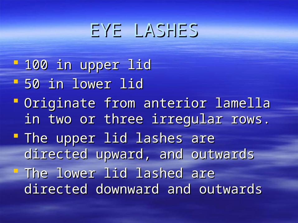
EYE LASHESEYE LASHES
100 in upper lid100 in upper lid 50 in lower lid50 in lower lid Originate from anterior lamella in two or Originate from anterior lamella in two or
three irregular rows.three irregular rows. The upper lid lashes are directed upward, The upper lid lashes are directed upward,
and outwards and outwards The lower lid lashed are directed downward The lower lid lashed are directed downward
and outwards and outwards
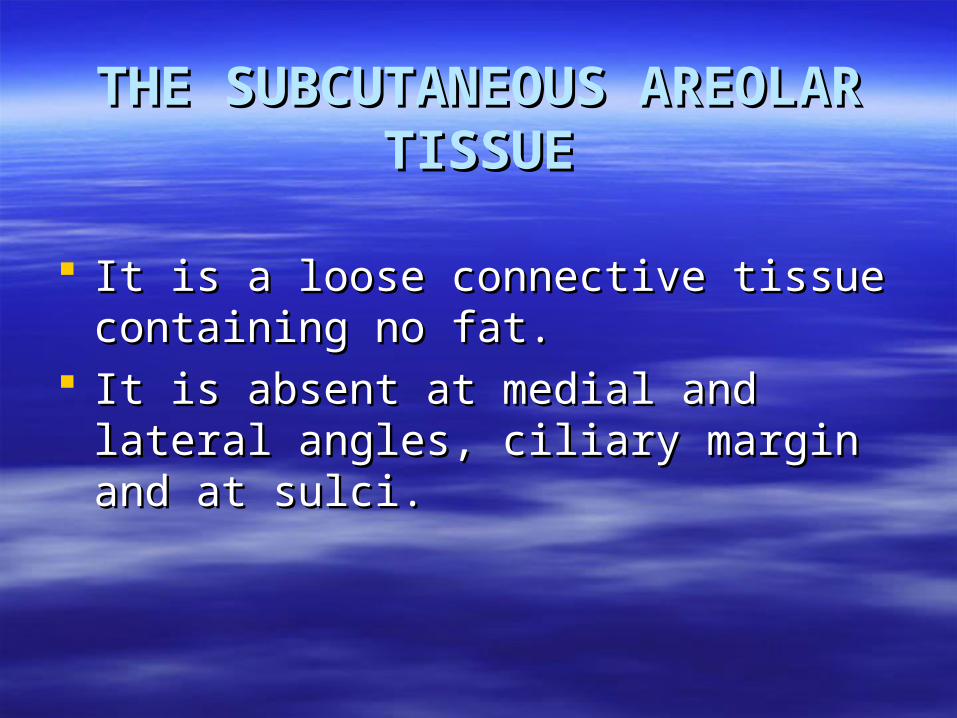
THE SUBCUTANEOUS THE SUBCUTANEOUS AREOLAR TISSUEAREOLAR TISSUE
It is a loose connective tissue containing It is a loose connective tissue containing no fat.no fat.
It is absent at medial and lateral angles, It is absent at medial and lateral angles, ciliary margin and at sulci.ciliary margin and at sulci.
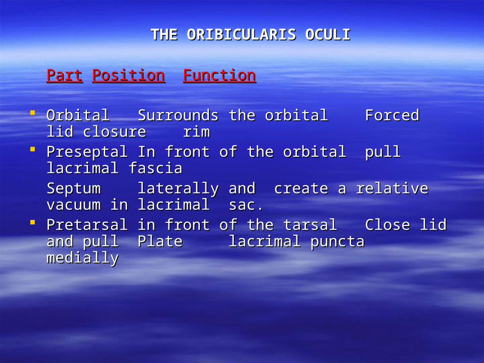
THE ORIBICULARIS OCULITHE ORIBICULARIS OCULI
PartPart PositionPosition FunctionFunction
Orbital Orbital Surrounds the orbital Surrounds the orbital Forced lid Forced lid closureclosure rim rim
PreseptalPreseptal In front of the orbitalIn front of the orbital pull lacrimal pull lacrimal fascia fascia
SeptumSeptum laterally and laterally and create a relative create a relative vacuum in vacuum in
lacrimal lacrimal sac.sac. PretarsalPretarsal in front of the tarsalin front of the tarsal Close lid and Close lid and
pull pull PlatePlate lacrimal puncta lacrimal puncta medially medially
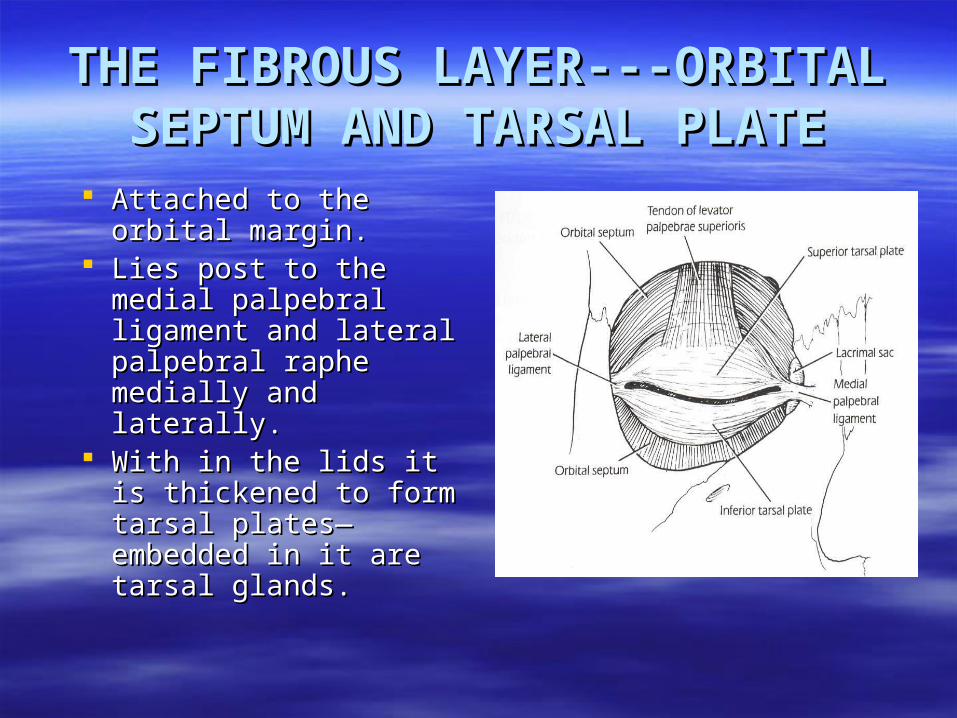
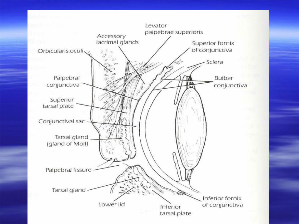
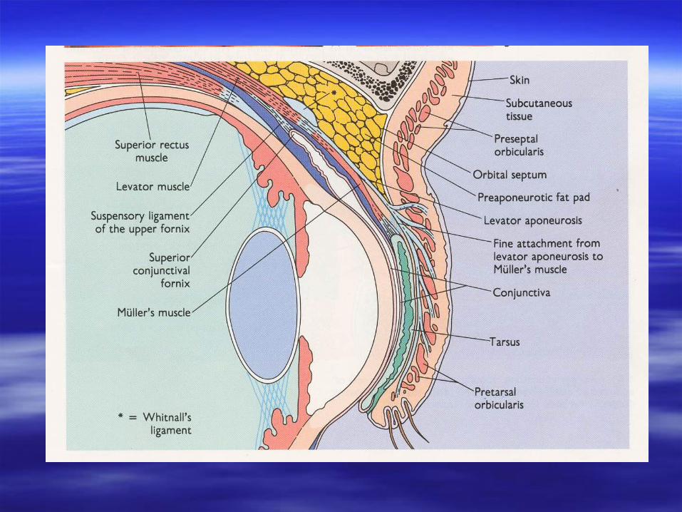
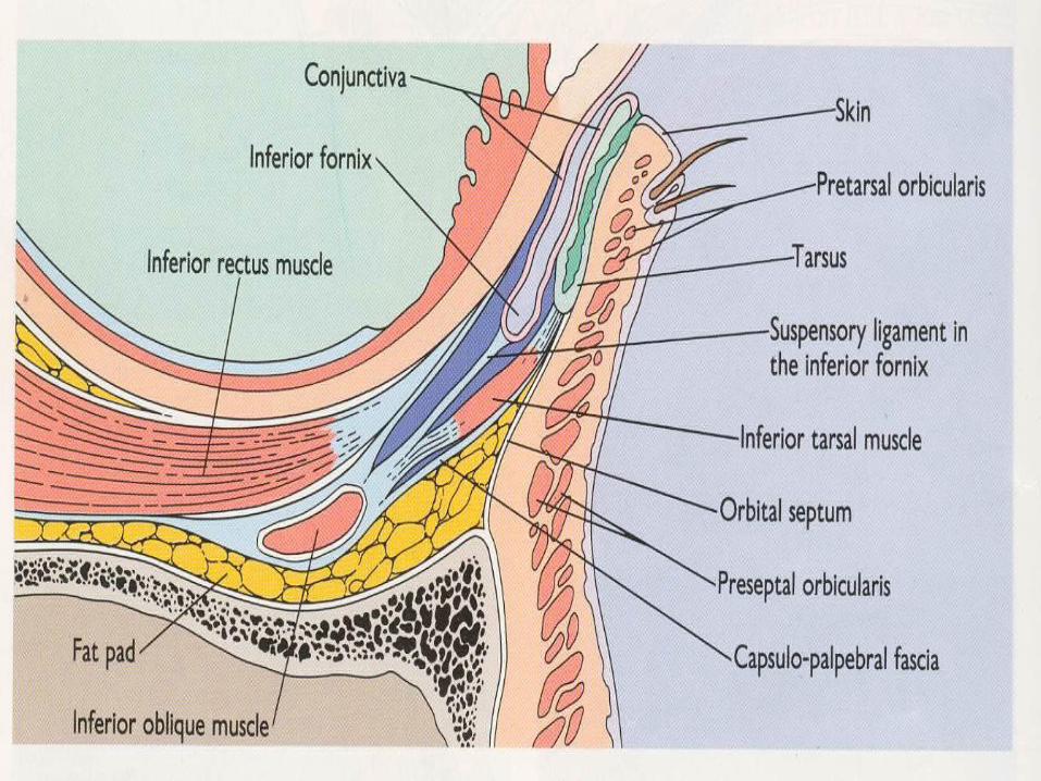
THE FIBROUS LAYER---ORBITAL THE FIBROUS LAYER---ORBITAL SEPTUM AND TARSAL PLATESEPTUM AND TARSAL PLATE
Attached to the orbital Attached to the orbital margin.margin.
Lies post to the medial Lies post to the medial palpebral ligament and palpebral ligament and lateral palpebral raphe lateral palpebral raphe medially and laterally.medially and laterally.
With in the lids it is With in the lids it is thickened to form tarsal thickened to form tarsal plates—embedded in it plates—embedded in it are tarsal glands.are tarsal glands.
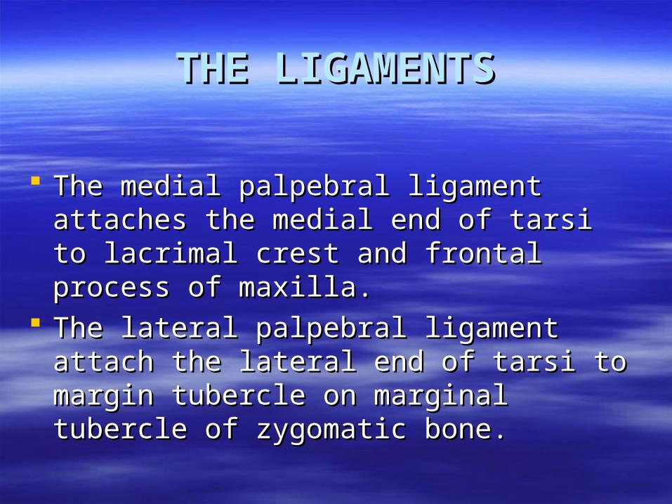
THE LIGAMENTSTHE LIGAMENTS
The medial palpebral ligament attaches the The medial palpebral ligament attaches the medial end of tarsi to lacrimal crest and medial end of tarsi to lacrimal crest and frontal process of maxilla.frontal process of maxilla.
The lateral palpebral ligament attach the The lateral palpebral ligament attach the lateral end of tarsi to margin tubercle on lateral end of tarsi to margin tubercle on marginal tubercle of zygomatic bone.marginal tubercle of zygomatic bone.
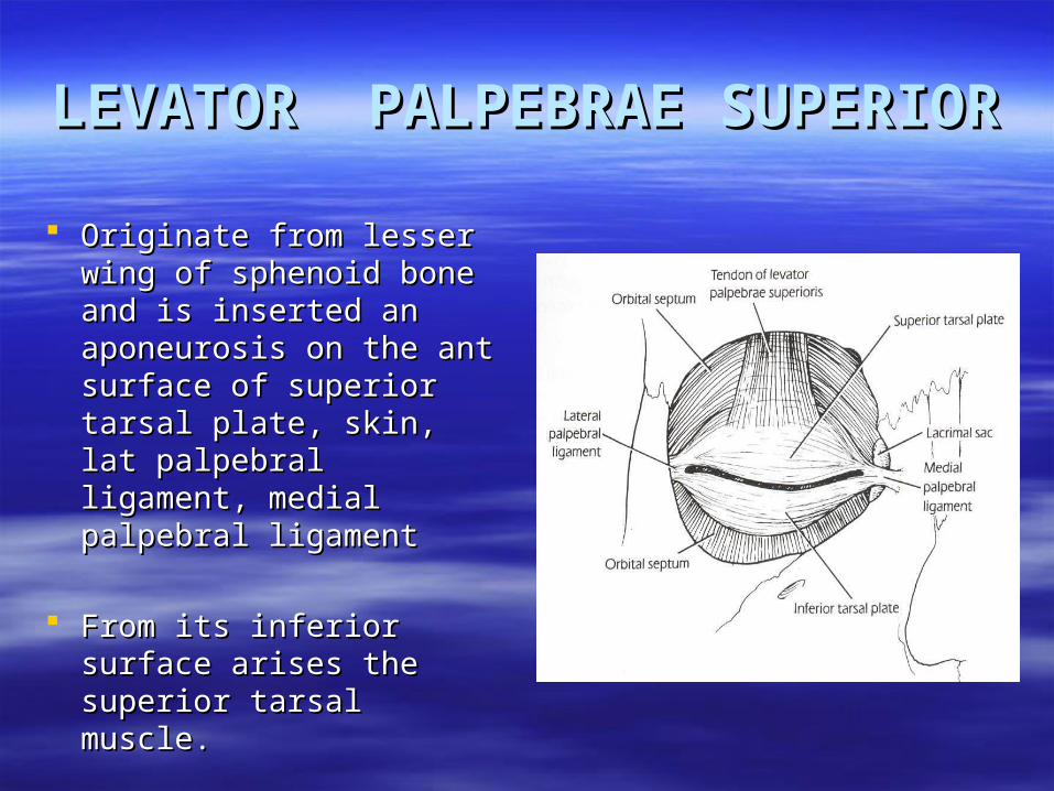
LEVATOR PALPEBRAE LEVATOR PALPEBRAE SUPERIORSUPERIOR
Originate from lesser wing Originate from lesser wing of sphenoid bone and is of sphenoid bone and is inserted an aponeurosis inserted an aponeurosis on the ant surface of on the ant surface of superior tarsal plate, skin, superior tarsal plate, skin, lat palpebral ligament, lat palpebral ligament, medial palpebral ligamentmedial palpebral ligament
From its inferior surface From its inferior surface arises the superior tarsal arises the superior tarsal muscle.muscle.
CONJUNCTIVACONJUNCTIVA
Thin mucous membrane lined by non Thin mucous membrane lined by non keratinized stratified squamous keratinized stratified squamous epithelium.epithelium.
It has a richly vascularized Substantia It has a richly vascularized Substantia propriapropria
At the margin of the eyelids it is At the margin of the eyelids it is continuous with the skin.continuous with the skin.
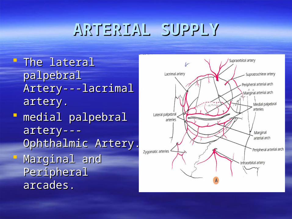
ARTERIAL SUPPLYARTERIAL SUPPLY
The lateral palpebral The lateral palpebral Artery---lacrimal artery.Artery---lacrimal artery.
medial palpebral medial palpebral artery---Ophthalmic artery---Ophthalmic Artery.Artery.
Marginal and Marginal and Peripheral arcades.Peripheral arcades.
VENOUS DRANAGEVENOUS DRANAGE
Medially – Ophthalmic and angular vein Medially – Ophthalmic and angular vein Laterally-superficial temporal veinLaterally-superficial temporal vein
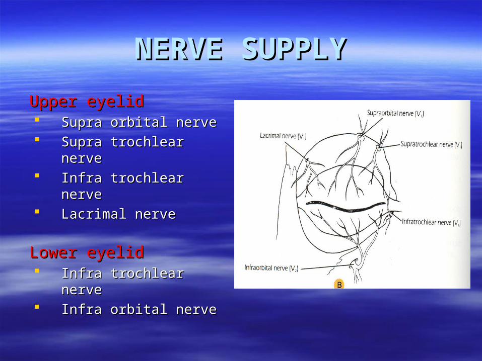
NERVE SUPPLYNERVE SUPPLY
Upper eyelidUpper eyelid Supra orbital nerve Supra orbital nerve Supra trochlear nerveSupra trochlear nerve Infra trochlear nerveInfra trochlear nerve Lacrimal nerveLacrimal nerve
Lower eyelidLower eyelid Infra trochlear nerveInfra trochlear nerve Infra orbital nerveInfra orbital nerve

LYMPHATIC DRAINAGELYMPHATIC DRAINAGE
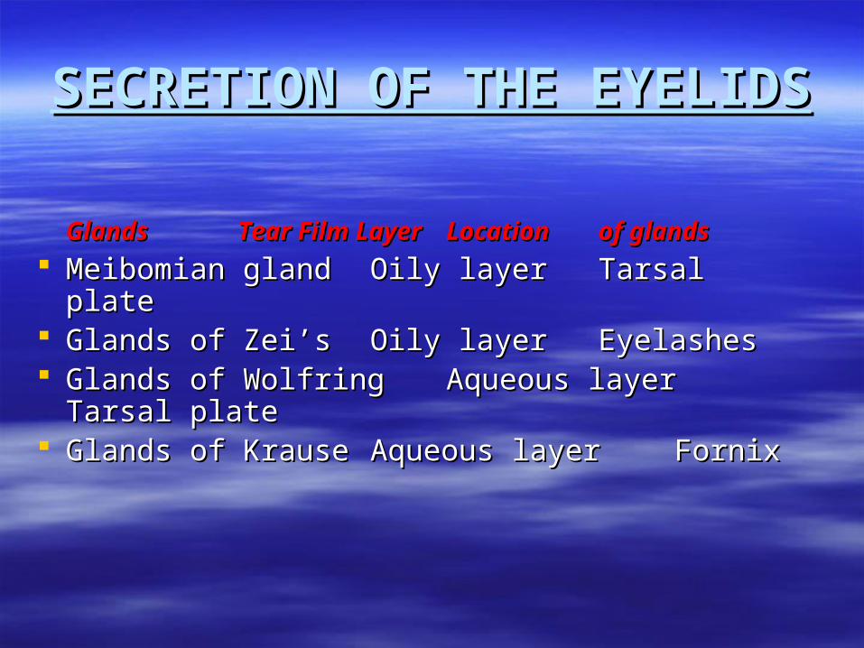
SECRETION OF THE EYELIDSSECRETION OF THE EYELIDS
GlandsGlands Tear Film Layer Tear Film Layer Location Location of glandsof glands
Meibomian glandMeibomian gland Oily layerOily layer Tarsal Tarsal plateplate
Glands of Zei’sGlands of Zei’s Oily layer Oily layer EyelashesEyelashes Glands of WolfringGlands of Wolfring Aqueous layerAqueous layer Tarsal plateTarsal plate Glands of Krause Glands of Krause Aqueous layerAqueous layer FornixFornix
BlinkingBlinking
There are two main types of blinking There are two main types of blinking
Reflex blinkingReflex blinking Spontaneous BlinkingSpontaneous Blinking
Reflex BlinkingReflex Blinking
TactileTactile Corneal Touch Corneal Touch Cortical ConnectionCortical Connection Dimunation of sensitivity in contact lens Dimunation of sensitivity in contact lens
wearerwearer
DazzleDazzle Bright lightBright light Optic nerve--- Superior colliculusOptic nerve--- Superior colliculus Associated fiber to facial nucleiAssociated fiber to facial nuclei
MenaceMenace Sudden presence of near objectSudden presence of near object Optic nerve--- Cortical ConnectionOptic nerve--- Cortical Connection Predominantly cortical in naturePredominantly cortical in nature
Spontaneous BlinkingSpontaneous Blinking
– Occurs at regular basis without an apparent external Occurs at regular basis without an apparent external stimuli.stimuli.
– Mechanics facilitates the drainage of tear film.Mechanics facilitates the drainage of tear film.
– Present in blind as no retinal stimuli are required.Present in blind as no retinal stimuli are required.
BlephrospasmBlephrospasm
Simultaneous forcible contraction of Simultaneous forcible contraction of orbiculars oculi.orbiculars oculi.
Forcible closure of the lids.Forcible closure of the lids. Its role in surgical procedures.Its role in surgical procedures. Anterior segment injury.Anterior segment injury.

THANK YOU