Embed Size (px)
Citation preview
The Enteric Nervous System: A Second Brain MICHAEL D. GERSHON Columbia University
Once dismissed as a simple collection of relay ganglia, the enteric nervous system is now recognized as a complex, integrative brain in its own right. Although we still are unable to relate complex behaviors such as gut motility and secretion to the activity of individual neurons, work in that area is proceeding briskly--and will lead to rapid advances in the management of functional bowel disease.
Dr. Gershon is Professor and Chair, Department of Anatomy and Cell Biology, Columbia University College of Physicians and Surgeons, New York. In addition to numerous scientific publications, he is the author of The Second Brain (Harper Collins, New York, 1998).
Structurally and neurochemically, the enteric nervous system (ENS) is a brain unto itself. Within those yards of tubing lies a complex web of microcircuitry driven by more neurotransmitters and neuromodulators than can be found anywhere else in the peripheral nervous system. These allow the ENS to perform many of its tasks in the absence of central nervous system (CNS) control--a unique endowment that has permitted enteric neurobiologists to investigate nerve cell ontogeny and chemical mediation of reflex behavior in a laboratory setting. Recognition of the importance of this work as a basis for developing effective therapies for functional bowel disease, coupled with the recent, unexpected discovery of major enteric defects following the knockout of murine genes not previously known to affect the gut, has produced a groundswell of interest that has attracted some of the best investigators to the field. Add to this that the ENS provides the closest thing we have to a window on the brain, and one begins to understand why the bowel--the second brain--is finally receiving the attention it deserves.
Discovery of the ENS
The field of neurogastroenterology dates back to the nineteenth-century English investigators William M. Bayliss and Ernest H. Starling, who demonstrated that application of pressure to the intestinal lumen of anesthetized dogs resulted in oral contraction and anal relaxation, followed by a propulsive wave (which they referred to as the "law of the intestine" and we now call the peristaltic reflex) of sufficient strength to propel food through the digestive tract. Because the reflex persisted even after all of the extrinsic nerves to the gut had been severed, Bayliss and Starling deduced--correctly--that the ENS was a self-contained hub of neuronal activity that operated largely independent of CNS input.
Eighteen years later, the German scientist Paul Trendelenburg confirmed these findings by demonstrating that the peristaltic reflex could be elicited in vitro in the isolated gut of a guinea pig, without participation of the brain, spinal cord, dorsal root, or cranial ganglia. Trendelenburg knew this finding was unique; no other peripheral organ had such a highly developed intrinsic neural apparatus. Cut the connection linking the bladder or the skeletal muscles to the CNS, and all motor activity ceases. Cut the connection to the gut, however, and function persists.
Trendelenburg's results were published in 1917. That they were accepted by at least some of his contemporaries is evident from the description of the ENS contained in John N. Langley's classic textbook, The Autonomic Nervous System, published in 1921. Like Trendelenburg, Langley knew that intestinal function must involve not only excitatory and inhibitory motor neurons to innervate the smooth muscle, glands, and blood vessels but also primary afferent neurons to detect increases in pressure, as well as interneurons to coordinate the wave of activity down the length of the bowel. The brain could not perform these complex functions alone, he reasoned, because the gut is innervated by only a few thousand motor fibers. Logic dictated that the nerve cells in the bowel--which Langley suspected, and we now know, number in the millions--had to have their own separate innervation. Thus, when he described the autonomic nervous system, it was as three distinct parts: the sympathetic, the parasympathetic, and the enteric.
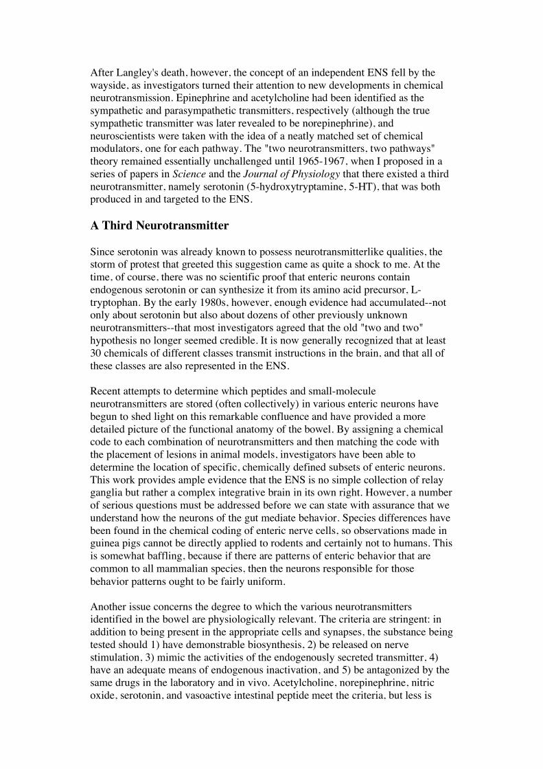
Unfortunately, Langley, who was owner and editor of the Journal of Physiology, alienated many of his colleagues. After his death, editorship of the Journal passed to the Physiological Society, whose members reclassified the enteric neurons as parasympathetic relay ganglia, part of the vagal supply that directs gut motility. To an extent, of course, they were right. The vagus nerve is normally responsible for commanding the vast microcircuits of the ENS to carry out their appointed tasks. What it cannot do, as Langley and his predecessors intuitively grasped, is tell them how to carry them out. That is strictly an inside job, and one that the gut is marvelously capable of performing. In addition to propulsion, the ENS bears primary responsibility for self-cleaning, regulating the luminal environment, working with the immune system to defend the bowel, and modifying the rate of proliferation and growth of mucosal cells. Neurons emanating from the gut also innervate ganglia in neighboring organs, such as the gallbladder and pancreas (Figure 1).
After Langley's death, however, the concept of an independent ENS fell by the wayside, as investigators turned their attention to new developments in chemical neurotransmission. Epinephrine and acetylcholine had been identified as the sympathetic and parasympathetic transmitters, respectively (although the true sympathetic transmitter was later revealed to be norepinephrine), and neuroscientists were taken with the idea of a neatly matched set of chemical modulators, one for each pathway. The "two neurotransmitters, two pathways" theory remained essentially unchallenged until 1965-1967, when I proposed in a series of papers in Science and the Journal of Physiology that there existed a third neurotransmitter, namely serotonin (5-hydroxytryptamine, 5-HT), that was both produced in and targeted to the ENS.
A Third Neurotransmitter
Since serotonin was already known to possess neurotransmitterlike qualities, the storm of protest that greeted this suggestion came as quite a shock to me. At the time, of course, there was no scientific proof that enteric neurons contain endogenous serotonin or can synthesize it from its amino acid precursor, L-tryptophan. By the early 1980s, however, enough evidence had accumulated--not only about serotonin but also about dozens of other previously unknown neurotransmitters--that most investigators agreed that the old "two and two" hypothesis no longer seemed credible. It is now generally recognized that at least 30 chemicals of different classes transmit instructions in the brain, and that all of these classes are also represented in the ENS.
Recent attempts to determine which peptides and small-molecule neurotransmitters are stored (often collectively) in various enteric neurons have begun to shed light on this remarkable confluence and have provided a more detailed picture of the functional anatomy of the bowel. By assigning a chemical code to each combination of neurotransmitters and then matching the code with the placement of lesions in animal models, investigators have been able to determine the location of specific, chemically defined subsets of enteric neurons. This work provides ample evidence that the ENS is no simple collection of relay ganglia but rather a complex integrative brain in its own right. However, a number of serious questions must be addressed before we can state with assurance that we understand how the neurons of the gut mediate behavior. Species differences have been found in the chemical coding of enteric nerve cells, so observations made in guinea pigs cannot be directly applied to rodents and certainly not to humans. This is somewhat baffling, because if there are patterns of enteric behavior that are common to all mammalian species, then the neurons responsible for those behavior patterns ought to be fairly uniform.
Another issue concerns the degree to which the various neurotransmitters identified in the bowel are physiologically relevant. The criteria are stringent: in addition to being present in the appropriate cells and synapses, the substance being tested should 1) have demonstrable biosynthesis, 2) be released on nerve stimulation, 3) mimic the activities of the endogenously secreted transmitter, 4) have an adequate means of endogenous inactivation, and 5) be antagonized by the same drugs in the laboratory and in vivo. Acetylcholine, norepinephrine, nitric oxide, serotonin, and vasoactive intestinal peptide meet the criteria, but less is
known about other candidate molecules.
Anatomy of the ENS
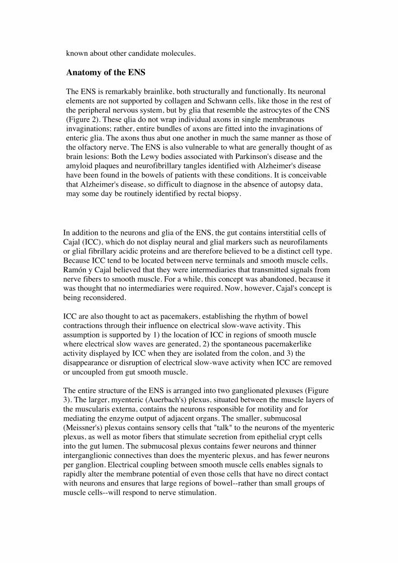
The ENS is remarkably brainlike, both structurally and functionally. Its neuronal elements are not supported by collagen and Schwann cells, like those in the rest of the peripheral nervous system, but by glia that resemble the astrocytes of the CNS (Figure 2). These qlia do not wrap individual axons in single membranous invaginations; rather, entire bundles of axons are fitted into the invaginations of enteric glia. The axons thus abut one another in much the same manner as those of the olfactory nerve. The ENS is also vulnerable to what are generally thought of as brain lesions: Both the Lewy bodies associated with Parkinson's disease and the amyloid plaques and neurofibrillary tangles identified with Alzheimer's disease have been found in the bowels of patients with these conditions. It is conceivable that Alzheimer's disease, so difficult to diagnose in the absence of autopsy data, may some day be routinely identified by rectal biopsy.
In addition to the neurons and glia of the ENS, the gut contains interstitial cells of Cajal (ICC), which do not display neural and glial markers such as neurofilaments or glial fibrillary acidic proteins and are therefore believed to be a distinct cell type. Because ICC tend to be located between nerve terminals and smooth muscle cells, Ramón y Cajal believed that they were intermediaries that transmitted signals from nerve fibers to smooth muscle. For a while, this concept was abandoned, because it was thought that no intermediaries were required. Now, however, Cajal's concept is being reconsidered.
ICC are also thought to act as pacemakers, establishing the rhythm of bowel contractions through their influence on electrical slow-wave activity. This assumption is supported by 1) the location of ICC in regions of smooth muscle where electrical slow waves are generated, 2) the spontaneous pacemakerlike activity displayed by ICC when they are isolated from the colon, and 3) the disappearance or disruption of electrical slow-wave activity when ICC are removed or uncoupled from gut smooth muscle.
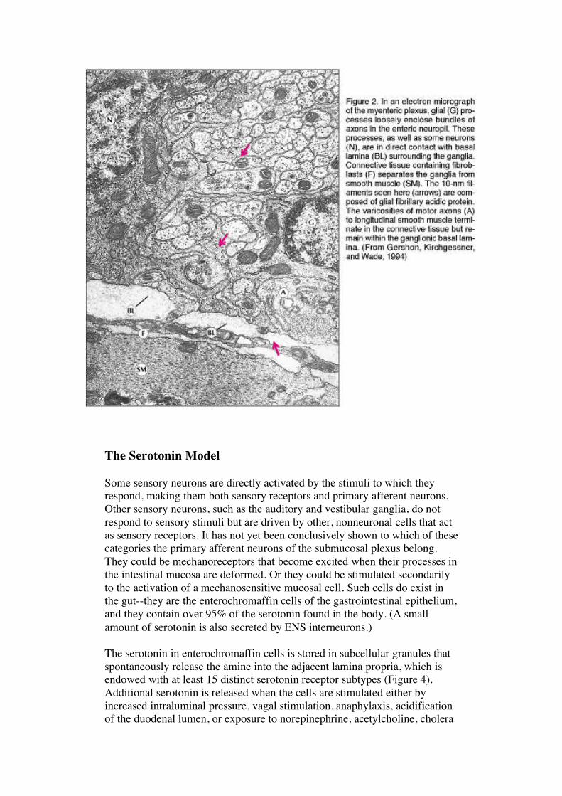
The entire structure of the ENS is arranged into two ganglionated plexuses (Figure 3). The larger, myenteric (Auerbach's) plexus, situated between the muscle layers of the muscularis externa, contains the neurons responsible for motility and for mediating the enzyme output of adjacent organs. The smaller, submucosal (Meissner's) plexus contains sensory cells that "talk" to the neurons of the myenteric plexus, as well as motor fibers that stimulate secretion from epithelial crypt cells into the gut lumen. The submucosal plexus contains fewer neurons and thinner interganglionic connectives than does the myenteric plexus, and has fewer neurons per ganglion. Electrical coupling between smooth muscle cells enables signals to rapidly alter the membrane potential of even those cells that have no direct contact with neurons and ensures that large regions of bowel--rather than small groups of muscle cells--will respond to nerve stimulation.
The Serotonin Model
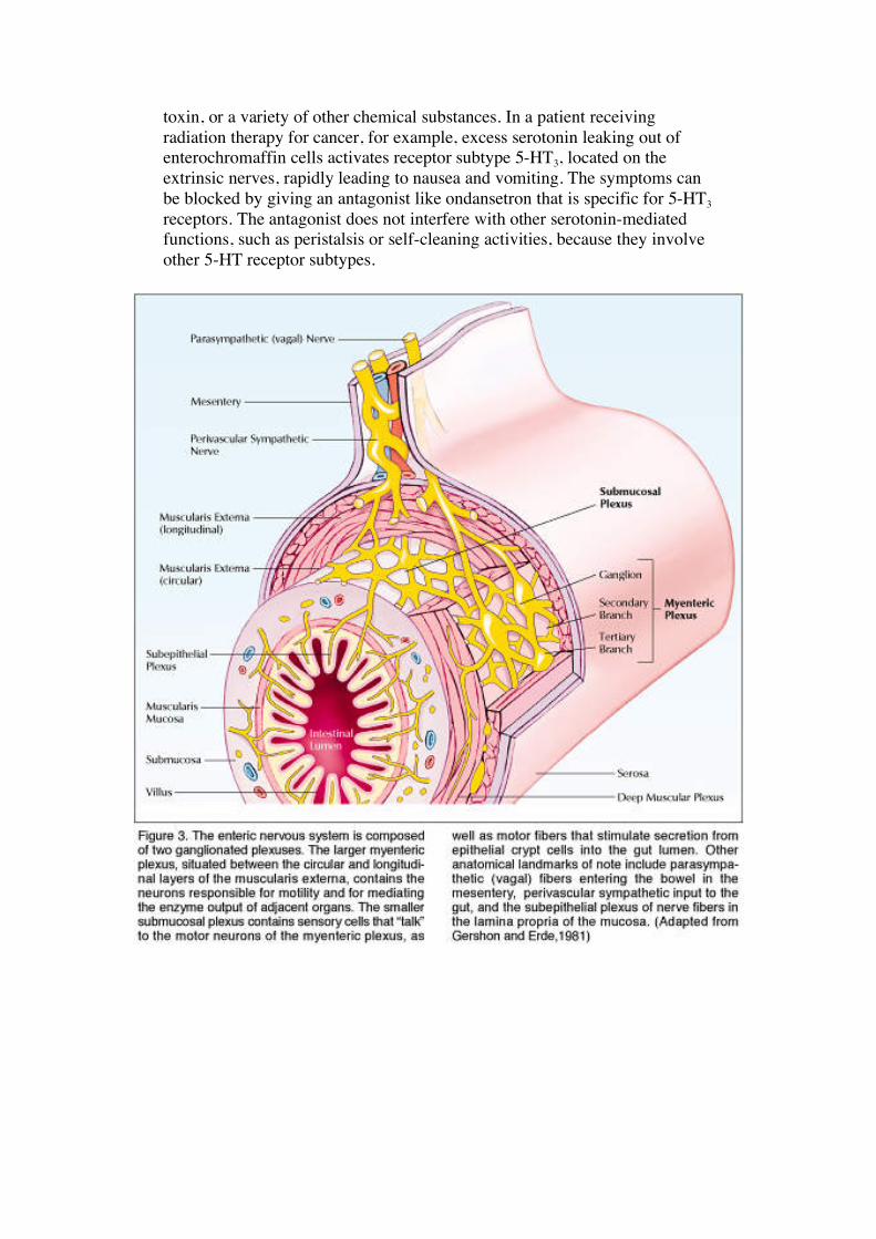
Some sensory neurons are directly activated by the stimuli to which they respond, making them both sensory receptors and primary afferent neurons. Other sensory neurons, such as the auditory and vestibular ganglia, do not respond to sensory stimuli but are driven by other, nonneuronal cells that act as sensory receptors. It has not yet been conclusively shown to which of these categories the primary afferent neurons of the submucosal plexus belong. They could be mechanoreceptors that become excited when their processes in the intestinal mucosa are deformed. Or they could be stimulated secondarily to the activation of a mechanosensitive mucosal cell. Such cells do exist in the gut--they are the enterochromaffin cells of the gastrointestinal epithelium, and they contain over 95% of the serotonin found in the body. (A small amount of serotonin is also secreted by ENS interneurons.)
The serotonin in enterochromaffin cells is stored in subcellular granules that spontaneously release the amine into the adjacent lamina propria, which is endowed with at least 15 distinct serotonin receptor subtypes (Figure 4). Additional serotonin is released when the cells are stimulated either by increased intraluminal pressure, vagal stimulation, anaphylaxis, acidification of the duodenal lumen, or exposure to norepinephrine, acetylcholine, cholera
toxin, or a variety of other chemical substances. In a patient receiving radiation therapy for cancer, for example, excess serotonin leaking out of enterochromaffin cells activates receptor subtype 5-HT3, located on the extrinsic nerves, rapidly leading to nausea and vomiting. The symptoms can be blocked by giving an antagonist like ondansetron that is specific for 5-HT3 receptors. The antagonist does not interfere with other serotonin-mediated functions, such as peristalsis or self-cleaning activities, because they involve other 5-HT receptor subtypes.
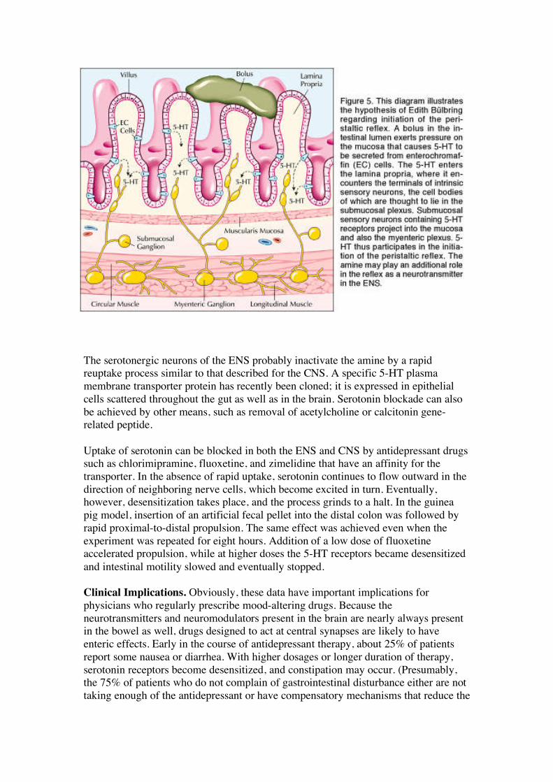
We now have extensive data (from studies of the serotonin antagonist 5-HTP-DP and anti-idiotypic antibodies that recognize 5-HT receptors) confirming that 1) serotonin stimulates the peristaltic reflex when it is applied to the mucosal surface of the bowel, 2) serotonin is released whenever the peristaltic reflex is initiated, and 3) the reflex is diminished when the mucosal source of serotonin is removed. Consequently, there is wide support for the hypothesis, first proposed by Edith Bülbring in 1958, that enterochromaffin cells act as pressure transducers and that the serotonin they secrete acts as a mediator to excite the mucosal afferent nerves, initiating the peristaltic reflex (Figure 5).
The serotonergic neurons of the ENS probably inactivate the amine by a rapid reuptake process similar to that described for the CNS. A specific 5-HT plasma membrane transporter protein has recently been cloned; it is expressed in epithelial cells scattered throughout the gut as well as in the brain. Serotonin blockade can also be achieved by other means, such as removal of acetylcholine or calcitonin gene-related peptide.
Uptake of serotonin can be blocked in both the ENS and CNS by antidepressant drugs such as chlorimipramine, fluoxetine, and zimelidine that have an affinity for the transporter. In the absence of rapid uptake, serotonin continues to flow outward in the direction of neighboring nerve cells, which become excited in turn. Eventually, however, desensitization takes place, and the process grinds to a halt. In the guinea pig model, insertion of an artificial fecal pellet into the distal colon was followed by rapid proximal-to-distal propulsion. The same effect was achieved even when the experiment was repeated for eight hours. Addition of a low dose of fluoxetine accelerated propulsion, while at higher doses the 5-HT receptors became desensitized and intestinal motility slowed and eventually stopped.
Clinical Implications. Obviously, these data have important implications for physicians who regularly prescribe mood-altering drugs. Because the neurotransmitters and neuromodulators present in the brain are nearly always present in the bowel as well, drugs designed to act at central synapses are likely to have enteric effects. Early in the course of antidepressant therapy, about 25% of patients report some nausea or diarrhea. With higher dosages or longer duration of therapy, serotonin receptors become desensitized, and constipation may occur. (Presumably, the 75% of patients who do not complain of gastrointestinal disturbance either are not taking enough of the antidepressant or have compensatory mechanisms that reduce the
impact of prolonged serotonin availability.) If these effects--which are not side effects per se but predictable consequences of transporter protein blockade--are not anticipated and carefully explained to the patient, they are likely to reduce adherence and limit the value of treatment.
On the other hand, the same drugs that tend to cause difficulty for patients who take them for emotional illness may be a godsend to those with functional bowel disease. Moreover, because the ENS reacts promptly to changes in serotonin availability, patients with chronic bowel problems often find their symptoms relieved at pharmacologic concentrations far below those used in conventional antidepressant therapy.
More About the Brain-Gut Connection
Provided that the vagus nerve is intact, a steady stream of messages flows back and forth between the brain and the gut. We all experience situations in which our brains cause our bowels to go into overdrive. But in fact, messages departing the gut outnumber the opposing traffic on the order of about nine to one. Satiety, nausea, the urge to vomit, abdominal pain--all are the gut's way of warning the brain of danger from ingested food or infectious pathogens. And while the brain normally responds with appropriate signals, the ENS can take over when necessary, as for example when vagal input has been surgically severed.
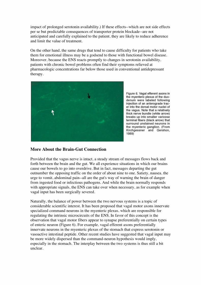
Naturally, the balance of power between the two nervous systems is a topic of considerable scientific interest. It has been proposed that vagal motor axons innervate specialized command neurons in the myenteric plexus, which are responsible for regulating the intrinsic microcircuits of the ENS. In favor of this concept is the observation that vagal motor fibers appear to synapse preferentially on certain types of enteric neuron (Figure 6). For example, vagal efferent axons preferentially innervate neurons in the myenteric plexus of the stomach that express serotonin or vasoactive intestinal peptide. Other recent studies have suggested that vagal input may be more widely dispersed than the command-neuron hypothesis would imply, especially in the stomach. The interplay between the two systems is thus still a bit unclear.
Correlation or Causation? Whatever the exact connection, the relationship between the cerebral and enteric brains is so close that it is easy to become confused about which is doing the talking. Until peptic ulcer was found to be an infectious disease, for example, physicians regarded anxiety as the chief cause. Now that we recognize Helicobacter pylori as the cause, it seems clear that the physical sensation of burning epigastric pain is generally responsible for the emotional symptoms, rather than the other way around. But because most ulcer patients, if questioned, will admit to feeling anxious, the misunderstanding persisted for decades. Another illustration is ulcerative colitis, which was considered the prototypic psychosomatic disease when I was in medical school. There were even lectures on the "ulcerative colitis personality." The ulcerative colitis personality, if indeed there is one, is a consequence of living with a disabling autoimmune disease that prevents patients from feeling relaxed and comfortable in social situations. It is altogether possible that with passage of time, many of the ailments currently labeled as functional bowel diseases will prove to have similarly identifiable physiologic causes.
Embryonic Development: New Insights
In order to better appreciate ENS functioning, it is helpful to know something about its embryonic development. Which sites in the embryo give rise to the precursors of enteric neurons and glia? What impels these precursors to migrate to the bowel? And what features of the enteric microenvironment ultimately cause these incipient nerve cells to arrest their journey and undergo phenotypic differentiation?
The neural and glial precursor cells of the ENS are the descendants of émigrés from the vagal, rostral-truncal, and sacral levels of the neural crest. Of these three, the vagal crest is the most influential, because its cells colonize the entire gut. The rostral-truncal crest colonizes only the esophagus and adjacent stomach, whereas the sacral crest colonizes the postumbilical bowel.
It might be assumed that premigratory cells in each of these regions are already programmed to locate their appropriate portion of the gut and differentiate as enteric neurons or glia. However, that idea has been shown to be incorrect. The premigratory crest population is multipotent--so much so that whole regions of the crest can be interchanged in avian embryos without interfering with ENS formation. Furthermore,
even the group of crest-derived cells that are destined to colonize the bowel contains pluripotent precursors with a number of "career" options. Terminal differentiation does not take place until the émigrés have reached the gut wall and interacted with the enteric microenvironment via a number of specific chemical growth factor-receptor combinations. If these molecules are unavailable, the migration of the crest-derived cells will be cut short, and aganglionosis of the remaining bowel will result.
Nerve cell lineages are defined by their common dependence on particular growth factors or genes. For example, there is a very large lineage defined by dependence on stimulation of the Ret receptor by glial cell-derived neurotrophin factor (GDNF) and its binding molecule, GFR-alpha1. This so-called first precursor gives rise to essentially all of the neurons of the bowel, with the exception of those of the rostral foregut. Partial loss of GDNF-Ret may result in a precursor pool that is too small to colonize the entire gut, while complete loss of either GDNF or Ret eliminates the possibility of nerve cells below the level of the esophagus.
A second lineage depends on Mash-1, a member of the basic helix-loop-helix family of transcriptional regulators. These neurons, which include those of the rostral foregut as well as a subset of cells in the remainder of the bowel, are transiently catechola-minergic, develop early (enteric neurons develop in successive waves), and generate the entire set of enteric serotonergic neurons. A third lineage is independent of Mash-1, develops later, and gives rise to peptidergic neurons such as those that contain calcitonin gene-related peptide.
Sublineages of enteric neurons include those dependent on neurotrophin 3 (NT-3) and endothelin 3 (ET-3). The peptide-receptor combination ET-3-ETB is particularly interesting because it appears to act as a brake that prevents migrating cells from differentiating prematurely--before colonization of the gastrointestinal tract has been completed. Absence of ET-3 results in loss of nerve cells in the terminal portion of the bowel. In humans, this condition, known as Hirsch-sprung's disease (congenital megacolon), occurs in roughly one in 5,000 live births. Without innervation, intestinal traffic is blocked, and the colon becomes enormously dilated above the blockage. Surgery is extremely difficult because the aganglionic portion of the infant's intestine must be removed without damaging functioning ganglionic tissue. One experimental model for this disease, the lethal spotted mouse, lacks ET-3, while another laboratory strain, the piebald mouse, lacks the endothelin receptor ETB. In either case, the result is a mouse with the equivalent of Hirschsprung's disease. (The link between ET-3 deficiency and aganglionosis was discovered quite by accident, when Masashi Yanagisawa knocked out genes coding for ET-3 and ETB to study their effect on blood pressure regulation. The animals had such severe bowel abnormalities that they did not live long enough to manifest cardiovascular problems.)
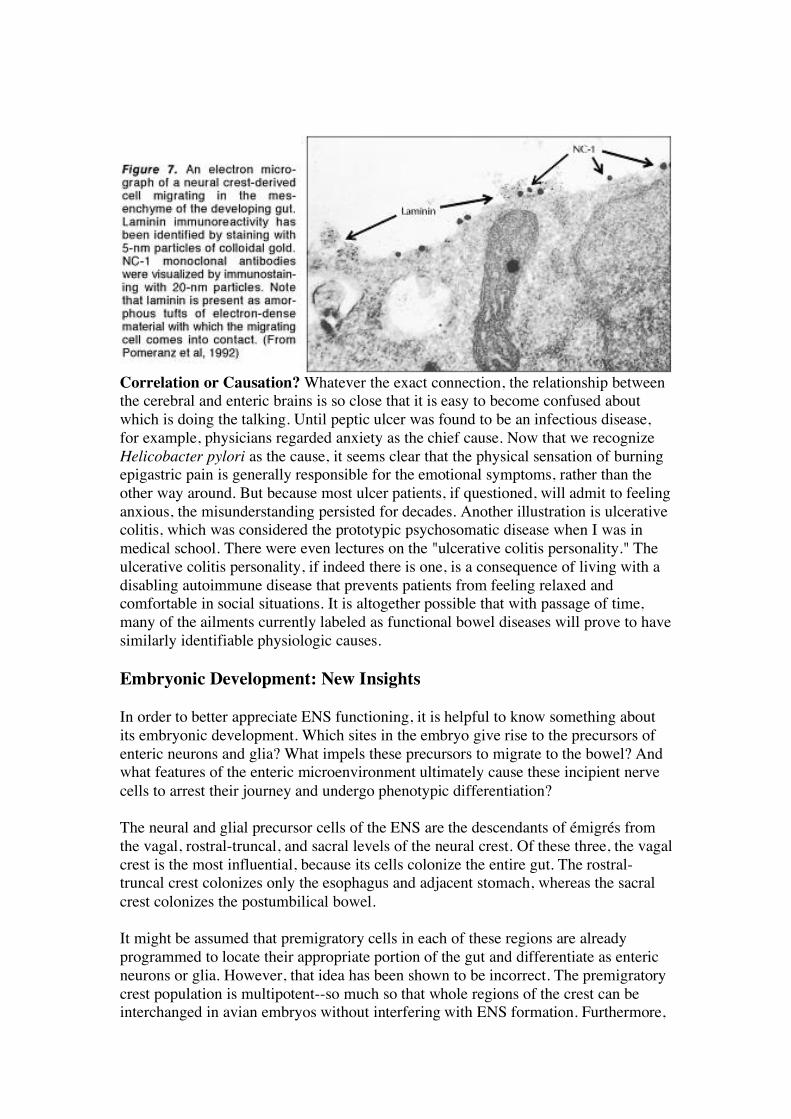
Our laboratory is currently attempting to define exactly where the endothelins are expressed, as well as to clarify the role of another putative factor in the pathogenesis of Hirschsprung's disease, laminin-1. This is an extracellular matrix protein excreted by smooth muscle precursors that both encourages adhesion of migrating cells and promotes their differentiation into neurons (Figure 7). We are trying to produce a transgenic mouse that overexpresses laminin in the gut, and anticipate that Hirschsprung's disease equivalent will result.
We also are studying an interesting group of molecules called netrins, which are expressed in both gut epithelium and the CNS. Netrins are attraction molecules that appear to guide migrating axons in the developing CNS and neuronal precursors in the bowel and may be especially important in forming the submucosal plexus. The attraction they create is so powerful that if netrin-expressing cells are placed next to the gut, neuronal precursors will migrate out of the bowel in search of the netrin-expressing cells. Two potential receptors for the netrins have been identified,
neogenin and DCC (deleted in colorectal cancer). Antibodies to DCC will counter the attraction of netrins and cause nerve cell precursors to suspend their migration. Other teams are studying avoidance molecules called sema-phorins that are the opposite of the attraction molecules (i.e., they repel the enteric precursors).
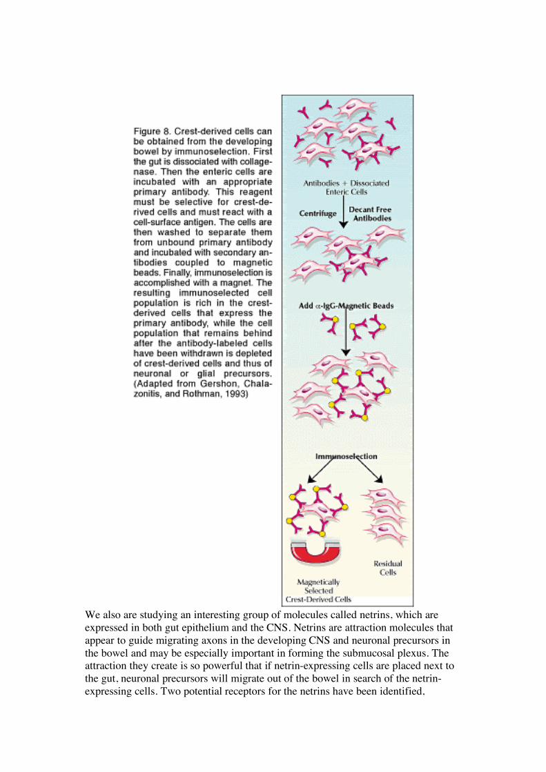
Mention should also be made of the important role that technology has played in accelerating scientific progress in this area. In particular, the ability to isolate crest-derived cell populations by magnetic immunoselection and then to culture them in defined media has made it possible to test the direct effects of putative growth factors on the precursors of neurons and glia, as well as to analyze cell receptors, transcription factors, and other developmentally relevant molecules (Figure 8). The alternative--carrying out experiments with mixed populations of enteric precursor cells or cells cultured in serum-containing media--would have produced unreliable results because of the uncontrolled interaction of crest-derived and non-crest-derived cells in media of unknown content.
Future Directions
Clearly, much has been accomplished since the days when the ENS was dismissed as an inconsequential collection of relay ganglia. Although we still are unable to relate such complex behaviors as gut motility and secretion to the activity of individual neurons, work in that area is proceeding briskly. Similarly, we are moving toward an overarching picture of how the CNS interacts with the microcircuits of the bowel to produce coordinated responses. Finally, it seems inevitable that advancement of basic knowledge about the ENS will be followed by related clinical applications, so that the next generation of medical practitioners and patients will find fewer ailments listed under the catch-all heading of functional bowel disease.
Selected Reading
Costa M et al: Neurochemical classification of myenteric neurons in the guinea-pig ileum. Neuroscience 75:949, 1996
Furness JB et al: Intrinsic primary afferent neurons of the intestine. Prog Neurobiol 54:1, 1998
Gershon MD: Genes, lineages, and tissue interactions in the development of the enteric nervous system. Am J Physiol 275:G869, 1998
Gershon MD: The Second Brain. Harper Collins, New York, 1998
Gershon MD, Chalazonitis A, Rothman TP: From neural crest to bowel: Development of the enteric nervous system. J Neurobiol 24:199, 1993
Gershon MD, Erde SM: The nervous system of the gut. Gastroenterology 80:1571, 1981
Gershon MD, Kirchgessner AL, Wade PR: Functional anatomy of the enteric nervous system. In Physiology of the Gastrointestinal Tract, 3rd ed, vol 1, Johnson LR et al (Eds). Raven Press, New York, 1994, pp 381-422
Kirchgessner AL, Gershon MD: Identification of vagal efferent fibers and putative target neurons in the enteric nervous system of the rat. J Comp Neurol 285:38, 1989
Kirchgessner AL, Gershon MD: Innervation of the pancreas by neurons in the gut. J Neurosci 10:1626, 1990
Pomeranz HD et al: Expression of a neurally related laminin binding protein by neural crest-derived cells that colonize the gut: Relationship to the formation of enteric ganglia. J Comp Neurol 313:625, 1991
Rosenthal A: The GDNF protein family: Gene ablation studies reveal what they really do and how. Neuron 22:201, 1999