Embed Size (px)
Citation preview
Toxicology and Applied Pharmacology 245 (2010) 134–142
Contents lists available at ScienceDirect
Toxicology and Applied Pharmacology
j ourna l homepage: www.e lsev ie r.com/ locate /ytaap
Review
The discovery and development of proteomic safety biomarkers for the detection ofdrug-induced liver toxicity
David E. AmacherP.0. Box 254, Hadlyme, CT 06439, USA
0041-008X/$ – see front matter © 2010 Elsevier Inc. Adoi:10.1016/j.taap.2010.02.011
a b s t r a c t
a r t i c l e i n f oArticle history:Received 23 December 2009Revised 14 February 2010Accepted 19 February 2010Available online 26 February 2010
Keywords:BiomarkersProteomicsHepatotoxicityMass spectrometryMultiple reaction monitoringPreclinical toxicology
Biomarkers are biometric measurements that provide critical quantitative information about the biologicalcondition of the animal or individual being tested. In drug safety studies, established toxicity biomarkers are usedalong with other conventional study data to determine dose-limiting organ toxicity, and to define speciessensitivity for newchemical entities intended for possible use as humanmedicines. A continuing goal of drug safetyscientists in the pharmaceutical industry is to discover and develop better trans-species biomarkers that can beused to determine target organ toxicities for preclinical species in short-term studies at dose levels that are somemultiple of the intended human dose and again later in full development for monitoring clinical trials at lowertherapeutic doses. Of particular value are early, predictive, noninvasive biomarkers that have in vitro, in vivo, andclinical transferability. Such translational biomarkers bridge animal testing used in preclinical science and humanstudies that are part of subsequent clinical testing. Although suitable for in vivo preclinical regulatory studies,conventional hepatic safety biomarkers are basically confirmatorymarkers because they signal organ toxicity aftersome pathological damage has occurred, and are therefore not well-suited for short-term, predictive screeningassays early in the discovery-to-development progression of new chemical entities (NCEs) available in limitedquantities. Efforts between regulatory agencies and the pharmaceutical industry are underway for the coordinateddiscovery, qualification, verification and validation of early predictive toxicity biomarkers. Early predictive safetybiomarkers are those that are detectable and quantifiable prior to the onset of irreversible tissue injury andwhichare associated with a mechanism of action relevant to a specific type of potential hepatic injury. Potential drugtoxicity biomarkers are typically endogenous macromolecules in biological fluids with varying immunoreactivitywhich can present bioanalytical challenges when first discovered. The potential success of these efforts is greatlyenhanced by recent advances in two closely linked technologies, toxicoproteomics and targeted, quantitativemassspectrometry. This review focuses on the examination of the current status of these technologies as they relate tothe discovery and development of novel preclinical biomarkers of hepatotoxicity. A critical assessment of thecurrent literature reveals two distinct lines of safety biomarker investigation, (1) peripheral fluid biomarkers oforgan toxicity and (2) tissue or cell-based toxicity signatures. Improved peripheral fluid biomarkers should allowthe sensitive detection of potential organ toxicity prior to the onset of overt organ pathology. Advancements intissue or cell-based toxicity biomarkerswill provide sensitive in vitro or ex vivo screening systems based on toxicitypathwaymarkers. An examination of the current practices in clinical pathology and the critical evaluation of somerecently proposed biomarker candidates in comparison to the desired characteristics of an ideal toxicity biomarkerlead this author to conclude that a combination of selected biomarkerswill bemore informative if not predictive ofpotential animal organ toxicity than any singlebiomarker, newor old. For the practical assessment of combinationsof conventional and/or novel toxicity biomarkers in rodent and large animal preclinical species,mass spectrometryhas emerged as the premier analytical tool compared to specific immunoassays or functional assays. Selected andmultiple reactionmonitoringmass spectrometry applicationsmake it possible for this same basic technology to beused in the progressive stages of biomarker discovery, development, and more importantly, routine studyapplications without the use of specific antibody reagents. This technology combined with other “omics”technologies can provide added selectivity and sensitivity in preclinical drug safety testing.
ll rights reserved.
© 2010 Elsevier Inc. All rights reserved.
Contents
Introduction . . . . . . . . . . . . . . . . . . . . . . . . . . . . . . . . . . . . . . . . . . . . . . . . . . . . . . . . . . . . . . . . 135Current preclinical biomarkers for liver toxicity. . . . . . . . . . . . . . . . . . . . . . . . . . . . . . . . . . . . . . . . . . . . . . . . 135Biomarker characteristics . . . . . . . . . . . . . . . . . . . . . . . . . . . . . . . . . . . . . . . . . . . . . . . . . . . . . . . . . . 136
135D.E. Amacher / Toxicology and Applied Pharmacology 245 (2010) 134–142
Potential sources for biomarker discovery . . . . . . . . . . . . . . . . . . . . . . . . . . . . . . . . . . . . . . . . . . . . . . . . . . 137Biofluids . . . . . . . . . . . . . . . . . . . . . . . . . . . . . . . . . . . . . . . . . . . . . . . . . . . . . . . . . . . . . . . . 137Cells and tissues . . . . . . . . . . . . . . . . . . . . . . . . . . . . . . . . . . . . . . . . . . . . . . . . . . . . . . . . . . . . 137
The application of toxicogenomics. . . . . . . . . . . . . . . . . . . . . . . . . . . . . . . . . . . . . . . . . . . . . . . . . . . . . . 137The nature of the proteome. . . . . . . . . . . . . . . . . . . . . . . . . . . . . . . . . . . . . . . . . . . . . . . . . . . . . . . . . 138The application of mass spectrometry . . . . . . . . . . . . . . . . . . . . . . . . . . . . . . . . . . . . . . . . . . . . . . . . . . . . 138The application of targeted quantitative mass spectrometry. . . . . . . . . . . . . . . . . . . . . . . . . . . . . . . . . . . . . . . . . . 139Stages of biomarker discovery and development . . . . . . . . . . . . . . . . . . . . . . . . . . . . . . . . . . . . . . . . . . . . . . . 139Selected and multiple reaction monitoring. . . . . . . . . . . . . . . . . . . . . . . . . . . . . . . . . . . . . . . . . . . . . . . . . . 140Clinical applications . . . . . . . . . . . . . . . . . . . . . . . . . . . . . . . . . . . . . . . . . . . . . . . . . . . . . . . . . . . . 140Concluding remarks . . . . . . . . . . . . . . . . . . . . . . . . . . . . . . . . . . . . . . . . . . . . . . . . . . . . . . . . . . . . 140Conflict of interest statement . . . . . . . . . . . . . . . . . . . . . . . . . . . . . . . . . . . . . . . . . . . . . . . . . . . . . . . . 141References . . . . . . . . . . . . . . . . . . . . . . . . . . . . . . . . . . . . . . . . . . . . . . . . . . . . . . . . . . . . . . . . . 141
Introduction
Drug or chemical mediated hepatic injury is the most commonmanifestation of drug toxicity (Lee, 2003a). Classifications of liver injuryinclude: (1) acute or chronic hepatitis/cholestasis, (2) zonal ornonzonalprimary hepatic necrosis, (3) reversible hepatic changes such assteatosis, glycogen accumulation, or centrilobular hypertrophy due toP450 enzyme induction, (4) preneoplastic/neoplastic patterns ofhepatic injury, (5) mixed histological patterns due to a combination oflesions, and (6) nonspecific changes that are secondary to othersystemic and metabolic diseases (Ramaiah, 2007). While hepatocytesare the predominant cell type, other liver cells can be targets of druginjury or serve as modulators of an incipient reaction. Kupffer cellsactivate cytokines that can amplify injury. Injury to sinusoidalendothelial cells can lead to veno-occlusive disease. Stellate cells ormacrophages can augment injury, produce fibrosis, or form granulomas(Lee, 2003b). In preclinical drug safety studies, drug-induced liver injury(DILI) is routinely assessed by elevations in well-established serum orplasma toxicity biomarkers in the clinical chemistry lab and eventuallyconfirmed by histopathology (Amacher, 2002). The acceptance andutilization of these conventional safety biomarkers are based on long-established, professional, preclinical and clinical consensus. Morerecently, the Predictive Safety Testing Consortium (PSTC) has beenestablished in order to qualify a new generation of predictivebiomarkers with the long-term goal of translating these biomarkerdiscoveries into clinical applications (Goodsaid et al., 2008). The currentavailability of targeted, quantitative mass spectrometry technologieswill play a major role in the discovery, development and application ofanynewhepatotoxicity biomarkers. Thiswill require close collaborationbetween preclinical toxicologists, clinical pathologists, histopatholo-gists, bioinformaticians, and analytic chemists specializing in massspectrometry. The purpose of this review is two-fold: (1) to describehow these technologies will provide pharmaceutical toxicologists withthe sensitive and accurate tools necessary for toxicity biomarkerdiscovery, qualification, verification, and validation in biofluids, and(2) to delineate how these technologies can be used for in vitro studieswith cells or ex vivo studies with liver tissues from exposed preclinicalanimals to reveal off-target mechanisms and altered biochemicalpathways that may result in liver toxicity.
Current preclinical biomarkers for liver toxicity
Liver function tests are based on the condition of the primary livermetabolic and excretory functions under treatment conditions (ordisease state) versus reference values or experimental control condi-tions. Conventional liver function assays include serumbilirubin, serumalbumin, and plasma prothrombin time. In addition to these, selectedhepatic enzymes that are representative of cellular disruption ordamage to organelles when elevated in the plasma are used routinelyas conventional preclinical biomarkers of DILI. These include enzyme
biomarkers of cholestasis such as alkaline phosphatase (ALP), 5′-nucleotidase and gamma-glutamyl transferase (GGT); and enzymesthat are indicative of hepatocellular cytotoxic effects such as alanineaminotransferase (ALT), aspartate aminotransferase (AST) and sorbitoldehydrogenase (SDH) (Evans, 2009). These enzymes are measured bystandardized photometric methods that can be applied across specieswith readily available reagents. A non-parenchymal cell in the liver alsocan be injured by toxicants and provide toxicity biomarkers followingdrug-induced liver injury. Serumhyaluronic acid (HA) has been used asa marker of hepatic sinusoidal endothelial function. Williams et al.(2003) has shown that damage to hepatic sinusoidal endothelial cellsassessed by human serum HA was greater in non-survivors ofparacetamol-induced toxicity than survivors. Additional plasma mar-kers of sinusoidal endothelial cell function include von Willebrand'sfactor, tissue type plasminogen activator (t-PA) and plasma activity ofangiotensin converting enzyme (Scrobohaci et al., 1988). Othercommonly assayed liver toxicity biomarkers include endogenousmetabolites that serve as indicators of altered liver function whenpresent in the blood at levels outside the relevant reference ranges andconcurrent control levels. For guidance in drug safety studies, theInternational Committee for Harmonization of Clinical PathologyTesting (IHCPT) has suggested measurements be taken for two of thefive established hepatocellular tests that include ALT, AST, SDH,glutamate dehydrogenase (GLDH), total bile acids and from two of thefive established hepatobiliary tests that include ALP, GGT, 5′-nucleotid-ase, total bilirubin, and total bile acids (Boone et al., 2005).
Recently, some other established but non-routine predictive biomar-kers of hepatotoxicity have received attention. For example, rodentstudies have demonstrated that significant increases and peak levels ofthe liver-specific urea cycle enzymes cytosolic type-I arginase and themitochondrial ornithine carbamoyl transferase (OCT) were detected inserumprior to similar elevations of ALT or AST (Murayama et al., 2008a).In a multiple-dose safety study, early detection of organ toxicity is animportantgoal. The rapid releaseof adetectable proteinbiomarker that isspecific to the liver provides enhanced sensitivity. The presumedsuperiority of OCT has also been demonstrated after alcohol inducedliver injury (Murayamaet al., 2009) alongwith themitochondria-derivedenzyme GLDH for non-alcoholic steatohepatitis in rats (Murayama et al.,2008b). Cytochrome c is anotherpotentialmitochondrialmarker of drug-induced injury which has been shown to reach peak elevations in serumalongwithALT andAST in acetaminophenandD-galactosaminedosed ratliver injury studies (Miller et al., 2008). ALT, AST, GLDH, OCT, GGT andARG catalyze reactions in or linked with the gamma-glutamyl cycle orureametabolism of the liver. Glutathione-S-transferase isoformsα andπhave also been proposed to monitor short-term pathological change(MacGregor, 2003). While generally useful for preclinical studies, someinterspecies differences are known for these biomarkers, and not all havebeen applied to routine human clinical testing.
While clearly useful, many of the conventional and experimentalhepatotoxicity biomarkers are not exclusively liver-specific, or their
136 D.E. Amacher / Toxicology and Applied Pharmacology 245 (2010) 134–142
presence or activity in serum or plasma may be influenced by factorsother than liver injury. Although conventional assays measure totalserum ALT activity, there are two known forms of ALT encoded byseparate genes. In normal human tissue, ALT1 is highly expressed inliver, skeletal muscle and kidney, and high ALT2 reactivity isdetectable in heart and skeletal muscle (Linblom et al., 2007). Indogs, immunoblot analyses showed that liver ALT1Nskeletal muscleALT1Nskeletal muscle ALT2 (Miyazaki et al., 2009); however previousevaluation of various canine tissues by RT-PCR had revealed that ALT1expression is in the order of heartN liverN fat (Rajamohan et al., 2006).In rats, qRT-PCR analysis showed that ALT1 messenger is widelydistributed and mainly expressed in intestine, liver, fat tissues, colon,muscle, and heart in the order of high to low expression level whichagreed with the tissue distributions of the cytosolic ALT1 protein(Yang et al., 2009). Using animal models of hepatotoxicity induced bycarbon tetrachloride and acetaminophen, those authors found thatboth serum ALT1 and the mitochondrial ALT2 protein levels weresignificantly elevated and correlated with ALT activity. Thus, inanimal models, ALT1 may not be as specific for the liver as inhumans. In the dog and rat, plasma total ALT can increase ordecrease following microsomal enzyme induction (Amacher et al.,1998, 2001), and can be elevated in the rat following dexamethasonetreatment in the absence of adverse liver pathology (Jackson et al.,2008). The highest GGT concentration is in the kidney and pancreasfollowed by the liver. Plasma GGT levels are very low in the ratresulting in reduced assay sensitivity in that species. Hepatic GGTsynthesis can be induced in the liver by some xenobiotics (Evans,2009) resulting in increased serum GGT activity not necessarilyassociated with liver injury (Teschke et al., 1983). The mitochondrialand cytosolic enzyme AST is widely distributed in tissues such ascardiac, skeletal, hepatic, and renal tissues. Accordingly, AST is alsoused as a clinical biomarker for cardiac and skeletal muscle injury. Astypically measured, plasma ALP consists of a mixture of hepatic,osseous, intestinal, and placental isozymes (Evans, 2009). Therelative proportions of these ALP isoenzymes vary among species.OCT leakage into and clearance from circulation can be influenced bythe activation state of Kupffer cells (Murayama et al., 2007), andstudies with hepatotoxic fungal toxins and the peroxisome prolif-erator ciprofibrate have indicated that ALT was consistently morepredictive of hepatotoxicity than OCT (Bondy et al., 2000). Cytosolicarginase-I is almost exclusively a (periportal) liver enzyme, and theactivity of arginase-I in serum is considered to be an exact test ofliver function (Ashamiss et al., 2004). However, the activity of asecond mitochondrial isozyme, arginase-II, localized in perivenoushepatocytes, is increased in human cirrhotic liver while arginase-I isdecreased (Chrzanowska et al., 2009). Those authors suggested thatsuch changes in both the expression and activity of the two arginaseisozymes in cirrhotic liver compared to control tissue may allowcompensation of ammonia detoxification in various zones of hepaticacinus in cirrhotic livers. SDH has a short half-life and is relativelyunstable in plasma at room temperature so care must be taken toprevent deactivation prior to analysis (Evans, 2009). Because it is aninducible enzyme, the utility of GLDH as a marker of liver cellnecrosis has been questioned in humans during excessive alcoholintake (Jenkens et al., 1982). GLDH may appear in blood withoutappreciable evidence of hepatic necrosis either by cytoplasmicenzyme synthesis or mitochondrial material within releasedcytoplasmic membrane blebs (Solter, 2005). GLDH loses its diag-nostic sensitivity with age in rats (O'Brien et al., 2002) diminishingin the tissues but increasing in plasma over the lifetime of untreatedrats (Lindena et al., 1980). Synthesis of ALP is increased in liver,kidneys and intestinal mucosa of dogs in response to treatment withthe glucocorticoid prednisone (Wiedmeyer et al., 2002), and otherstudies in dogs have suggested that serum ALP, ALT, and GGTincreases may reflect enzyme induction rather than hepatic injuryduring phenobarbital treatment (Müller et al., 2000).
One current goal of biomarker discovery is to find novelbiomarkers that are superior to the conventional selection of clinicalchemistry assays in regard to sensitivity, selectivity, and organspecificity for both preclinical and clinical applications and that areindicative of specific toxicities and amenable to adaptation to theclinical pathology laboratories. These biomarkers would be found inbiofluids from study animals. A second goal is to find interlinked orassociated biomarkers that can be analyzed simultaneously in panelsto reveal patterns or toxicity signatures/profiles that are indicative ofspecific types of DILI in preclinical species. These protein biomarkerswould be found in drug-treated cells in culture or in cellularcomponents harvested from a drug-dosed animal. Toxicity signaturescould also be constructed from a combination of altered metaboliteprofiles and marker proteins from cells, tissues and/or biofluids.
Biomarker characteristics
Biomarkers are biometric measurements that provide informationabout the biological condition of the subject being tested. Idealserum/plasma biomarkers can be used in preclinical animal studiesfor establishing target organ toxicities and then again later in duringearly clinical trials for monitoring any adverse effects. Biomarkers fallinto several categories and provide varied information dependingupon the category of interest. Major categories include biomarkers ofexposure, biomarkers of susceptibility, and biomarkers of response.The aforementioned established clinical chemistry parameters repre-sent biomarkers of response to a toxicant. Factors that determine theserum or plasma concentration of a biomarker for hepatic damage arethe tendency for release during liver damage, the half-life and stabilityof the marker in plasma, and its relative abundance in the livercompared to other organ sources. In blood, clinically useful biomar-kers are present at nanogram per milliliter levels or below. Therefore,the detection and quantitation of plasma or serum proteins at ng/mllevels or lower are critical for the discovery and evaluation of any newprotein toxicity biomarkers. But biological specimens such as tissues,urine, or plasma are complex and very heterogeneous, and circulatingproteins not normally a functional component of blood are rapidlycleared. The dynamic range of proteins expressed in complexbiological mixtures such as plasma or serum can exceed six ordersof magnitude in cells and ten orders of magnitude in body fluids (Wuand Han, 2006). Consequently, one of the greatest challenges inanalyzing the plasma proteins is the dynamic range problem.
Another challenge is the observation that most plasma proteins aswell as proteins secreted or shed from cell surfaces are glycosylated(Spiro, 2002). Target protein fragments called N-linked glycopeptidescommonly make up cell-surface receptors and thus are more likely tobe shed into the blood stream. Protein phosphorylation is another keypost-translational event in signaling pathways and in proteindegradation (Merrick and Tomer, 2003). Reversible phosphorylationand 0- or N-linked glycosylation are the most common forms of post-translational modifications (PTMs), but these are not distinguished byspecific immunoassays or functional assays. To date, over 350 distinctPTMs have been reported (Wu and Han, 2006). In recent years, twoimportant technical developments have dominated the search fornovel biomarkers of toxicity. These are the application of toxicoge-nomic techniques for biomarker discovery and advancements in massspectrometry methods for targeted detection and quantification ofthese biomarkers. For non-regulatory studies in preclinical species,these methodologies provide biomarker discovery and monitoringtools not previously available.
Contemporary hepatotoxicity biomarker discovery efforts havetypically focused on marker proteins or metabolites obtained fromeither (1) animal or human biofluids or (2) animal tissue extracts/supernatants or from primary hepatocyte cell cultures. Each sourceoffers potentially useful information to the investigator in regard totarget organ toxicity or signaling pathways that mediate the toxicity.
137D.E. Amacher / Toxicology and Applied Pharmacology 245 (2010) 134–142
Potential sources for biomarker discovery
Biofluids
Preclinical toxicology studies typically focus on easily obtainedbiofluids such as urine, the blood components serum and plasma, oralternate biofluids. These fluids provide noninvasive and repetitivesampling from the same animal or individual over time. This is animportant monitoring feature for multiple-dose toxicity studies inorder determine no observed adverse effect levels (NOAELs) or tomonitor recovery if dosing is suspended. For the clinical chemist, theultimate goal of biomarker discovery is the development of simple,standardized blood tests that can be applied to preclinical species and,ideally, translated to clinical testing. Serum is potentially the mostvaluable source of biomarkers, but low abundant proteins, the mostlikely source of toxicity biomarkers, make up only about 1% of theentire human serum proteome, with the remaining 99% containingonly 22 proteins (Tirumalai et al., 2003). Also, regulatory proteins andtranscription factors are expressed in very low copy numbers. Somebiomarkers can only be accurately quantitated in plasma, as forexample, those that are susceptible to proteolysis or are involved inthe coagulation pathway or platelet activation. To date, human plasmahas been incompletely characterized, but is thought to contain at least10,000 core proteins spanning 10 orders of magnitude in proteinabundance from albumin to the cytokines (Anderson and Anderson,2002; Rifai and Gerszten, 2006; Rifai et al., 2006).
The protein content of plasma has been characterized succinct-ly as: (1) proteins secreted by solid tissues and that act in plasma,(2) immunoglobulins, (3) “long distance” receptor ligands (e.g.,hormones), (4) “local” receptor ligands (e.g., cytokines), (5) tempo-rary passengers (e.g., some lysosomal proteins), (6) tissue leakageproducts (e.g., transaminases), (7) aberrant secretions (e.g., tumorcell proteins), and (8) foreign proteins (Anderson and Anderson,2002). On the basis of abundance, only ∼10% of plasma proteins arethought to be informative to the novel biomarker investigator(Merrick, 2008). To add to the complexity, blood proteins can varyas cleavage products or by PTM. These challenges require the use ofsophisticated technologies that allow the investigation of low-abundance, transient markers and their absolute quantification.Some examples of currently under-developed but potentially usefulbiomarkers of hepatotoxicity include: α glutathione-S-transferase,serum F protein, regucalcin (senescence marker protein 30), arginase1, ornithine carbamoyl transferase (Amacher, 2002), paraoxonase(PON1) (Amacher et al., 2005), ALT isoforms (Miyazaki et al., 2009),liver-specific death proteins such as calpain (Limaye et al., 2003) andsPLA2 (Bhave et al., 2008a,b), and the liver-specific isoform of ALP(Amacher et al., 1987).
Cells and tissues
The liver is the only organ that can fully regenerate in mammalsafter injury (Beyer et al., 2008). Liver injury causes significant changesin the expression and activity of a variety of signal mediatorsproduced by hepatic cells, endocrine glands and platelets. Liverregeneration progresses through several distinct phases that include apriming phase, a proliferative phase, a remodeling phase, and aterminating phase (Zheng et al., 2009). The progression of liverregeneration is highly coordinated by signal communication betweenhepatocytes and non-parenchymal cells. The signal communicationthat accompanies these progressive phases of liver regeneration canprovide a rich source of biomarkers such as growth factors,extracellular proteases and protease inhibitors, cytokines, and others.
In addition to biofluids, cell homogenates, tissue lysates, or solublefactors released from primary cell cultures have been used to monitorthe toxicity of chemicals. Wetmore and Merrick (2004) havetabulated a number of published proteomic studies that involved
the 2-dimensional gel electrophoresis–mass spectrometry (2DGE-MS) or liquid chromatography tandem mass spectrometry (LC-MS/MS) analyses of rat liver, mouse liver, rat liver endoplasmic reticulum(ER), and human hepatocytes. When cells in culture, as for exampleprimary hepatocytes, are used, biologically significant alterations canbe monitored without relying on whole animals and when onlylimited quantities of the NCE are available. In such models, mechan-isms of hepatotoxicity cited by Lee (2003b) such as alterations ofmitochondrial function, altered production of reactive oxygen species,alteration of transporter proteins, alterations of actin fibrils andfilaments, disruption of intracellular calcium homeostasis, andstimulation of apoptosis can provide potential toxicity biomarkersor toxicity signatures.
Preclinical biomarker studies with primary human hepatocytesmay be particularly useful to detect species-specific toxicities notrevealed in preclinical species. For example, the antidiabetic trogli-tazone, which was withdrawn from themarket following severe casesof hepatotoxicity in men, suffered from limited predictivity inpreclinical animal studies. A recent troglitazone study (Lauer et al.,2009) with human hepatocytes confirmed the increased susceptibilityof the human cells in vitro accompanied by strong CYP induction and amore pronounced oxidative stress response (HMOX-1, HSP70-1a,thioredoxin, GADD45a) compared to similarly treated rat hepato-cytes. Such studies suggest that early in vitro testing with humanhepatocytes for perturbations of what have been termed cellularstress response pathways through which cells mount a homeostaticresponse to toxicants may provide early indications of potentialtoxicity (Simmons et al., 2009). At the same time, human hepatocytescan also be monitored for phase I and phase II enzyme induction.
The cellular stress response pathways provide a set of integrativepathways through which cells mount a homeostatic response totoxicants or participate in cell fate/death decisions (Simmons et al.,2009). For environmental toxicants these pathways include oxidativestress, the heat shock response, DNA damage response, hypoxia, ERstress, metal stress, osmotic stress, and inflammation. The adaptivestress response pathways culminate with the intracellular liberationor activation of transcription factors followed by the up-regulation oftarget genes. This results in the eventual release of enzymatic proteinswhich may serve as biomarkers when using in vitro or in vivo modelsystems following exposure to potential toxicants. Some examples ofcellular stress response pathways and the associated biomarkers aresummarized in Table 1. Proprietary software for establishing thepredominant interaction networks and functions of differentiallyexpressed proteins is available from the Ingenuity Pathways Knowl-edge Base (www.ingenuity.com). It should be noted, however, thattissue biomarkers are not necessarily released to the biofluids. Or ifreleased, some low-abundance, stable tissue biomarkers may bebound to abundant carrier proteins such as albumin. Overall, analysesof cells or tissues following drug exposure can provide importantinformation on liver-specific toxicity mechanisms in early preclinicalstudies, provide comparisons with on-target effects, and yield otherimportant information such as microsomal enzyme induction or thetriggering of an apoptosis cascade.
The application of toxicogenomics
Aardema and MacGregor (2002) have defined toxicogenomics asthe study of the relationship between the structure and activity of thegenome and the adverse biological effects of exogenous agents. Theterm toxicogenomics encompasses transcriptomics, metabonomics,and proteomics. The transcriptome consists of all mature mRNA at agiven time and can be analyzed to monitor the level of expression ofindividual genes in response to the exposure to a toxic substance. Butthe amount of mRNA formed is thought to differ 20- to 30-fold fromthe proteins they produce, possibly due to the rapid degradation ofmRNA transcripts that are a part of gene silencing (Dail et al., 2008), or
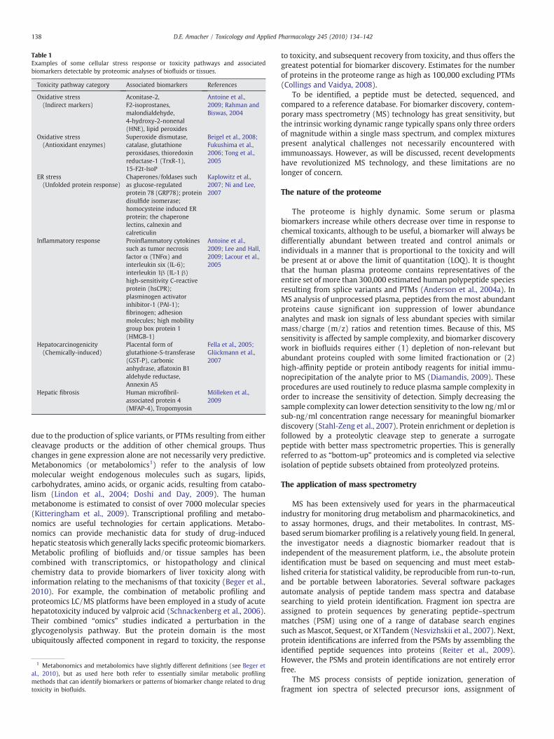
Table 1Examples of some cellular stress response or toxicity pathways and associatedbiomarkers detectable by proteomic analyses of biofluids or tissues.
Toxicity pathway category Associated biomarkers References
Oxidative stress(Indirect markers)
Aconitase-2,F2-isoprostanes,malondialdehyde,4-hydroxy-2-nonenal(HNE), lipid peroxides
Antoine et al.,2009; Rahman andBiswas, 2004
Oxidative stress(Antioxidant enzymes)
Superoxide dismutase,catalase, glutathioneperoxidases, thioredoxinreductase-1 (TrxR-1),15-F2t-IsoP
Beigel et al., 2008;Fukushima et al.,2006; Tong et al.,2005
ER stress(Unfolded protein response)
Chaperones/foldases suchas glucose-regulatedprotein 78 (GRP78); proteindisulfide isomerase;homocysteine induced ERprotein; the chaperonelectins, calnexin andcalreticulin
Kaplowitz et al.,2007; Ni and Lee,2007
Inflammatory response Proinflammatory cytokinessuch as tumor necrosisfactor α (TNFα) andinterleukin six (IL-6);interleukin 1β (IL-1 β)high-sensitivity C-reactiveprotein (hsCPR);plasminogen activatorinhibitor-1 (PAI-1);fibrinogen; adhesionmolecules; high mobilitygroup box protein 1(HMGB-1)
Antoine et al.,2009; Lee and Hall,2009; Lacour et al.,2005
Hepatocarcinogenicity(Chemically-induced)
Placental form ofglutathione-S-transferase(GST-P), carbonicanhydrase, aflatoxin B1aldehyde reductase,Annexin A5
Fella et al., 2005;Glückmann et al.,2007
Hepatic fibrosis Human microfibril-associated protein 4(MFAP-4), Tropomyosin
Mölleken et al.,2009
138 D.E. Amacher / Toxicology and Applied Pharmacology 245 (2010) 134–142
due to the production of splice variants, or PTMs resulting from eithercleavage products or the addition of other chemical groups. Thuschanges in gene expression alone are not necessarily very predictive.Metabonomics (or metabolomics1) refer to the analysis of lowmolecular weight endogenous molecules such as sugars, lipids,carbohydrates, amino acids, or organic acids, resulting from catabo-lism (Lindon et al., 2004; Doshi and Day, 2009). The humanmetabonome is estimated to consist of over 7000 molecular species(Kitteringham et al., 2009). Transcriptional profiling and metabo-nomics are useful technologies for certain applications. Metabo-nomics can provide mechanistic data for study of drug-inducedhepatic steatosis which generally lacks specific proteomic biomarkers.Metabolic profiling of biofluids and/or tissue samples has beencombined with transcriptomics, or histopathology and clinicalchemistry data to provide biomarkers of liver toxicity along withinformation relating to the mechanisms of that toxicity (Beger et al.,2010). For example, the combination of metabolic profiling andproteomics LC/MS platforms have been employed in a study of acutehepatotoxicity induced by valproic acid (Schnackenberg et al., 2006).Their combined “omics” studies indicated a perturbation in theglycogenolysis pathway. But the protein domain is the mostubiquitously affected component in regard to toxicity, the response
1 Metabonomics and metabolomics have slightly different definitions (see Beger etal., 2010), but as used here both refer to essentially similar metabolic profilingmethods that can identify biomarkers or patterns of biomarker change related to drugtoxicity in biofluids.
to toxicity, and subsequent recovery from toxicity, and thus offers thegreatest potential for biomarker discovery. Estimates for the numberof proteins in the proteome range as high as 100,000 excluding PTMs(Collings and Vaidya, 2008).
To be identified, a peptide must be detected, sequenced, andcompared to a reference database. For biomarker discovery, contem-porary mass spectrometry (MS) technology has great sensitivity, butthe intrinsic working dynamic range typically spans only three ordersof magnitude within a single mass spectrum, and complex mixturespresent analytical challenges not necessarily encountered withimmunoassays. However, as will be discussed, recent developmentshave revolutionized MS technology, and these limitations are nolonger of concern.
The nature of the proteome
The proteome is highly dynamic. Some serum or plasmabiomarkers increase while others decrease over time in response tochemical toxicants, although to be useful, a biomarker will always bedifferentially abundant between treated and control animals orindividuals in a manner that is proportional to the toxicity and willbe present at or above the limit of quantitation (LOQ). It is thoughtthat the human plasma proteome contains representatives of theentire set of more than 300,000 estimated human polypeptide speciesresulting from splice variants and PTMs (Anderson et al., 2004a). InMS analysis of unprocessed plasma, peptides from the most abundantproteins cause significant ion suppression of lower abundanceanalytes and mask ion signals of less abundant species with similarmass/charge (m/z) ratios and retention times. Because of this, MSsensitivity is affected by sample complexity, and biomarker discoverywork in biofluids requires either (1) depletion of non-relevant butabundant proteins coupled with some limited fractionation or (2)high-affinity peptide or protein antibody reagents for initial immu-noprecipitation of the analyte prior to MS (Diamandis, 2009). Theseprocedures are used routinely to reduce plasma sample complexity inorder to increase the sensitivity of detection. Simply decreasing thesample complexity can lower detection sensitivity to the low ng/ml orsub-ng/ml concentration range necessary for meaningful biomarkerdiscovery (Stahl-Zeng et al., 2007). Protein enrichment or depletion isfollowed by a proteolytic cleavage step to generate a surrogatepeptide with better mass spectrometric properties. This is generallyreferred to as “bottom-up” proteomics and is completed via selectiveisolation of peptide subsets obtained from proteolyzed proteins.
The application of mass spectrometry
MS has been extensively used for years in the pharmaceuticalindustry for monitoring drug metabolism and pharmacokinetics, andto assay hormones, drugs, and their metabolites. In contrast, MS-based serum biomarker profiling is a relatively young field. In general,the investigator needs a diagnostic biomarker readout that isindependent of the measurement platform, i.e., the absolute proteinidentification must be based on sequencing and must meet estab-lished criteria for statistical validity, be reproducible from run-to-run,and be portable between laboratories. Several software packagesautomate analysis of peptide tandem mass spectra and databasesearching to yield protein identification. Fragment ion spectra areassigned to protein sequences by generating peptide–spectrummatches (PSM) using one of a range of database search enginessuch as Mascot, Sequest, or X!Tandem (Nesvizhskii et al., 2007). Next,protein identifications are inferred from the PSMs by assembling theidentified peptide sequences into proteins (Reiter et al., 2009).However, the PSMs and protein identifications are not entirely errorfree.
The MS process consists of peptide ionization, generation offragment ion spectra of selected precursor ions, assignment of

139D.E. Amacher / Toxicology and Applied Pharmacology 245 (2010) 134–142
fragment ion spectra to peptide sequences, and the assembly ofprotein identity from the identified peptides. Matrix-assisted laserdesorption/ionization (MALDI) and electrospray ionization (ESI) aretwo common soft ionization techniques used for the generation ofpeptide/protein ions (molecular ions) for subsequent mass measure-ments (Aebersold and Mann, 2003). MALDI has an advantage of highsensitivity, and mass spectrometry imaging using MALDI (MALDI-MSItechnology) has found application in clinical proteomics for thedetection of disease biomarkers (Franck et al., 2009). MALDI allowsthe non-destructive interrogation of samples. Because it operates atatmospheric pressure, ESI is more commonly applied to complexpeptide/protein mixtures and is easily coupled on-line with apreceding liquid chromatography (LC) or capillary electrophoresisseparation step. After ionization, isolated proteins or peptides aresubjected to fragmentation by methods such as collision-induceddissociation (CID), electron capture dissociation (ECD), electrontransfer dissociation (ETD), sustained off-resonance irradiation(SORI) fragmentation, or infrared multiphoton dissociation (IRMPD)(Gao et al., 2009).
Applied to biomarker discovery, a critical step is the absolutequantification of proteins. Quantification based on strategies involv-ing stable-isotope dilution for the absolute quantification of proteinabundances have been reviewed by Brun et al. (2009). These methodsinvolve the addition of defined quantities of stable-isotope labeledstandards (e.g. 13C and/or 15N-labeled) that exhibit chromatographicproperties identical to native compounds. Isotope dilution methodsare generally targeted, i.e., focused on a restricted set of proteins.
The application of targeted quantitative mass spectrometry
The initial approach to proteomic analysis for toxicity biomarkers isbased on a global protein analysis (Zhou and Yu, 2003), also referred toas shotgun proteomics, which strives to maximize the coverage of theentire proteome. Shotgun proteomics data can provide a relatively highthroughput assessment of changes in protein expression, but the dataare highly redundant and insensitive to isoforms. A more discrete levelof protein analysis termed targeted proteomics (Dongre et al., 2001)focuses on a smaller number of proteins that convey specificinformation on pathological processes or response to toxic injury.After somepreliminary steps, global proteomics involves the extraction,enzymaticdigestion, and fractionationof proteins prior toanalysis of theresulting peptides by tandem mass spectrometry (MS/MS). Aspreviously discussed, serumandplasma require a preliminary depletion(or enrichment) step. High-abundance protein affinity depletion can becompleted via 2DLC (sequential ion-exchange and reversed phase
Table 2Some advantages and limitations of mass spectrometry-based (LC-MS/MS) targeted protei
Advantages L
Capable of the detection and absolute quantitation of a wide spectrum of proteins. Idealfor identification and quantitation of novel panels of protein markers for whichquality immuno-based reagents are not available.
Sim
Ease of assembling multiplex detection (e.g. N50) in a single measurement. Themultiplexing capability of LC-QqQ-MS platforms for measuring peptides in complexdigests is substantial.
As
Selected and MRM applications allow this technology to be used in the progressivestages of biomarker discovery, development, and routine study applications withoutthe use of any specific antibody reagents.
Mime
The instrumentation used to measure peptide MRMs is very similar to existingplatforms used for the high throughput quantitative measurement of plasma drugmetabolites.
Ss
Unlike immunoassays, peptide MRM results should be insensitive to alterations inprotein folding and subunit alterations.
Ttq
Analytical precision for multiple assays with human plasma proteins has beendemonstrated as b10% with in-run coefficients of variation 2–20%.
Adapted from Anderson and Hunter (2006); Kuzyk et al. (2009b), and other sources.
chromatography). Examples of a global approach include 2DGE-MSanalyses (Amacher et al., 2005; Yamamoto et al., 2006) or 2DLC-ESI-MSanalysis (Dail et al., 2008) of rat liver following exposure to varioustoxicants ordrugs.Meneses-Lorente et al. (2004, 2006) have used global2DE-MS procedures to identify proteomic signatures associated withhepatocellular steatosis in rat liver, serum. Similarly, Chen et al. (2007)have reported the global protein analysis of human liver using RPLC-MS/MS.
Targeted proteomics typically is facilitated by the SRM or, whenmultiplexed, themultiple reactionmonitoring (MRM)MS application.Selective reaction monitoring uses a scan table to monitor selectedions for quantification. Targeted proteomics can include the selectivefocus on a subproteome such as the glycoproteome (Scheiss et al.,2009) or specific proteins of interest. Pan et al. (2009) havesummarized 20 recent studies using MS-based targeted proteinquantitation for specimens including plasma, serum, cerebral spinalfluid, urine, tissue, and cell lysate. A list of candidate biomarkers fortargeted proteomics has been compiled by Polanski and Anderson(2006). An absolute quantification strategy is typically used that isbased on stable-isotope dilution as discussed above. Table 2 sum-marizes the advantages and disadvantages of these LC-MS/MStechnologies in comparison to other methods such as immunoassays.
Stages of biomarker discovery and development
Four distinct stages for the biomarker discovery and developmentprocess have been identified as discovery, qualification, verification, andvalidation with verification being the rate-limiting step for proteinbiomarker development (Berna and Ackermann, 2009). The three stepsin biomarker discovery include identification of markers, prioritizationof identified markers, and the preliminary qualification of prioritizedmarkers (Gao et al., 2005). MS-based techniques such as LC-MS/MS arelow throughput, but ideal for biomarker discovery. Qualification refersto the process whereby a biomarker is linked to a preclinical or clinicalend point or to a biological process in a specific context (Goodsaid andFrueh, 2007; Wagner, 2008). The verification of candidate biomarkersrequires demonstration of the reproducibility and transferability ofthese assays between laboratories, a formidable process. For mostbiomarker candidates, verification can be done by multiple reactionmonitoring/stable-isotope dilution MS (MRM/SID-MS) using a triple-quadrupole mass spectrometer. The substantial multiplexing providedby MRM technology means that 30–100 candidate proteins can besimultaneously targeted andmeasured. Sensitivity is always somewhatconstrainedby sample complexity, but as previouslymentioned, affinitydepletion (Keshishian et al., 2007) or enrichment (Anderson et al.,
n quantification.
imitations
erial procedures are used resulting in low throughput (e.g. 1–3 days) compared tomunoassays.
lthough applicable to PTM peptides, the PTM modification must be known orpecifically hypothesized prior to the assay.
ost current MS-based platforms require multiple sample preparations such asmunosubtraction, multidimensional LC separation, or immunoaffinity or solid phase
xtraction to enhance the analytical dynamic range and detection sensitivity.ome proteins may not readily produce usable peptides because they are either toohort or too variable (e.g. immunoglobulins).
ranslation of any novel serum/plasma biomarker by preclinical LC-MS/MSechnology to clinical monitoring would require completion of an extensiveualification process and FDA approval.
140 D.E. Amacher / Toxicology and Applied Pharmacology 245 (2010) 134–142
2004b; Stahl-Zeng et al., 2007) can be used as a preliminary step toeliminate high-abundance proteins in serum or plasma that are not ofinterest, thus decreasing complexity and increasing sensitivity. Valida-tion refers to the formal, technical procedure to prove the suitability ofan analytical assay and involves the determination of analyticalaccuracy, precision, limits of quantification, and stability of an analytein a matrix (Muller and Dieterle, 2009). Subsequently, validation androutine monitoring of protein biomarkers are most readily achieved byligand binding assays in diagnostic and pharmaceutical labs. The mostprevalent format is the enzyme-linked immunosorbent assay (ELISA),but this, of course, requires the availability of high quality and low-costantibodies against the candidate biomarker. In practice, the advantagesof using multiplexed panels of biomarkers include specimen conserva-tion, limited sample handling, and cost and time savings, but thechallenges include the need to optimize assay format for each proteinand selecting common dilution factors.
Selected and multiple reaction monitoring
MRM is a targeted MS scan mode unique to triple-quadrupole MSinstrumentation and requiring knowledge of an analyte's molecularweight and its fragmentation behavior under collision-induceddissociation (Kuzyk et al., 2009b). Unlike a full scan, the SRM orMRM scan is narrowly focused on the precursor ion(s) and specificproduct ion(s). The method provides high selectivity by monitoringchromatographic co-elution of multiple transitions for a given peptide(Wolf-Yadlin et al., 2007). It is capable of highly reproducibleconcentration determinations when stable-isotope labeled internalstandards (SIS) are included. The combination of triple-quadrupoleMS instrumentationwith nanoliter flow-rate high-performance liquidchromatography and nano-electrospray ionization is ideal for bio-marker discovery or qualification. The sensitivity limits for LC-MS/MSusually lie within the range of fg–pg of analyte per LC on-columninjection (Winnik and Kitchin, 2008). MRM-LC-MS/MS in electro-spray mode combined with stable-isotope dilution is well-suited fordirect quantification of proteins in plasma. For example, it hasrecently been reported that when combined with the use ofisotopically-labeled synthetic peptide standards, MRM analysis iscapable of sensitive (attomole level) and absolute determination ofpeptide concentrations across a wide concentration scale spanning adynamic range of 103 to 104 (Kuzyk et al., 2009a). In a recent multi-site assessment of MRM-based measurements of proteins in plasma,Addona et al. (2009) have demonstrated that these assays can behighly reproducible within and across laboratories and instrumentplatforms, and are sensitive to low mug/ml protein concentrations inunfractionated plasma.
While biofluids are more commonly analyzed in the search forspecific organ toxicity biomarkers that can eventually be used in bothpreclinical and clinical applications, the proteome of animal tissues orprimary cells in culture can also be evaluated for toxicity biomarkersfollowing exposure to xenobiotics. The advantages of these applica-tions include (1) the potential discovery of toxicity signatures, i.e.,linked changes or patterns in the proteome and/or metabonome thatare characteristic of and thus potentially predictive of specific types oftoxic injury or (2) a relatively quick way to compare close analogs ofNCEs for potential off-target effects before committing to longer termanimal testing. This is possible because at the tissue or cellular level,toxicological pathways, i.e. those cellular response pathways that canresult in adverse health effects when sufficiently perturbed, can bemonitored. These include signaling cascades which regulate many cellbiological processes that include proliferation, differentiation, migra-tion, and cell death. Other pathways and processes affected bytoxicants can include cell matrix adhesion, amino acid transporteractivity, fatty acid biosynthetic process, cellular defense responses,chemokine activity, organic ion transporter activity, sulfate transport,cytoskeletal organization, positive regulation of transcription, and
carbohydrate transport among others. Toxicant-induced mechanisticchanges in glucose and lipid metabolism can also result in potentialmetabonomic biomarkers. Other protein changes can include adducts,i.e., proteins and specific protein sites covalently bound by reactivechemicals or their intermediate metabolites (Merrick, 2008). Forcomplete information on procedures used for MRM targetedproteomics methodology, the reader should refer publications byHan and Higgs, 2008; Kitteringham et al., 2009; Keshishian et al.,2007; Pan et al., 2009; and Stahl-Zeng et al., 2007.
Clinical applications
Once identified in preclinical studies, the application of any novelplasma or serum biomarker to clinical practice will not be easy. Theprocess of transferring basic research at the preclinical level intopractical clinical applications involves many sequential processessuch as development and validation of clinical assays, release ofreagents and systems by diagnostic companies, evaluation of theanalytical and clinical performances of the commercial assays in thefield, and reliable implementation into clinical training throughtraining laboratory professionals and refining interpretation andutilization of the new information by all medical personnel (Plebaniand Marincola, 2006). In humans, the disease state, age, life-style, co-medications, and other variables not found in preclinical studies cancomplicate biomarker responses. But multiplexed signature assaysmay well be the future direction for clinical testing, especially todetect isoform variants, and MRM is an especially attractivetechnology at the clinical level (Diamandis, 2009). Triple-quadrupoleLC-MS/MS instrumentation is already well-established in hospitallaboratories. (Chace et al., 2006). Some particular advantages of MSover ELISA methods are for: (1) the analysis of proteins whereinterferences in the immunoassay are known (see: Hoofnagle et al.,2008), and (2) for new panels of protein biomarkers where theimmuno-based reagents do not exist or are not of sufficient quality.The ultimate goal for any preclinical toxicity biomarker discovery anddevelopment effort in biofluid analysis is translational medicinewhich refers to the translation of basic research into practical clinicalapplications. Much like the drug discovery/development/registrationpipeline itself, the discovery and development of novel toxicitybiomarkers is a long process with a high attrition rate.
The FDA has established a pilot process for biomarker qualification.The goal for the pilot process is a consensus at the FDA on theinterpretation of biomarker measurements submitted with IND/BLA/NDA. A nonclinical submission for qualification needs individual animaldata as well as summary tables (Goodsaid et al., 2008). Ultimate FDAapproval and clinical acceptance of MS-based biomarker assaysincluding MRM-derived assays will require formal evaluation in aCollege of American Pathologists (CAP) approved Clinical LaboratoriesImprovement Amendment-certified (CLIA) reference laboratory.
Concluding remarks
In preclinical/animal toxicology studies, the goal for identifyingand using novel qualified predictive safety biomarkers is to assist inselecting drug candidates that are more likely to be safe and well-tolerated in humans. In practice, hepatotoxicity biomarker discoveryand development has proceeded along two distinct lines of investi-gation. The first involves studies that are focused on the identificationof toxicity biomarkers in biofluids that would augment or even besuperior to those in current practice. Ideally, these would be highlyselective, very sensitive, indicative of a specific type of organ injury,and eventually could be applied to both preclinical and clinicalsamples. This type of biomarker would be released from the affectedtissue or organ just prior to or coincidental with initial toxicant-induced liver injury and could be confirmed by histopathology orother established indicators of altered organ function upon continued
141D.E. Amacher / Toxicology and Applied Pharmacology 245 (2010) 134–142
dosing. A second line of investigation involves the examination oftissue samples from preclinical species or primary hepatocyte cellcultures for multiple inter-related biomarkers that are associatedwithestablished toxicity mechanisms, as for example, oxidative or ERstress. The latter would conform to distinct toxicity signatures orpatterns that are indicative of complex cellular responses toperturbation of homeostatic maintenance. There is a growingconsensus that panels of biomarkers, for example 4–10, may be ableto supply the specificity and sensitivity that individual markers lack(Polanski and Anderson, 2006).
MRM-LC-MS/MS has become the preferred platform for thetargeted and quantitative discovery and development of novelbiomarkers. Emphasis is shifting toward performing full biomarkerdiscovery, qualification, and quantification on the same technologyplatform. Multiplexing (MRM), use of stable-isotope labeling, and thedevelopment of technology platforms for the determination of PTMshas enhanced sensitivity and specificity of MS assays. PTMs present achallenge for the investigator. But the current literature suggests thatthese have been resolved through the application of triple-quadrupoleor hybrid triple-quadrupole linear ion trap instruments. The hybridlinear ion trap (Q TRAP) mass spectrometer combines very sensitivetriple-quadrupole based tandem mass spectrometry (MS/MS) scanswith very sensitive ion trap product ion scans which are ideal forproteomics applications. (LeBlanc et al., 2003). This allows rapididentification of peptides at low concentrations derived from PTMs onchromatographic time scales.
Conflict of interest statement
The author has no financial or personal relationships with otherpeople or organizations that have influenced the work submitted forpublication. The work was not supported by any funding source.
References
Aardema, M.J., MacGregor, J.T., 2002. Toxicology and genetic toxicology in the new eraof “toxicogenomics”: impact of “omics” technologies. Mutat. Res. 499, 13–25.
Addona, T.A., Abbatiello, S.E., Schilling, B., Skates, S.J., Mani, D.R., Bunk, D.M.,Spiegelman, C.H., Zimmerman, L.J., Ham, A.J., Keshishian, H., Hall, S.C., Allen, S.,Blackman, R.K., Borchers, C.H., Buck, C., Cardasis, H.L., Cusack, M.P., Dodder, N.G.,Gibson, B.W., Held, J.M., Hiltke, T., Jackson, A., Johansen, E.B., Kinsinger, C.R., Li, J.,Mesri, M., Neubert, T.A., Niles, R.K., Pulsipher, T.C., Ransohoff, D., Rodriguez, H.,Rudnick, P.A., Smith, D., Tabb, D.L., Tegeler, T.J., Variyath, A.M., Vega-Montoto, L.J.,Wahlander, A., Waldemarson, S., Wang, M., Whiteaker, J.R., Zhao, L., Anderson, N.L.,Fisher, S.J., Liebler, D.C., Paulovich, A.G., Regnier, F.E., Tempst, P., Carr, S.A., 2009.Multi-site assessment of the precision and reproducibility of multiple reactionmonitoring-based measurements of proteins in plasma. Nat. Biotechnol. 27,633–641.
Aebersold, R., Mann, M., 2003. Mass spectrometry-based proteomics. Nature 422, 198–207.Amacher, D.E., 2002. A toxicologist's guide to biomarkers of hepatic response. Hum.
Exp. Toxicol. 21, 253–262.Amacher, D.E., Smith, D.J., Martz, L.K., Hoffmann, W.E., 1987. Characterization of
alkaline phosphatase in canine serum. Enzyme 37, 141–149.Amacher, D.E., Schomaker, S.J., Burkhardt, J.E., 1998. The relationship among
microsomal enzyme induction, liver weight, and histologic change in rat toxicologystudies. Food Chem. Toxicol. 36, 831–839.
Amacher, D.E., Schomaker, S.J., Burkhardt, J.E., 2001. The relationship amongmicrosomal enzyme induction, liver weight, and histological change in beagledog toxicology studies. Food Chem. Toxicol. 39, 817–825.
Amacher, D.E., Adler, R., Herath, A., Townsend, R.R., 2005. The use of proteomicmethodsto identify serum biomarkers associated with rat liver toxicity or hypertrophy. Clin.Chem. 51, 1796–1803.
Anderson, N.L., Anderson, N.G., 2002. The human plasma proteome: history, character,and diagnostic prospects. Mol. Cell. Proteomics 1, 845–867.
Anderson, L., Hunter, C.L., 2006. Quantitative mass spectrometric multiple reactionmonitoring assays for major plasma proteins. Mol. Cell. Proteomics 5, 573–588.
Anderson, N.L., Polanski, M., Pieper, R., Gatlin, T., Tirumalai, R.S., Conrads, T.P., Veenstra,T.D., Adkins, J.N., Pounds, J.G., Fagan, R., Lobley, A., 2004a. The human proteome: anonredundant list developed by combination of four separate sources. Mol. Cell.Proteomics 3, 311–326.
Anderson, N.L., Anderson, N.G., Haines, L.R., Hardie, D.B., Olafson, R.W., Pearson, T.W., 2004b.Mass spectrometric quantitation of peptides andproteins using Stable IsotopeStandardsAnd Capture by Anti-Peptide Antibodies (SISCAPA). J. Proteome Res. 3, 235–244.
Antoine, D.J., Mercer, A.E., Williams, D.P., Park, B.K., 2009. Mechanism-based bioanalysisand biomarkers for hepatic chemical stress. Xenobiotics 39, 565–577.
Ashamiss, F., Wierzbicki, Z., Chrzanowska, A., Scibior, D., Pacholczyk, M., Kosieradzki,M., Lagiewska, B., Porembska, Z., Rowiński, W., 2004. Clinical significance ofarginase after liver transplantation. Ann. Transplant. 9, 58–60.
Beger, R.D., Sun, J., Schnackenberg, L.K., 2010. Metabolomics approaches for discoveringbiomarkers of drug-induced hepatotoxicity and nephrotoxicity. Toxicol. Appl.Pharmacol. 243 (2), 154–166.
Beigel, J., Fella, K., Kramer, P.J., Kroeger, M., Hewitt, P., 2008. Genomics and proteomicsanalysis of cultured primary rat hepatocytes. Toxicol. In Vitro 22, 171–181.
Berna, M., Ackermann, B., 2009. Increased throughput for low-abundance proteinbiomarker verification by liquid chromatography/tandem mass spectrometry.Anal. Chem. 81, 3950–3956.
Beyer, T.A., Xu,W., Teupser, D., auf dem Keller, U., Bugnon, P., Hildt, E., Thiery, J., Kan, Y.W.,Werner, S., 2008. Impaired liver regeneration in Nrf2 knockout mice: role of ROS-mediated insulin/IGF-1 resistance. EMBO J. 27, 212–223.
Bhave, V.S., Donthamsetty, S., Latendresse, J.R., Muskhelishvili, L., Mehendale, H.M.,2008a. Secretory phospholipase A2mediates progression of acute liver injury in theabsence of sufficient cyclooxygenase-2. Toxicol. Appl. Pharmacol. 228, 225–238.
Bhave, V.S., Donthamsetty, S., Latendresse, J.R., Mehendale, H.M., 2008b. Inhibition ofcyclooxygenase-2 aggravates secretory phospholipase A2-mediated progression ofacute liver injury. Toxicol. Appl. Pharmacol. 228, 239–246.
Bondy, G.S., Armstrong, C.L., Curran, I.H., Barker, M.G., Mehta, R., 2000. Retrospectiveevaluation of serum ornithine carbamyltransferase activity as an index ofhepatotoxicity in toxicological studies with rats. Toxicol. Lett. 114, 163–171.
Boone, L., Meyer, D., Cusick, P., Ennulat, D., Bolliger, A.P., Everds, N., Meador, V., Elliott,G., Honor, D., Bounous, D., Jordan, H., 2005. Selection and interpretation of clinicalpathology indicators of hepatic injury in preclinical studies. Vet. Clin. Pathol. 34,182–188.
Brun, V., Masselon, C., Garin, J., Dupuis, A., 2009. Isotope dilution strategies for absolutequantitative proteomics. J. Proteomics 72, 740–749.
Chace, D.H., Barr, J.R., Duncan, M.W., Matern, D., Morris, M.R., Palmer-Toy, D.E.,Rockwood, A.L., Siuzdak, G., Urbani, A., Yergey, A.L., Chan, Y.M., 2006. Massspectrometry in the clinical laboratory: general principles and guidance. Ap-proved Guideline. Clinical and Laboratory Standards Institute, Wayne, Pennsylva-nia, pp. 1–94.
Chen, M., Ying, W., Song, Y., Liu, X., Yang, B., Wu, S., Jiang, Y., Cai, Y., He, F., Qian, X., 2007.Analysis of human liver proteome using replicate shotgun strategy. Proteomics 7,2479–2488.
Chrzanowska, A., Gajewska, B., Barańczyk-Kuźma, A., 2009. Arginase isoenzymes inhuman cirrhotic liver. Acta Biochem. Pol. 56, 465–469.
Collings, F.B., Vaidya, V.S., 2008. Novel technologies for the discovery and quantitationof biomarkers of toxicity. Toxicology 245, 167–174.
Dail, M.B., Shack, L.A., Chambers, J.E., Burgess, S.C., 2008. Global liver proteomics of ratsexposed for 5 days to Phenobarbital identifies changes associated with cancer andwith CYP metabolism. Toxicol. Sci. 102, 556–569.
Diamandis, E.P., 2009. Protein quantification bymass spectrometry: is it ready for primetime? Clin. Chem. 55, 1427–1430.
Dongre, A.R., Opiteck, G., Cosand,W.L., Hefta, S.A., 2001. Proteomics in the post-genomeage. Biopolymers 60, 206–211.
Doshi, R., Day, J.R., 2009. Metabolomics of serum peptides. Protein Pept. Lett. 16, 460–466.Evans, G.O., 2009. Animal Clinical Chemistry. A Practical Guide for Toxicologists and
Biomedical Researchers, second ed. CRC Press, Boca Raton.Fella, K., Glückmann, M., Hellmann, J., Karas, M., Kramer, P.-J., Kröger, M., 2005. Use of
two-dimensional gel electrophoresis in predictive toxicology: identification ofpotential early protein biomarkers in chemically induced hepatocarcinogenesis.Proteomics 5, 1914–1927.
Franck, J., Arafah, K., Elayed, M., Bonnel, D., Vergara, D., Jacquet, A., Vinatier, D.,Wisztorski, M., Day, R., Fournier, I., Salzet, M., 2009. MALDI imaging massspectrometry: state of the art technology in clinical proteomics. Mol. Cell.Proteomics. 8, 2023–2033.
Fukushima, T., Kikkawa, R., Hamada, Y., Horii, I., 2006. Genomic cluster and networkanalysis for predictive screening for hepatotoxicity. J. Toxicol. Sci. 31, 419–432.
Gao, J., Garulacan, L.A., Storm, S.M., Opiteck, G.J., Dubaquie, Y., Hefta, S.A., Dambach, D.M.,Dongre, A.R., 2005. Biomarker discovery in biological fluids. Methods 35, 291–302.
Gao, R.D., Holland, D., Yu, L.-R., 2009. Quantitative proteomics for drug toxicity. Brief.Funct. Genomics Proteomics 8, 158–166.
Glückmann, M., Fella, K., Waidelich, D., Merkel, D., Kruft, V., Kramer, P.J., Walter, Y.,Hellmann, J., Karas,M., Kröger,M., 2007. Prevalidation of potential protein biomarkersin toxicology using iTRAQ reagent technology. Proteomics 7, 1564–1574.
Goodsaid, F., Frueh, F., 2007. Biomarker qualification pilot process at the US Food andDrug Administration. AAPS J. 9, E105–E108.
Goodsaid, F.M., Frueh, F.W., Mattes, W., 2008. Strategic paths for biomarkerqualification. Toxicology 245, 219–223.
Han, B., Higgs, R.E., 2008. Proteomics: from hypothesis to quantitative assay on a singleplatform. Guidelines for developing MRM assays using ion trap mass spectrometers.Brief. Funct. Genomics Proteomics 7, 340–354.
Hoofnagle, A.N., Becker, J.O., Wener, M.H., Heinecke, J.W., 2008. Quantification ofthyroglobin, a low-abundance serum protein, by immunoaffinity peptide enrichmentand tandem mass spectrometry. Clin. Chem. 54, 1796–1804.
Jackson, E.R., Kilroy, C., Joslin, D.L., Schomaker, S.J., Pruimboom-Brees, I., Amacher, D.E.,2008. The early effects of short-term dexamethasone administration on hepaticand serum alanine aminotransferase in the rat. Drug Chem. Toxicol. 31, 427–445.
Jenkens, W.J., Rosalki, S.B., Foo, Y., Scheuer, P.J., Nemesanszky, E., Sherlock, S., 1982.Serum glutamate dehydrogenase is not a reliable marker of liver cell necrosis inalcoholics. J. Clin. Pathol. 35, 207–210.
Kaplowitz, N., Than, T.A., Shinohara, M., Ji, C., 2007. Endoplasmic reticulum stress andliver injury. Semin. Liver Dis. 27, 367–377.
142 D.E. Amacher / Toxicology and Applied Pharmacology 245 (2010) 134–142
Keshishian, H., Addona, T., Burgess, M., Kuhn, E., Carr, S.A., 2007. Quantitative,multiplexed assays for low abundance proteins in plasma by targeted massspectrometry and stable isotope dilution. Mol. Cell. Proteomics 6, 2212–2229.
Kitteringham, N.R., Jenkins, R.E., Lane, C.S., Elliot, V.L., Park, B.K., 2009. Multiple reactionmonitoring for quantitative biomarker analysis in proteomics and metabolomics.J. Chromatogr. B Anal. Technol. Biomed. Life Sci. 877, 1229–1239.
Kuzyk, M.A., Ohlund, L.B., Elliott, M.H., Smith, D., Oian, H., Delaney, A., Hunter, C.L.,Borchers, C.H., 2009a. A comparison of MS/MS-based, stable-isotope-labeled,quantitation performance on ESI-quadrupole TOF and MALDI-TOF/TOF massspectrometers. Proteomics 9, 3328–3340.
Kuzyk, M.A., Smith, D., Yang, J., Cross, T., Jackson, A.M., Hardie, D.B., Anderson, N.L.,Borchers, C.H., 2009b. MRM-based, multiplexed, absolute quantitation of 45proteins in human plasma. Mol. Cell. Proteomics 8, 1860–1877.
Lacour, S., Gautier, J.C., Pallardy, M., Roberts, R., 2005. Cytokines as potential biomarkersof liver toxicity. Cancer Biomark. 1, 29–39.
Lauer, B., Tuschi, G., Kling,M., Mueller, S.O., 2009. Species-specific toxicity of diclofenac andtroglitazone inprimaryhumanandrathepatocytes. Chemico-Biol. Interact. 179, 17–24.
LeBlanc, J.C., Hager, J.W., Ilisiu, A.M., Hunter, C., Zhong, F., Chu, I., 2003. Unique scanningcapabilities of a new hybrid linear ion trap mass spectrophotometer (Q TRAP) usedfor high sensitivity proteomics applications. Proteomics 3, 859–869.
Lee, W.M., 2003a. Acute liver failure in the United States. Semin. Liver Dis. 23, 217–226.Lee, W.M., 2003b. Drug-induced hepatotoxicity. N. Engl. J. Med. 349, 474–485.Lee, J.W., Hall, M., 2009. Method validation of protein biomarkers in support of drug
development or clinical diagnosis/prognosis. J. Chromatogr. B. Anal. Technol.Biomed. Life Sci. 877, 1259–1271.
Limaye, P.B., Apte, U.M., Shankar, K., Bucci, T.J., Warbritton, A., Mehendale, H.M., 2003.Calpain released from dying hepatocytes mediates progression of acute liver injuryinduced by model hepatotoxicants. Toxicol. Appl. Pharmacol. 191, 211–226.
Linblom, P., Rafter, I., Copley, C., Andersson, U., Hedberg, J.J., Berg, A.-L., Samuelsson, A.,Hellmold, H., Cotgreave, I., Glinghammar, B., 2007. Isoforms of alanine amino-transferases in human tissues and serum—differential tissue expression usingnovel antibodies. Arch. Biochem. Biophys. 466, 66–77.
Lindena, J., Friedel, R., Rapp, K., Sommerfeld, U., Trautschold, I., Deerberg, F., 1980. Long-term observation of plasma and tissue enzyme activities in the rat. Mech. AgeingDev. 14, 379–407.
Lindon, J.C., Holmes, E., Nicholson, J.K., 2004. Toxicological applications of magneticresonance. Prog. Nucl. Magn. Reson. Spectrosc. 45, 109–143.
MacGregor, J.T., 2003. The future of regulatory toxicology: impact of the biotechnologyrevolution. Toxicol. Sci. 75, 236–248.
Meneses-Lorente, G., Guest, P.C., Lawrence, J., Muniappa, N., Knowles, M.R., Skynner, H.A.,Salim, K., Cristea, I., Mortishire-Smith, R., Gaskell, S.J., Watt, A., 2004. A proteomicinvestigation of drug-induced steatosis in rat liver. Chem. Res. Toxicol. 17, 605–612.
Meneses-Lorente, G., Watt, A., Salim, K., Gaskell, S.J., Muniappa, N., Lawrence, J., Guest,P.C., 2006. Identification of early proteomic markers for hepatic steatosis. Chem.Res. Toxicol. 19, 986–998.
Merrick, B.A., 2008. The plasma proteome, adductome and idiosyncratic toxicity intoxicoproteomics research. Brief. Funct. Genomics Proteomics 7, 35–49.
Merrick, B.A., Tomer, K.B., 2003. Toxicoproteomics: a parallel approach to identifyingbiomarkers. Environ. Health Perspect. 111, A578–A579.
Miller, T.J., Knapton, A., Adeyemo, O., Noory, L., Hanig, J.P., 2008. Cytochrome c: a non-invasive biomarker of drug-induced liver injury. J. Appl. Toxicol. 28, 815–828.
Miyazaki, M., Rosenblum, J.S., Kasahara, Y., Nakagawa, I., Patricelli, M.P., 2009.Determination of enzymatic source of alanine aminotransferase activity in serumfrom dogs with liver injury. Pharmacol. Toxicol. Meth. Sept. 11.
Mölleken, C., Sitek, B., Porschmann, G., Sipos, B., Wiesse, S., Warscheid, B., Broelsch, C.,Reiser, M., Friedman, S.L., Tornoe, I., Schosser, A., Klöppel, G., Schmiegel, W., Meyer,H.E., Holmskov, U., Stuhler, K., 2009. Detection of novel biomarkers of liver cirrhosisby proteomic analysis. Hepatology 49, 1257–1266.
Muller, P.Y., Dieterle, F., 2009. Tissue-specific, non-invasive toxicity biomarkers:translation from preclinical safety assessment to clinical safety monitoring. ExpertOpin. Drug Metab. Toxicol. 5, 1–16.
Müller, P.B., Taboada, J., Hosgood, G., Partington, B.P., VanSteenhouse, J.L., Taylor, H.W.,Wolfsheimer, K.J., 2000. Effects of long-term Phenobarbital treatment on the liverin dogs. J. Vet. Intern. Med. 14, 165–171.
Murayama, H., Ikemoto, M., Fukuda, Y., Tsunekawa, S., Nagata, A., 2007. Serum level ofornithine carbamoyltransferase is influenced by the state of Kupffer cells. Clin.Chim. Acta 380, 170–174.
Murayama, H., Ikemoto, M., Fukuda, Y., Nagata, A., 2008a. Superiority of serum type-aarginase and ornithine carbamyltransferase in the detection of toxicant-inducedacute hepatic injury in rats. Clin. Chim. Acta 391, 31–35.
Murayama, H., Ikemoto,M., Nagata, A., 2008b.Marked elevation of serummitochondrion-derived markers in mild models of non-alcoholic steatohepatitis in rats.J. Gastroenterol. Hepatol. 16, 38–45.
Murayama, H., Ikemoto, M., Hamaoki, M., 2009. Ornithine carbamyltransferase is asensitive marker for alcohol-induced liver injury. Clin. Chim. Acta 401, 100–104.
Nesvizhskii, A.I., Vitek, O., Aebersold, R., 2007. Analysis of proteomic data generated bytandem mass spectrometry. Nat. Meth. 4, 787–797.
Ni, M., Lee, A.S., 2007. ER chaperones in mammalian development and human diseases.FEBS Lett. 581, 3641–3651.
O'Brien, P.J., Slaughter, M.R., Polley, S.R., Kramer, K., 2002. Advantages of glutamatedehydrogenase as a blood biomarker of acute hepatic injury in rats. Lab. Anim. 36,313–321.
Pan, S., Aebersold, R., Chen, R., Rush, J., Goodlett, D.R., McIntosh, M.W., Zhang, J.,Brentnall, T.A., 2009. Mass spectrometry based targeted protein quantification:methods and applications. J. Proteome Res. 8, 787–797.
Plebani, M., Marincola, F.M., 2006. Research translation: a new frontier for clinicallaboratories. Clin. Chem. Lab. Med. 44, 1303–1312.
Polanski, M., Anderson, N.L., 2006. A list of candidate cancer biomarkers for targetedproteomics. Biomark. Insights 1, 1–48.
Rahman, I., Biswas, S.K., 2004. Non-invasive biomarkers of oxidative stress: reproducibilityand methodological issues. Redox. Rep. 9, 125–143.
Rajamohan, F., Nelms, L., Joslin, D.L., Lu, B., Reagan, W.J., Lawton, M., 2006. cDNAcloning, expression, purification, distribution, and characterization of biologicallyactive canine alanine aminotransferase-1. Protein Expr. Purf. 48, 81–89.
Ramaiah, S.K., 2007. A toxicologist guide to the diagnostic interpretation of hepaticbiochemical parameters. Food Chem. Toxicol. 45, 1551–1557.
Reiter, L., Classen, M., Schrimpf, S.P., Jovanoic, M., Schmidt, A., Buhmann, J.M.,Hengartner, M.O., Aebersold, R., 2009. Protein identification false positive ratesfor very large proteomics datasets generated by tandem mass spectrometry. Mol.Cell. Proteomics Jul 16.
Rifai, N., Gerszten, R.E., 2006. Biomarker discovery and validation. Clin. Chem. 52,3635–3637.
Rifai, N., Gillette, M.A., Carr, S.A., 2006. Protein biomarker discovery and validation: thelong and uncertain path to clinical utility. Nat. Biotechnol. 24, 971–983.
Scheiss, R., Wollscheid, B., Aebersold, R., 2009. Targeted proteomic strategy for clinicalbiomarker discovery. Mol. Oncol. 3, 33–44.
Schnackenberg, L.K., Jones, R.C., Thyparambil, S., Taylor, J.T., Han, T., Tong, W., Hansen,D.K., Fuscoe, J.C., Edmondson, R.D., Beger, R.D., Dragan, Y.P., 2006. An integratedstudy of acute effects of valproic acid in the liver using metabonomics, proteomics,and transcriptomics platforms. OMICS 10, 1–14.
Scrobohaci, M.L., Drouet, L., Baudin, B., 1988. Hemostasis tests as markers of hepatic andendothelial toxicity in chemotherapy. Nouv. Rev. Fr. Hematol. 30, 109–114.
Simmons, S.O., Fan, C.-Y., Ramabhadran, R., 2009. Cellular stress response pathwaysystem as a sentinel ensemble in toxicological screening. Toxicol. Sci. 111, 202–225.
Solter, P.F., 2005. Clinical pathology approaches to hepatic injury. Toxicol. Pathol. 33,9–16.
Spiro, R.G., 2002. Protein glycosylation: nature, distribution, enzymatic formation, anddisease implication of glycopeptide bonds. Glycobiology 12, 43R–56R.
Stahl-Zeng, J., Lange, V., Ossola, R., Eckhardt, K., Krek,W., Aebersold, R., Domon, B., 2007.High sensitivity detection of plasma proteins by multiple reaction monitoring ofN-glycosides. Mol. Cell. Protomics 6, 1809–1817.
Teschke, R., Neuefeind, M., Nishimura, M., Strohmeyer, G., 1983. Hepatic gamma-glutamyltransferase activity in alcoholic fatty liver: comparison with other liverenzyme in man and rats. Gut 24, 625–630.
Tirumalai, R.S., Chan, K.C., Prieto, D.A., Issaq, H.J., Conrads, T.P., Veenstra, T.D., 2003.Characterization of low molecular weight human serum proteome. Mol. Cell.Proteomics 2, 1096–1103.
Tong, V., Teng, X.W., Chang, T.K., Abbott, F.S., 2005. Valproic acid I: time course of lipidperoxidation biomarkers, liver toxicity, and valproic acid metabolite levels in rats.Toxicol Sci. 86, 427–435.
Wagner, J.A., 2008. Strategic approach to fit-for-purpose biomarkers in drugdevelopment. Annu. Rev. Pharmacol. Toxicol. 48, 631–651.
Wetmore, B.A., Merrick, B.A., 2004. Toxicoproteomics: proteomics applied to toxicologyand pathology. Toxicol. Pathol. 32, 619–642.
Wiedmeyer, C.E., Solter, P.E., Hoffmann, W.E., 2002. Alkaline phosphatase expression intissues from glucocorticoid-treated dogs. Am. J. Vet. Res. 63, 1083–1088.
Williams, A.M., Langley, P.G., Osei-Hwediah, J., Wendon, J.A., Hughes, R.D., 2003.Hyaluronic acid and endothelial damage due to paracetamol-induced hepatotox-icity. Liver Int. 23, 110–115.
Winnik, W.M., Kitchin, K.T., 2008. Measurement of oxidative stress parameters usingliquid chromatograph-tandem mass spectrometry (LC-MS/MS). Toxicol. Appl.Pharmacol. 233, 100–106.
Wolf-Yadlin, A., Hautaniemi, S., Lauffenburger, D.A., White, F.M., 2007. Multiplereaction monitoring for robust quantitative proteomic analysis of cellular signalingnetworks. Proc. Natl. Acad. Sci. U. S. A. 104, 5860–5865.
Wu, L., Han, D.K., 2006. Overcoming the dynamic range problem in mass spectrometry-based shotgun proteomics. Expert Rev. Proteomics 3, 611–619.
Yamamoto, T., Kikkawa, R., Yamada, H., Horii, I., 2006. Investigation of proteomicbiomarkers in in vivo hepatotoxicity study of rat liver: toxicity differentiation inhepatotoxicants. J. Toxicol. Sci. 31, 49–60.
Yang, R.Z., Park, S., Reagan, W.J., Goldstein, R., Zhong, S., Lawton, M., Rajamohan, F.,Qian, K., Liu, L., Gong, D.W., 2009. Alanine aminotransferase isoenzymes: molecularcloning and quantitative analysis of tissue expression in rats and serum elevation inliver toxicity. Hepatology 49, 598–607.
Zheng, A.-Y., Weng, S.-Y., Yu, Y., 2009. Signal molecule-mediated hepatic communi-cation during liver regeneration. World J. Gastroenterol. 15, 5776–5783.
Zhou, M., Yu, L.R., 2003. Proteomic analysis by two-dimensional polyacrylamide gelelectrophoresis. Adv. Protein Chem. 65, 57–84.