Embed Size (px)
Citation preview

The Digestive SystemThe Digestive System
Lindsey BilyAnatomy & Physiology
Austin High School
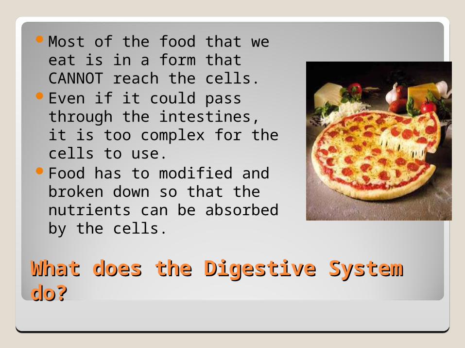
What does the Digestive System What does the Digestive System do?do?
Most of the food that we eat is in a form that CANNOT reach the cells.
Even if it could pass through the intestines, it is too complex for the cells to use.
Food has to modified and broken down so that the nutrients can be absorbed by the cells.
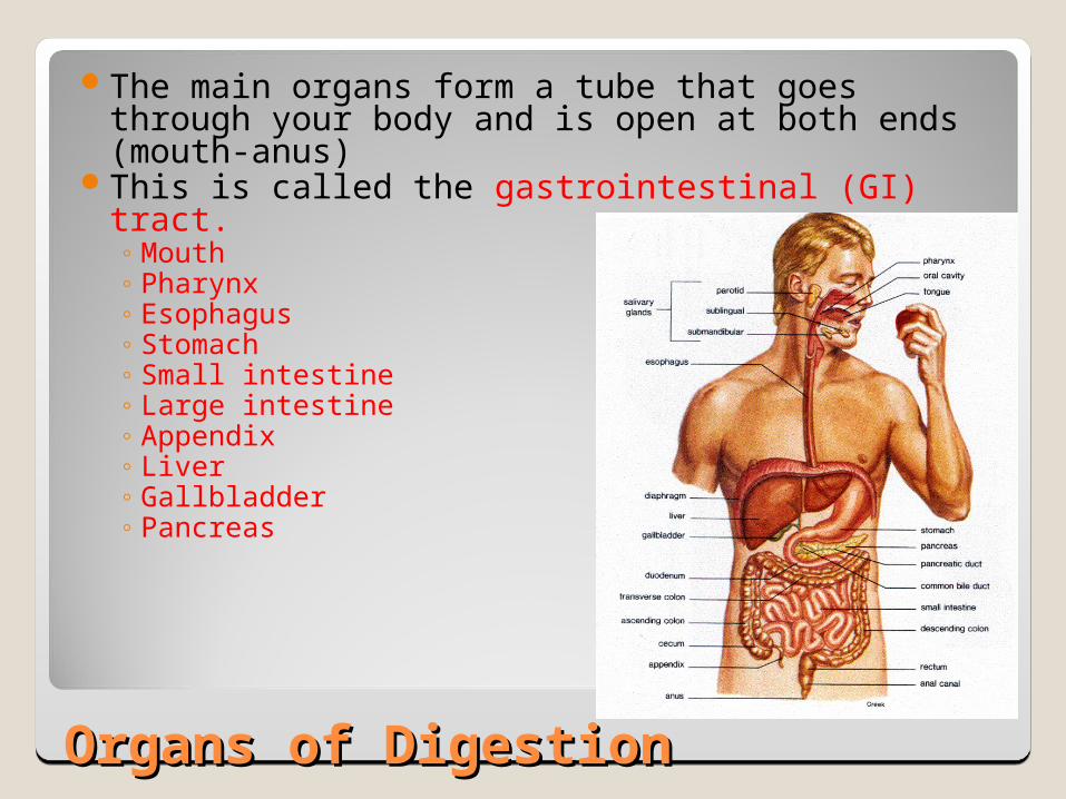
Organs of DigestionOrgans of Digestion
The main organs form a tube that goes through your body and is open at both ends (mouth-anus)
This is called the gastrointestinal (GI) tract.◦ Mouth◦ Pharynx◦ Esophagus◦ Stomach◦ Small intestine◦ Large intestine◦ Appendix◦ Liver◦ Gallbladder◦ Pancreas
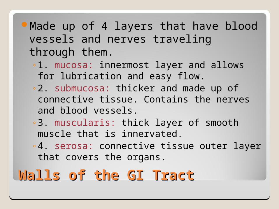
Walls of the GI TractWalls of the GI Tract
Made up of 4 layers that have blood vessels and nerves traveling through them.◦1. mucosa: innermost layer and allows for
lubrication and easy flow.◦2. submucosa: thicker and made up of
connective tissue. Contains the nerves and blood vessels.
◦3. muscularis: thick layer of smooth muscle that is innervated.
◦4. serosa: connective tissue outer layer that covers the organs.
The Mouth
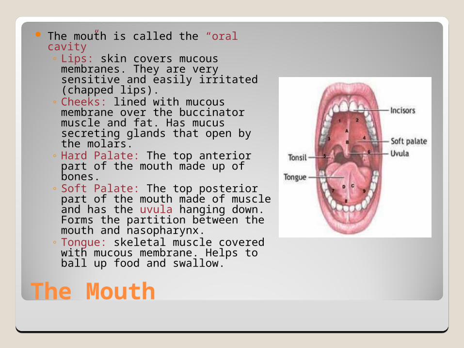
The mouth is called the “oral cavity”◦ Lips: skin covers mucous
membranes. They are very sensitive and easily irritated (chapped lips).
◦ Cheeks: lined with mucous membrane over the buccinator muscle and fat. Has mucus secreting glands that open by the molars.
◦ Hard Palate: The top anterior part of the mouth made up of bones.
◦ Soft Palate: The top posterior part of the mouth made of muscle and has the uvula hanging down. Forms the partition between the mouth and nasopharynx.
◦ Tongue: skeletal muscle covered with mucous membrane. Helps to ball up food and swallow.
The Mouth
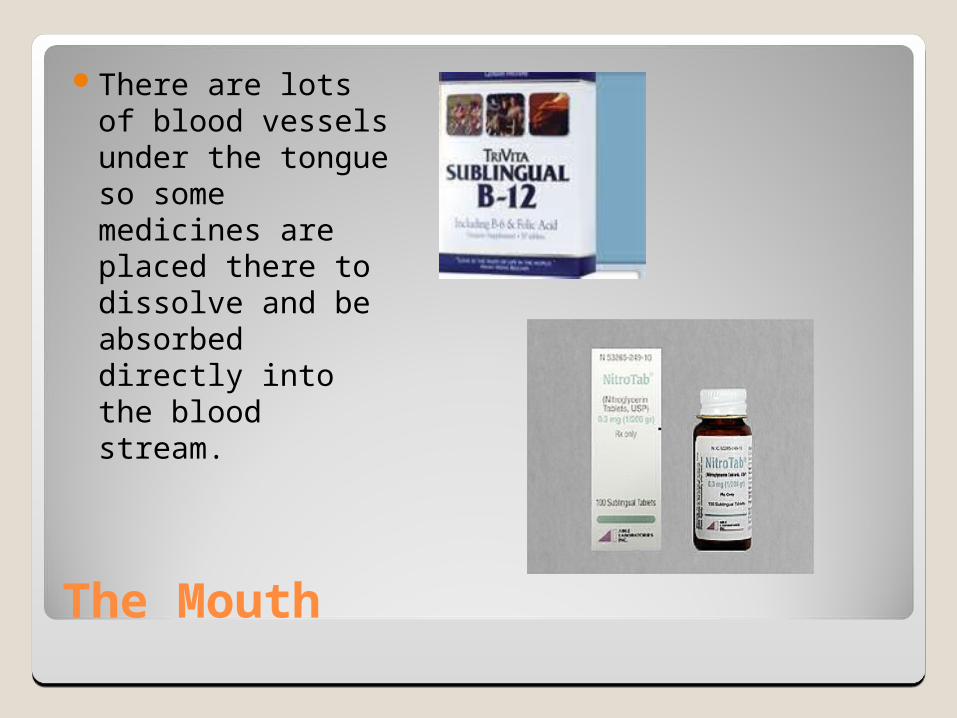
There are lots of blood vessels under the tongue so some medicines are placed there to dissolve and be absorbed directly into the blood stream.
The Mouth
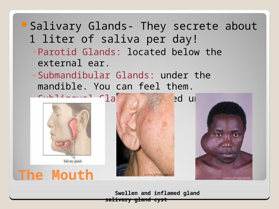
Salivary Glands- They secrete about 1 liter of saliva per day!◦Parotid Glands: located below the external ear.◦Submandibular Glands: under the mandible.
You can feel them.◦Sublingual Glands: located under the tongue.
Swollen and inflamed gland salivary gland cyst

The Teeth
Teeth allow us to masticate (to chew).They can cut, tear and grind our food so
that it ground into small bits which increases the surface area.
Increased surface area allows more surface for the digestive enzymes to get to.
The Teeth
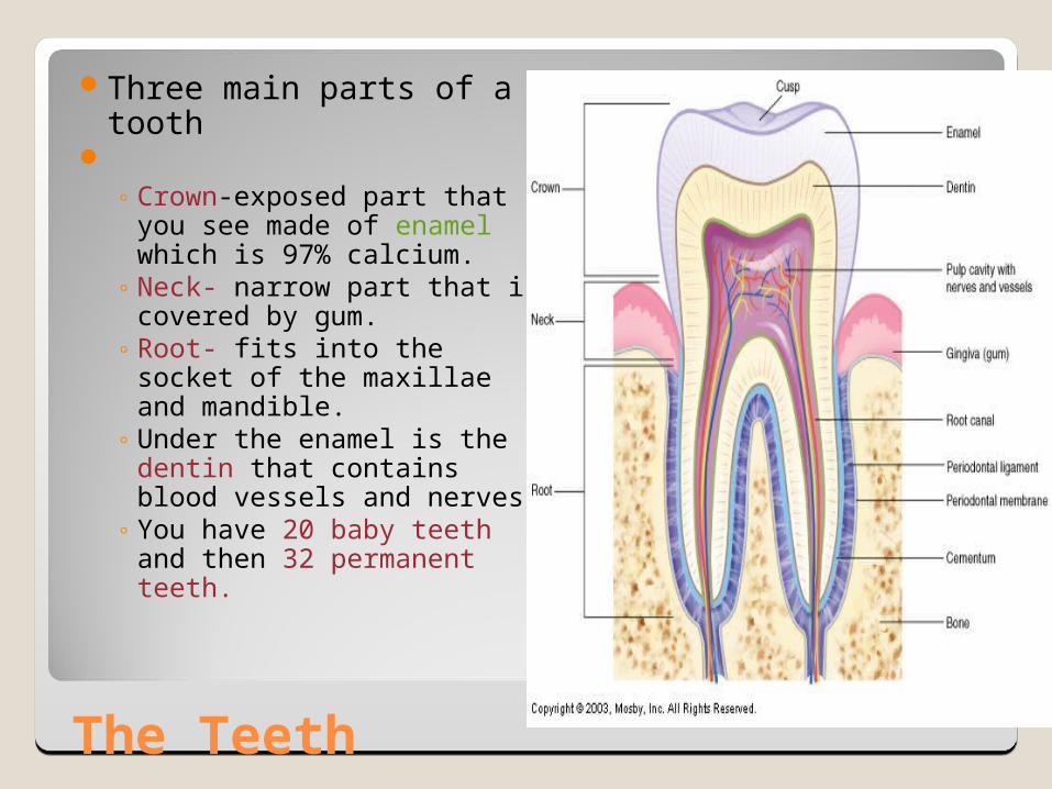
Three main parts of a tooth
◦ Crown-exposed part that
you see made of enamel which is 97% calcium.
◦ Neck- narrow part that is covered by gum.
◦ Root- fits into the socket of the maxillae and mandible.
◦ Under the enamel is the dentin that contains blood vessels and nerves.
◦ You have 20 baby teeth and then 32 permanent teeth.
Pharynx
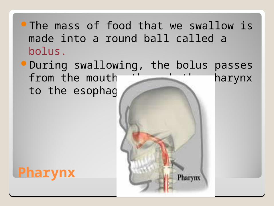
The mass of food that we swallow is made into a round ball called a bolus.
During swallowing, the bolus passes from the mouth, through the pharynx to the esophagus.

Esophagus
About 25 cm long and goes from the pharynx to the stomach through the diaphragm.
Lies posterior to the tracheaGuarded by 2 sphincters at each end
◦Upper esophageal sphincter: makes sure you don’t get air in your esophagus.
◦Lower esophageal sphincter (cardiac sphincter): opening of the esophagus and stomach.
Esophagus
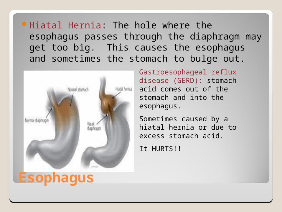
Hiatal Hernia: The hole where the esophagus passes through the diaphragm may get too big. This causes the esophagus and sometimes the stomach to bulge out.
Gastroesophageal reflux disease (GERD): stomach acid comes out of the stomach and into the esophagus.
Sometimes caused by a hiatal hernia or due to excess stomach acid.
It HURTS!!
The Stomach
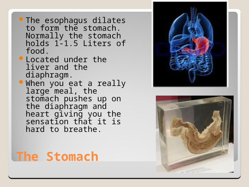
The esophagus dilates to form the stomach. Normally the stomach holds 1-1.5 Liters of food.
Located under the liver and the diaphragm.
When you eat a really large meal, the stomach pushes up on the diaphragm and heart giving you the sensation that it is hard to breathe.
Stomach
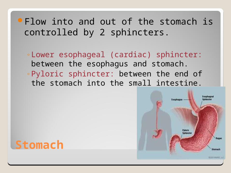
Flow into and out of the stomach is controlled by 2 sphincters.
◦Lower esophageal (cardiac) sphincter: between the esophagus and stomach.
◦Pyloric sphincter: between the end of the stomach into the small intestine.
Stomach Functions
Stores food until it is partially digested and ready to move to the intestines.
Secretes gastric juice that contains acid and enzymes to help digest food.
Churns the food which breaks it into the small particles and mixes it with the gastric juice.
Secretes intrinsic factor that protects vitamin B12 from being destroyed.
Absorbs some nutrients (some drugs, water, alcohol, and some short chain fatty acids).
Produces hormones gastrin (regulates digestion) and ghrelin (increases appetite).
Destroys some pathogens.

Small Intestine
It is 1 inch wide and 20 feet long and fills most of the abdominal cavity!
Has three divisions…◦duodenum: part where the stomach attaches.◦Jejunum: part where it turns abruptly forward
and downward. It is about 8 feet long.◦Ileum: The remaining 12 feet.
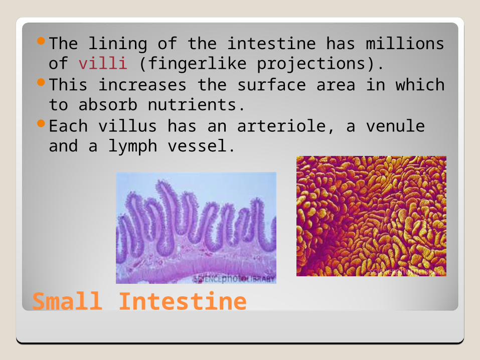
Small Intestine
The lining of the intestine has millions of villi (fingerlike projections).
This increases the surface area in which to absorb nutrients.
Each villus has an arteriole, a venule and a lymph vessel.
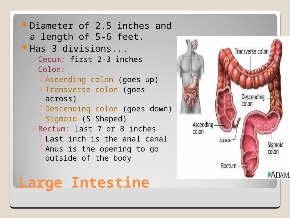
Large Intestine
Diameter of 2.5 inches and a length of 5-6 feet.
Has 3 divisions...◦ Cecum: first 2-3 inches◦ Colon:
Ascending colon (goes up) Transverse colon (goes across) Descending colon (goes down) Sigmoid (S Shaped)
◦ Rectum: last 7 or 8 inches Last inch is the anal canal Anus is the opening to go
outside of the body

Vermiform Appendix
Vermis means “worm” and forma means “shape”. The appendix is a wormlike tubular structure.
3-4 inches longIt connects with the
cecum of the large intestine.
Believed to be a “breeding ground” for the bacteria that live in your large intestine.
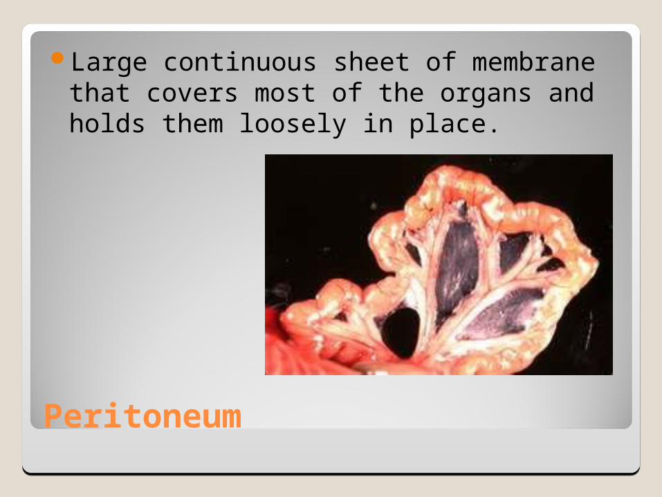
Peritoneum
Large continuous sheet of membrane that covers most of the organs and holds them loosely in place.
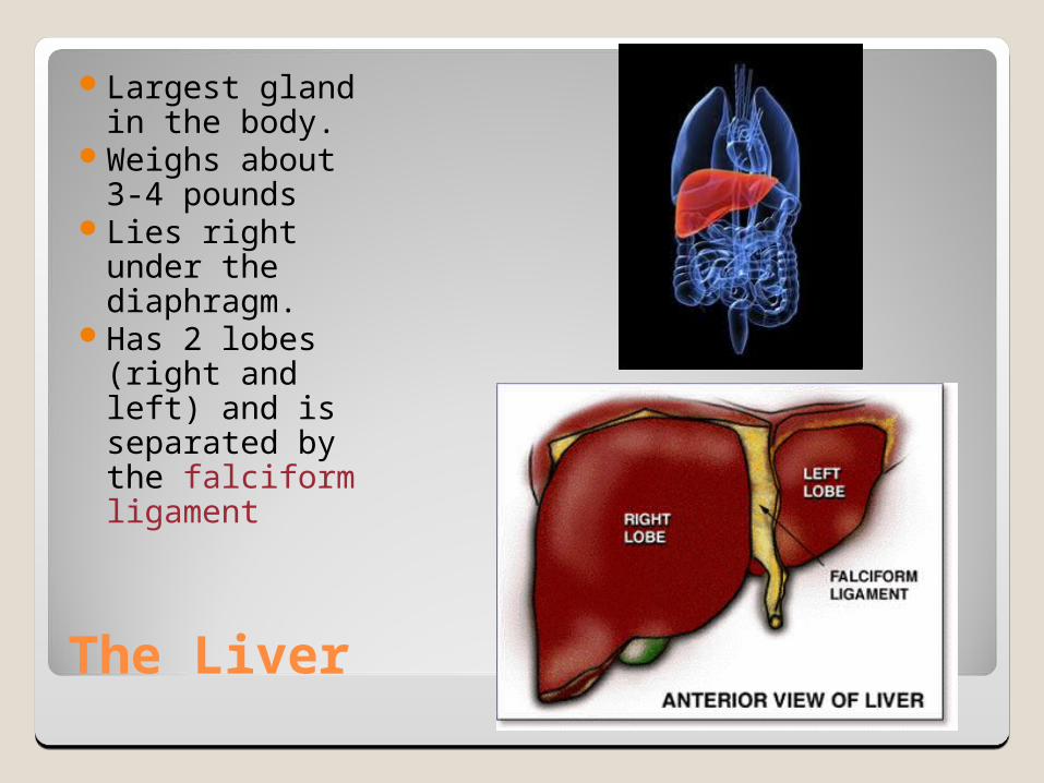
The Liver
Largest gland in the body.
Weighs about 3-4 pounds
Lies right under the diaphragm.
Has 2 lobes (right and left) and is separated by the falciform ligament
The Liver
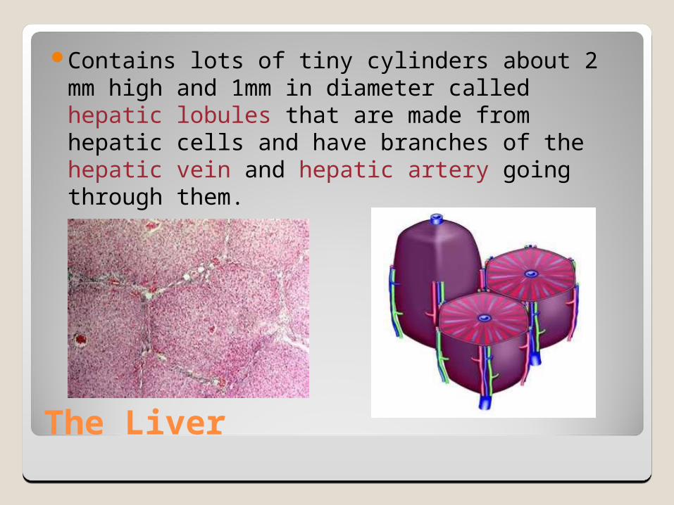
Contains lots of tiny cylinders about 2 mm high and 1mm in diameter called hepatic lobules that are made from hepatic cells and have branches of the hepatic vein and hepatic artery going through them.

The Liver
Blood comes into the hepatic lobules…◦From hepatic artery: cells get oxygen◦From hepatic portal vein: blood gets
“inspected”. Phagocytic cells eat bacteria, old RBCs, and other
foreign particles. Vitamins, minerals, and nutrients are absorbed by
the hepatic cells. Toxins are absorbed into the hepatic cells and are
detoxified (made harmless).
The Liver-Bile Ducts
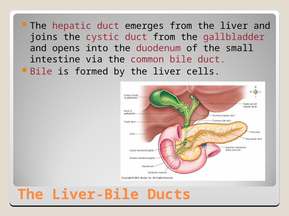
The hepatic duct emerges from the liver and joins the cystic duct from the gallbladder and opens into the duodenum of the small intestine via the common bile duct.
Bile is formed by the liver cells.
Liver-Functions
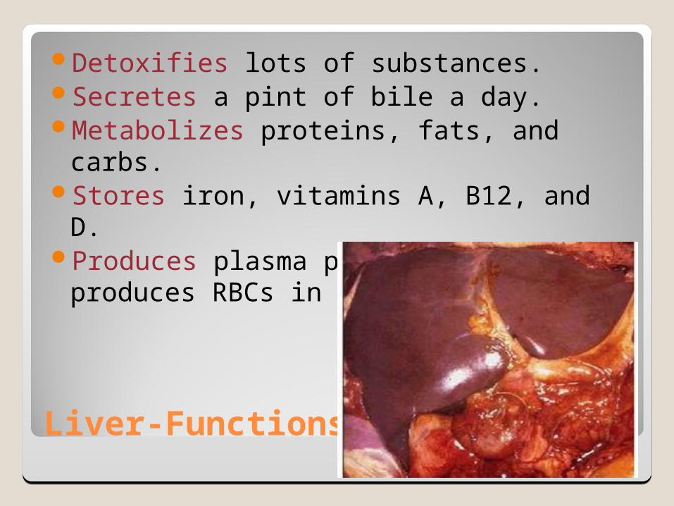
Detoxifies lots of substances.Secretes a pint of bile a day.Metabolizes proteins, fats, and carbs.Stores iron, vitamins A, B12, and D.Produces plasma proteins and produces
RBCs in the fetus.
Bile
Bile is made up of bile salts, bile pigments, and cholesterol.
Bile salts are made from cholesterol by the liver
Aid in absorption of fats. If you don’t have bile, you get really sick after eating a high fat meal.
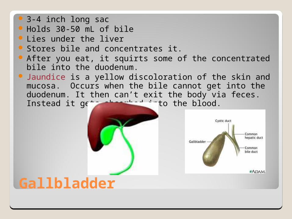
Gallbladder
3-4 inch long sac Holds 30-50 mL of bile Lies under the liver Stores bile and concentrates it. After you eat, it squirts some of the concentrated bile into
the duodenum. Jaundice is a yellow discoloration of the skin and mucosa.
Occurs when the bile cannot get into the duodenum. It then can’t exit the body via feces. Instead it gets absorbed into the blood.

Gallbladder Disorders
Cholecystitis: inflammation of the gallbladder.
Cholelithiasis: gallstone formation. They are solid clumps of mostly cholesterol.
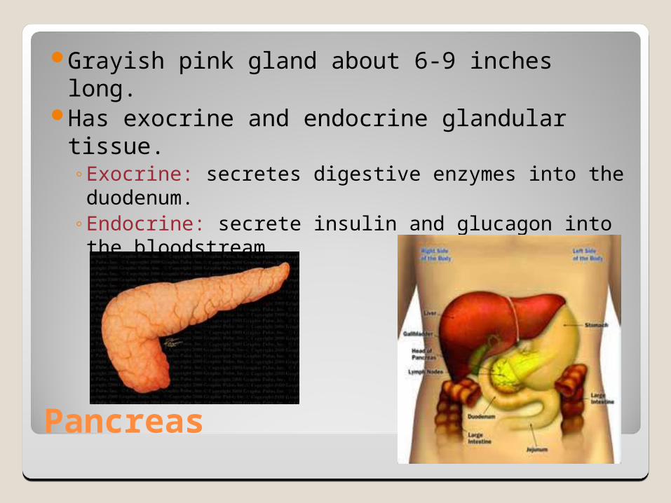
Pancreas
Grayish pink gland about 6-9 inches long.Has exocrine and endocrine glandular
tissue.◦Exocrine: secretes digestive enzymes into the
duodenum.◦Endocrine: secrete insulin and glucagon into
the bloodstream.
Digestive Disorders
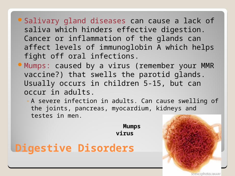
Salivary gland diseases can cause a lack of saliva which hinders effective digestion. Cancer or inflammation of the glands can affect levels of immunoglobin A which helps fight off oral infections.
Mumps: caused by a virus (remember your MMR vaccine?) that swells the parotid glands. Usually occurs in children 5-15, but can occur in adults. ◦ A severe infection in adults. Can cause swelling of the
joints, pancreas, myocardium, kidneys and testes in men.
Mumps virus
Digestive Disorders
Tooth decay: occurs on the tooth surfaces where food debris, acid-secreting bacteria, and plaque accumulate causing a cavity. If not treated, you can get an infection and loss of teeth.
Gingivitis: inflammation of the gums. Usually caused by poor hygiene (poor flossing or brushing) but also by diabetes, vitamin deficiency, or pregnancy.

Tooth decay
gingivitis

Periodontis
Inflammation of the periodontal ligament that holds the tooth in the jaw.
Usually occurs with untreated gingivitis that spreads into the bony tissue of the jaw.
Leading cause of tooth loss in adults.

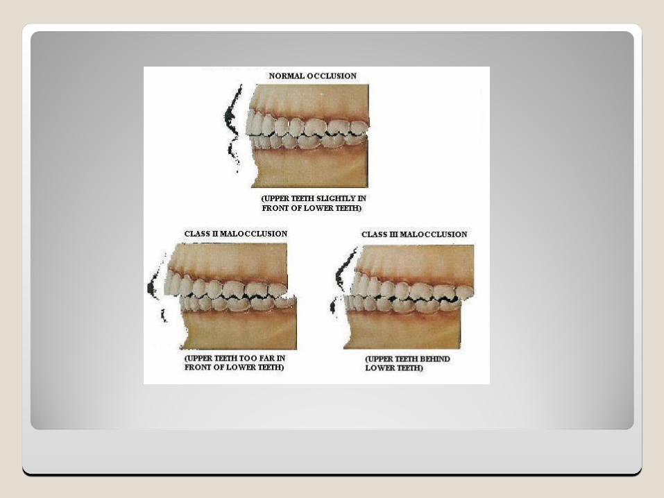
Malocclusion
Wide spaces within teeth, overlapping teeth, overbite or underbite.
Contributes to headaches or proper mastication of food.
Corrected by orthodontics

Gastroesophageal Reflux Disease (GERD)
Affects more than 60 million AmericansCalled heartburn or acid indigestionBackflow of gastric juices into the esophagus.Usually symptoms are mild and occur
infrequently.Avoid smoking, caffeine, alcohol, spicy foods,
acidic foods, chocolate or losing weight to fix the problem.
Antacids (TUMS) or H2-receptor antagonists (Pepcid, Zantac, or Tagament)

GERD
Severe cases can cause asthma attacks, severe chest pain, bleeding or erosive esophagitis.
Proton pump inhibitor drugs decrease the production of stomach acid (Nexium, Prilosec, Prevacid)
Barrett’s esophagus is a precancerous condition when GERD is left untreated.

Ulcers
Craterlike wound or sore caused by tissue destruction.
1 in 10 Americans will have a gastric or duodenal ulcer in their lifetime.
Cause burning pain and may result in hemorrhage, perforation, widespread inflammation, or scarring.
Most are caused by an infection of the bacteria H. pylori.
The bacteria burrows in the mucosa lining and impairs the body to produce mucus.
Can also be caused by long term use of NSAIDs (aspirin and ibuprofen). They interfere with the prostaglandin function of regulating the mucus lining of the GI Tract.
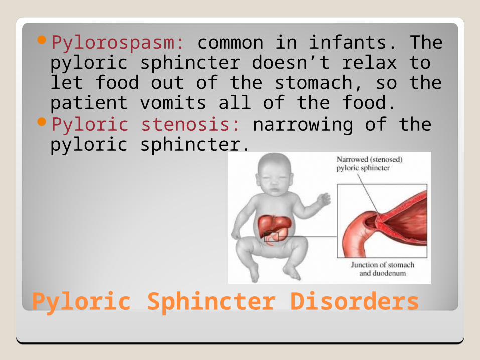
Pyloric Sphincter Disorders
Pylorospasm: common in infants. The pyloric sphincter doesn’t relax to let food out of the stomach, so the patient vomits all of the food.
Pyloric stenosis: narrowing of the pyloric sphincter.
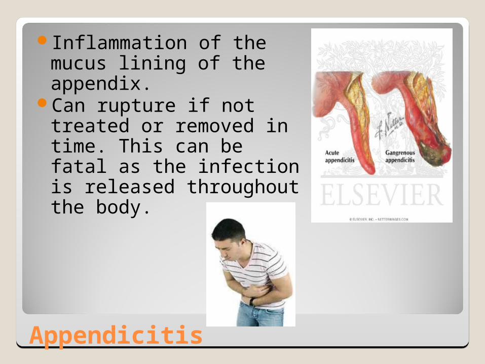
Appendicitis
Inflammation of the mucus lining of the appendix.
Can rupture if not treated or removed in time. This can be fatal as the infection is released throughout the body.
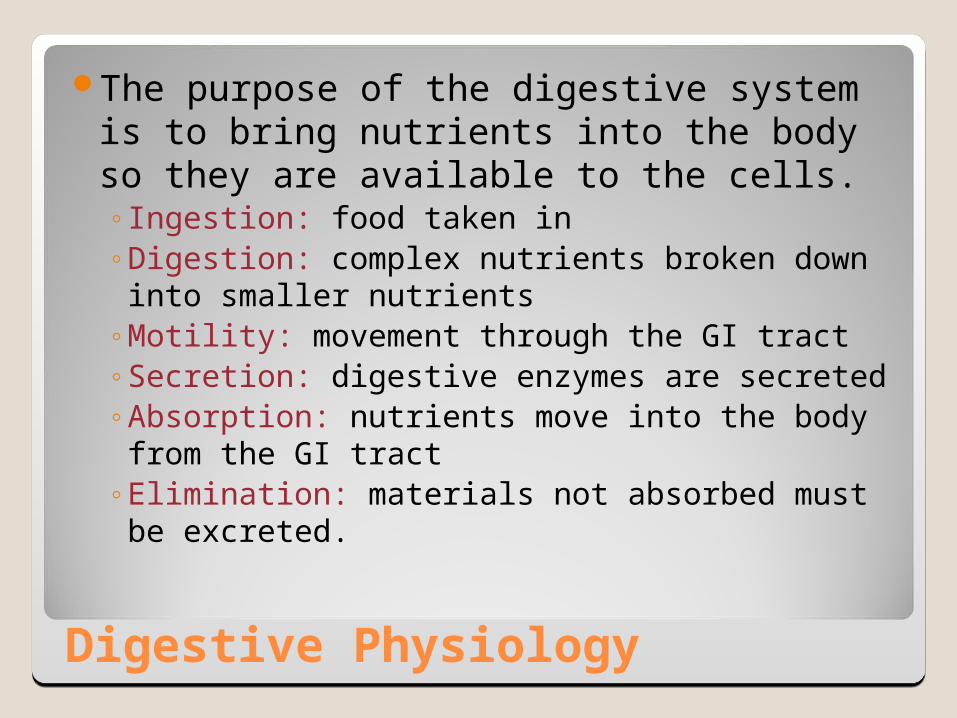
Digestive Physiology
The purpose of the digestive system is to bring nutrients into the body so they are available to the cells.◦Ingestion: food taken in◦Digestion: complex nutrients broken down into
smaller nutrients◦Motility: movement through the GI tract◦Secretion: digestive enzymes are secreted◦Absorption: nutrients move into the body from
the GI tract◦Elimination: materials not absorbed must be
excreted.

Mechanical Digestion
Includes all movement of food through the GI tract.◦ Mastication: chewing. Particle size is reduced.◦ Deglutition: swallowing◦ Peristalsis: the smooth muscles of the GI tract produce a
wavelike ripple of movement to propel the food forward through the tract.
◦ Segmentation: Forward and back movement in one region to break apart the food.
peristalsis
Segmentation

Food Movement
It takes anywhere from 2-6 hours for food to empty the stomach after a meal.
Food is churned and mixed with gastric juices to form a milky substance called chyme.
This slowly is squirted into the duodenum.It takes about 5 hours to travel through
the small intestine.

Chemical Digestion
The food undergoes hydrolysis reactions (combines with water) to split into smaller compounds.
Digestive enzymes are organic catalysts that speed up the breakdown of food.
Most of the organs in the system secrete some kind of enzyme.

Carbohydrate Digestion
Carbs are made up of saccharides Polysaccharides- starch and glycogen Disaccharides- sucrose, lactose, maltose Monosaccharides- glucose, fructose, galactose
Amylases are enzymes that break apart polysaccharides into disaccharides. (salivary amylase and pancreatic amylase)
Maltase, sucrase, and lactase break down the disaccharides into monosaccharides.
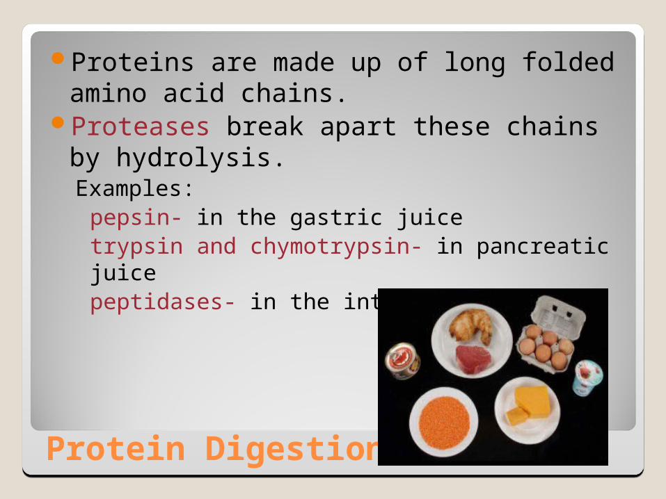
Protein Digestion
Proteins are made up of long folded amino acid chains.
Proteases break apart these chains by hydrolysis.Examples:
pepsin- in the gastric juicetrypsin and chymotrypsin- in pancreatic juicepeptidases- in the intestine
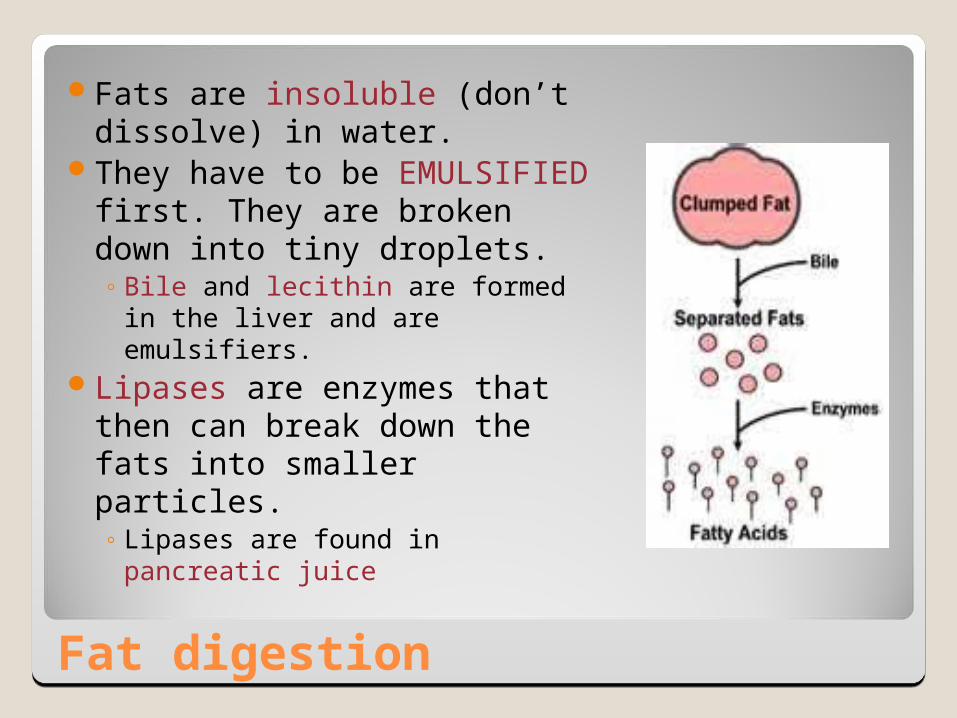
Fat digestion
Fats are insoluble (don’t dissolve) in water.
They have to be EMULSIFIED first. They are broken down into tiny droplets.◦ Bile and lecithin are formed in the
liver and are emulsifiers.Lipases are enzymes that then
can break down the fats into smaller particles.◦ Lipases are found in pancreatic
juice

Saliva
Mostly water that helps to liquefy the food
Contains salivary amylase to break down polysaccharides.
Also contains lipase but really doesn’t have enough time to do any digestion of fats.
Contains NaHCO3 which makes it slightly basic. Amylase works best is a higher pH.
Contains mucus so the food can slide through the GI tract easier
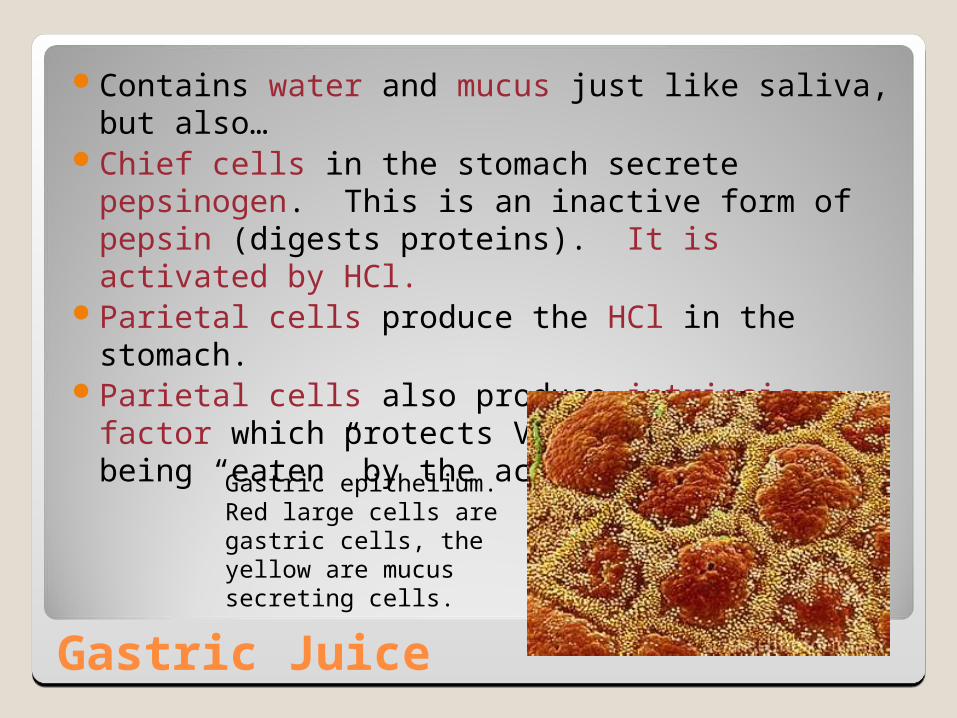
Gastric Juice
Contains water and mucus just like saliva, but also…
Chief cells in the stomach secrete pepsinogen. This is an inactive form of pepsin (digests proteins). It is activated by HCl.
Parietal cells produce the HCl in the stomach. Parietal cells also produce intrinsic factor which
protects Vitamin B12 from being “eaten” by the acid and enzymes.
Gastric epithelium. Red large cells are gastric cells, the yellow are mucus secreting cells.
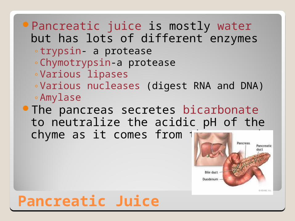
Pancreatic Juice
Pancreatic juice is mostly water but has lots of different enzymes◦trypsin- a protease◦Chymotrypsin-a protease◦Various lipases◦Various nucleases (digest RNA and DNA)◦Amylase
The pancreas secretes bicarbonate to neutralize the acidic pH of the chyme as it comes from the stomach.
Intestinal Juice
The intestines secrete bicarbonate to keep the buffer the pH of the chyme.
Also contains mucus.
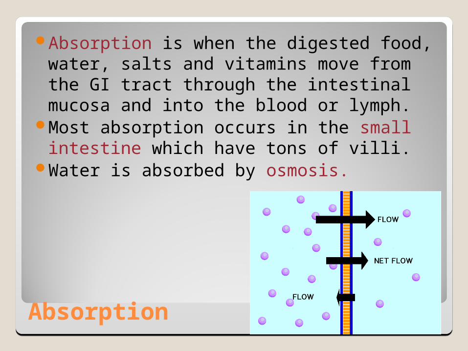
Absorption
Absorption is when the digested food, water, salts and vitamins move from the GI tract through the intestinal mucosa and into the blood or lymph.
Most absorption occurs in the small intestine which have tons of villi.
Water is absorbed by osmosis.
Absorption
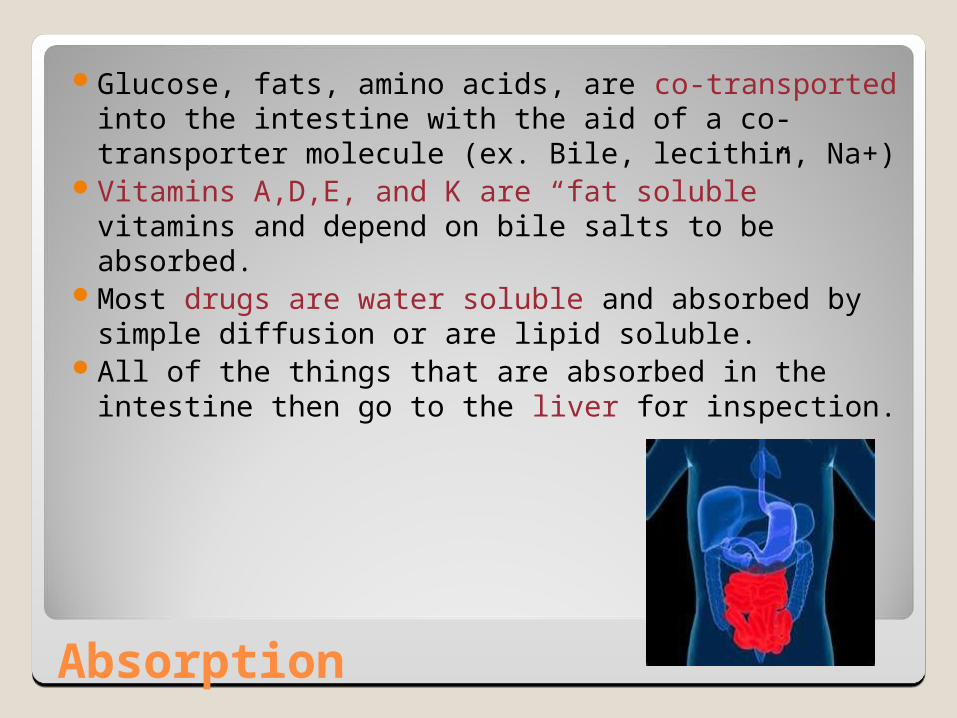
Glucose, fats, amino acids, are co-transported into the intestine with the aid of a co-transporter molecule (ex. Bile, lecithin, Na+)
Vitamins A,D,E, and K are “fat soluble” vitamins and depend on bile salts to be absorbed.
Most drugs are water soluble and absorbed by simple diffusion or are lipid soluble.
All of the things that are absorbed in the intestine then go to the liver for inspection.
Elimination
Elimination is the getting rid of the residues of digestion called feces.
Expelling of the feces is called defecation.It is a reflex caused by the stimulation of
receptors in the rectum as fecal matter is moved to the rectum.
Elimination
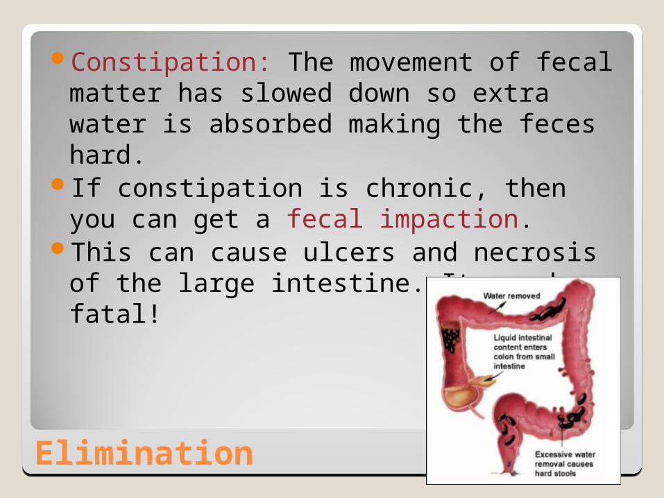
Constipation: The movement of fecal matter has slowed down so extra water is absorbed making the feces hard.
If constipation is chronic, then you can get a fecal impaction.
This can cause ulcers and necrosis of the large intestine. It can be fatal!

Disorders of the GI Tract
Gastroenteritis (aka gastritis): stomach inflammation Nausea: unpleasant feeling that often leads to vomiting Emesis: vomiting Diarrhea: elimination of liquid feces, sometime with
abdominal cramps. Stomach cancer: linked to excessive alcohol consumption,
chewing tobacco, eating smoked or heavily preserved food.◦ Usually the cancer has already spread before the patient is
diagnosed. The early warning signs are belching, heartburn and nausea, so patients usually treat themselves.
◦ Later symptoms are chronic indigestion, vomiting, anorexia, stomach pain, and blood in the feces.

Disorders of the GI Tract
Malabsorption Syndrome: general term for anything that causes the small intestine to not absorb nutrients properly.
Diverticulosis: abnormal saclike pouches that come out of the large intestinal wall. Common in people over 50.
The diverticuli can become inflamed, called diverticulitis (pain, tenderness, fever, blood in stool).
Disorders of the GI Tract
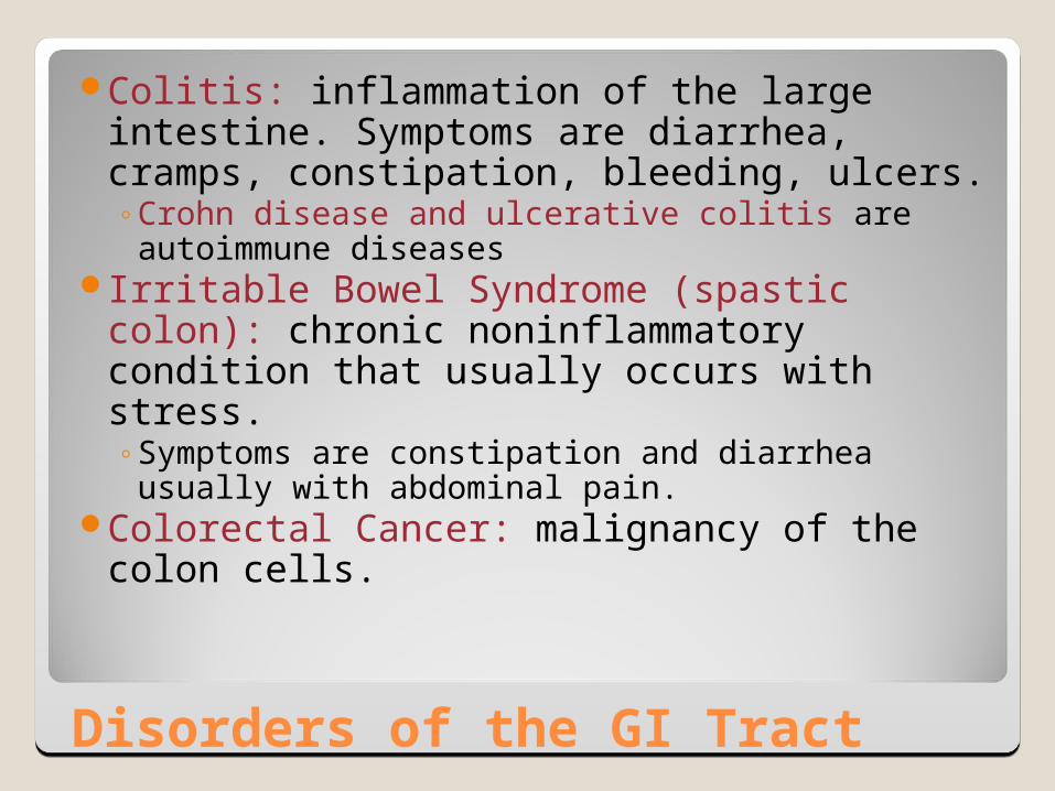
Colitis: inflammation of the large intestine. Symptoms are diarrhea, cramps, constipation, bleeding, ulcers.◦Crohn disease and ulcerative colitis are
autoimmune diseasesIrritable Bowel Syndrome (spastic colon):
chronic noninflammatory condition that usually occurs with stress. ◦Symptoms are constipation and diarrhea
usually with abdominal pain.Colorectal Cancer: malignancy of the
colon cells.
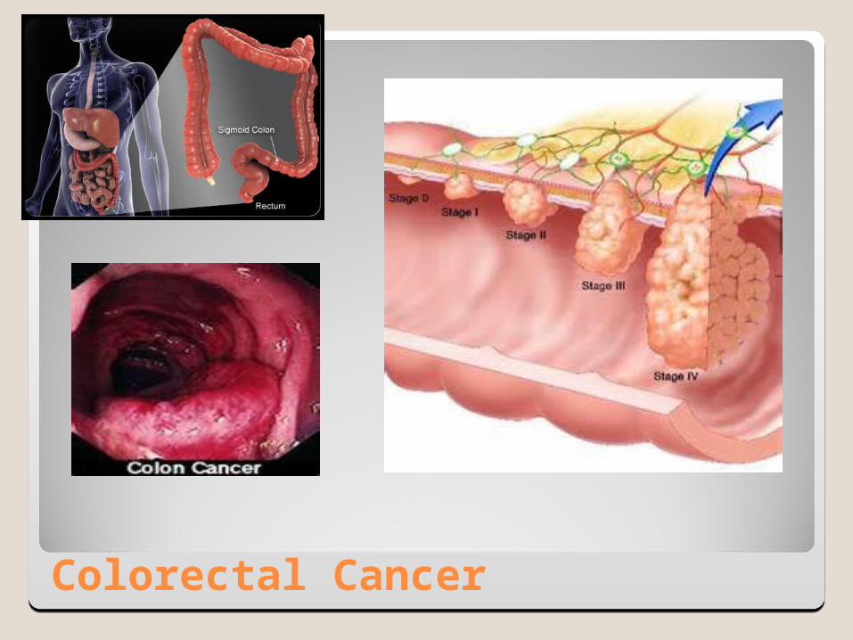
Colorectal Cancer