Embed Size (px)
Citation preview
Connective tissue
Part 1
The Digestive System
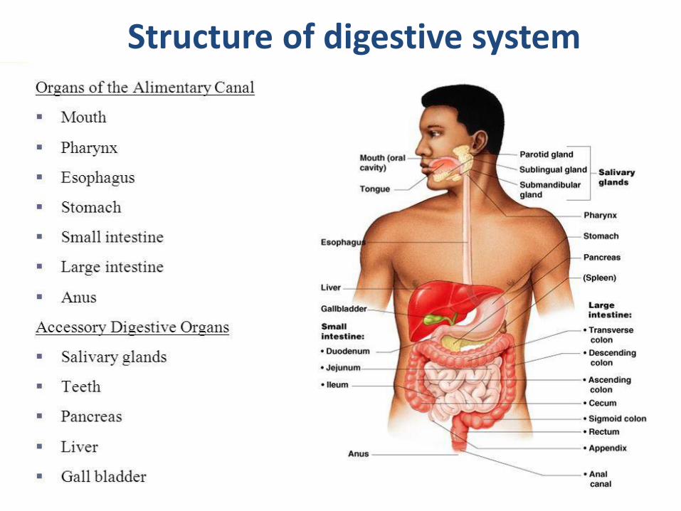
Structure of digestive system
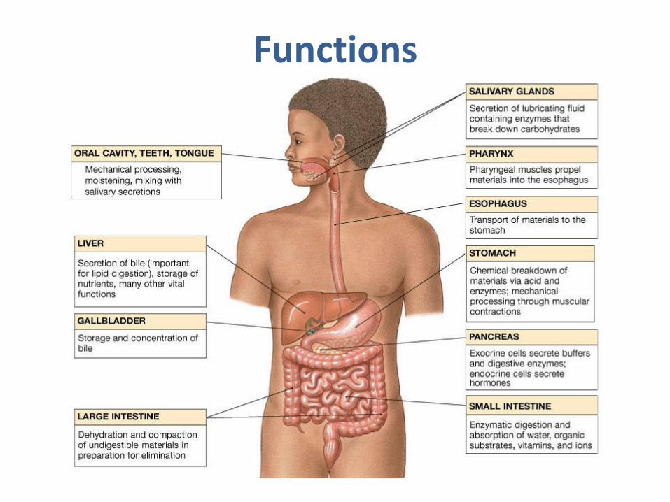
Functions
Basic Structure of the Alimentary Canal Wall
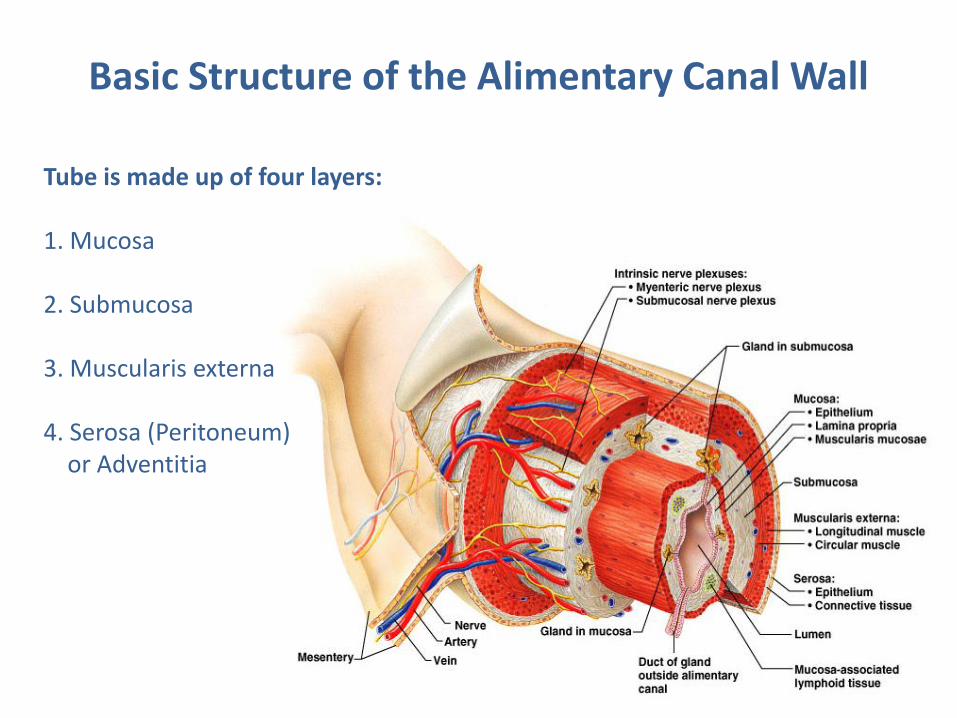
Tube is made up of four layers: 1. Mucosa 2. Submucosa 3. Muscularis externa 4. Serosa (Peritoneum) or Adventitia
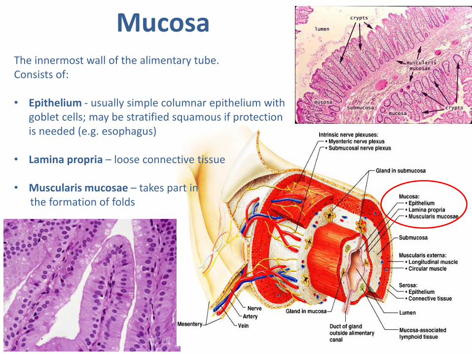
Mucosa The innermost wall of the alimentary tube. Consists of: • Epithelium - usually simple columnar epithelium with
goblet cells; may be stratified squamous if protection is needed (e.g. esophagus)
• Lamina propria – loose connective tissue • Muscularis mucosae – takes part in the formation of folds
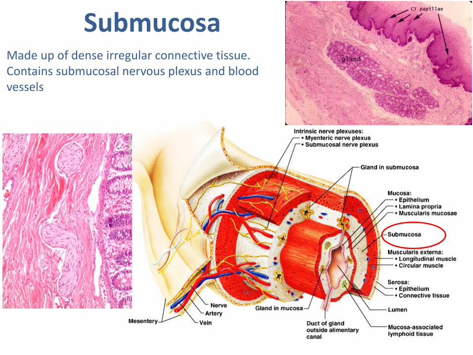
Made up of dense irregular connective tissue. Contains submucosal nervous plexus and blood vessels
Submucosa
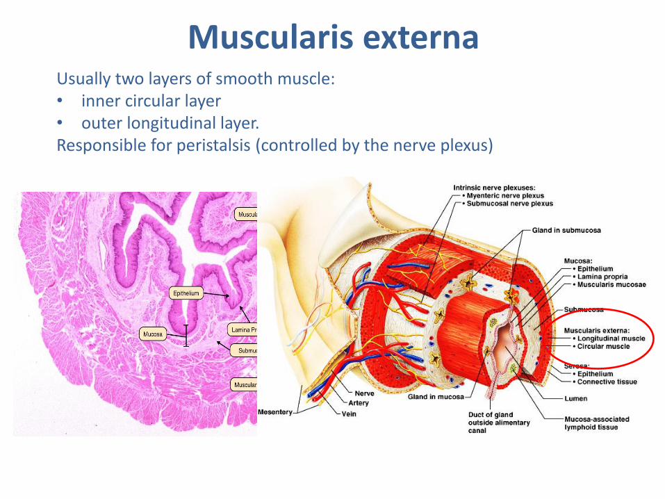
Muscularis externa Usually two layers of smooth muscle: • inner circular layer • outer longitudinal layer. Responsible for peristalsis (controlled by the nerve plexus)
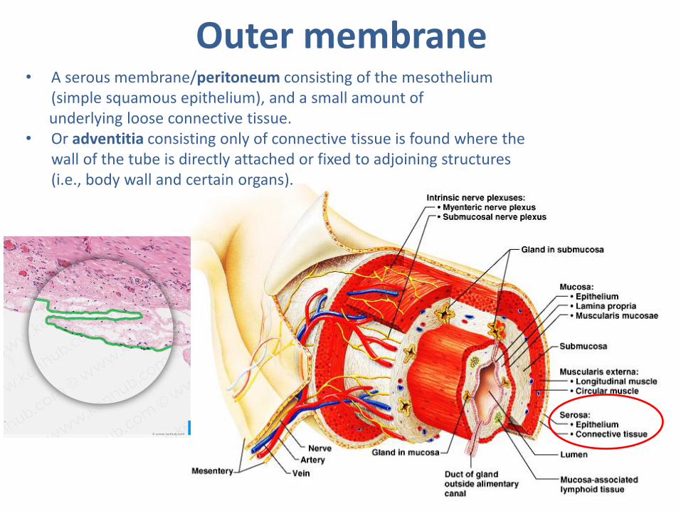
• A serous membrane/peritoneum consisting of the mesothelium (simple squamous epithelium), and a small amount of
underlying loose connective tissue. • Or adventitia consisting only of connective tissue is found where the
wall of the tube is directly attached or fixed to adjoining structures (i.e., body wall and certain organs).
Outer membrane
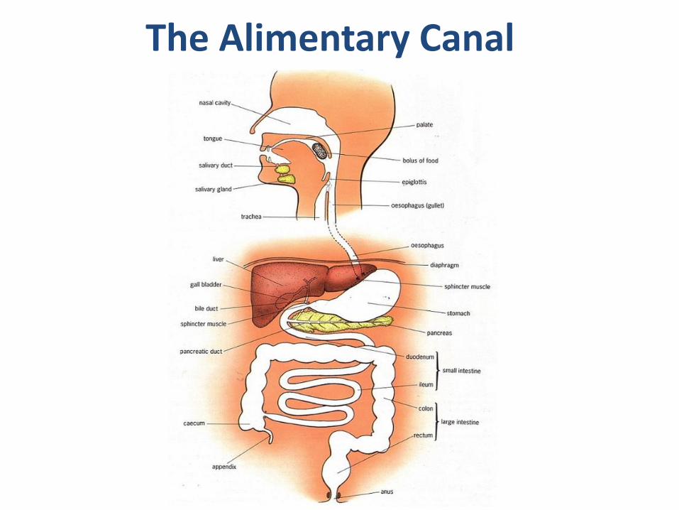
The Alimentary Canal
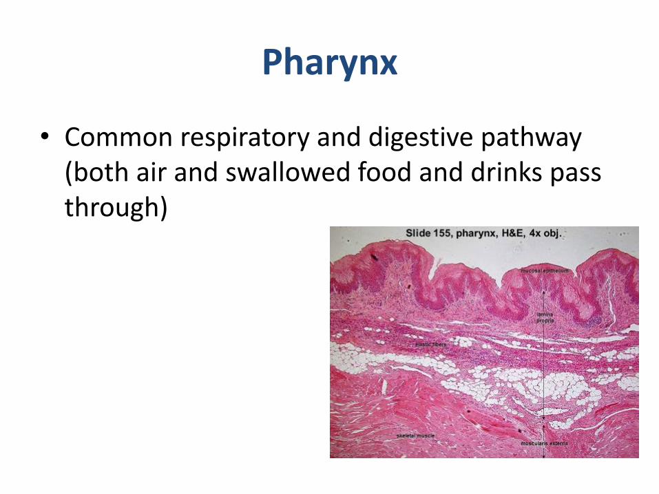
Pharynx
• Common respiratory and digestive pathway (both air and swallowed food and drinks pass through)
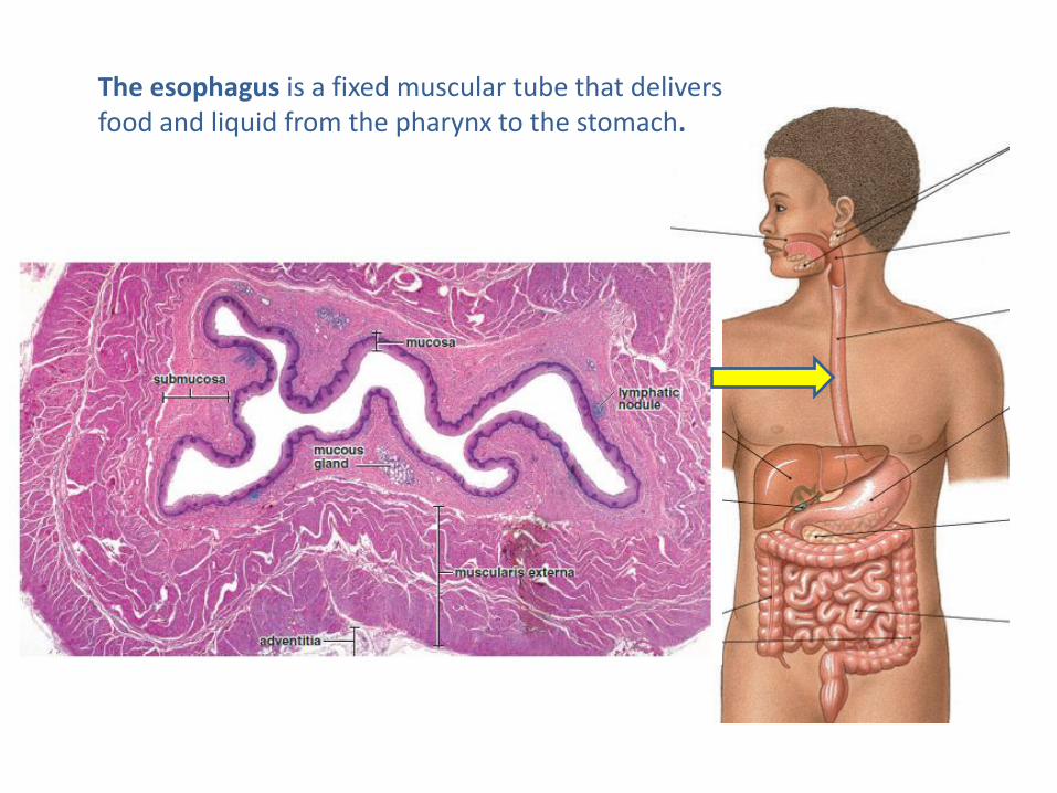
The esophagus is a fixed muscular tube that delivers food and liquid from the pharynx to the stomach.
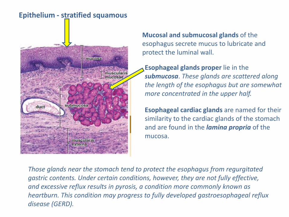
Mucosal and submucosal glands of the esophagus secrete mucus to lubricate and protect the luminal wall.
Esophageal glands proper lie in the submucosa. These glands are scattered along the length of the esophagus but are somewhat more concentrated in the upper half.
Esophageal cardiac glands are named for their similarity to the cardiac glands of the stomach and are found in the lamina propria of the mucosa.
Those glands near the stomach tend to protect the esophagus from regurgitated gastric contents. Under certain conditions, however, they are not fully effective, and excessive reflux results in pyrosis, a condition more commonly known as heartburn. This condition may progress to fully developed gastroesophageal reflux disease (GERD).
Epithelium - stratified squamous
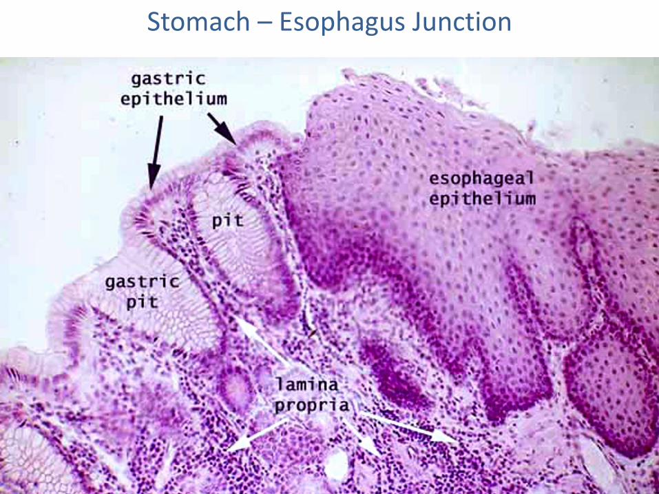
Stomach – Esophagus Junction
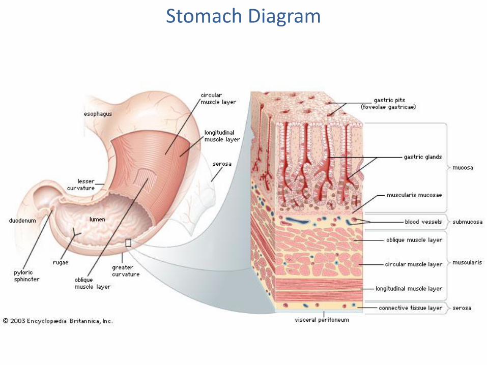
Stomach Diagram
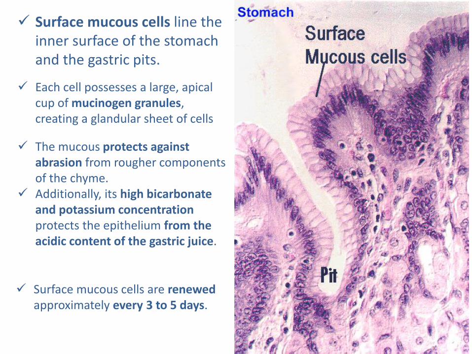
Surface mucous cells line the inner surface of the stomach and the gastric pits.
Each cell possesses a large, apical cup of mucinogen granules, creating a glandular sheet of cells
The mucous protects against abrasion from rougher components of the chyme.
Additionally, its high bicarbonate and potassium concentration protects the epithelium from the acidic content of the gastric juice.
Surface mucous cells are renewed approximately every 3 to 5 days.
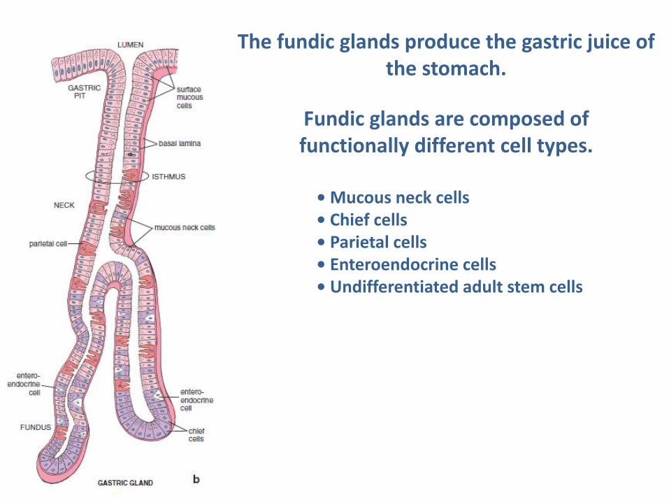
Fundic glands are composed of functionally different cell types.
• Mucous neck cells • Chief cells • Parietal cells • Enteroendocrine cells • Undifferentiated adult stem cells
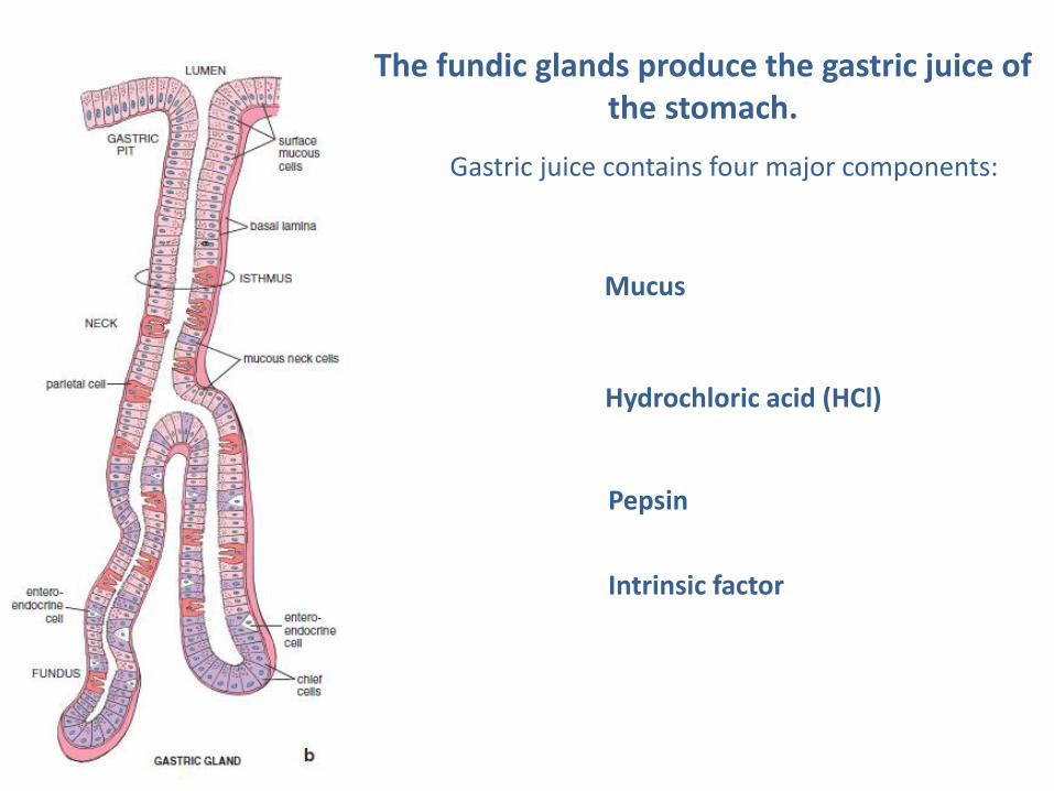
The fundic glands produce the gastric juice of the stomach.
The fundic glands produce the gastric juice of the stomach.
Gastric juice contains four major components:
Hydrochloric acid (HCl)
Pepsin
Mucus
Intrinsic factor
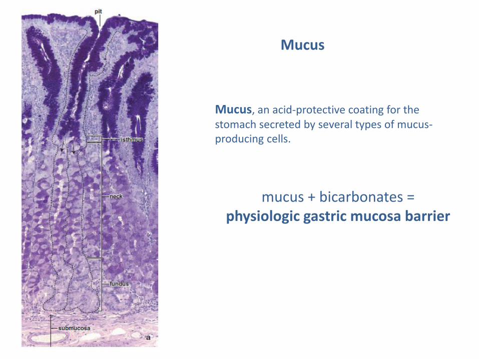
Mucus
Mucus, an acid-protective coating for the stomach secreted by several types of mucus-producing cells.
mucus + bicarbonates = physiologic gastric mucosa barrier
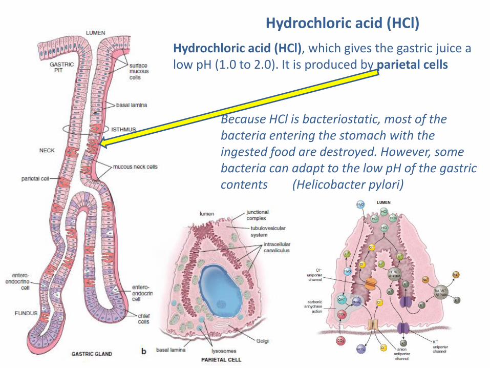
Hydrochloric acid (HCl)
Hydrochloric acid (HCl), which gives the gastric juice a low pH (1.0 to 2.0). It is produced by parietal cells
Because HCl is bacteriostatic, most of the bacteria entering the stomach with the ingested food are destroyed. However, some bacteria can adapt to the low pH of the gastric contents (Helicobacter pylori)
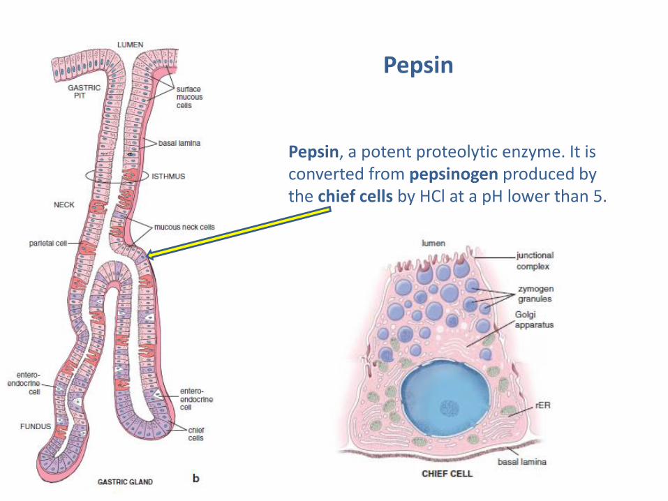
Pepsin
Pepsin, a potent proteolytic enzyme. It is converted from pepsinogen produced by the chief cells by HCl at a pH lower than 5.
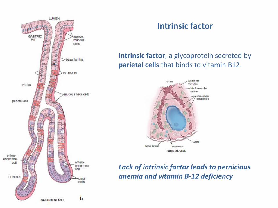
Intrinsic factor
Intrinsic factor, a glycoprotein secreted by parietal cells that binds to vitamin B12.
Lack of intrinsic factor leads to pernicious anemia and vitamin B-12 deficiency
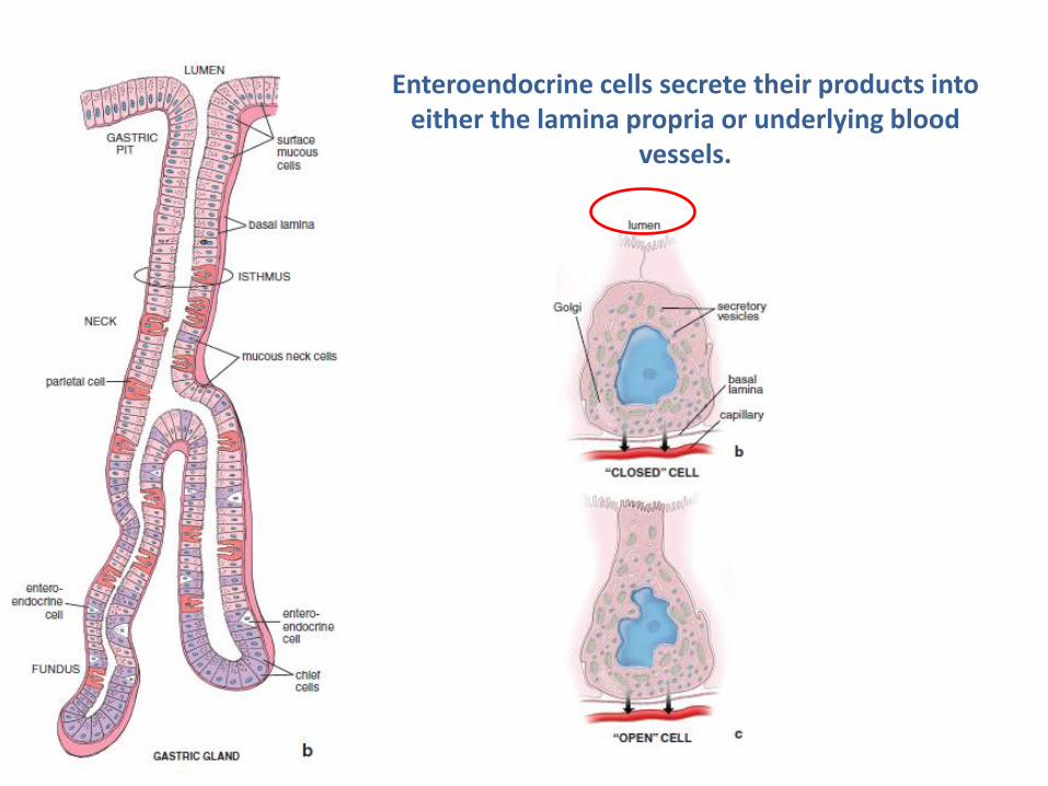
Enteroendocrine cells secrete their products into either the lamina propria or underlying blood
vessels.
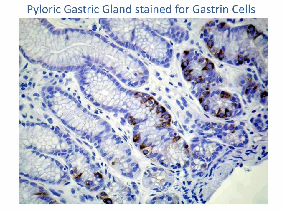
Pyloric Gastric Gland stained for Gastrin Cells
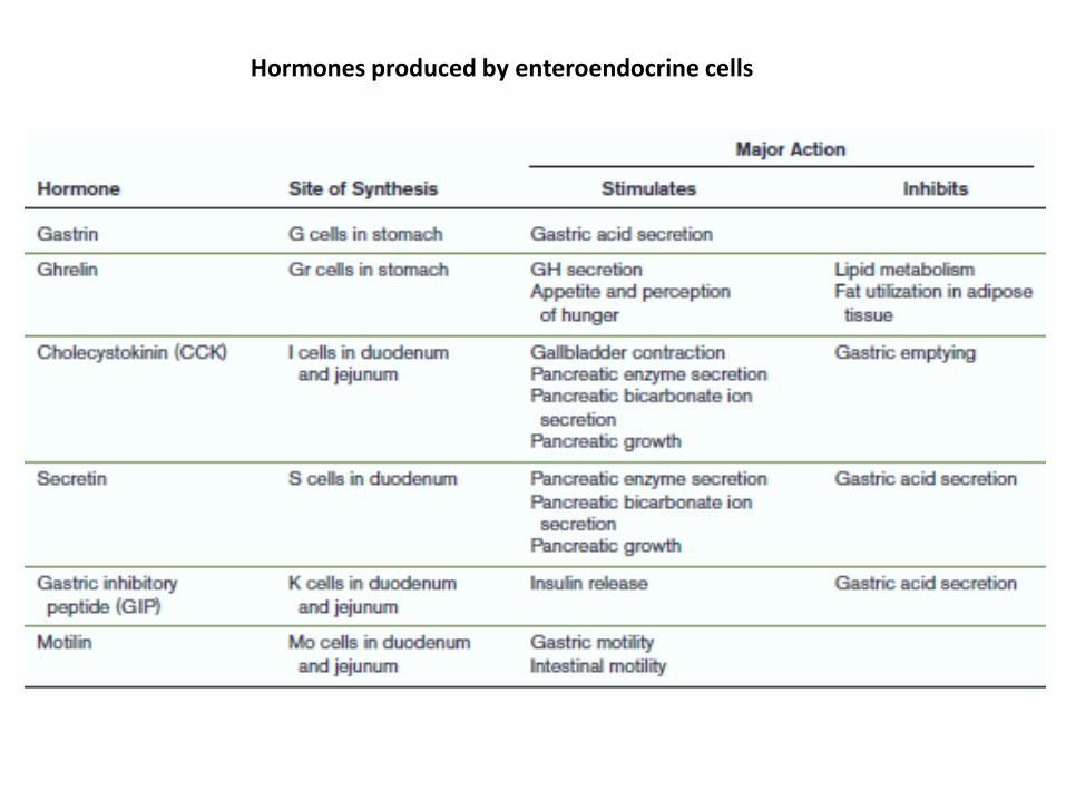
Hormones produced by enteroendocrine cells

Pyloric Stomach
Long pits (P)
Short Glands
Fundic Stomach
Short pits (P)
Long Glands

The Small Intestine
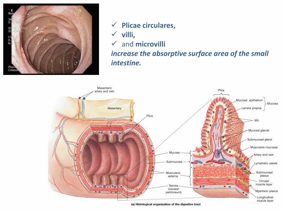
Plicae circulares, villi, and microvilli increase the absorptive surface area of the small intestine.
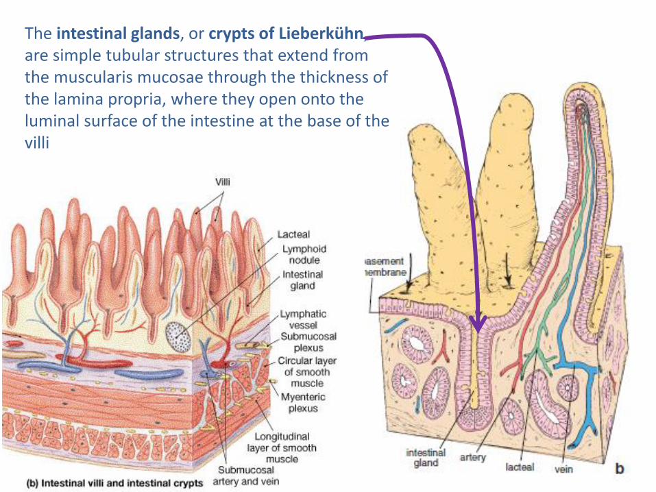
The intestinal glands, or crypts of Lieberkühn, are simple tubular structures that extend from the muscularis mucosae through the thickness of the lamina propria, where they open onto the luminal surface of the intestine at the base of the villi
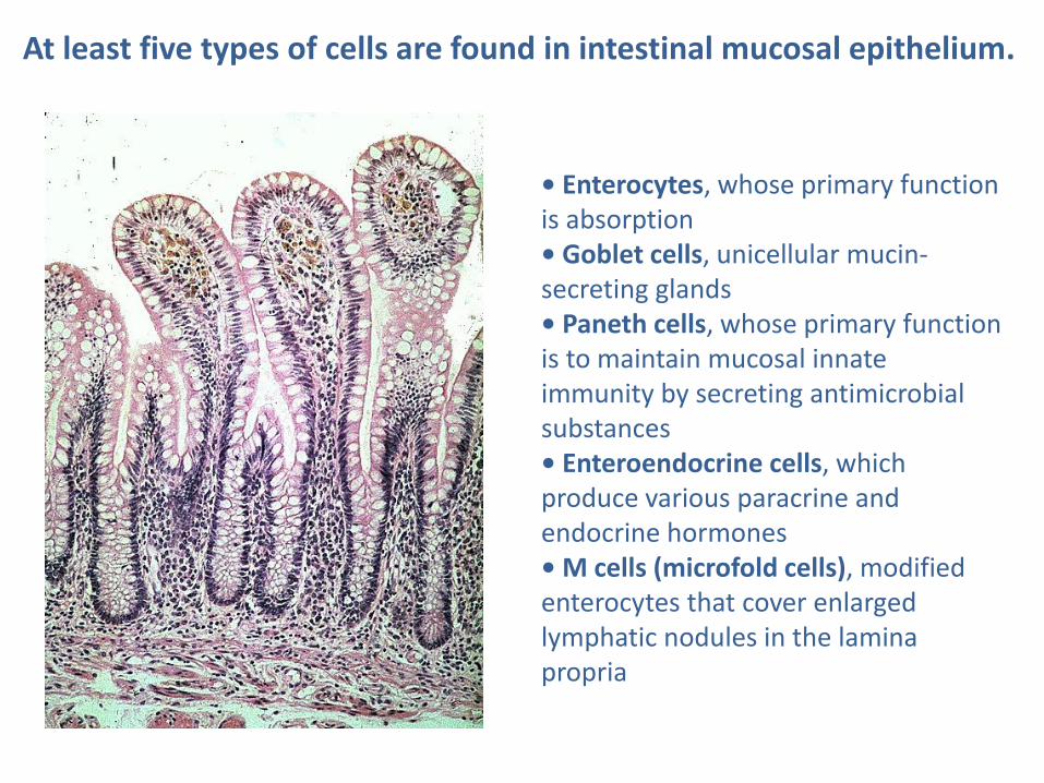
At least five types of cells are found in intestinal mucosal epithelium.
• Enterocytes, whose primary function is absorption • Goblet cells, unicellular mucin-secreting glands • Paneth cells, whose primary function is to maintain mucosal innate immunity by secreting antimicrobial substances • Enteroendocrine cells, which produce various paracrine and endocrine hormones • M cells (microfold cells), modified enterocytes that cover enlarged lymphatic nodules in the lamina propria

Enterocytes
Goblet cells represent unicellular glands
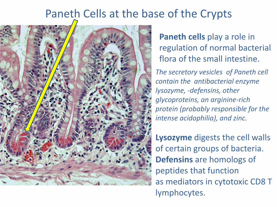
Paneth Cells at the base of the Crypts
Paneth cells play a role in regulation of normal bacterial flora of the small intestine.
The secretory vesicles of Paneth cell contain the antibacterial enzyme lysozyme, -defensins, other glycoproteins, an arginine-rich protein (probably responsible for the intense acidophilia), and zinc.
Lysozyme digests the cell walls of certain groups of bacteria. Defensins are homologs of peptides that function as mediators in cytotoxic CD8 T lymphocytes.
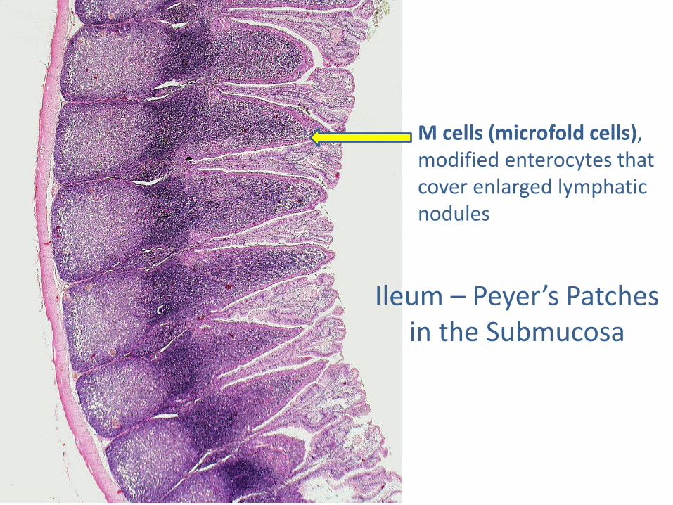
M cells (microfold cells), modified enterocytes that cover enlarged lymphatic nodules
Ileum – Peyer’s Patches in the Submucosa
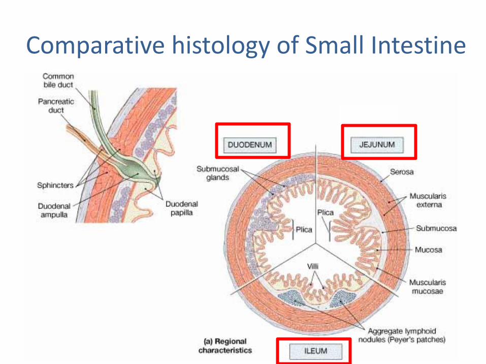
Comparative histology of Small Intestine

The large intestine The principal functions of the large intestine are reabsorption of electrolytes and water and elimination of undigested food and waste.
The mucosal epithelium of the large intestine contains the same cell types as the small intestine except Paneth cells, which are normally absent in humans.
The mucosa of the large intestine has a “smooth” surface; neither plicae circulares nor villi are present. It contains numerous straight tubular intestinal glands (crypts of Lieberkühn) that extend through the full thickness of the mucosa
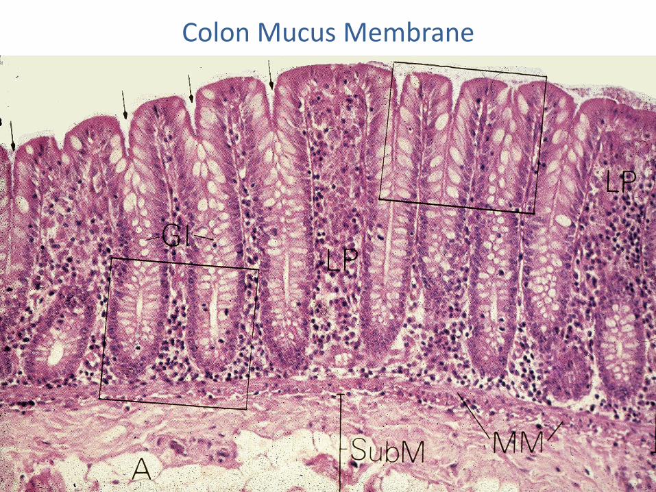
Colon Mucus Membrane
Accessory digestive organs
Salivary glands
Liver and gallbladder
The pancreas
Oral Cavity
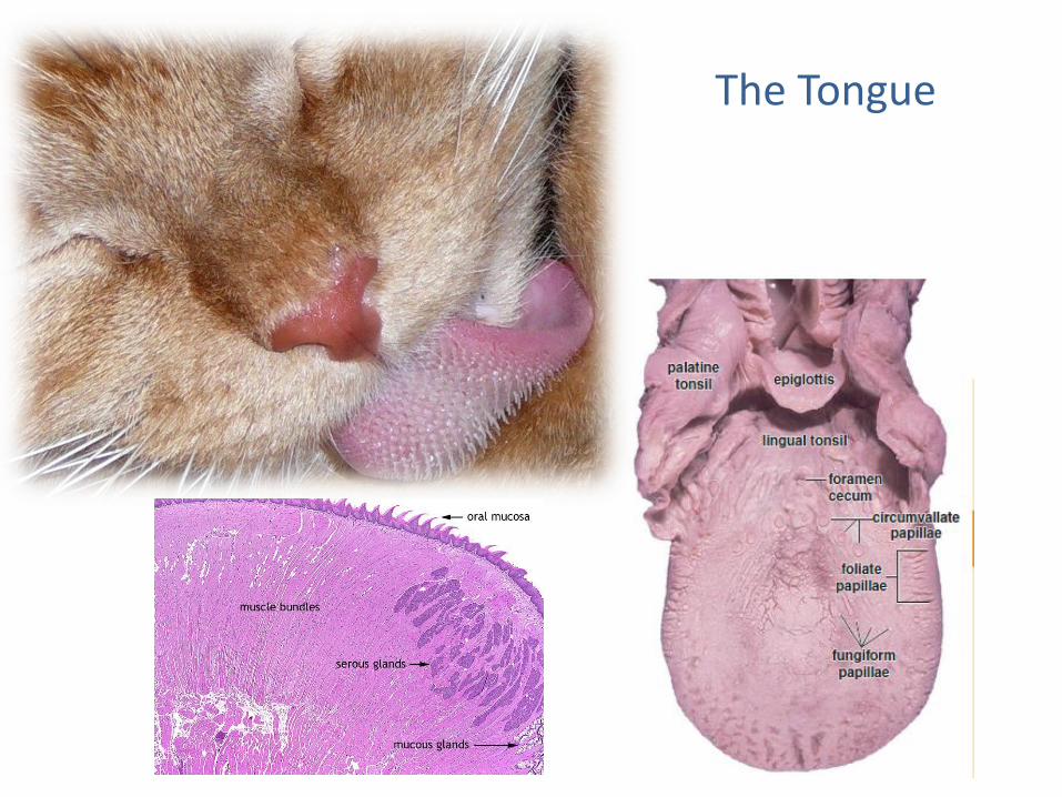
The Tongue
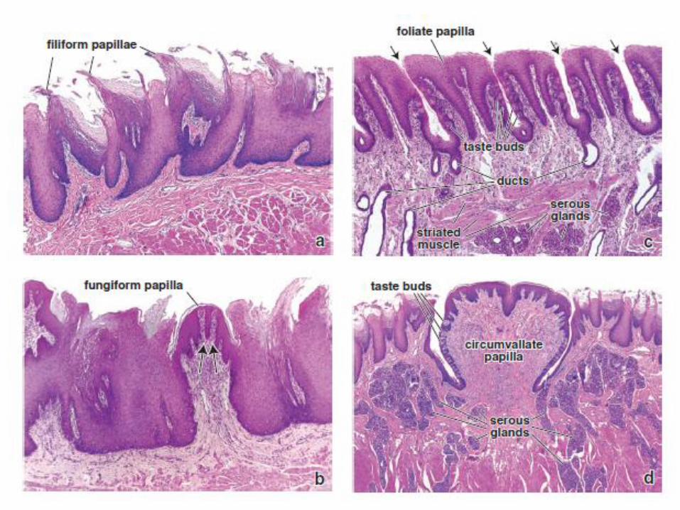
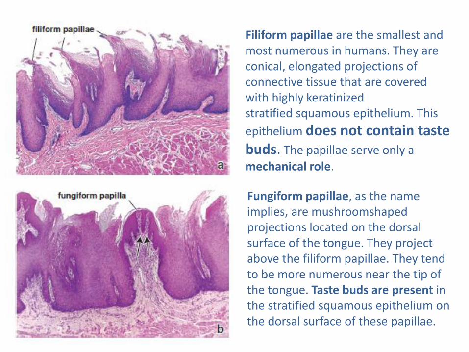
Filiform papillae are the smallest and most numerous in humans. They are conical, elongated projections of connective tissue that are covered with highly keratinized stratified squamous epithelium. This
epithelium does not contain taste buds. The papillae serve only a
mechanical role.
Fungiform papillae, as the name implies, are mushroomshaped projections located on the dorsal surface of the tongue. They project above the filiform papillae. They tend to be more numerous near the tip of the tongue. Taste buds are present in the stratified squamous epithelium on the dorsal surface of these papillae.
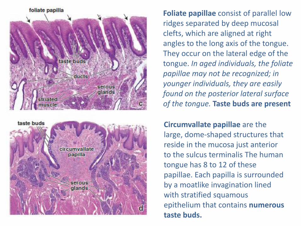
Foliate papillae consist of parallel low ridges separated by deep mucosal clefts, which are aligned at right angles to the long axis of the tongue. They occur on the lateral edge of the tongue. In aged individuals, the foliate papillae may not be recognized; in younger individuals, they are easily found on the posterior lateral surface of the tongue. Taste buds are present
Circumvallate papillae are the large, dome-shaped structures that reside in the mucosa just anterior to the sulcus terminalis The human tongue has 8 to 12 of these papillae. Each papilla is surrounded by a moatlike invagination lined with stratified squamous epithelium that contains numerous taste buds.
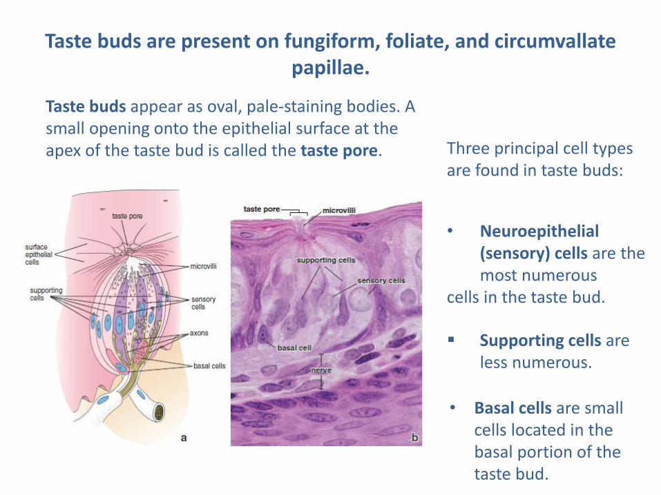
Taste buds appear as oval, pale-staining bodies. A small opening onto the epithelial surface at the apex of the taste bud is called the taste pore.
Taste buds are present on fungiform, foliate, and circumvallate papillae.
Three principal cell types are found in taste buds:
• Neuroepithelial (sensory) cells are the most numerous
cells in the taste bud.
Supporting cells are less numerous.
• Basal cells are small cells located in the basal portion of the taste bud.
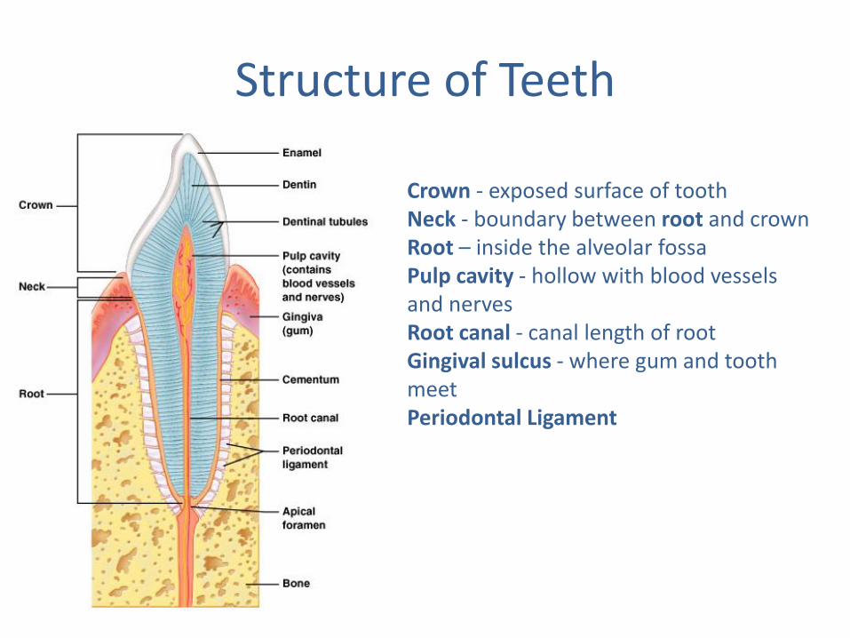
Crown - exposed surface of tooth Neck - boundary between root and crown Root – inside the alveolar fossa Pulp cavity - hollow with blood vessels and nerves Root canal - canal length of root Gingival sulcus - where gum and tooth meet Periodontal Ligament
Structure of Teeth
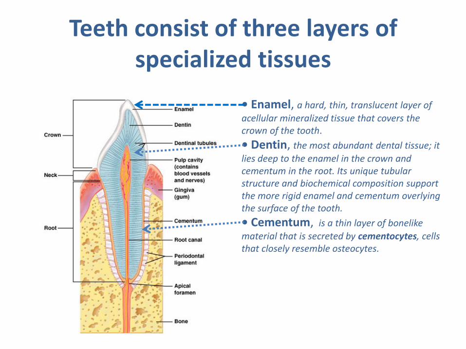
Teeth consist of three layers of specialized tissues
• Enamel, a hard, thin, translucent layer of
acellular mineralized tissue that covers the crown of the tooth.
• Dentin, the most abundant dental tissue; it
lies deep to the enamel in the crown and cementum in the root. Its unique tubular structure and biochemical composition support the more rigid enamel and cementum overlying the surface of the tooth.
• Cementum, is a thin layer of bonelike
material that is secreted by cementocytes, cells that closely resemble osteocytes.
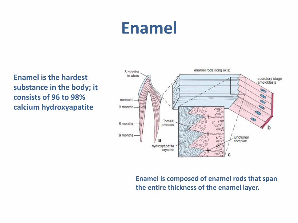
Enamel
Enamel is the hardest substance in the body; it consists of 96 to 98% calcium hydroxyapatite
Enamel is composed of enamel rods that span the entire thickness of the enamel layer.
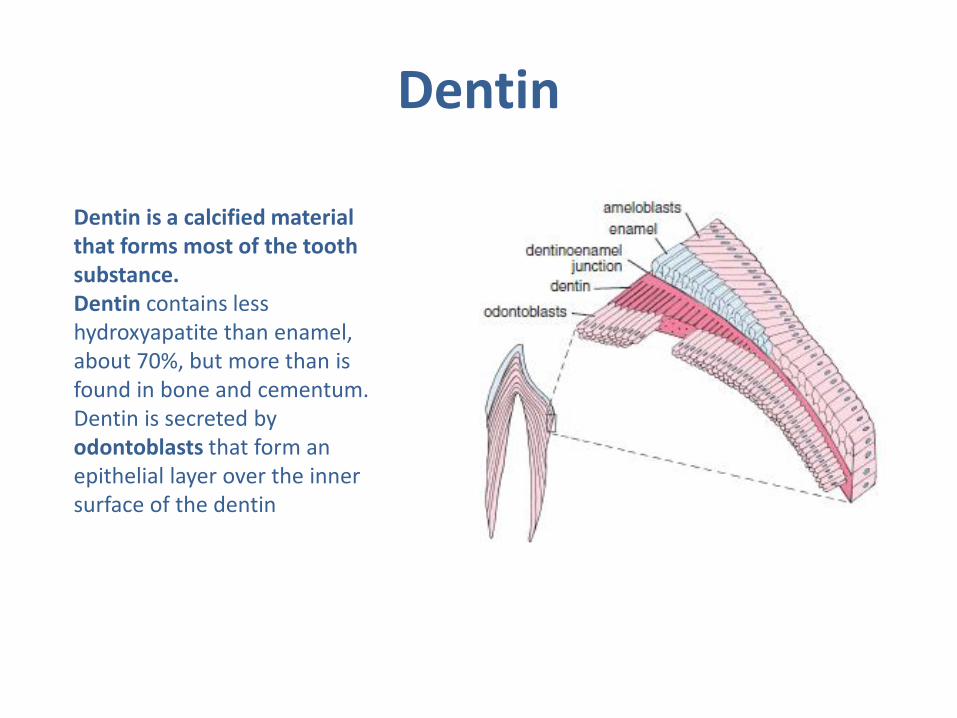
Dentin
Dentin is a calcified material that forms most of the tooth substance. Dentin contains less hydroxyapatite than enamel, about 70%, but more than is found in bone and cementum. Dentin is secreted by odontoblasts that form an epithelial layer over the inner surface of the dentin
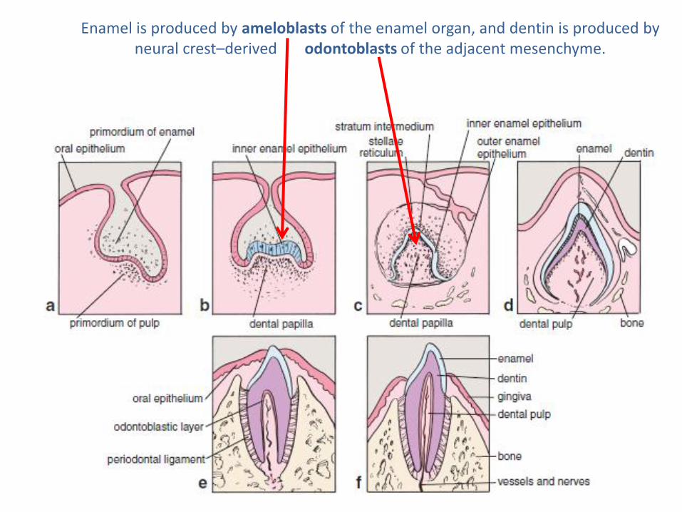
Enamel is produced by ameloblasts of the enamel organ, and dentin is produced by neural crest–derived odontoblasts of the adjacent mesenchyme.
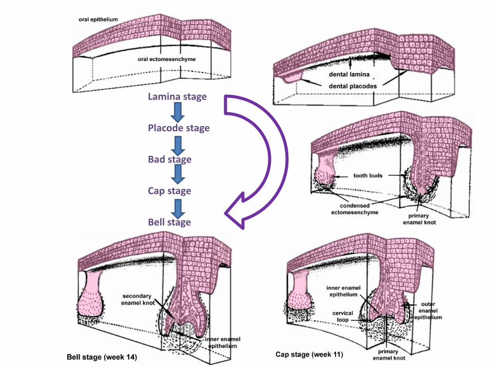
Lamina stage
Placode stage
Bad stage
Cap stage
Bell stage
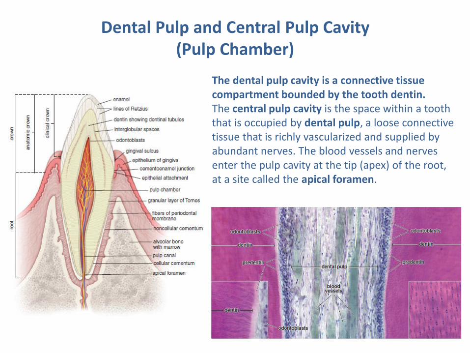
Dental Pulp and Central Pulp Cavity (Pulp Chamber)
The dental pulp cavity is a connective tissue compartment bounded by the tooth dentin. The central pulp cavity is the space within a tooth that is occupied by dental pulp, a loose connective tissue that is richly vascularized and supplied by abundant nerves. The blood vessels and nerves enter the pulp cavity at the tip (apex) of the root, at a site called the apical foramen.
Salivary glands
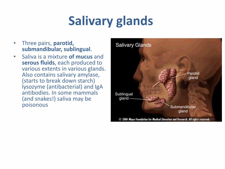
• Three pairs, parotid, submandibular, sublingual.
• Saliva is a mixture of mucus and serous fluids, each produced to various extents in various glands. Also contains salivary amylase, (starts to break down starch) lysozyme (antibacterial) and IgA antibodies. In some mammals (and snakes!) saliva may be poisonous
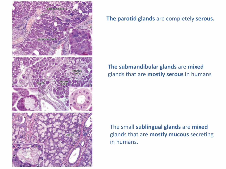
The parotid glands are completely serous.
The submandibular glands are mixed glands that are mostly serous in humans
The small sublingual glands are mixed glands that are mostly mucous secreting in humans.
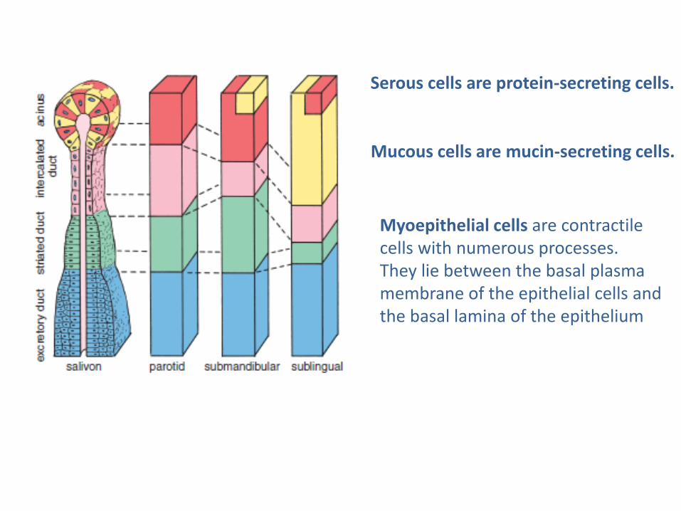
Serous cells are protein-secreting cells.
Mucous cells are mucin-secreting cells.
Myoepithelial cells are contractile cells with numerous processes. They lie between the basal plasma membrane of the epithelial cells and the basal lamina of the epithelium

The lumen of the salivary acinus is continuous with that of a duct system that may have as many as three sequential segments: • Intercalated duct, which leads from the acinus • Striated duct, so-called because of the presence of “striations,” the infoldings of the basal plasma membrane of the columnar cells that form the duct • Excretory ducts, which are the larger ducts that empty into the oral cavity
Salivary Ducts
Mumps
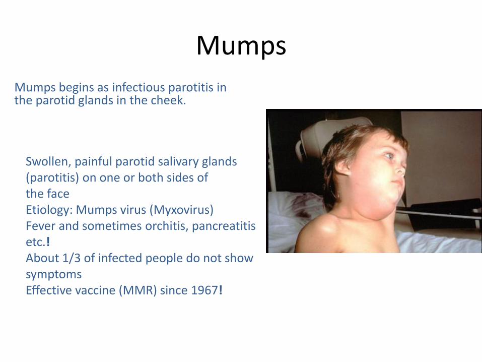
Swollen, painful parotid salivary glands (parotitis) on one or both sides of the face Etiology: Mumps virus (Myxovirus) Fever and sometimes orchitis, pancreatitis etc.! About 1/3 of infected people do not show symptoms Effective vaccine (MMR) since 1967!
Mumps begins as infectious parotitis in the parotid glands in the cheek.
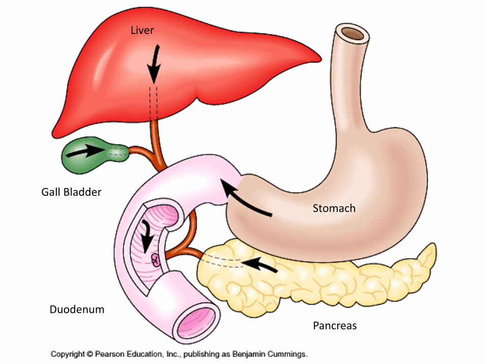
Gall Bladder
Duodenum
Liver
Pancreas
Stomach
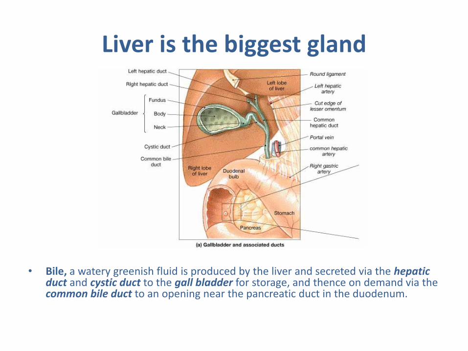
Liver is the biggest gland
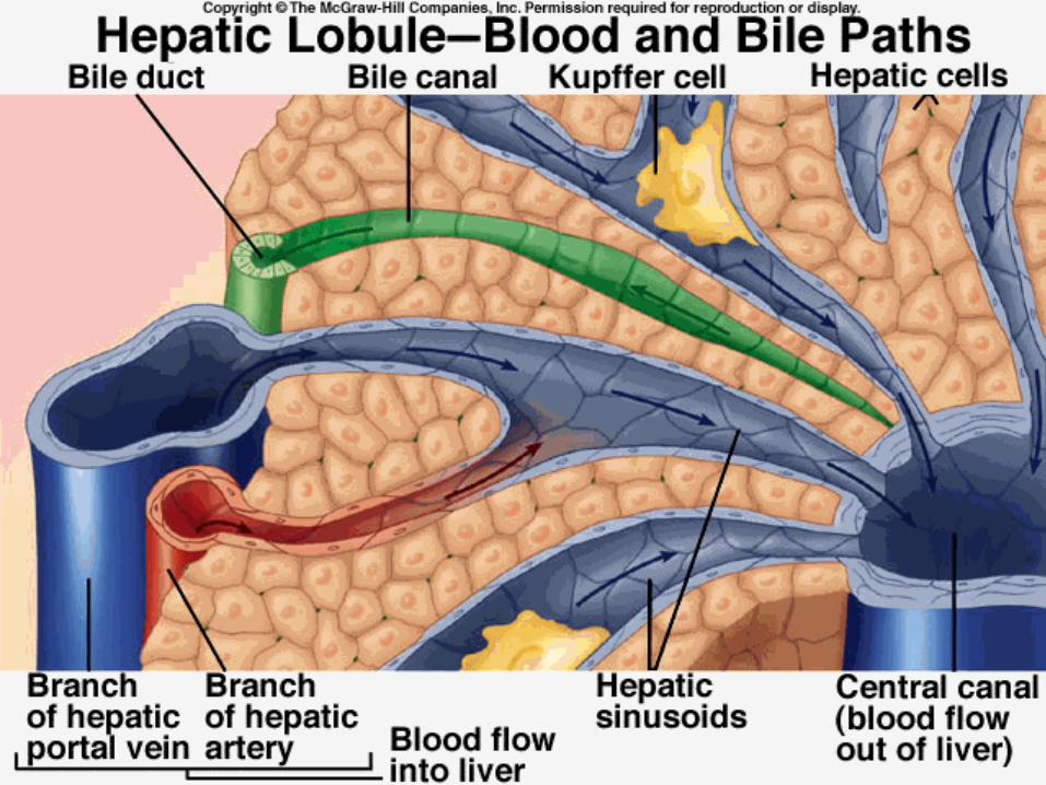
• Bile, a watery greenish fluid is produced by the liver and secreted via the hepatic duct and cystic duct to the gall bladder for storage, and thence on demand via the common bile duct to an opening near the pancreatic duct in the duodenum.
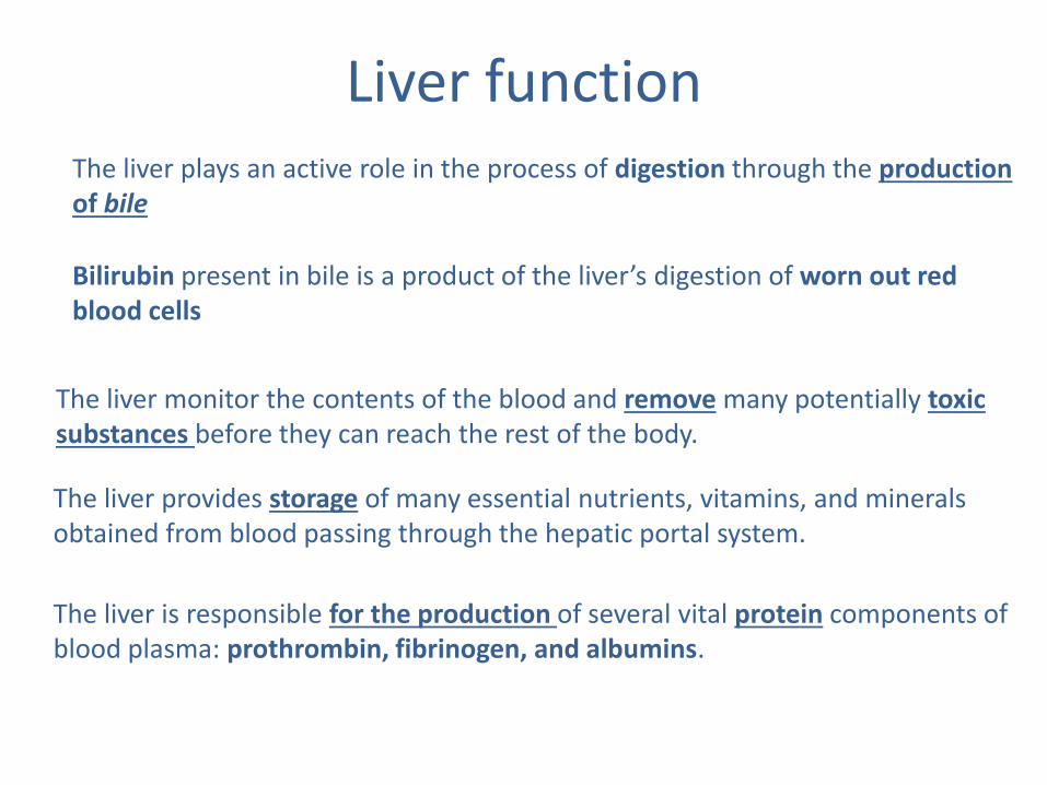
The liver provides storage of many essential nutrients, vitamins, and minerals obtained from blood passing through the hepatic portal system.
The liver is responsible for the production of several vital protein components of blood plasma: prothrombin, fibrinogen, and albumins.
The liver monitor the contents of the blood and remove many potentially toxic substances before they can reach the rest of the body.
Liver function The liver plays an active role in the process of digestion through the production of bile
Bilirubin present in bile is a product of the liver’s digestion of worn out red blood cells
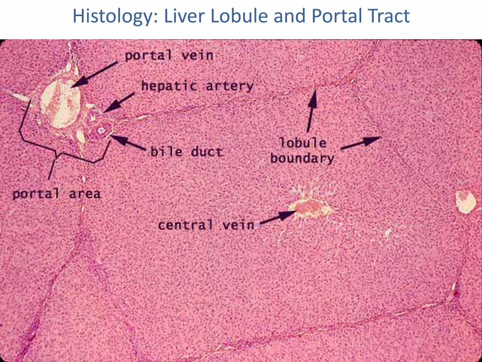
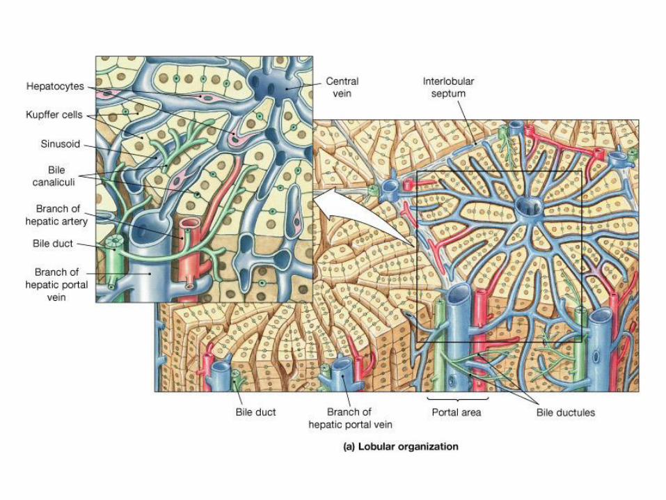
Histology: Liver Lobule and Portal Tract
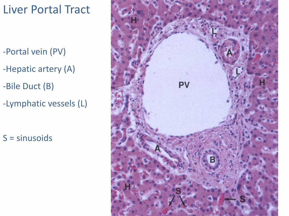
Liver Portal Tract
-Portal vein (PV)
-Hepatic artery (A)
-Bile Duct (B)
-Lymphatic vessels (L)
S = sinusoids

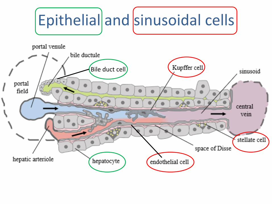
Bile duct cell
Epithelial and sinusoidal cells
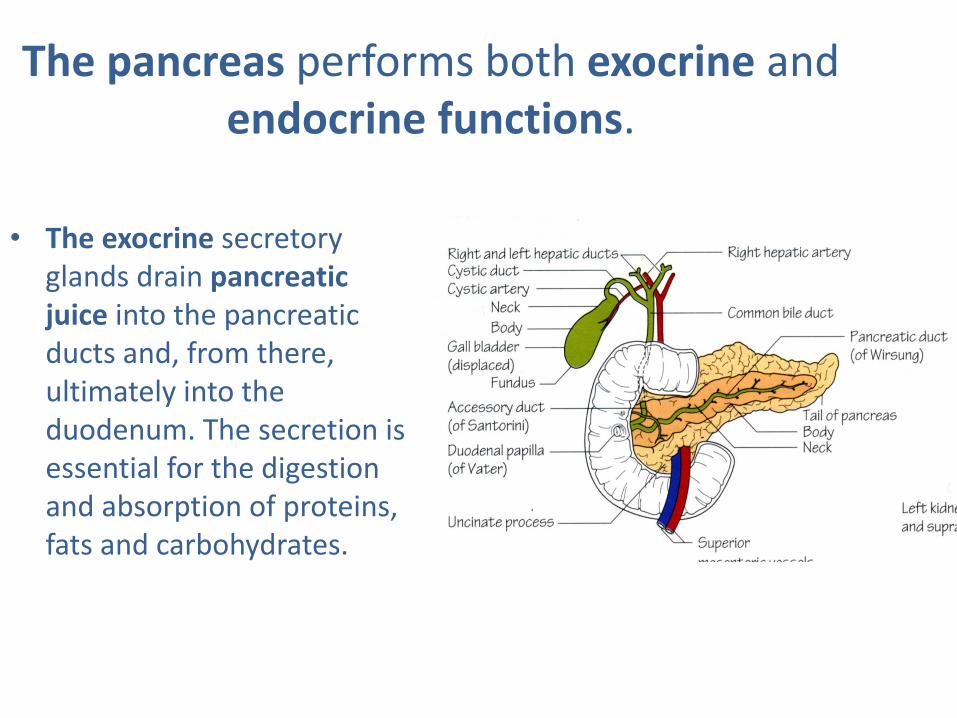
The pancreas performs both exocrine and endocrine functions.
• The exocrine secretory glands drain pancreatic juice into the pancreatic ducts and, from there, ultimately into the duodenum. The secretion is essential for the digestion and absorption of proteins, fats and carbohydrates.
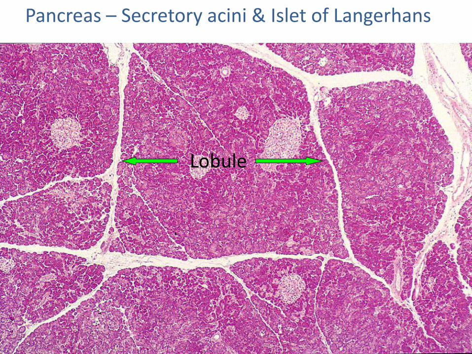
Pancreas – Secretory acini & Islet of Langerhans
Lobule
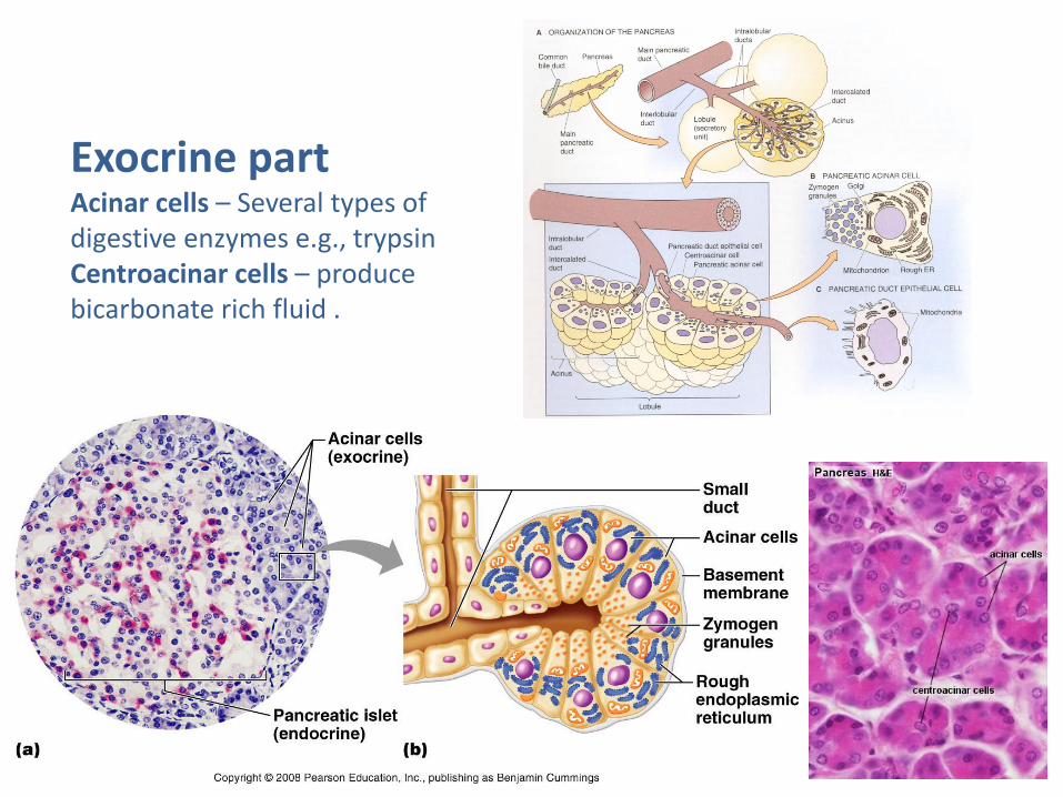
Exocrine part Acinar cells – Several types of digestive enzymes e.g., trypsin Centroacinar cells – produce bicarbonate rich fluid .
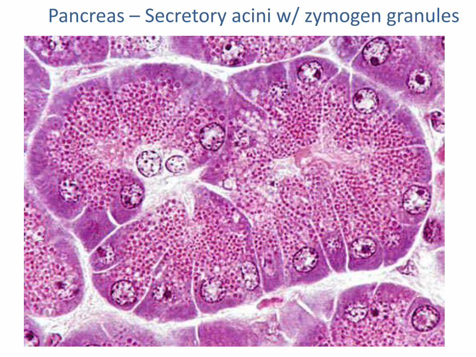
Pancreas – Secretory acini w/ zymogen granules
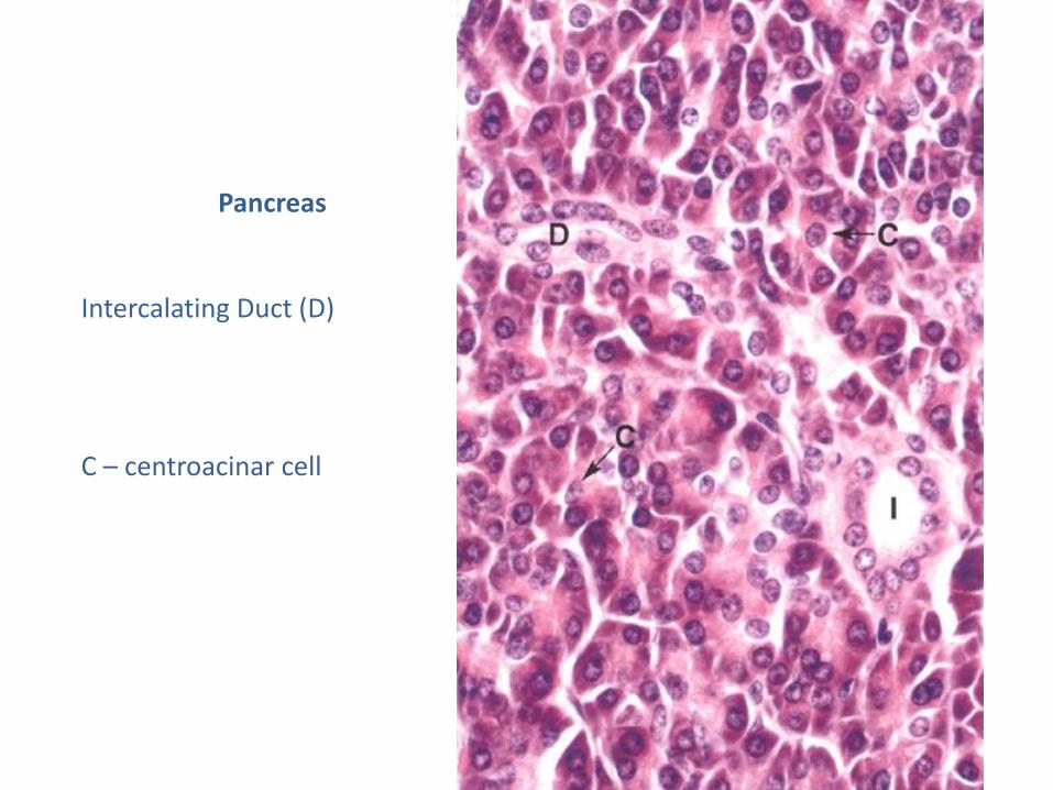
Pancreas
Intercalating Duct (D)
C – centroacinar cell
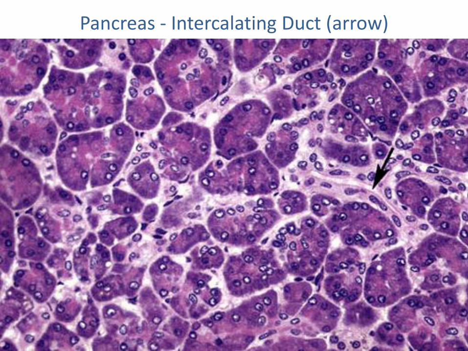
Pancreas - Intercalating Duct (arrow)
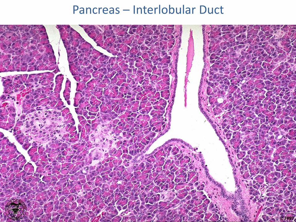
Pancreas – Interlobular Duct
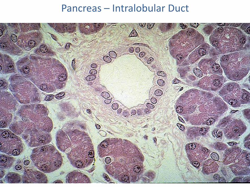
Pancreas – Intralobular Duct
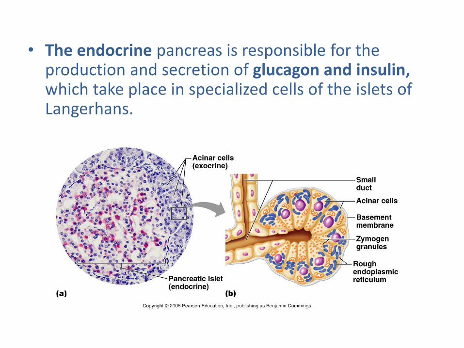
• The endocrine pancreas is responsible for the production and secretion of glucagon and insulin, which take place in specialized cells of the islets of Langerhans.

Thank you for attention