Embed Size (px)
Citation preview
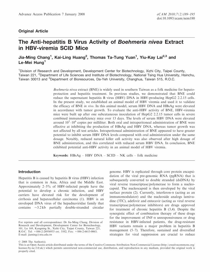
Advance Access Publication 7 January 2008 eCAM 2010;7(2)189–195doi:10.1093/ecam/nem180
Original Article
The Anti-hepatitis B Virus Activity of Boehmeria nivea Extractin HBV-viremia SCID Mice
Jia-Ming Chang1, Kai-Ling Huang2, Thomas Ta-Tung Yuan1, Yiu-Kay Lai2,3 andLe-Mei Hung1
1Division of Research and Development, Development Center for Biotechnology, Xizhi City, Taipei County,Taiwan 221, 2Department of Life Sciences and Institute of Biotechnology, National Tsing Hua University, Hsinchu,Taiwan 30013 and 3Department of Bioresources, Da-Yeh University, Changhua, Taiwan 515, R.O.C.
Boehmeria nivea extract (BNE) is widely used in southern Taiwan as a folk medicine for hepato-protection and hepatitis treatment. In previous studies, we demonstrated that BNE couldreduce the supernatant hepatitis B virus (HBV) DNA in HBV-producing HepG2 2.2.15 cells.In the present study, we established an animal model of HBV viremia and used it to validatethe efficacy of BNE in vivo. In this animal model, serum HBV DNA and HBsAg were elevatedin accordance with tumor growth. To evaluate the anti-HBV activity of BNE, HBV-viremiamice were built up after one subcutaneous inoculation of HepG2 2.2.15 tumor cells in severecombined immunodeficiency mice over 13 days. The levels of serum HBV DNA were elevatedaround 105–106 copies per milliliter. Both oral and intraperitoneal administration of BNE wereeffective at inhibiting the production of HBsAg and HBV DNA, whereas tumor growth wasnot affected by all test articles. Intraperitoneal administration of BNE appeared to have greaterpotential to inhibit serum HBV DNA levels compared with oral administration under the samedosage. Notably, reduced natural killer cell activity was also observed after high dosage ofBNE administration, and this correlated with reduced serum HBV DNA. In conclusion, BNEexhibited potential anti-HBV activity in an animal model of HBV viremia.
Keywords: HBsAg – HBV DNA – SCID – NK cells – folk medicine
Introduction
Hepatitis B is caused by hepatitis B virus (HBV) infection
that is common in Asia, Africa and the Middle East.
Approximately 2–5% of HBV-infected people have the
potential to develop a chronic infection, and HBV
carriers have elevated risk for the development of
cirrhosis and hepatocellular carcinoma (1). HBV is an
enveloped DNA virus of the hepadnaviridae family that
contains �3.2 kb partially double-stranded circular
genome. HBV is replicated through core protein encapsi-
dation of the viral pre-genomic RNA (pgRNA) that issubsequently converted to double stranded (dsDNA) byviral reverse transcriptase/polymerase to form a nucleo-capsid. The nucleocapsid is then enveloped by the viralsurface protein (2). Currently, interferon-a (acting as animmunomodulator) and the nucleoside analogs lamivu-
dine (3TC), adefovir and entecavir (acting as viral reversetranscriptase/polymerase inhibitors) are drugs approvedfor treatment of chronic hepatitis B (3,4). Despite thesynergistic effect of combination therapy of these drugsfor the improvement of INF-a unresponsiveness or drug
resistance in HBV-infected patients, the drug-resistantHBV variants remain a major problem in hepatitis Bmanagement (5–7). Therefore, sustained and diversifiedstrategies for viral suppression are crucial to reduce
For reprints and all correspondence: Dr Jia-Ming Chang, Division ofResearch and Development, Development Center for Biotechnology,101, Ln 169, Kangning St., Xizhi City, Taipei County, Taiwan 221,R.O.C. Tel: +886-2-26956933 ext. 5102; Fax: +886-2-6615-0063;E-mail: [email protected]
� 2008 The Author(s).This is an Open Access article distributed under the terms of the Creative Commons Attribution Non-Commercial License (http://creativecommons.org/licenses/by-nc/2.0/uk/) which permits unrestricted non-commercial use, distribution, and reproduction in any medium, provided the original work isproperly cited.
hepatic inflammation, progression of liver fibrosis, andhepatoma (8).Approaches for the treatment of chronic HBV infection
have been in development for the past decade, and whencombined with a number of anti-viral drugs thesetreatments may have potential to improve the effective-ness of anti-HBV therapy. Chinese medicinal herbs havebeen used for centuries to treat liver disease (9). Recently,the flavonoid ellagic acid from woody dicotyledonousplants has been found to block hepatitis B e antigen(HBeAg) secretion either in an HBV-infected cell line orin HBeAg transgenic mice (10). In addition, sesquiterpenelactones from Senecio species suppress the expressionof hepatitis B s antigen (HBsAg) and HBeAg (11), andan ethanol extract of Polygonum cuspidatum inhibitsHBV production in an HBV-producing cell line (12). Anextract of the genus Phyllanthus has a positive effect onthe clearance of HBsAg in clinical trials conducted onchronic HBV infections, and the extract has a synergisticeffect when administered with IFN-a (9). Our previousstudies demonstrated that crude root extract ofBoehmeria nivea had anti-HBV activity via the inhibitionof HBV production in HepG2 2.2.15 cells (13). ThisB. nivea extract (BNE) suppressed the production ofinfectious virus but not intracellular HBV DNA replica-tion, and its anti-HBV mechanism appears to differ fromthat of the nucleoside analogs. Thus, it is conceivablethat BNE might work synergistically with other anti-viralcompounds for the treatment of HBV infection.HBV replicates through an RNA intermediate by its
reverse transcriptase/polymerase. The viral reverse tran-scriptase/polymerase lacks a proofreading function, how-ever, thus leading to the formation of drug-resistantmutants that in turn cause serious problems with currentmanagement of hepatitis B infection (14). Therefore,effective treatment strategies with different pharmacolo-gical modes are urgently needed to lessen the enormousburden of viral hepatitis on health care worldwide.A major obstacle to the development of anti-HBV
drugs is the lack of an efficient in vitro cell model or aneasy-to-use conventional animal model that is promisingfor natural viral infection and replication (15). Avail-ability of laboratory animal models of HBV infection isimperative for the development of effective methods totreat these diseases. One of the few animal models forHBV infection is the chimpanzee, which is extensivelyused to evaluate the safety and immunogenicity of HBVvaccines (16). However, the limited availability and thehigh cost of these primates severely restrict their usefor such purposes. Investigators have attempted to estab-lish conventional animal models to mimic human HBVreplication in rats (17), nude mice (18) and transgenicmice (19). Other animal models, such as woodchuckhepatitis virus in woodchuck and duck hepatitis virus induck, have also been developed to assess anti-viral drugs(20). However, testing of anti-viral agents, including
nucleoside analogs, in these non-human HBV modelsmay produce aberrant results as a consequence of virus-specific differential susceptibility of the viral polymeraseto these agents (21). Thus, establishment of a rapid,convenient and less expensive animal model wouldgreatly facilitate this area of research. In this study, weestablished an HBV-viremia animal model and studiedthe efficacy of BNE in vivo.
Subjects and Methods
Experimental Animals and Cell Culture
Male severe combined immunodeficiency (SCID) mice4–6 weeks of age, C.B17/icr-scid, were purchased fromthe experimental animal center of National TaiwanUniversity (Taipei, Taiwan, R.O.C.). The human hepa-toma HepG2 cell line was purchased from AmericanType Culture Center (ATCC, USA), and the HepG22.2.15 cell line was kindly given by Dr Ho, MS(Academia Sinica, Taipei, Taiwan, R.O.C.). Thesehepatoma cells were maintained in minimum essentialmedium (Gibco, USA) containing 10% fetal bovineserum (Hyclone, USA), 1.5 g l�1 sodium bicarbonate,0.1mM non-essential amino acids, 1.0mM sodiumpyruvate, and 100Uml�1 penicillin G (Gibco, USA)and 100 mgml�1 streptomycin (Gibco, USA). A finalconcentration of 200 mgml�1 G418 (Gibco, USA) wasincluded in the medium for the maintenance of HepG22.2.15 cells. YAC-1 cells (ATCC, USA) for the naturalkiller (NK) cell activity assay were cultured in RPMI1640 medium containing 2mM L-glutamine, 1.5 g l�1
sodium bicarbonate, 10mM HEPES and 10% fetalbovine serum.
Anti-HBV Drugs and BNE
To prepare the B. nivea (L.) Gaudich plant extract(i.e. BNE) utilized in our experiments, the roots of theplants were collected and dried (13). Briefly, 100 g of thedried roots was cut into pieces �0.5 cm in length beforeboiling in 1 l of 20% ethanol under boiling reflux for 3 h.The decoction was filtered through a 0.22 mm filterand lyophilized into powder. 3TC was purchased fromGlaxoSmithKline (Holland).
Establishment of an HBV-Viremia Animal Model
To establish HBV-viremia animals, the 20 experimentalSCID mice were divided into three groups: HepG2 group(n=8), HepG2 2.2.15 group (n=8) and one vehiclecontrol group (n=4). The SCID mice were inoculatedwith 107 HepG2 or HepG2 2.2.15 cells in 100 mlphosphate-buffered saline (PBS) into the flank subcuta-neously. The control group was inoculated likewise with
190 Anti-HBV activity of B. nivea extract in vivo
PBS. Tumors were measured using a caliper, and tumorweight was calculated with the equation: tumor weight(in mg)= length (in mm)�width (in mm2)/2 (22). Onpost-inoculation Day 0 and Day 14, blood was collectedby retro-orbital sampling. On Day 28, the mice weresacrificed and blood was collected directly from the heart.Serum HBsAg and HBV DNA were analyzed asdescribed subsequently.
Determination of Serum Alanine Aminotransferase
(ALT) and Serum HBsAg
A blood sample was withdrawn from tumor-bearing ani-mals at the indicated times (Days 0, 14 and 28), and liverfunction was evaluated based on the levels of ALT. ALTactivity was determined with the DTSC II Moduledetection system (Johnson-Johnson Co., USA) usingthe Vitros ALT/SGPT kit (Johnson-Johnson) as per theuser manual. HBsAg levels in mouse serum were deter-mined semi-quantitatively by enzyme-linked immunosor-bent assay (ELISA; General Biologicals Corp., Taiwan,R.O.C.) according to the user manual. The cutoffvalue was calculated by the formula: cutoff value=ODnagative control+0.025. An OD450 value higher than thecutoff value implies an HBV-positive result.
Determination of HBV DNA by the Quantitative
Real-time Polymerase Chain Reaction (PCR)
Blood (500ml) was withdrawn from test animals at theindicated times (Days 13 and 24). Blood was allowed toclot for 30min and then centrifuged at 800g for 15min atroom temperature. Serum was collected and stored at�70�C prior to analysis of HBsAg and HBV DNA.Serum HBV DNA was extracted by a QIAamp DNABlood Mini kit (Qiagen, USA). HBV DNA was quan-titated with the ABI 7500 Sequence Detection Systemusing the HBV RealQuant PCR kit (General BiologicalsCorp.). Briefly, PCR was performed with initial denatur-ing steps at 50�C for 2min and 95�C for 10min, followedby 45 cycles at 95�C for 15 s and annealing/extending at58�C for 1min.
Validation of the Efficacy of BNE in
HBV-viremia Animals
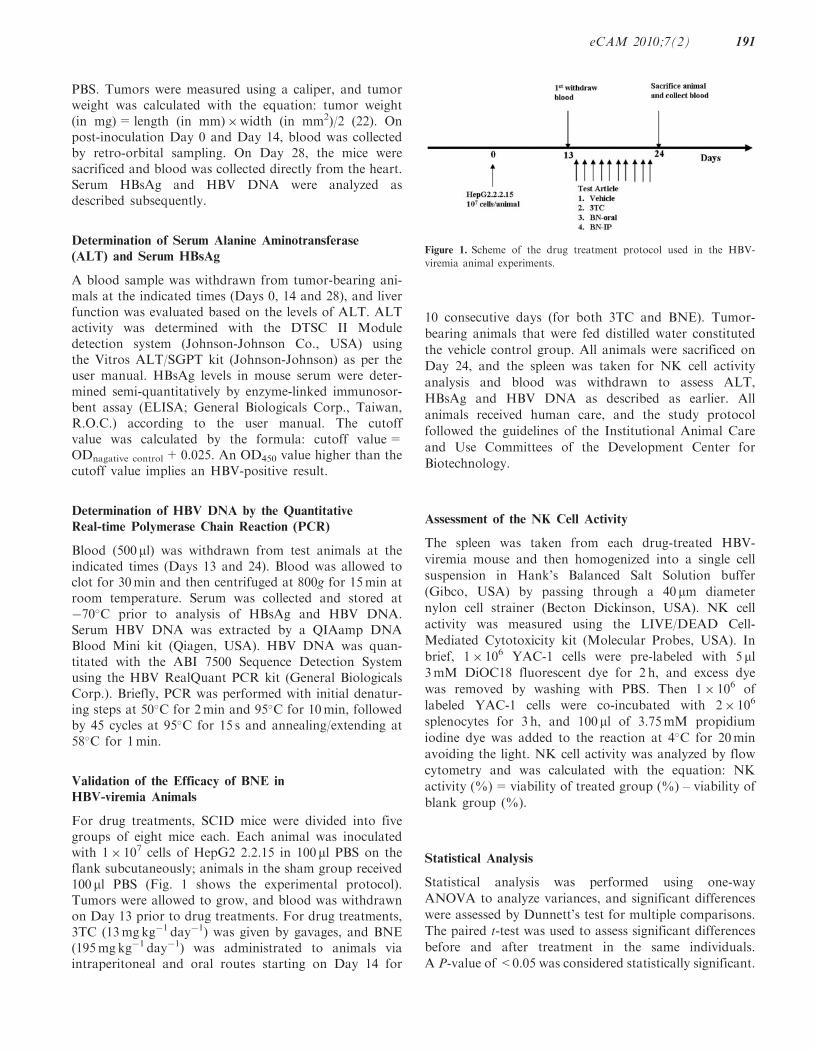
For drug treatments, SCID mice were divided into fivegroups of eight mice each. Each animal was inoculatedwith 1� 107 cells of HepG2 2.2.15 in 100 ml PBS on theflank subcutaneously; animals in the sham group received100 ml PBS (Fig. 1 shows the experimental protocol).Tumors were allowed to grow, and blood was withdrawnon Day 13 prior to drug treatments. For drug treatments,3TC (13mgkg�1 day�1) was given by gavages, and BNE(195mgkg�1 day�1) was administrated to animals viaintraperitoneal and oral routes starting on Day 14 for
10 consecutive days (for both 3TC and BNE). Tumor-
bearing animals that were fed distilled water constituted
the vehicle control group. All animals were sacrificed on
Day 24, and the spleen was taken for NK cell activity
analysis and blood was withdrawn to assess ALT,
HBsAg and HBV DNA as described as earlier. All
animals received human care, and the study protocol
followed the guidelines of the Institutional Animal Care
and Use Committees of the Development Center for
Biotechnology.
Assessment of the NK Cell Activity
The spleen was taken from each drug-treated HBV-
viremia mouse and then homogenized into a single cell
suspension in Hank’s Balanced Salt Solution buffer
(Gibco, USA) by passing through a 40 mm diameter
nylon cell strainer (Becton Dickinson, USA). NK cell
activity was measured using the LIVE/DEAD Cell-
Mediated Cytotoxicity kit (Molecular Probes, USA). In
brief, 1� 106 YAC-1 cells were pre-labeled with 5 ml3mM DiOC18 fluorescent dye for 2 h, and excess dye
was removed by washing with PBS. Then 1� 106 of
labeled YAC-1 cells were co-incubated with 2� 106
splenocytes for 3 h, and 100 ml of 3.75mM propidium
iodine dye was added to the reaction at 4�C for 20min
avoiding the light. NK cell activity was analyzed by flow
cytometry and was calculated with the equation: NK
activity (%)=viability of treated group (%) – viability of
blank group (%).
Statistical Analysis
Statistical analysis was performed using one-way
ANOVA to analyze variances, and significant differences
were assessed by Dunnett’s test for multiple comparisons.
The paired t-test was used to assess significant differences
before and after treatment in the same individuals.
A P-value of <0.05 was considered statistically significant.
Figure 1. Scheme of the drug treatment protocol used in the HBV-
viremia animal experiments.
eCAM 2010;7(2) 191
Results
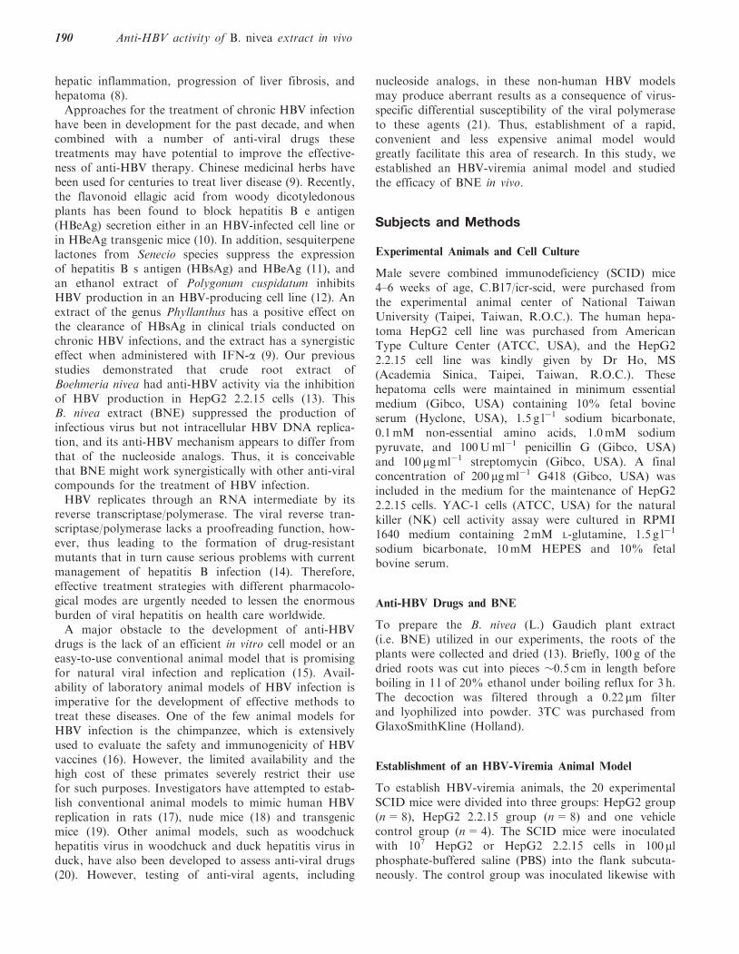
Tumor Formation Caused by HepG2 and HepG2 2.2.15
Cells in SCID Mice
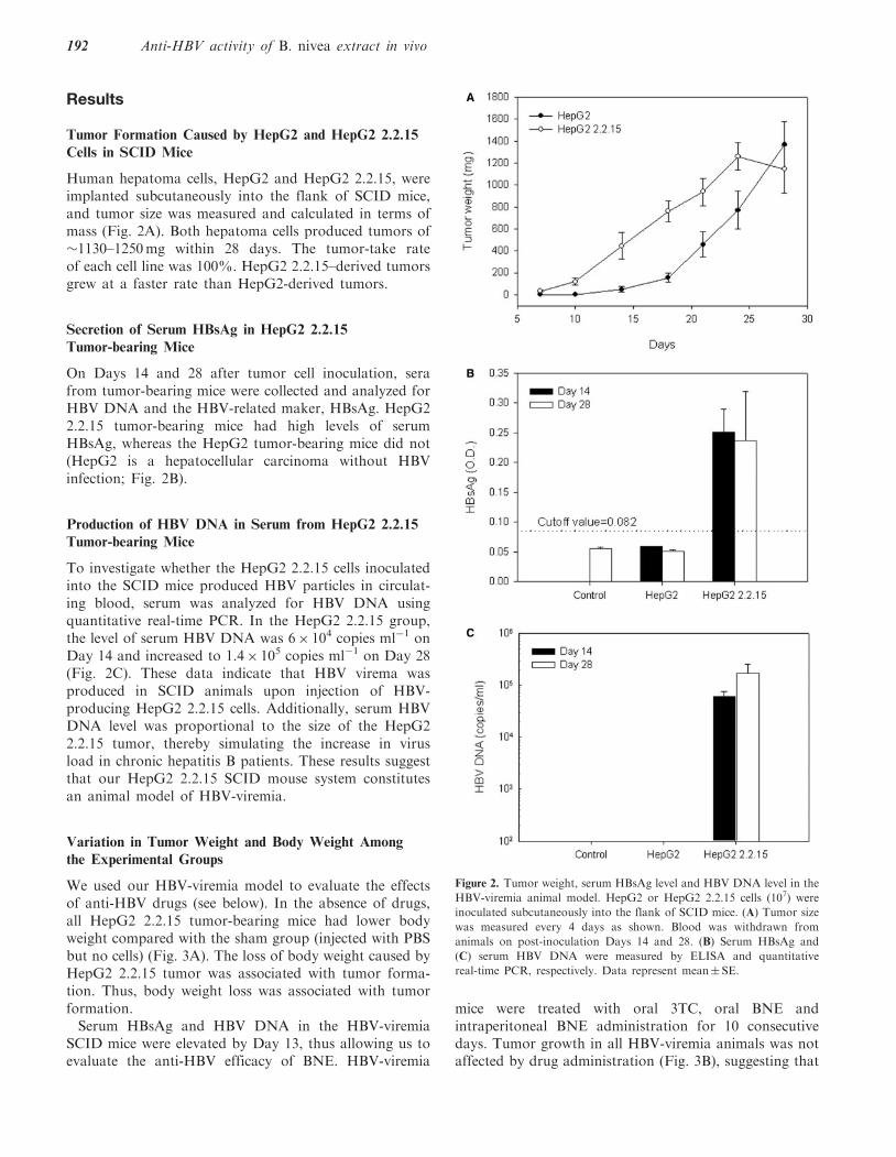
Human hepatoma cells, HepG2 and HepG2 2.2.15, wereimplanted subcutaneously into the flank of SCID mice,and tumor size was measured and calculated in terms ofmass (Fig. 2A). Both hepatoma cells produced tumors of�1130–1250mg within 28 days. The tumor-take rateof each cell line was 100%. HepG2 2.2.15–derived tumorsgrew at a faster rate than HepG2-derived tumors.
Secretion of Serum HBsAg in HepG2 2.2.15
Tumor-bearing Mice
On Days 14 and 28 after tumor cell inoculation, serafrom tumor-bearing mice were collected and analyzed forHBV DNA and the HBV-related maker, HBsAg. HepG22.2.15 tumor-bearing mice had high levels of serumHBsAg, whereas the HepG2 tumor-bearing mice did not(HepG2 is a hepatocellular carcinoma without HBVinfection; Fig. 2B).
Production of HBV DNA in Serum from HepG2 2.2.15
Tumor-bearing Mice
To investigate whether the HepG2 2.2.15 cells inoculatedinto the SCID mice produced HBV particles in circulat-ing blood, serum was analyzed for HBV DNA usingquantitative real-time PCR. In the HepG2 2.2.15 group,the level of serum HBV DNA was 6� 104 copies ml�1 onDay 14 and increased to 1.4� 105 copies ml�1 on Day 28(Fig. 2C). These data indicate that HBV virema wasproduced in SCID animals upon injection of HBV-producing HepG2 2.2.15 cells. Additionally, serum HBVDNA level was proportional to the size of the HepG22.2.15 tumor, thereby simulating the increase in virusload in chronic hepatitis B patients. These results suggestthat our HepG2 2.2.15 SCID mouse system constitutesan animal model of HBV-viremia.
Variation in Tumor Weight and Body Weight Among
the Experimental Groups
We used our HBV-viremia model to evaluate the effectsof anti-HBV drugs (see below). In the absence of drugs,all HepG2 2.2.15 tumor-bearing mice had lower bodyweight compared with the sham group (injected with PBSbut no cells) (Fig. 3A). The loss of body weight caused byHepG2 2.2.15 tumor was associated with tumor forma-tion. Thus, body weight loss was associated with tumorformation.Serum HBsAg and HBV DNA in the HBV-viremia
SCID mice were elevated by Day 13, thus allowing us toevaluate the anti-HBV efficacy of BNE. HBV-viremia
mice were treated with oral 3TC, oral BNE andintraperitoneal BNE administration for 10 consecutivedays. Tumor growth in all HBV-viremia animals was notaffected by drug administration (Fig. 3B), suggesting that
Figure 2. Tumor weight, serum HBsAg level and HBV DNA level in the
HBV-viremia animal model. HepG2 or HepG2 2.2.15 cells (107) were
inoculated subcutaneously into the flank of SCID mice. (A) Tumor size
was measured every 4 days as shown. Blood was withdrawn from
animals on post-inoculation Days 14 and 28. (B) Serum HBsAg and
(C) serum HBV DNA were measured by ELISA and quantitative
real-time PCR, respectively. Data represent mean� SE.
192 Anti-HBV activity of B. nivea extract in vivo
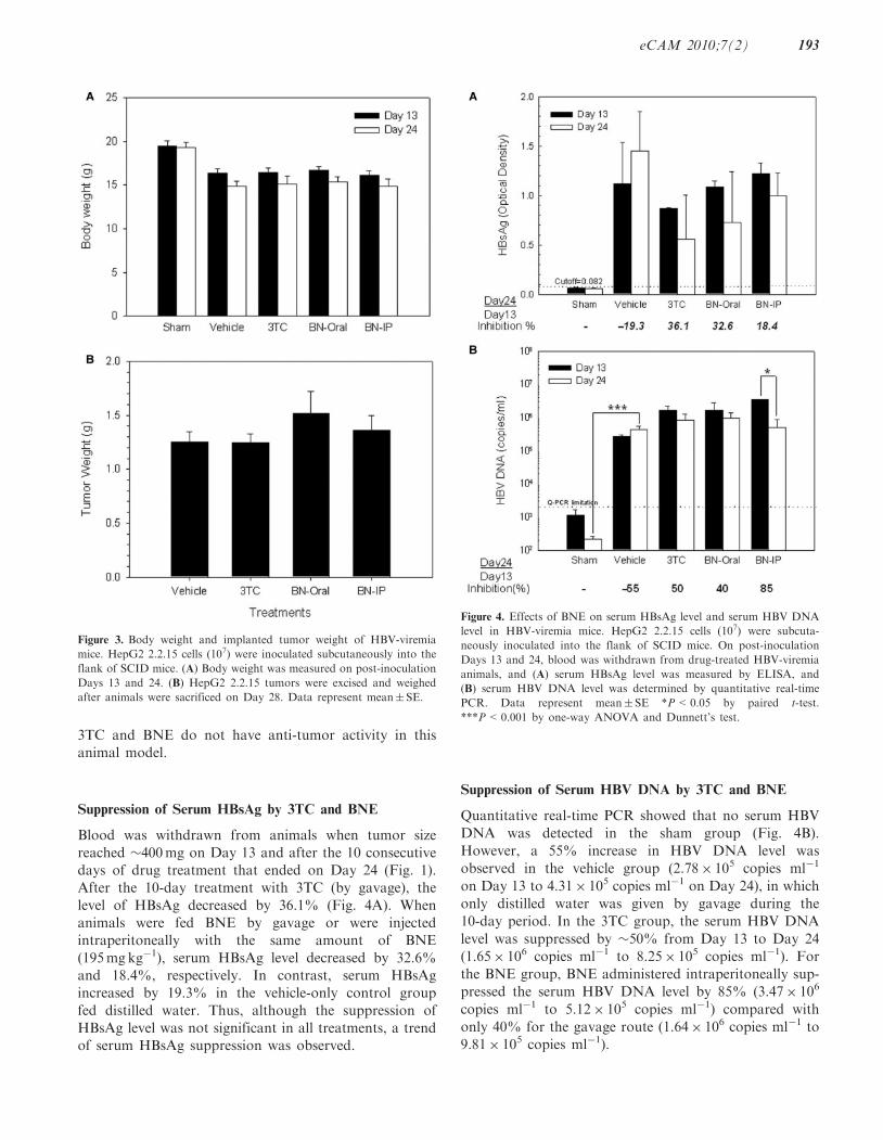
3TC and BNE do not have anti-tumor activity in thisanimal model.
Suppression of Serum HBsAg by 3TC and BNE
Blood was withdrawn from animals when tumor size
reached �400mg on Day 13 and after the 10 consecutivedays of drug treatment that ended on Day 24 (Fig. 1).After the 10-day treatment with 3TC (by gavage), the
level of HBsAg decreased by 36.1% (Fig. 4A). Whenanimals were fed BNE by gavage or were injectedintraperitoneally with the same amount of BNE
(195mgkg�1), serum HBsAg level decreased by 32.6%and 18.4%, respectively. In contrast, serum HBsAgincreased by 19.3% in the vehicle-only control group
fed distilled water. Thus, although the suppression ofHBsAg level was not significant in all treatments, a trendof serum HBsAg suppression was observed.
Suppression of Serum HBV DNA by 3TC and BNE
Quantitative real-time PCR showed that no serum HBV
DNA was detected in the sham group (Fig. 4B).
However, a 55% increase in HBV DNA level was
observed in the vehicle group (2.78� 105 copies ml�1
on Day 13 to 4.31� 105 copies ml�1 on Day 24), in which
only distilled water was given by gavage during the
10-day period. In the 3TC group, the serum HBV DNA
level was suppressed by �50% from Day 13 to Day 24
(1.65� 106 copies ml�1 to 8.25� 105 copies ml�1). For
the BNE group, BNE administered intraperitoneally sup-
pressed the serum HBV DNA level by 85% (3.47� 106
copies ml�1 to 5.12� 105 copies ml�1) compared with
only 40% for the gavage route (1.64� 106 copies ml�1 to
9.81� 105 copies ml�1).
Figure 4. Effects of BNE on serum HBsAg level and serum HBV DNA
level in HBV-viremia mice. HepG2 2.2.15 cells (107) were subcuta-
neously inoculated into the flank of SCID mice. On post-inoculation
Days 13 and 24, blood was withdrawn from drug-treated HBV-viremia
animals, and (A) serum HBsAg level was measured by ELISA, and
(B) serum HBV DNA level was determined by quantitative real-time
PCR. Data represent mean� SE *P<0.05 by paired t-test.
***P<0.001 by one-way ANOVA and Dunnett’s test.
Figure 3. Body weight and implanted tumor weight of HBV-viremia
mice. HepG2 2.2.15 cells (107) were inoculated subcutaneously into the
flank of SCID mice. (A) Body weight was measured on post-inoculation
Days 13 and 24. (B) HepG2 2.2.15 tumors were excised and weighed
after animals were sacrificed on Day 28. Data represent mean� SE.
eCAM 2010;7(2) 193
Assessment of ALT Activity in the Experimental Groups
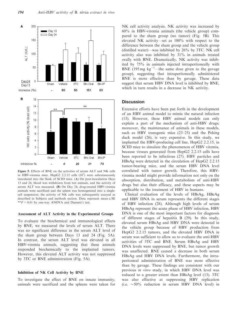
To evaluate the biochemical and immunological effectsby BNE, we measured the levels of serum ALT. Therewas no significant difference in the serum ALT level ofthe sham group between Days 13 and 24 (Fig. 5A).In contrast, the serum ALT level was elevated in allHBV-viremia animals, suggesting that these animalsresponded biochemically to the implanted tumors.However, this elevated ALT activity was not suppressedby 3TC or BNE administration (Fig. 5A).
Inhibition of NK Cell Activity by BNE
To investigate the effect of BNE on innate immunity,animals were sacrificed and the spleens were taken for
NK cell activity analysis. NK activity was increased by60% in HBV-viremia animals (the vehicle group) com-pared to the sham group (no tumor) (Fig. 5B). Thiselevated NK activity—set as 100% with respect to thedifference between the sham group and the vehicle group(distilled water)—was inhibited by 26% by 3TC. NK cellactivity also was inhibited by 31% in animals treatedorally with BNE. Dramatically, NK activity was inhib-ited by 75% in animals injected intraperitoneally withBNE (195mg kg�1—the same dose given to the gavagegroup), suggesting that intraperitoneally administeredBNE is more effective than by gavage. These datasuggest that serum HBV DNA level is inhibited by BNE,which in turn results in a decrease in NK activity.
Discussion
Extensive efforts have been put forth in the developmentof an HBV animal model to mimic the natural infection(15). However, these HBV animal models can onlyexplain a part of the mechanism of anti-HBV drugs;moreover, the maintenance of animals in these models,such as HBV transgenic mice (23–25) and the Pekingduck model (26), is very expensive. In this study, weimplanted the HBV-producing cell line, HepG2 2.2.15, inSCID mice to simulate the phenomenon of HBV viremia,because viruses generated from HepG2 2.2.15 cells havebeen reported to be infectious (27). HBV particles andHBsAg were detected in the circulation of HepG2 2.2.15tumor-bearing mice, and the serum HBV DNA levelcorrelated with tumor growth. Therefore, this HBV-viremia model might provide information not only on theabsorption, distribution, and metabolism of anti-HBVdrugs but also their efficacy, and these aspects may beapplicable to the treatment of HBV in humans.Clinical evaluation of the levels of HBsAg, HBeAg
and HBV DNA in serum represents the different stagesof HBV infection (28). Although high levels of serumHBsAg represent the acute phase of HBV infection, HBVDNA is one of the most important factors for diagnosisof different stages of hepatitis B (29). In this study,elevated serum HBsAg and HBV DNA were detected inthe vehicle group because of HBV production fromHepG2 2.2.15 tumors, and the elevated HBV DNA inserum was sufficient to allow us to evaluate the anti-HBVactivities of 3TC and BNE. Serum HBsAg and HBVDNA levels were suppressed by BNE, but tumor growthwas unaffected. BNE caused a decrease in both serumHBsAg and HBV DNA levels. Furthermore, the intra-peritoneal administration of BNE was more effectivethan by gavage. These findings are consistent with ourprevious in vitro study, in which HBV DNA level wasreduced to a greater extent than HBsAg level (13). 3TCwas also effective at suppressing HBV replication(i.e. �50% reduction in serum HBV DNA level) in
Figure 5. Effects of BNE on the activities of serum ALT and NK cells
in HBV-viremia mice. HepG2 2.2.15 cells (107) were subcutaneously
inoculated into the flank of SCID mice. (A) On post-inoculation Days
13 and 24, blood was withdrawn from test animals, and the activity of
serum ALT was measured. (B) On Day 24, drug-treated HBV-viremia
animals were sacrificed and the spleen was homogenized into a single-
cell suspension; the activity of NK cells was subsequently assayed as
described in Subjects and methods section. Data represent mean� SE
**P<0.01 by one-way ANOVA and Dunnett’s test.
194 Anti-HBV activity of B. nivea extract in vivo
HBV-viremia animals, and this efficacy may improvewith prolonged therapy.Although SCID mice lack adaptive immunity (functio-
nal T and B cells), they retain innate immunity in C.B17/icr species (e.g. functional granulocytes and NK cells).Thus, liver damage that might be caused by adaptiveimmunity in normal mice might not be observed in SCIDmice (30). Apparent suppression of NK cell activity wasobserved after intraperitoneal administration of BNE.NK cells play a critical role in host innate defense againstviruses and may be partly responsible for liver injuryresulting from anti-viral responses. However, the exactrole of NK cells in liver injury remains unclear (31).In this model, the observed increase in ALT level couldnot be conclusively attributed to the mouse liver cells ortumor cells. Nonetheless, histological examination of theliver of all mice revealed no apparent damage (data notshown), suggesting that the elevated serum ALT activityprobably was a result of expression from the implantedtumor cells. Taken together, the data support the anti-HBV effects of BNE and the chemical composition ofBNE is subject to for further investigation.
Acknowledgment
We thank Dr Chang DTM for polishing the English andgiving us comments on this article.
References1. Nakamoto Y, Guidotti LG, Kuhlen CV, Fowler P, Chisari FV.
Immune pathogenesis of hepatocellular carcinoma. J Exp Med1998;188:341–50.
2. Ganem D, Varmus HE. The molecular biology of the hepatitis Bviruses. Annu Rev Biochem 1987;56:651–93.
3. Ocama P, Opio CK, Lee WM. Hepatitis B virus infection: currentstatus. Am J Med 2005;118:1413.
4. Mailliard ME, Gollan JL. Emerging therapeutics for chronichepatitis B. Annu Rev Med 2006;57:155–66.
5. Tenney DJ, Levine SM, Rose RE, Walsh AW, Weinheimer SP,Discotto L, et al. Clinical emergence of entecavir-resistant hepatitisB virus requires additional substitutions in virus already resistant tolamivudine. Antimicrob Agents Chemother 2004;48:3498–507.
6. Lee SK, Wong CK, Poon PM, Ip PS, Che CT, Fung KP, et al.In vitro immunomodulatory activities of a newly concoctedtraditional Chinese medicine formula: VI-28. Phytother Res2006;20:883–8.
7. Fischer KP, Gutfreund KS, Tyrrell DL. Lamivudine resistance inhepatitis B: mechanisms and clinical implications. Drug ResistUpdat 2001;4:118–28.
8. Fattovich G, Stroffolini T, Zagni I, Donato F. Hepatocellularcarcinoma in cirrhosis: incidence and risk factors. Gastroenterology2004;127:S35–50.
9. Liu J, Lin H, McIntosh H. Genus Phyllanthus for chronic hepatitisB virus infection: a systematic review. J Viral Hepat 2001;8:358–66.
10. Kang EH, Kown TY, Oh GT, Park WF, Park SI, Park SK, et al.The flavonoid ellagic acid from a medicinal herb inhibits hostimmune tolerance induced by the hepatitis B virus-e antigen.Antiviral Res 2006;72:100–6.
11. Li H, Zhou C, Zhou L, Chen Z, Yang L, Bai H, et al. In vitroantiviral activity of three enantiomeric sesquiterpene lactones from
Senecio species against hepatitis B virus. Antivir Chem Chemother2005;16:277–82.
12. Chang JS, Liu HW, Wang KC, Chen MC, Chiang LC, Hua YC,et al. Ethanol extract of Polygonum cuspidatum inhibits hepatitis Bvirus in a stable HBV-producing cell line. Antiviral Res2005;66:29–34.
13. Huang KL, Lai YK, Lin CC, Chang JM. Inhibition of hepatitis Bvirus production by Boehmeria nivea root extract in HepG2 2.2.15cells. World J Gastroenterol 2006;12:5721–5.
14. Locarnini S. Molecular virology and the development of resistantmutants: implications for therapy. Semin Liver Dis 2005;25 (Suppl1): 9–19.
15. Guha C, Mohan S, Roy-Chowdhury N, Roy-Chowdhury J. Cellculture and animal models of viral hepatitis. Part I: hepatitis B. LabAnim 2004;33:37–46.
16. Prince AM, Brotman B. Perspectives on hepatitis B studies withchimpanzees. Ilar J 2001;42:85–8.
17. Takahashi H, Fujimoto J, Hanada S, Isselbacher KJ. Acutehepatitis in rats expressing human hepatitis B virus transgenes.Proc Natl Acad Sci USA 1995;92:1470–4.
18. Zhai WR, Vajta G, Acs G, Paronetto F. A nude mouse model forthe in vivo production of hepatitis B virus. Gastroenterology1990;98:470–7.
19. Sato H, Goto W, Yamamura J, Kurokawa M, Kageyama S,Takahara T, et al. Therapeutic basis of glycyrrhizin on chronichepatitis B. Antiviral Res 1996;30:171–7.
20. Dandri M, Volz TK, Lutgehetmann M, Petersen J. Animal modelsfor the study of HBV replication and its variants. J Clin Virol2005;34 (Suppl 1): S54–62.
21. Condreay LD, Jansen RW, Powdrill TF, Johnson LC,Selleseth DW, Paff MT, et al. Evaluation of the potent anti-hepatitis B virus agent (-) cis-5-fluoro-1-[2-(hydroxymethyl)-1,3-oxathiolan-5-yl]cytosine in a novel in vivo model. AntimicrobAgents Chemother 1994;38:616–9.
22. Euhus DM, Hudd C, LaRegina MC, Johnson FE. Tumormeasurement in the nude mouse. J Surg Oncol 1986;31:229–34.
23. Sitia G, Iannacone M, Muller S, Bianchi ME, Guidotti LG.Treatment with HMGB1 inhibitors diminishes CTL-induced liverdisease in HBV transgenic mice. J Leukoc Biol 2007;81:100–7.
24. Morrey JD, Bailey KW, Korba BE, Sidwell RW. Utilizationof transgenic mice replicating high levels of hepatitis Bvirus for antiviral evaluation of lamivudine. Antiviral Res1999;42:97–108.
25. Morrey JD, Korba BE, Sidwell RW. Transgenic mice as achemotherapeutic model for hepatitis B virus infection. AntivirTher 1998;3:59–68.
26. Jilbert AR, Botten JA, Miller DS, Bertram EM, Hall PM,Kotlarski J, et al. Characterization of age- and dose-relatedoutcomes of duck hepatitis B virus infection. Virology1998;244:273–82.
27. Liu N, Zhu B, Huang Z, Zhu Y, Chen Q, Guo X, et al.[The inhibitive effects of the ethanol extract from Radix et RhizomaRhei on the secretion of HBsAg and HBeAg]. Zhong Yao Cai2004;27:419–21.
28. Ganem D, Prince AM. Hepatitis B virus infection–natural historyand clinical consequences. N Engl J Med 2004;350:1118–29.
29. Mommeja-Marin H, Mondou E, Blum MR, Rousseau F. SerumHBV DNA as a marker of efficacy during therapy for chronic HBVinfection: analysis and review of the literature. Hepatology2003;37:1309–19.
30. Sun Y, Chen HY, Xin SJ. Effect of IL-18 on peripheral bloodmononuclear cells of chronic hepatitis B and hepatitis B virus DNAreleased by HepG2.2.15 cell lines. Hepatobiliary Pancreat Dis Int2004;3:230–4.
31. Sjolin H, Tomasello E, Mousavi-Jazi M, Bartolazzi A, Karre K,Vivier E, et al. Pivotal role of KARAP/DAP12 adaptor molecule inthe natural killer cell-mediated resistance to murine cytomegalovirusinfection. J Exp Med 2002;195:825–34.
Received April 30, 2007; accepted December 5, 2007
eCAM 2010;7(2) 195

Submit your manuscripts athttp://www.hindawi.com
Stem CellsInternational
Hindawi Publishing Corporationhttp://www.hindawi.com Volume 2014
Hindawi Publishing Corporationhttp://www.hindawi.com Volume 2014
MEDIATORSINFLAMMATION
of
Hindawi Publishing Corporationhttp://www.hindawi.com Volume 2014
Behavioural Neurology
EndocrinologyInternational Journal of
Hindawi Publishing Corporationhttp://www.hindawi.com Volume 2014
Hindawi Publishing Corporationhttp://www.hindawi.com Volume 2014
Disease Markers
Hindawi Publishing Corporationhttp://www.hindawi.com Volume 2014
BioMed Research International
OncologyJournal of
Hindawi Publishing Corporationhttp://www.hindawi.com Volume 2014
Hindawi Publishing Corporationhttp://www.hindawi.com Volume 2014
Oxidative Medicine and Cellular Longevity
Hindawi Publishing Corporationhttp://www.hindawi.com Volume 2014
PPAR Research
The Scientific World JournalHindawi Publishing Corporation http://www.hindawi.com Volume 2014
Immunology ResearchHindawi Publishing Corporationhttp://www.hindawi.com Volume 2014
Journal of
ObesityJournal of
Hindawi Publishing Corporationhttp://www.hindawi.com Volume 2014
Hindawi Publishing Corporationhttp://www.hindawi.com Volume 2014
Computational and Mathematical Methods in Medicine
OphthalmologyJournal of
Hindawi Publishing Corporationhttp://www.hindawi.com Volume 2014
Diabetes ResearchJournal of
Hindawi Publishing Corporationhttp://www.hindawi.com Volume 2014
Hindawi Publishing Corporationhttp://www.hindawi.com Volume 2014
Research and TreatmentAIDS
Hindawi Publishing Corporationhttp://www.hindawi.com Volume 2014
Gastroenterology Research and Practice
Hindawi Publishing Corporationhttp://www.hindawi.com Volume 2014
Parkinson’s Disease
Evidence-Based Complementary and Alternative Medicine
Volume 2014Hindawi Publishing Corporationhttp://www.hindawi.com