Embed Size (px)
Citation preview
Thalamus807
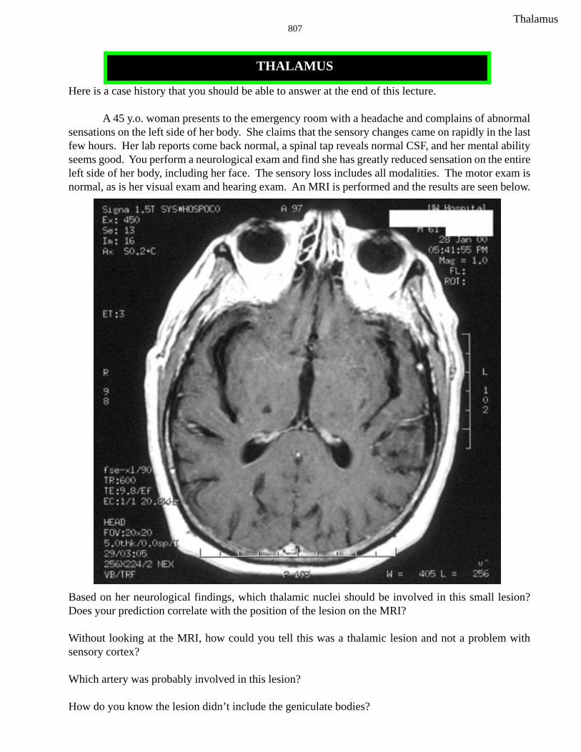
Here is a case history that you should be able to answer at the end of this lecture.
A 45 y.o. woman presents to the emergency room with a headache and complains of abnormalsensations on the left side of her body. She claims that the sensory changes came on rapidly in the lastfew hours. Her lab reports come back normal, a spinal tap reveals normal CSF, and her mental abilityseems good. You perform a neurological exam and find she has greatly reduced sensation on the entireleft side of her body, including her face. The sensory loss includes all modalities. The motor exam isnormal, as is her visual exam and hearing exam. An MRI is performed and the results are seen below.
Based on her neurological findings, which thalamic nuclei should be involved in this small lesion?Does your prediction correlate with the position of the lesion on the MRI?
Without looking at the MRI, how could you tell this was a thalamic lesion and not a problem withsensory cortex?
Which artery was probably involved in this lesion?
How do you know the lesion didn’t include the geniculate bodies?
THALAMUS
Thalamus808
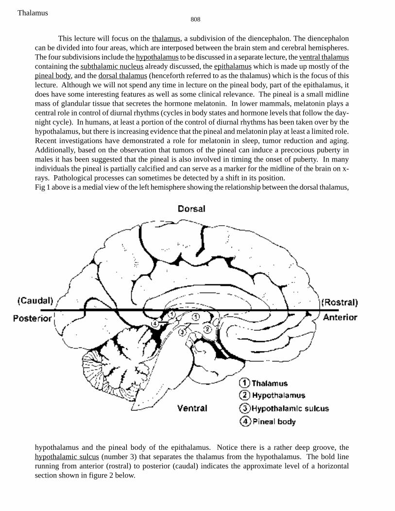
This lecture will focus on the thalamus, a subdivision of the diencephalon. The diencephaloncan be divided into four areas, which are interposed between the brain stem and cerebral hemispheres.The four subdivisions include the hypothalamus to be discussed in a separate lecture, the ventral thalamuscontaining the subthalamic nucleus already discussed, the epithalamus which is made up mostly of thepineal body, and the dorsal thalamus (henceforth referred to as the thalamus) which is the focus of thislecture. Although we will not spend any time in lecture on the pineal body, part of the epithalamus, itdoes have some interesting features as well as some clinical relevance. The pineal is a small midlinemass of glandular tissue that secretes the hormone melatonin. In lower mammals, melatonin plays acentral role in control of diurnal rhythms (cycles in body states and hormone levels that follow the day-night cycle). In humans, at least a portion of the control of diurnal rhythms has been taken over by thehypothalamus, but there is increasing evidence that the pineal and melatonin play at least a limited role.Recent investigations have demonstrated a role for melatonin in sleep, tumor reduction and aging.Additionally, based on the observation that tumors of the pineal can induce a precocious puberty inmales it has been suggested that the pineal is also involved in timing the onset of puberty. In manyindividuals the pineal is partially calcified and can serve as a marker for the midline of the brain on x-rays. Pathological processes can sometimes be detected by a shift in its position.Fig 1 above is a medial view of the left hemisphere showing the relationship between the dorsal thalamus,
hypothalamus and the pineal body of the epithalamus. Notice there is a rather deep groove, thehypothalamic sulcus (number 3) that separates the thalamus from the hypothalamus. The bold linerunning from anterior (rostral) to posterior (caudal) indicates the approximate level of a horizontalsection shown in figure 2 below.
Thalamus809
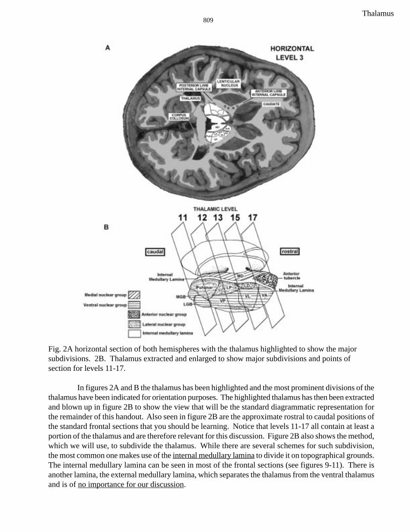
Fig. 2A horizontal section of both hemispheres with the thalamus highlighted to show the majorsubdivisions. 2B. Thalamus extracted and enlarged to show major subdivisions and points ofsection for levels 11-17.
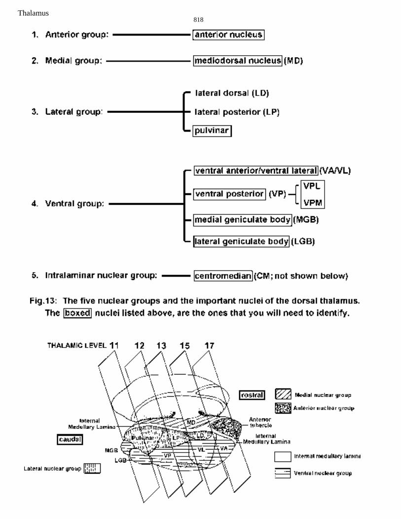
In figures 2A and B the thalamus has been highlighted and the most prominent divisions of thethalamus have been indicated for orientation purposes. The highlighted thalamus has then been extractedand blown up in figure 2B to show the view that will be the standard diagrammatic representation forthe remainder of this handout. Also seen in figure 2B are the approximate rostral to caudal positions ofthe standard frontal sections that you should be learning. Notice that levels 11-17 all contain at least aportion of the thalamus and are therefore relevant for this discussion. Figure 2B also shows the method,which we will use, to subdivide the thalamus. While there are several schemes for such subdivision,the most common one makes use of the internal medullary lamina to divide it on topographical grounds.The internal medullary lamina can be seen in most of the frontal sections (see figures 9-11). There isanother lamina, the external medullary lamina, which separates the thalamus from the ventral thalamusand is of no importance for our discussion.
Thalamus810
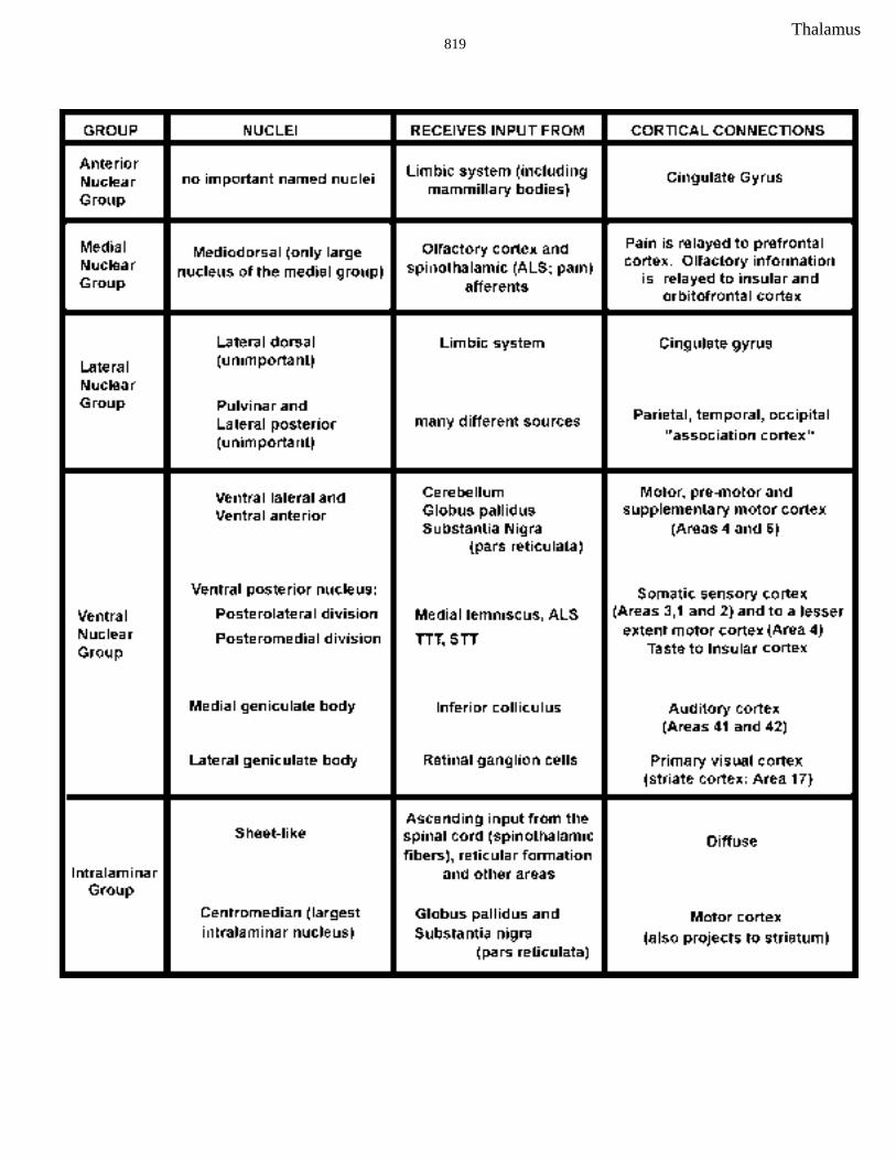
The internal medullary lamina is a thin sheet of white matter that runs longitudinally throughthe thalamus, separating it into medial and lateral nuclear masses. The medial mass consists of themedial nuclear group; the lateral mass contains the lateral nuclear group and the ventral nuclear group.In the rostral part of the thalamus the internal medullary lamina splits to form a partial capsule aroundthe anterior nuclear group. Finally, a fifth nuclear group, termed the intralaminar nuclear group, isfound within the confines of the internal medullary lamina and is therefore not seen in this diagrammaticrepresentation. The different nuclear groups are indicated by capitol letters and by different shadingpatterns in Fig. 2B. In the remainder of the handout each of the nuclear groups will be briefly discussed.
In the spinal cord and brain stem portions of the course you learned about certain “relay” nucleiof the thalamus that transfer information from sub-cortical structures to the cerebral cortex. By virtueof these relay functions that encompass the major senses and motor systems, the thalamus is oftenreferred to as the gateway to the cortex. You should bear in mind, however, that the thalamus does farmore than relay sensory and motor information. In the first place, it does not simply relay information,but integrates it and regulates its transfer in complex ways. Secondly, the thalamus is involved in manyfunctions that cannot be considered as sensory or motor. Although we have not emphasized it yet, youshould know that the projection from the thalamus to the cortex uses the internal capsule, both theanterior and posterior limbs. ). This portion of the internal capsule is known as the thalamic radiation.In order to help you learn the thalamus I will start with the thalamic nuclei relevant to the major sensoryand motor systems, which you already know (see figure 3). For each of these nuclei, we will considerthe inputs, outputs and position in the thalamus starting from the posterior of the thalamus (the sectionabove your last brain stem level. Notice that all six of these nuclei are found in the ventral nucleargroup of the lateral mass. We will then come back to the thalamic nuclei that are new to you.
Thalamus811
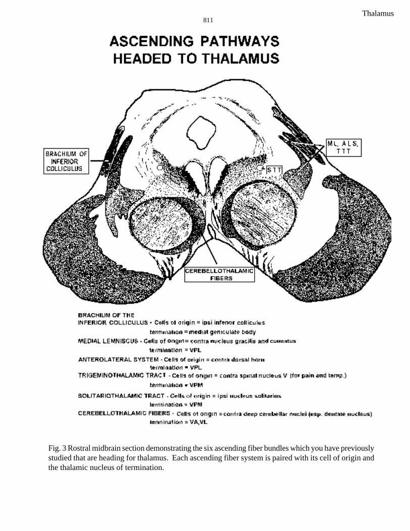
Fig. 3 Rostral midbrain section demonstrating the six ascending fiber bundles which you have previouslystudied that are heading for thalamus. Each ascending fiber system is paired with its cell of origin andthe thalamic nucleus of termination.
Thalamus812
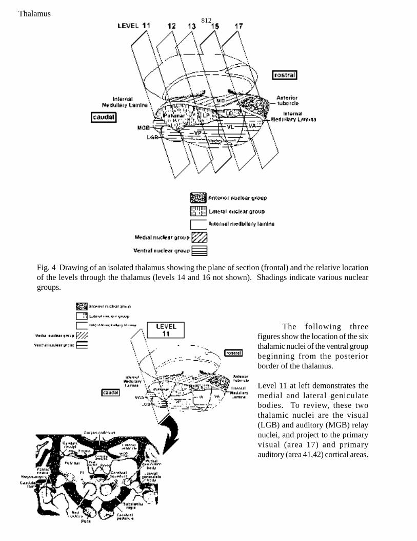
Fig. 4 Drawing of an isolated thalamus showing the plane of section (frontal) and the relative locationof the levels through the thalamus (levels 14 and 16 not shown). Shadings indicate various nucleargroups.
The following threefigures show the location of the sixthalamic nuclei of the ventral groupbeginning from the posteriorborder of the thalamus.
Level 11 at left demonstrates themedial and lateral geniculatebodies. To review, these twothalamic nuclei are the visual(LGB) and auditory (MGB) relaynuclei, and project to the primaryvisual (area 17) and primaryauditory (area 41,42) cortical areas.

Thalamus813
Ventral group nuclei seen in level12 include the VPM and VPL. Thesetwo nuclei should be very familiar toyou. Recall that they receive inputsfrom the DC-ML, LSTT to the VPL, andthe TTT and STT to VPM. They in turnproject to the Primary sensory cortex,area 3,1,2.
Ventral group nuclei seen in level15 include the VA and VL nuclei. It isdifficult to tell the difference betweenVA and VL so we are going to groupthem together. Recall that these twonuclei are the motor relay nuclei,receiving inputs from the cerebellumand the basal ganglia. Do youremember the names of the pathwaysthat project to these nuclei? There aretwo landmarks that indicate that youare nearing the anterior pole of thethalamus which will help you identifythe VA/VL complex. They are theMTT and the IML as it wraps aroundthe anterior nucleus.
Thalamus814
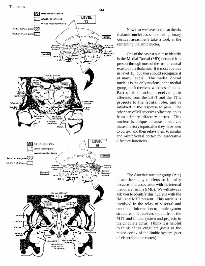
Now that we have looked at the sixthalamic nuclei associated with primarycortical areas, let’s take a look at theremaining thalamic nuclei.
One of the easiest nuclei to identifyis the Medial Dorsal (MD) because it ispresent through most of the rostral-caudalextent of the thalamus. It is most obviousin level 13, but you should recognize itat many levels. The medial dorsalnucleus is the only nucleus in the medialgroup, and it receives two kinds of inputs.Part of this nucleus receives painafferents from the LSTT and the TTT,projects to the frontal lobe, and isinvolved in the response to pain. Theother part of MD receives olfactory inputsfrom primary olfactory cortex. Thisnucleus is unique because it receivesthese olfactory inputs after they have beento cortex, and then relays them to insularand orbitofrontal cortex for associativeolfactory functions.
The Anterior nuclear group (Ant)is another easy nucleus to identifybecause of its association with the internalmedullary lamina (IML). We will alwaysask you to identify this nucleus with theIML and MTT present. This nucleus isinvolved in the relay of visceral andemotional information to limbic systemstructures. It receives inputs from theMTT and limbic system and projects tothe cingulate gyrus. I think it is helpfulto think of the cingulate gyrus as themotor cortex of the limbic system (sortof visceral motor cortex).
Thalamus815
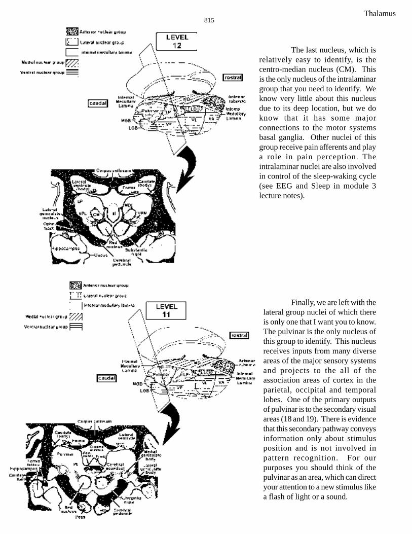
The last nucleus, which isrelatively easy to identify, is thecentro-median nucleus (CM). Thisis the only nucleus of the intralaminargroup that you need to identify. Weknow very little about this nucleusdue to its deep location, but we doknow that it has some majorconnections to the motor systemsbasal ganglia. Other nuclei of thisgroup receive pain afferents and playa role in pain perception. Theintralaminar nuclei are also involvedin control of the sleep-waking cycle(see EEG and Sleep in module 3lecture notes).
Finally, we are left with thelateral group nuclei of which thereis only one that I want you to know.The pulvinar is the only nucleus ofthis group to identify. This nucleusreceives inputs from many diverseareas of the major sensory systemsand projects to the all of theassociation areas of cortex in theparietal, occipital and temporallobes. One of the primary outputsof pulvinar is to the secondary visualareas (18 and 19). There is evidencethat this secondary pathway conveysinformation only about stimulusposition and is not involved inpattern recognition. For ourpurposes you should think of thepulvinar as an area, which can directyour attention to a new stimulus likea flash of light or a sound.
Thalamus816
You probably noticed that we left out the Lateral Dorsal (LD) and the Lateral Posterior (LP)nuclei from our discussion. We will not hold you responsible for these two nuclei as we know verylittle about them.
Although I did not discuss the reticular nuclei you should at least be familiar with the name.This group of nuclei is involved in the regulation of sleep wake cycles, and has diffuse projections to allother thalamic nuclei, but is the only thalamic nucleus that does not project to the cortex.
Symptoms Following Lesions of the Thalamus
There are two considerations that must be taken into account when attempting to diagnoselesions of the thalamus: 1) thalamic nuclei are small so that lesions producing highly specific effectsare uncommon (although they do occur), and 2) the thalamus is immediately bounded by the internalcapsule and is in close proximity to the deep motor nuclei of the cerebral hemisphere (putamen, caudateand globus pallidus) so that thalamic lesions frequently are accompanied by symptoms from damage tothese other structures (most commonly from hemorrhage from the striate arteries—discussed in cortexlectures).
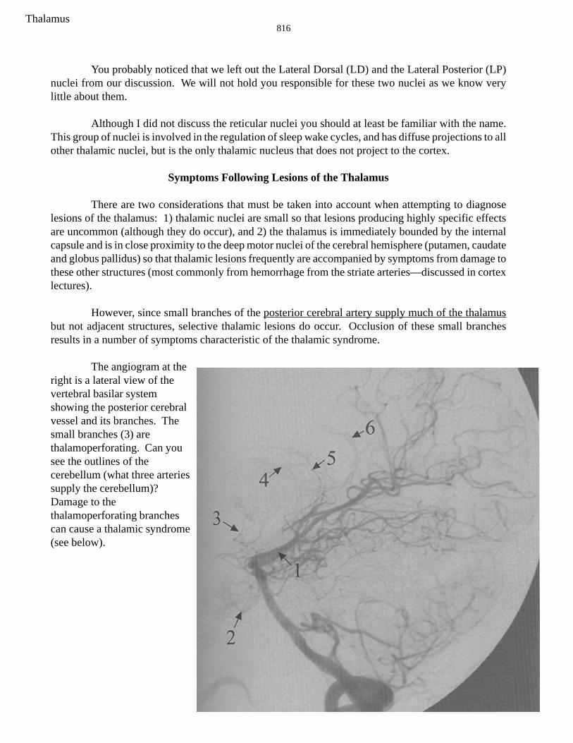
However, since small branches of the posterior cerebral artery supply much of the thalamusbut not adjacent structures, selective thalamic lesions do occur. Occlusion of these small branchesresults in a number of symptoms characteristic of the thalamic syndrome.
The angiogram at theright is a lateral view of thevertebral basilar systemshowing the posterior cerebralvessel and its branches. Thesmall branches (3) arethalamoperforating. Can yousee the outlines of thecerebellum (what three arteriessupply the cerebellum)?Damage to thethalamoperforating branchescan cause a thalamic syndrome(see below).
Thalamus817
1) If the damage includes VPL and VPM a contralateral hemianesthesia usually results. Typically, allsomatic sensory modalities are affected: light touch, conscious proprioception, 2-point discrimination& vibration, and pain & temperature. This loss of all somatic sensory modalities is an importantdiagnostic sign for thalamic damage (lesions of the internal capsule or cortex that impair somaticsensory function typically affect different modalities to different extents, often leaving painsensation unchanged).
2.) Sometimes seen after a period of recovery from damage to VPL and VPM (days to months) ishyperalgesia (an exaggerated unpleasant or painful sensation resulting from mild cutaneous stimulation)or in some cases spontaneous pain with no apparent stimulation (causalgia). Such pain can be severeand intractable. Hyperalgesia and spontaneous pain do not occur with lesions confined to the cerebralhemispheres (cortex, internal capsule, or deep nuclei). Obviously, damage to the postero-lateral part ofthe thalamus also will involve other nuclei such as the pulvinar and lateral posterior, but unilateralinfarcts in these higher order “association” nuclei typically result in no obvious deficits.
3.) If the LGB is affected there is a contralateral homonymous hemianopsia.
4) If the damage extends into the VA/VL nuclei complex movement disorders can result. The movementdisorders can be reminiscent of cerebellar damage (ataxia and intention tremor) and/or basal gangliadamage (choreoathetoid movements). This reflects, in part, the fact that both the cerebellum andbasal ganglia project to VA and VL. All such problems occur contralateral to the side of the lesion.
Thalamus818
Thalamus819
Thalamus820
SUMMARY OF IMPORTANT POINTS FROM THALAMUS LECTURES
Points you should concentrate on are as follows:
1) Spatial relationships between the diencephalon and the different components of the cerebralhemispheres (internal capsule, caudate, putamen, globus pallidus, ventricles, etc.). This is, of course,essential for interpretation of C-T scans, and diagnosing problems resulting from lesions involvingmore than one of these areas.
2) Organization of the dorsal thalamus. It is important for you to retain a sense of the 3-D relationshipsbetween different thalamic nuclei and I have, therefore, stressed the subdivisions of the thalamus intonuclear groups and positions of these groups with respect to the internal and external medullary laminaeand other landmarks. Since nuclei within the different subdivisions of the diencephalon and nucleargroups of the dorsal thalamus tend to have similar connections and/or functions, knowing these groupswill also help you to remember details concerning individual nuclei. Don’t confuse the ventral nucleargroup of the dorsal thalamus with the ventral thalamus.
3) Another useful starting point in learning the details of connections and functions of individual nucleiin the dorsal thalamus is to categorize them according to functional types: VA and VL are motor “relay”nuclei; VP (VPL & VPM), MGB, LGB and MD are sensory “relay” nuclei (don’t forget the taste relayin the medial part of VPM); LP, Pulvinar and MD are connected with association areas of cortex; theanterior nuclear group nuclei and LD have connections with limbic structures; the thin sheet-like nucleiin the intralaminar group receive nociceptive spinothalamic fibers (so do VPL and MD) and are part ofthe reticular activating system; the CM nucleus of the intralaminar group has connections with motorareas of the brain.
4) Thalamic syndromes. It is important to know the different types of deficits resulting from damage tothe thalamus and to remember that the thalamus can be selectively damaged with little or no damage tothe internal capsule or deep motor nuclei by blockage of the thalamoperforating branches of the posteriorcerebral artery.
After you learn about the effects of lesions of the internal capsule and cerebral cortex in later lectures,think about how you would distinguish deficits resulting from thalamic lesions from those resultingfrom damage to these other areas.
Thalamus821
PRACTICE QUESTIONS - THALAMUS
multiple choice (one correct answer):
1. The diencephalon is divided into symmetrical halves by the:
A. fourth ventricleB. hypothalamic sulcusC. stria medullarisD. third ventricleE. lateral ventricle
2. The lateral surface of the diencephalon is bounded by the:
A. third ventricleB. lateral ventricleC. internal capsuleD. internal medullary lamina
3. Which of the following statements is FALSE concerning thalamic syndrome:
A. the pain involved may be very disagreeable and may become intractable to analgesics.B. the syndrome usually results from a lesion that is vascular in origin.C. the symptoms of the syndrome vary according to the location and extent of the lesion.D. the threshold for somatic sensory stimuli is usually raised on the same side of the body as the
lesion.E. blockage of the posterior cerebral or its thalamic branches can cause the thalamic syndrome.
4. The largest nucleus within the intralaminar group is:
A. centromedianB. pulvinarC. lateral dorsalD. mediodorsalE. ventral anterior
5. The mediodorsal (dorsomedial) nucleus projects to the_____________lobe.
A. frontalB. parietalC. occipitalD. temporalE. ear
6. Which of the following nuclei is (are) recognized in the ventral nuclear group of the thalamus?
A. ventral posteriorB. ventral lateralC. ventral anteriorD. lateral geniculateE. all of the above
Practice questions

Thalamus822
7. Which of the following nuclei relay(s) somatosensory information?
A. ventral posterior lateralB. medial geniculateC. reticularD. ventral anteriorE. lateral geniculate
8. Which of the following thalamic nuclei is(are) thought to have a predominantly motor function?
A. ventral posterior lateralB. mediodorsalC. ventral lateralD. lateral posteriorE. pulvinar
9. The lateral nuclear group of the thalamus includes the:
A. pulvinarB. centromedianC. anteriorD. mediodorsalE. ventral lateral
10. Which of the following nuclei of the thalamus are sensory relay nuclei?
A. ventral posterior medialB. medial geniculateC. lateral geniculateD. ventral posterior lateralE. all of the above
11. Which of the following nuclei receive nociceptive anterolateral input?
A. mediodorsalB. sheet-like nuclei in intralaminar groupC. VPLD. pulvinarE. two of the above
Practice questions

Thalamus823
NOTE: The following are practice questions, the weigert stained sections will not be labeled onthe exam
12. Which of the following statements is true regarding the shaded structure?
A. cells project to wide areas of parietal, occipital and temporal cortexB. is an association nucleusC. receives input from the optic nerveD. receives input from the olfactory nerveE. two of the above are true
Practice questions

Thalamus824
13. Which of the following statements is true regarding the shaded structure?
A. involved in melatonin productionB. calcifiesC. is part of the epithalamusD. tumors could cause Parinaud syndrome (integrate from the brain stem module; rememberthe point on the superior colliculi??)E. all of the above are true
Practice questions
Thalamus825
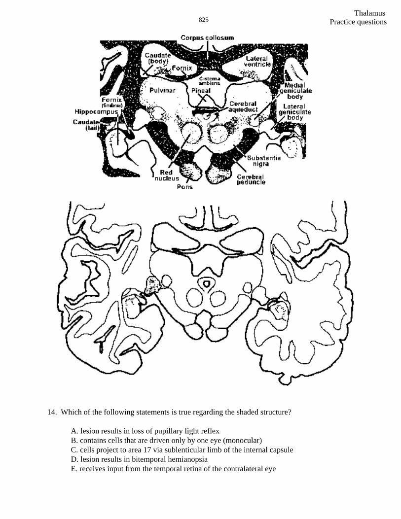
14. Which of the following statements is true regarding the shaded structure?
A. lesion results in loss of pupillary light reflexB. contains cells that are driven only by one eye (monocular)C. cells project to area 17 via sublenticular limb of the internal capsuleD. lesion results in bitemporal hemianopsiaE. receives input from the temporal retina of the contralateral eye
Practice questions
Thalamus826
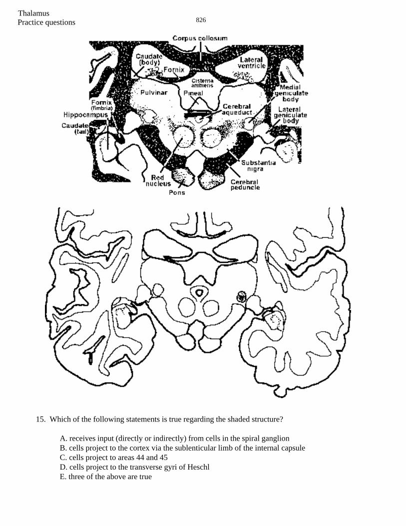
15. Which of the following statements is true regarding the shaded structure?
A. receives input (directly or indirectly) from cells in the spiral ganglionB. cells project to the cortex via the sublenticular limb of the internal capsuleC. cells project to areas 44 and 45D. cells project to the transverse gyri of HeschlE. three of the above are true
Practice questions

Thalamus827
16. Which of the following statements is true regarding the shaded structure?
A. receives input from the STTB. receives input from the TTTC. projects to cortical areas 3,1,2D. projects to cortex via posterior limb of the internal capsuleE. all of the above are true
Practice questions
Thalamus828
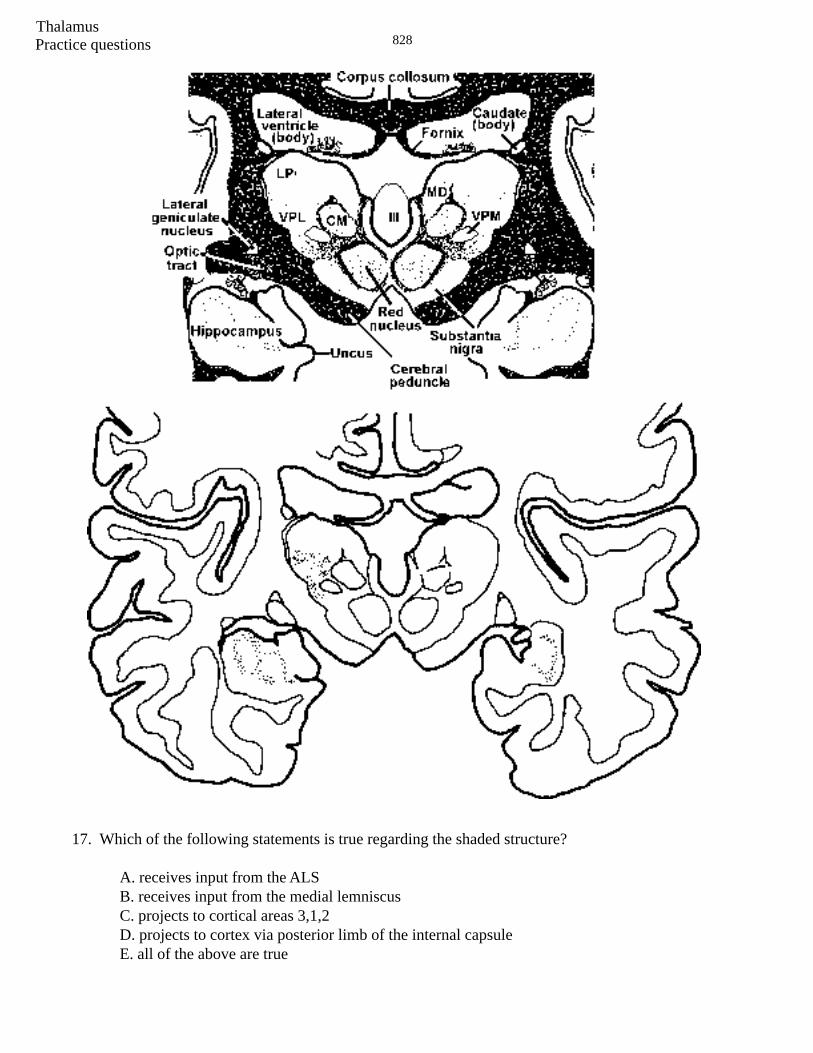
17. Which of the following statements is true regarding the shaded structure?
A. receives input from the ALSB. receives input from the medial lemniscusC. projects to cortical areas 3,1,2D. projects to cortex via posterior limb of the internal capsuleE. all of the above are true
Practice questions

Thalamus829
18. Which of the following statements is true regarding the shaded structure?
A. receives input from the STTB. receives input from the TTTC. projects to cortical areas 3,1,2D. projects to cortex via posterior limb of the internal capsuleE. two of the above are true
Practice questions
Thalamus830Practice questions
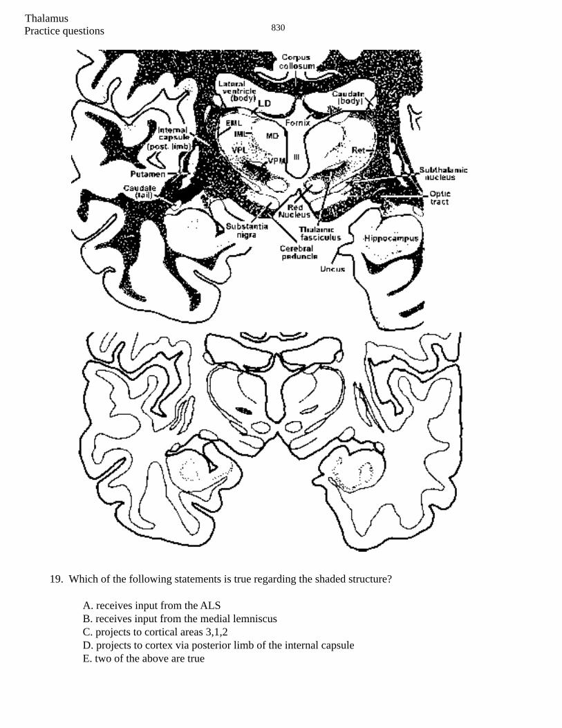
19. Which of the following statements is true regarding the shaded structure?
A. receives input from the ALSB. receives input from the medial lemniscusC. projects to cortical areas 3,1,2D. projects to cortex via posterior limb of the internal capsuleE. two of the above are true
Thalamus831 Practice questions
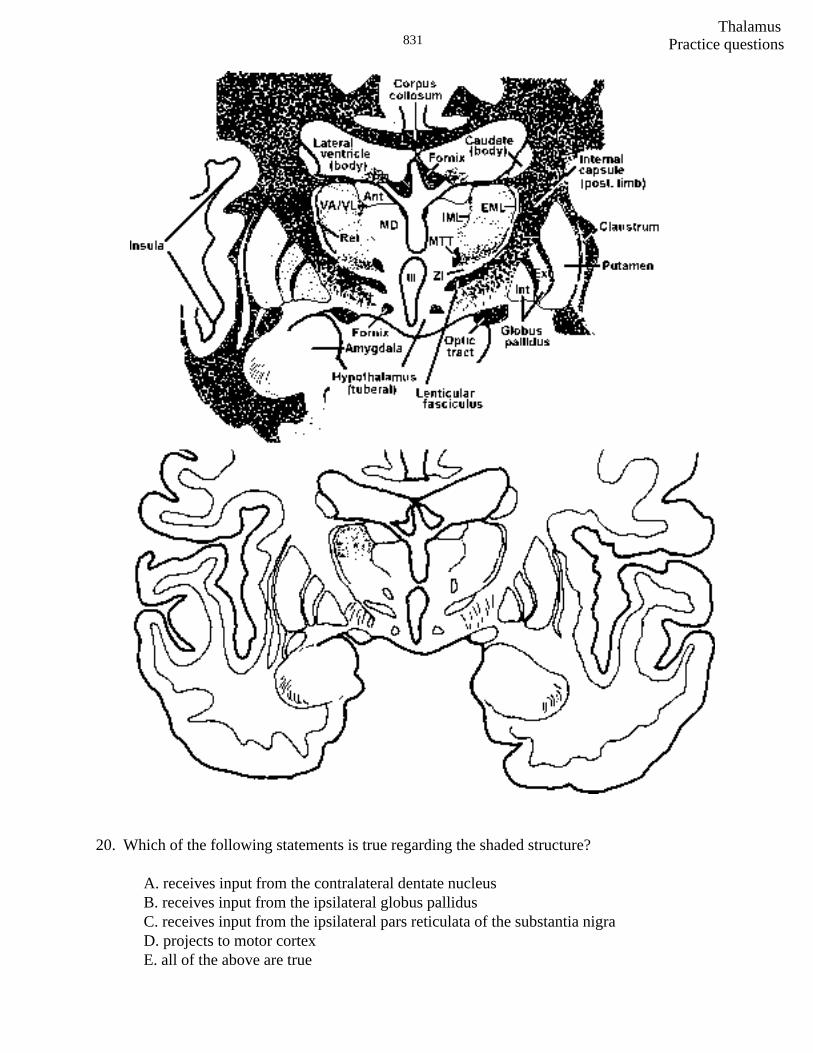
20. Which of the following statements is true regarding the shaded structure?
A. receives input from the contralateral dentate nucleusB. receives input from the ipsilateral globus pallidusC. receives input from the ipsilateral pars reticulata of the substantia nigraD. projects to motor cortexE. all of the above are true
Thalamus832
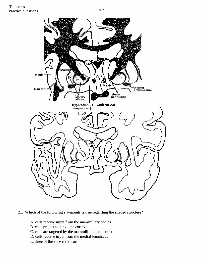
21. Which of the following statements is true regarding the shaded structure?
A. cells receive input from the mammillary bodiesB. cells project to cingulate cortexC. cells are targeted by the mammillothalamic tractD. cells receive input from the medial lemniscusE. three of the above are true
Practice questions
Thalamus833
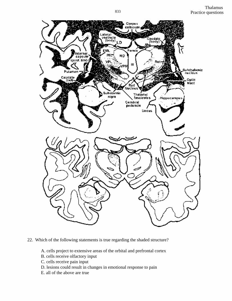
22. Which of the following statements is true regarding the shaded structure?
A. cells project to extensive areas of the orbital and prefrontal cortexB. cells receive olfactory inputC. cells receive pain inputD. lesions could result in changes in emotional response to painE. all of the above are true
Practice questions
Thalamus834
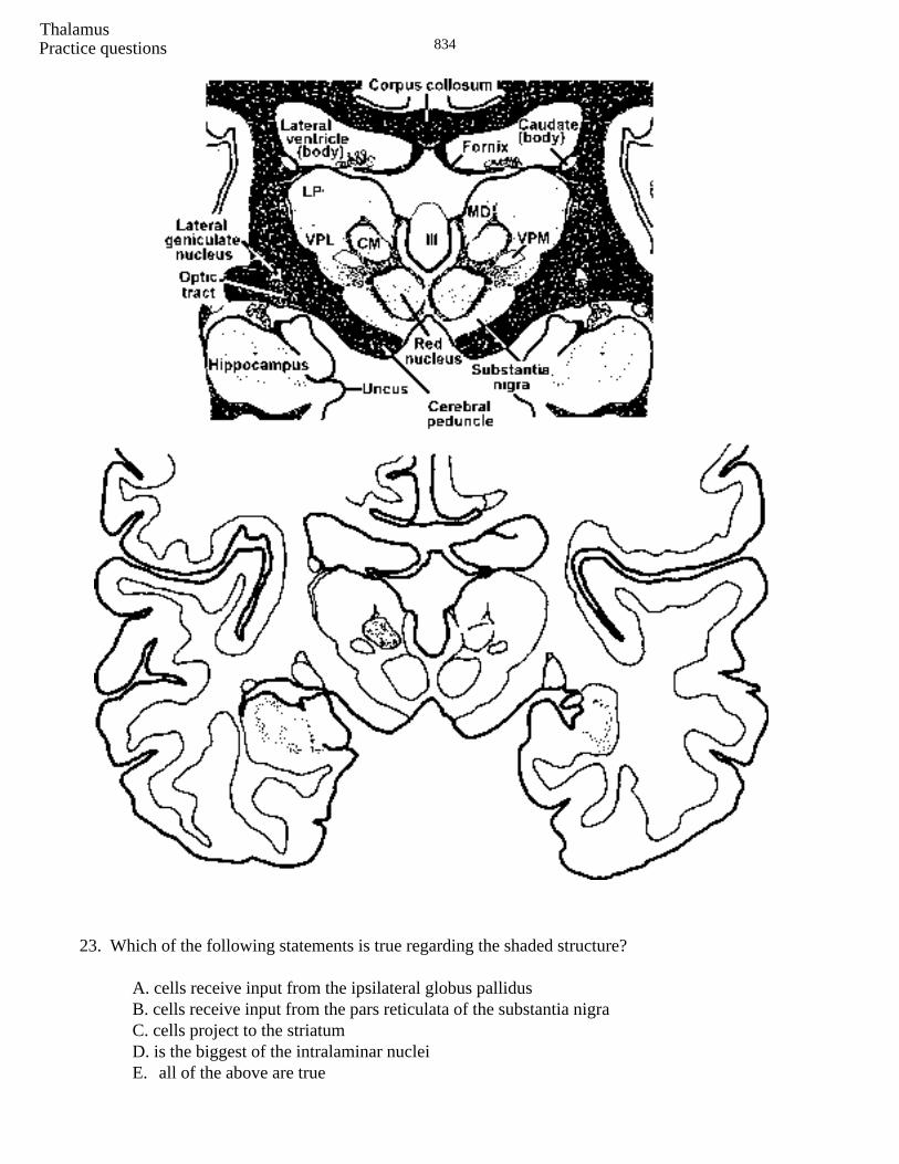
23. Which of the following statements is true regarding the shaded structure?
A. cells receive input from the ipsilateral globus pallidusB. cells receive input from the pars reticulata of the substantia nigraC. cells project to the striatumD. is the biggest of the intralaminar nucleiE. all of the above are true
Practice questions
Thalamus835
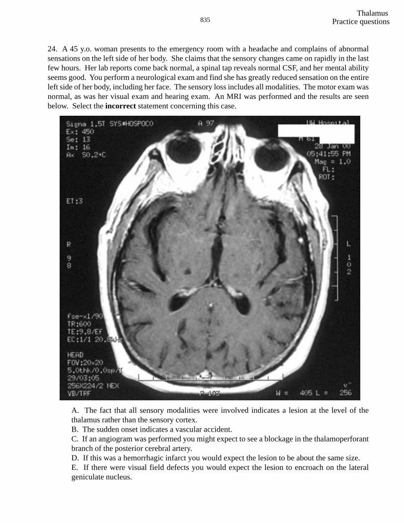
24. A 45 y.o. woman presents to the emergency room with a headache and complains of abnormalsensations on the left side of her body. She claims that the sensory changes came on rapidly in the lastfew hours. Her lab reports come back normal, a spinal tap reveals normal CSF, and her mental abilityseems good. You perform a neurological exam and find she has greatly reduced sensation on the entireleft side of her body, including her face. The sensory loss includes all modalities. The motor exam wasnormal, as was her visual exam and hearing exam. An MRI was performed and the results are seenbelow. Select the incorrect statement concerning this case.
A. The fact that all sensory modalities were involved indicates a lesion at the level of thethalamus rather than the sensory cortex.B. The sudden onset indicates a vascular accident.C. If an angiogram was performed you might expect to see a blockage in the thalamoperforantbranch of the posterior cerebral artery.D. If this was a hemorrhagic infarct you would expect the lesion to be about the same size.E. If there were visual field defects you would expect the lesion to encroach on the lateralgeniculate nucleus.
Practice questions
Thalamus836
1. D 2. C 3. D 4. A 5. A 6. E 7. A 8. C 9. A10. E11. E
12. E (A and B)13. E all14. B15. E (A, B and D)16. E all17. E all18. E (C and D)19. E (C and D)20. E all21. E (A, B and C)22. E all23. E all24. D
Practice question ANSWERS
SELF LEARNING Tuesday, March 30, 11AM-12
Make sure you have done the practice questions on Thalamus. Watch the CD-ROM on the thalamusand perhaps get an early start on the Cerebral Cortex. Read the www reading Brain Asymmetry andDyslexia and look over the Cerebral Blood Supply site.
Sorry to keep harping, but you should have read and understood the www reading regardingParkinson’s, Tourette Syndrome, Deep Brain Stimulation, Is Our Inverted Retina Really BadDesign?, Diabetic Retinopathy, Menieres’ Disease and Benign Paroxysmal Positional Vertigo.Moreover, the old stuff should be fixed in your brains. They are: 1) muscular dystrophy, 2)myasthenia gravis, 3) Guillain-Barre, 4) S1 radiculopathy, 5) amyotrophic lateral sclerosis (ALS), 6)Brown Sequard syndrome (spinal cord hemisection), 7) facial colliculus-vestibulo-cochlear, 8) lateralmedullary (Wallenberg’s) syndrome, 9) acoustic neuroma, 10) Weber Syndrome, 11) syringomyeliaand 12) subacute combined systems disease. Be familiar with the two cases (one Huntington’s andone Parkinson’s) in the practice questions following “Basal Ganglia.” Finally, look over the“thalamus/Cortex power point for quiz #9.
YOU CAN ALSO GO UP TO THE LABS AND LOOK ATTHE THALAMUS AND CORTEX