Embed Size (px)
Citation preview

ARTICLE
Received 9 Feb 2016 | Accepted 12 Apr 2016 | Published 7 Jun 2016
Th2 and eosinophil responses suppressinflammatory arthritisZhu Chen1,2,*, Darja Andreev1,*, Katharina Oeser3, Branislav Krljanac3, Axel Hueber1, Arnd Kleyer1,
David Voehringer3, Georg Schett1,** & Aline Bozec1,**
Th2–eosinophil immune responses are well known for mediating host defence against
helminths. Herein we describe a function of Th2–eosinophil responses in counteracting the
development of arthritis. In two independent models of arthritis, Nippostrongylus brasiliensis
infection leads to Th2 and eosinophil accumulation in the joints associated with robust
inhibition of arthritis and protection from bone loss. Mechanistically, this protective effect is
dependent on IL-4/IL-13-induced STAT6 pathway. Furthermore, we show that eosinophils
play a central role in the modulation of arthritis probably through the increase of
anti-inflammatory macrophages into arthritic joints. The presence of these pathways in
human disease is confirmed by detection of GATA3-positive cells and eosinophils in the joints
of rheumatoid arthritis patients. Taken together, these results demonstrate that eosinophils
and helminth-induced activation of the Th2 pathway axis effectively mitigate the course of
inflammatory arthritis.
DOI: 10.1038/ncomms11596 OPEN
1 Department of Internal Medicine 3, University Hospital Erlangen and Friedrich Alexander University of Erlangen-Nuremberg, Gluckstrasse 6, Erlangen 91054,Germany. 2 Department of Rheumatology and Immunology, Anhui Medical University Affiliated Provincial Hospital, Hefei 230001, China. 3 Department ofInfection Biology, University Hospital Erlangen and Friedrich-Alexander University Erlangen-Nuremberg (FAU), Erlangen 91054, Germany. * These authorscontributed equally to this work. ** These authors jointly supervised this work. Correspondence and requests for materials should be addressed to G.S.(email: [email protected]) or to A.B. (email: [email protected]).
NATURE COMMUNICATIONS | 7:11596 | DOI: 10.1038/ncomms11596 | www.nature.com/naturecommunications 1

Rheumatoid arthritis (RA) is a chronic autoimmune diseasecharacterized by synovial inflammation and bone erosion,which affects up to 1% of the population worldwide. It is
the paradigm of a chronic disease, which hardly resolves andusually accompanies patients during their entire life1,2. Thissituation is particularly challenging as RA can start early in life,even affecting children. The induction of type 1 (Th1) and type 17(Th17) helper T-cell responses in the context of underlyingautoimmunity are considered to play a key role in the initiationphase of RA1. Once inflammation is established in arthritis, theprocess turns out to be highly chronic without evidence forspontaneous resolution. Although the pro-inflammatorypathways in RA are well understood, immune mechanismscounteracting inflammation are yet to be characterized. Hence,finding intrinsic regulatory pathways fostering the impairedresolution processes in arthritis is of utmost importance.Discovery of such pathways may not only be critical inelucidating the pathophysiology of RA but may also uncoversome general principles why resolution is impaired in chronicinflammatory diseases.
Type 2 (Th2) immune responses are induced by helminthinfection in mice and in humans, and participate in the resolutionof inflammation and wound healing3–5. Helminth infections arefound in B1 billion people worldwide, mainly in underdevelopedcountries. Severe infections are rare and adults suffer of mild orasymptomatic disease. Helminths such as N. brasiliensis triggerrobust Th2 responses in vivo, characterized by increasedproduction of Th2 cytokines such as interleukin (IL)-4 andIL-13, accompanied with activation and expansion of CD4þTh2cells, eosinophilia, goblet and mucosal mast cell hyperplasia6–11.On the other hand, helminth infections also foster anti-inflammatory signals by inducing production of IL-10, Foxp3þ
regulatory T cells and alternatively activated macrophages12–16,which suppresses immune activation in the gut, the lungs and inautoimmune diabetes14,15,17. Hence, helminth infection andassociated Th2 responses may represent a promising pathwayfor fostering the resolution of arthritis. The exact mechanism,how such induction of Th2 responses by helminths infectioncould modulate arthritis, however, is still unknown.
Herein, we show that N. brasiliensis infection alleviated diseasein two models of inflammatory arthritis. At the molecular level,the attenuation of arthritis is dependent on IL-4/IL-13 secretionand STAT6 signalling pathway in haematopoietic cells. Moreover,hypereosinophilia triggered by N. brasiliensis infection alsocontributes to the resolution of arthritis by stimulating a shiftfrom pro- into anti-inflammatory macrophages in the joint.Hence, our findings indicate a crucial role of Th2 immuneresponses in inhibiting the development of arthritis.
ResultsN. brasiliensis infection suppresses inflammatory arthritis. Toassess the role of Th2 responses in arthritis, we performed serum-induced arthritis (SIA) in mice that were either untreated orinfected with N. brasiliensis. Interestingly, infected mice displayed asignificant reduction of arthritis as compared with non-infectedcontrols (Fig. 1a). Histologic analyses of the hind paws showeddecreased inflammation scores and protection from bone destruc-tion in N. brasiliensis-infected compared with non-infected mice(Fig. 1b,c). In accordance, expression levels of osteoclast markersand pro-inflammatory cytokines were decreased in the joints ofN. brasiliensis-infected compared with control mice (Fig. 1d,e),suggesting an effective inhibition of arthritis by worm infection.
N. brasiliensis inhibited arthritis associated with Th2 cells. Toconfirm that N. brasiliensis infection conducts a Th2-biased
immune response, T-cell subsets were quantified in the spleen ofN. brasiliensis-infected mice and non-infected controls. AlthoughCD4þ IFN-gþ (Th1) and CD4þ IL-17þ (Th17) cells remainedunchanged after N. brasiliensis infection, CD4þ IL-4þ (Th2) cellssignificantly increased in N. brasiliensis-infected compared withnon-infected mice (Fig. 1f). To characterize the kinetics of theTh2 response, cellular IL-4, IL-5 and IL-13 expression levels wereassessed at day 0, 3, 6 and 9 after induction of arthritis. IL-4- andIL-13-positive cells increased up to day 9 post N. brasiliensisinfection and remained high in the spleen and lymph nodes ofN. brasiliensis-infected mice (Fig. 1g,h). Furthermore, IL-5-producing cells were increased already early and returned tobaseline level 6 days after induction of arthritis (Fig. 1i). Alltogether, these data showed a profound Th2 response afterN. brasiliensis challenge during the onset of arthritis.
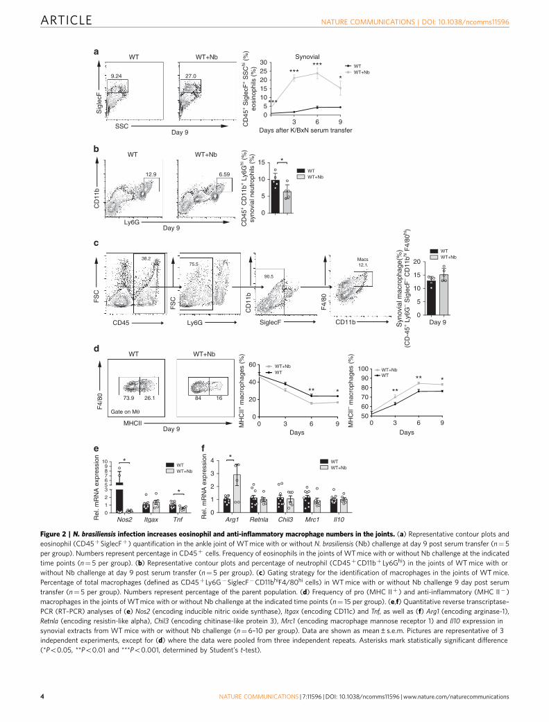
N. brasiliensis triggers eosinophil accumulation in arthritic joint.To examine whether N. brasiliensis-triggered Th2 responsesinduce respective effector cell accumulation, eosinophils,neutrophils and macrophages were quantified in the joints ofnon-infected and N. brasiliensis-infected mice. We set up thefluorescence-activated cell sorting strategy for quantifying eosi-nophils as CD45þLy6G�CD11bþSiglecFþ -positive cells usingDdblGATA mice for the negative control setting (SupplementaryFig. 1a). We could demonstrate that eosinophil numbersincreased in the joint of N. brasiliensis-infected mice. Time-course analyses revealed a rapid increase of eosinophils during theonset of arthritis (Fig. 2a). In contrast, neutrophils were decreasedin the joints of N. brasiliensis-infected mice (Fig. 2b). To definewhether Th2–eosinophil induction also affects macrophage pat-terning in the joints, the percentages of total and anti-inflam-matory macrophages were quantified in the joints. Althoughmacrophages defined as CD45þLy6G�SiglecF�CD11bhiF4/80hi
cells were unaffected in the joints of N. brasiliensis-infected mice(Fig. 2c), the proportion of previously described18 anti-inflammatory major histocompatibility complex (MHC) II�
macrophages increased, while pro-inflammatory MHC IIþ
macrophages decreased in the joints of N. brasiliensis-infectedarthritic mice (Fig. 2d). Time-course analyses revealed a rapidincrease of anti-inflammatory MHC II� macrophages in thejoints of N. brasiliensis-infected mice (Fig. 2d). In accordancewith the switch of the macrophage status, messenger RNAprofiling of pro- and anti-inflammatory markers showeddecreased Nos2 and Tnf but increased Arg1 levels (Fig. 2e,f).These data showed that N. brasiliensis-induced Th2 responsesaffect the cellular composition of the arthritic joint with increasednumbers of eosinophils and anti-inflammatory macrophages.
Consistent effects of N. brasiliensis in hTNFtg mice. To confirmthe inhibitory effect of N. brasiliensis-induced Th2 responses onarthritis, we examined a second model of arthritis and challenged5-week-old human tumour necrosis factor (TNF) transgenic(hTNFtg) mice with N. brasiliensis. Similar to the findings in SIAmodel, N. brasiliensis-infected hTNFtg mice displayed mitigatedarthritis compared with non-infected controls (Fig. 3a). Histo-pathological analyses revealed decreased articular inflammation,less bone erosion and a lower number of osteoclasts in the pawsof N. brasiliensis-challenged mice compared with controls(Fig. 3b). Furthermore, osteoclast numbers and sizes were alsolower in the tibia of N. brasiliensis-challenged hTNFtg mice(Supplementary Fig. 2a). Expression of osteoclast markers such asAcp5, but not Tnfrsf11a, Ctsk or Nfatc1 were found decreased(Supplementary Fig. 2b). Micro-computed tomography analysisof the tibial bones revealed an increased bone volume andtrabecular number in N. brasiliensis-infected hTNFtg mice
ARTICLE NATURE COMMUNICATIONS | DOI: 10.1038/ncomms11596
2 NATURE COMMUNICATIONS | 7:11596 | DOI: 10.1038/ncomms11596 | www.nature.com/naturecommunications

(Supplementary Fig. 2c,d). Taken together, these data showed thatinfection with N. brasiliensis also ameliorated TNFa-mediatedjoint inflammation and the resulting bone loss. Analysis of theimmune response showed significantly increased frequency ofCD4þ IL-4þ (Th2) cells without affecting CD4þ IFN-gþ (Th1)and CD4þ IL-17þ (Th17) cells (Fig. 3c) in hTNFtg micechallenged with N. brasiliensis. Accordingly, IL-4 and IL-5protein levels were increased in the serum (Fig. 3d). In addition,mRNA expression of Il4 and Il5 in the joints and the spleen of
N. brasiliensis-infected hTNFtg mice was increased comparedwith non-infected mice (Fig. 3e,f), confirming the induction ofTh2 responses in the joints after N. brasiliensis infection.
N. brasiliensis attenuated arthritis is IL-4/IL-13 dependent. Toexplore the molecular mechanisms involved in the anti-arthriticeffect of N. brasiliensis infection, we challenged Il4� /� Il13� /�
(4–13ko) mice and T cell-specific Il4� /� Il13� /� (4–13Tko)
a b
d e
0
0.2
0.4
0.6
0.8
1.0
Are
a (m
m2 )
Are
a (m
m2 )
***
0
0.05
0.10
0.15
0.20
0.25 **
0
10
20
30
40
50
Cel
ls p
er p
aw
**
WTWT+Nb
0
2
4
6
Rel
. mR
NA
exp
ress
ion
Rel
. mR
NA
exp
ress
ion*
Acp5
0
1
2
3 *Ctsk
0
1
2
3
4
5 *Tnfrsf11a
0
1
2
3
4Nfatc1
0
0.5
1.0
1.5
2.0 *Tnf
0
2
4
6
8 *Il1b
WTWT+Nb
0
0.5
1.0
1.5
2.0
2.5
Il6
WTWT+Nb
0 1 2 3 4 5 6 7 8 90
5
10
15
Days after K/BxN serum transfer
Art
hriti
s sc
ore
** *********************
WTWT+Nb
c
3 6 9Days after K/BxN serum transfer
g
IL-4
+ c
ells
(%
)
IL-4
+ c
ells
(%
)
IL-1
3+ c
ells
(%
)
IL-1
3+ c
ells
(%
)
3 6 9Days after K/BxN serum transfer
3 6 9Days after K/BxN serum transfer
h i
0
0.1
0.2
0.3
0.4*
3 6 9Days after K/BxN serum transfer
0
0.1
0.2
0.3
0.4
0.5
IL-5
+ c
ells
(%
)
IL-5
+ c
ells
(%
)
*
3 6 9Days after K/BxN serum transfer
WTWT+Nb
WTWT+Nb
WTWT+Nb
WT WT+Nb
Inflammation Erosion N.Oc
Spleen
Spleen Spleen
mLN
mLN mLN
H&E
TRAP
f
0
1
2
3
CD
4+IF
NG
+T
cel
ls (
%)
0
0.1
0.2
0.3
0.4
CD
4+ IL
-4+ T
-cel
ls (
%) ***
0
0.5
1.0
1.5
CD
4+ IL
-17+
T c
ells
(%
) WTWT+Nb
0
0.1
0.2
0.3
0.4
0.5
**
***
0.0
0.1
0.2
0.3 **
**
3 6 9Days after K/BxN serum transfer
0
0.05
0.10
0.15
**
***
0
0.05
0.10
0.15
**
Rel
. mR
NA
exp
ress
ion
pg m
l–1
*Tnf
Rel
. mR
NA
exp
ress
ion
Il1b
0
10
20
30
0
5
10
15
20 *
pg m
l–1
Figure 1 | N. brasiliensis infection inhibits K/BxN SIA. (a) Arthritis scores from WT mice infected or not with N. brasiliensis (Nb) (n¼6–10 per group).
(b,c) Hematoxylin and eosin (H&E) and tartrate-resistant acid phosphatase (TRAP) staining (b), and quantification of inflammation area, erosion area and
number of osteoclasts per paw (N.Oc per paw) (c) in the hind paw of WT mice with or without Nb challenge at day 9 post serum transfer (n¼ 6 per group).
Scale bar, 500 mm. (d,e) Analyses of Acp5 (encoding TRAP), Ctsk (encoding cathepsin K), Tnfrsf11a (encoding RANK), Nfatc1 (encoding NFATc1) (d), Tnf, Il1b
and Il6 (e) mRNA expression in synovial extracts and TNF and IL1b serum levels in WT mice with and without Nb challenge at day 9 post serum transfer
(n¼8–11 per group). (f) Frequency of CD4þ IFN-gþ (Th1), CD4þ IL-4þ (Th2) and CD4þ IL-17þ (Th17) cells in the spleen of WT mice with or without Nb
challenge at day 9 post serum transfer (n¼6–10 per group). (g–i) Frequency of IL-4þ (g), IL-13þ (h) and IL-5þ (i) lymphocytes in the spleen and in
mesenteric lymph node (mLN) from WT mice with or without Nb challenge at the indicated time points (n¼ 3–5 per group). Data are shown as
mean±s.e.m. Pictures are representative of 3 independent experiments. Asterisks mark statistically significant difference (*Po0.05, **Po0.01 and
***Po0.001 determined by Student’s t-test).
NATURE COMMUNICATIONS | DOI: 10.1038/ncomms11596 ARTICLE
NATURE COMMUNICATIONS | 7:11596 | DOI: 10.1038/ncomms11596 | www.nature.com/naturecommunications 3
SSC
Sig
lecF
b
c
Ly6G
CD
11b
d
Gate on Mθ
MHCII
F4/
80
WT WT+Nb
WT WT+Nb
WT WT+Nb
e f
CD
45+ S
igle
cF+ S
SC
hi (
%)
eosi
noph
ils (
%)
Syn
ovia
l mac
roph
age(
%)
(CD
-45+
Ly6
G– S
igle
cF– C
D11
bhi F
4/80
hi)
WTWT+Nb
CD
45+ C
D11
b+ L
y6G
hi (
%)
syno
vial
neu
trop
hils
(%
)
MH
CII+
mac
roph
ages
(%
)
0
1
2
356789
10
Rel
. mR
NA
exp
ress
ion
*
*
Nos2 Itgax Tnf0
1
2
3
4
Rel
. mR
NA
exp
ress
ion *
WTWT+Nb
WTWT+Nb
Arg1 Retnla Chil3 Mrc1 Il10
a
73.9 26.1 84 16
12.9 6.59
9.24 27.0
FS
C
CD45
FS
C
Ly6G
CD
11b
SiglecF CD11bF
4/80
0
5
10
15
20
Synovial
3 6 905
1015202530
******
*
***
Days after K/BxN serum transferDay 9
Day 9
30 6 950
60
70
80
90
100
**
** *** *
MH
CII– m
acro
phag
es (
%)
Days
00
20
40
60
3 6 9Days
WTWT+Nb
Day 9
Day 9
WTWT+NbWT
WT+Nb
WTWT+Nb
0
5
10
15 *
38.275.5
90.5
Macs12.1
Figure 2 | N. brasiliensis infection increases eosinophil and anti-inflammatory macrophage numbers in the joints. (a) Representative contour plots and
eosinophil (CD45þSiglecFþ ) quantification in the ankle joint of WT mice with or without N. brasiliensis (Nb) challenge at day 9 post serum transfer (n¼ 5
per group). Numbers represent percentage in CD45þ cells. Frequency of eosinophils in the joints of WT mice with or without Nb challenge at the indicated
time points (n¼ 5 per group). (b) Representative contour plots and percentage of neutrophil (CD45þCD11bþ Ly6Ghi) in the joints of WT mice with or
without Nb challenge at day 9 post serum transfer (n¼ 5 per group). (c) Gating strategy for the identification of macrophages in the joints of WT mice.
Percentage of total macrophages (defined as CD45þ Ly6G�SiglecF�CD11bhiF4/80hi cells) in WT mice with or without Nb challenge 9 day post serum
transfer (n¼ 5 per group). Numbers represent percentage of the parent population. (d) Frequency of pro (MHC IIþ ) and anti-inflammatory (MHC II� )
macrophages in the joints of WT mice with or without Nb challenge at the indicated time points (n¼ 15 per group). (e,f) Quantitative reverse transcriptase–
PCR (RT–PCR) analyses of (e) Nos2 (encoding inducible nitric oxide synthase), Itgax (encoding CD11c) and Tnf, as well as (f) Arg1 (encoding arginase-1),
Retnla (encoding resistin-like alpha), Chil3 (encoding chitinase-like protein 3), Mrc1 (encoding macrophage mannose receptor 1) and Il10 expression in
synovial extracts from WT mice with or without Nb challenge (n¼6–10 per group). Data are shown as mean±s.e.m. Pictures are representative of 3
independent experiments, except for (d) where the data were pooled from three independent repeats. Asterisks mark statistically significant difference
(*Po0.05, **Po0.01 and ***Po0.001, determined by Student’s t-test).
ARTICLE NATURE COMMUNICATIONS | DOI: 10.1038/ncomms11596
4 NATURE COMMUNICATIONS | 7:11596 | DOI: 10.1038/ncomms11596 | www.nature.com/naturecommunications

mice with N. brasiliensis before the induction of arthritis. WithoutN. brasiliensis infection, the level of arthritis was increased in4–13ko mice compared with wild-type (WT) controls (Fig. 4a).Moreover, the beneficial effect of N. brasiliensis infection on theonset of arthritis was virtually abolished in 4–13ko and 4–13Tkomice, showing almost identical induction of arthritis similar tothat in the non-infected controls (Fig. 4a). However, in contrastto WT controls some resolution of arthritis was still observed inNb-challenged 4–13ko and 4–13Tko mice, suggesting additionalregulatory processes being involved. This concept was alsoconfirmed by the histopathological analysis of arthritis scores,bone erosion scores and osteoclast numbers (Fig. 4b,c). Effects onbone and osteoclasts reflected the effects on inflammation,whereas no intrinsic differences in osteoclastogenesis and osteo-clast gene expression were observed between WT and 4–13komonocyte lineage cells (Supplementary Fig. 3a,b).
N. brasiliensis-attenuated arthritis is STAT6 dependent. Thesedata suggested that IL-4/IL-13 pathway is important formediating the inhibitory effects of N. brasiliensis infection on
arthritis. As STAT6 signalling pathway is essential for mediatingIL-4/IL-13-induced response19, we also induced arthritis inN. brasiliensis-infected Stat6� /� mice. N. brasiliensis-infectedStat6� /� mice developed comparable SIA than non-infectedmice (Fig. 4d and Supplementary Fig. 4a), confirming thedependence of the STAT6 signalling pathway in the mitigationof arthritis. To further evaluate the role of the STAT6-regulatedgenes in haematopoietic cells, Stat6� /� and WT recipient micewere reconstituted with WT bone marrow. These chimeric micewere then challenged with N. brasiliensis and induced forarthritis. Indeed, WT¼4Stat6� /� and WT¼4WT chimerasdisplayed similar inhibition of arthritis by N. brasiliensis (Fig. 4eand Supplementary Fig. 4b), suggesting that inhibitory effectsdepend on STAT6-regulated genes in haematopoietic cells.
Hypereosinophilia reduced arthritis in 4–13ko-infected mice.Although the induction phase of arthritis was comparable, diseasepartly resolved in the later stages of N. brasiliensis infected 4–13koand Stat6� /� mice (Fig. 4a–d). As the N. brasiliensis-inducedincrease of anti-inflammatory macrophages in the joints was
0
0.5
1.0
1.5
Are
a (m
m2 )
Are
a (m
m2 )
* * **
0
0.1
0.2
0.3
0
20
40
60
Cel
ls p
er p
aw
hTNFtghTNFtg+Nb
0
1
2
3
4
5
CD
4+ IF
NG
+ T
-cel
ls %
CD
4+ IL
-4+ T
-cel
ls %
CD
4+ IL
-17+
T-c
ells
%
0
0.2
0.40.4
0.2
0
0.6
0.8
0.8
0.6
1.0 * hTNFtghTNFtg+Nb
020406080
100120140160180200
pg m
l–1
*
0123
15
20
25
Rel
. mR
NA
exp
ress
ion
Rel
. mR
NA
exp
ress
ion
*
**
IL-4 IL-5 IL-10 IL-2 IFNγ GM-CSF
Joint JointSpleen
Il4
0
1
2
3
4Il5
5 6 7 8 90
1
2
3
Weeks
Art
hriti
s sc
ore
* * * **
hTNFtghTNFtg+Nb
hTNFtghTNFtg+Nb
hTNFtg hTNFtg+Nb
Inflammation Erosion N.Oc
H&E
TRAP
hTNFtghTNFtg+Nb
c
e f
d
ba
Figure 3 | N. brasiliensis infection induces Th2 response and alleviates arthritis in TNFa- mediated arthritis. (a) Arthritis score from 5-week-old hTNFtg
mice with or without N. brasiliensis (Nb) challenge. Data are pooled from two independent experiments (n¼ 9 per group). (b) Haematoxylin/eosin (H&E)
and tartrate-resistant acid phosphatase (TRAP) staining of the hind paw from 9-week-old hTNFtg mice with or without Nb challenge. Scale bar, 500mm.
Quantification of inflammation area, erosion area and osteoclast number (N.Oc) per paw in the hind paws of hTNFtg mice with or without Nb challenge
(n¼ 5–8 per group). (c) Frequency of CD4þ IFN-gþ (Th1), CD4þ IL-4þ (Th2) and CD4þ IL-17þ (Th17) cells in the spleen of hTNFtg mice with or without
Nb challenge (n¼4–5 per group). (d) Serum levels of IL-4, IL-5, IL-10, IL-2, IFN-g and granulocyte–macrophage colony-stimulating factor (GM-CSF) in
hTNFtg mice with or without Nb challenge (n¼4–5 per group). (e) Quantitative reverse transcriptase–PCR (RT–PCR) analyses of Il4 expression in the
spleen and synovial extracts from 9-week-old hTNFtg mice with or without Nb challenge (n¼4–8 per group). (f) Quantitative RT–PCR analyses of Il5
expression in synovial extracts of hTNFtg mice with or without Nb challenge (n¼4–8 per group). Data are expressed as mean±s.e.m. Pictures are
representative of 3 independent experiments. Asterisks mark statistically significant difference (*Po0.05 and **Po0.01 determined by Student’s t-test).
NATURE COMMUNICATIONS | DOI: 10.1038/ncomms11596 ARTICLE
NATURE COMMUNICATIONS | 7:11596 | DOI: 10.1038/ncomms11596 | www.nature.com/naturecommunications 5

a
c
d
e
f g
0 1 2 3 4 5 6 7 8 90
5
10
15
Days after K/BxN serum transfer
Art
hriti
s sc
ore
WT4–13ko+Nb4–13Tko+NbWT+Nb
**
* * * *
*
***** *** ***
****** *** ***
**
0
0.2
0.4
0.6
0.8
1.0
Are
a (m
m2 )
Are
a (m
m2 )
**
*** *** **
WT
4–13ko+Nb4–13Tko+Nb
WT+Nb
0
0.05
0.10
0.15
0.20
0.25
***
**
*** **
0
10
20
30
40
50
Cel
ls p
er p
aw
*** **
****
0 1 2 3 4 5 6 70
5
10
15
Days after K/BxN serum transfer
Art
hriti
s sc
ore
WTWT+NbStat6
–/–+Nb
* **** ** **
0 1 2 3 4 5 6 7 8 90
5
10
15
20
Days after K/BxN serum transfer
Art
hriti
s sc
ore
WTWT >WT sham chimera+NbWT > Stat6
–/– chimera+Nb
WT+Nb
*
** **
** **
**
**
***
***
***
****** **
***
0
5
10
15
20
CD
45+ s
igle
cF+S
SC
hi (
%) ***
***
WT
4–13ko+NbWT+Nb
4–13ko+Nb 4–13Tko+NbH&E
TRAP
Inflammation Erosion N.Oc
WT 4–13ko+Nb ΔdblGATA
*
**
* * * **
4–13ko
WT >WT sham chimera+Nb WT > Stat6–/– chimera+Nb
H&E
TRAP
H&E
TRAP
WT+Nb Stat6–/–+Nb
0
50
100
150
MB
P+
cel
ls p
er p
aw **
WT
4–13ko+Nb
ΔdblGATA
**
b
Figure 4 | Attenuation of arthritis by N. brasiliensis depends on the activation of the IL-4/IL-13/STAT6 pathway. (a) Arthritis score from WT, 4–13ko
and 4–13Tko with or without N. brasiliensis (Nb) challenge (n¼4–6 per group). (b,c) Haematoxylin/eosin (H&E) and tartrate-resistant acid phosphatase
(TRAP) staining and quantification of inflammation area, erosion area and number of osteoclasts (N.Oc) per paw on day 9 post K/BxN serum transfer in
the indicated group of mice (n¼4–6 per group); scale bar, 500mm. (d) Arthritis scores and corresponding H&E and TRAP stainings in unchallenged WT
mice and Nb-challenged WT and Stat6� /� mice (n¼ 3–5 per group); scale bar, 500 mm. (e) Arthritis score in unchallenged WT mice and Nb-challenged
WT¼4WT and WT¼4Stat6� /� chimeras (n¼ 5 per group); scale bar, 500mm. (f) Percentage of eosinophils on day 9 post serum transfer in
unchallenged WT and Nb-challenged WT and 4–13ko mice (n¼4–8 per group). (g) Major Basic protein (MBP) staining in the joints of WT, Nb-challenged
4–13ko mice and eosinophil-deficient DdblGATA mice (negative control) on day 9 post K/BxN serum transfer and respective quantification of staining. All
data are expressed as mean±s.e.m. Pictures are representative of 3 independent experiments. Asterisks mark statistically significant difference (*Po0.05,
**Po0.01 and ***Po0.001 determined by Student’s t-test for single comparison (a,d,e compared with WT without infection) or analysis of variance test for
multiple comparisons (c,f,g)).
ARTICLE NATURE COMMUNICATIONS | DOI: 10.1038/ncomms11596
6 NATURE COMMUNICATIONS | 7:11596 | DOI: 10.1038/ncomms11596 | www.nature.com/naturecommunications
abolished in 4–13ko mice (Supplementary Fig. 4c), other immunecells seem to be responsible, for this enhances resolution ofarthritis in N. brasiliensis-infected 4–13ko mice. As induction ofeosinophils is a hallmark of helminth infection, peripheral eosi-nophil frequency was measured during this resolution phase, 9days after induction of arthritis. Significant higher eosinophilswere found in both N. brasiliensis-infected WT, but also 4–13komice compared with non-infected controls (Fig. 4f). In addition,increased eosinophil infiltration of the joints of 4–13ko mice wasnoticed when staining for the eosinophil marker major basicprotein (Fig. 4g). Taken together, these findings suggest thateosinophils have an intrinsic inhibitory effect on the diseasecourse of arthritis.
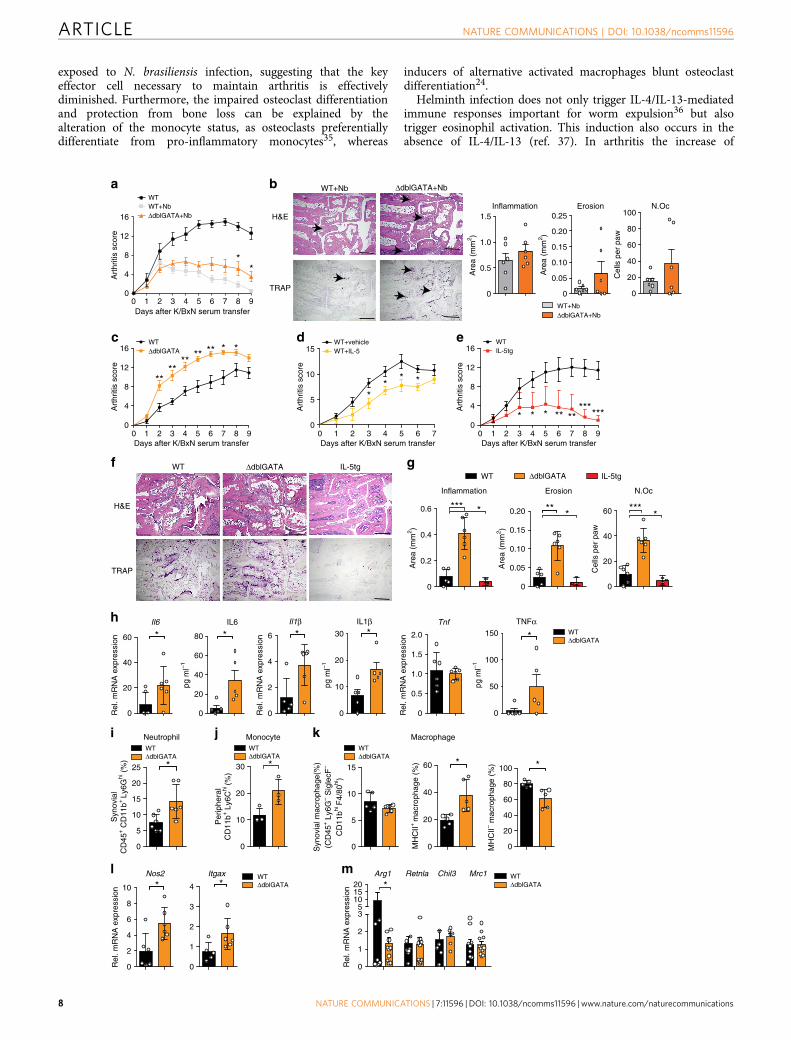
Eosinophil deficiency exacerbates arthritis. To further addressthe role of eosinophils in arthritis, controls, eosinophil-deficientDdblGATA mice and eosinophil-overexpressing Il5tg mice wereinduced for SIA. In addition, recombinant IL-5, known to induceeosinophils, was injected into WT mice before and duringinduction of arthritis. Strikingly, arthritis in N. brasiliensis-chal-lenged eosinophil-deficient DdblGATA mice was milder than innon-infected controls but lacked the resolution phase observedin WT mice challenged with N. brasiliensis (Fig. 5a,b). Theabsence of eosinophils was associated with a shift from anti- topro-inflammatory macrophages in the joints (SupplementaryFig. 5a,b). Furthermore, DdblGATA mice showed significantlyenhanced severity of arthritis per se (Fig. 5c). Re-introduction ofeosinophils into DdblGATA mice during the course of arthritisstimulated initiation of resolution and reduced arthritis scores tothe levels observed in WT mice (Supplementary Fig. 5c,d). Incontrast, Il5tg mice and, to a lesser extent, also IL-5 treatment ofWT mice led to significant reduction of arthritis scores (Fig. 5d,e).Accordingly, the extent of inflammation, bone erosion andnumber of osteoclasts were increased in DdblGATA mice anddecreased in Il5tg mice (Fig. 5f,g). Furthermore, pro-inflamma-tory cytokines such as Il6 and Il1b were increased at mRNA andprotein levels in DdblGATA arthritic mice compared with WTcontrols (Fig. 5h), suggesting that eosinophils play an importantrole in determining the severity of arthritis.
Next, the cellular environment in the joints of eosinophil-deficient arthritic mice was assessed: neutrophil numbers andneutrophil-associated chemokines Cxcl1 and Cxcl2 were signifi-cantly higher in the joints of arthritic DdblGATA mice comparedwith WT controls (Fig. 5i and Supplementary Fig. 5e). PeripheralLy6Chi inflammatory monocytes were also higher in DdblGATAmice than in controls (Fig. 5j), correlating with increased Ccr2expression in the synovium of DdblGATA mice (SupplementaryFig. 5f). The increased peripheral Ly6Chi inflammatory mono-cytes might promote the expression of neutrophil-associatedchemokines. Moreover, in the joints pro-inflammatory MHC IIþ
macrophages increased, while anti-inflammatory MHC II�
macrophages decreased in DdblGATA mice compared with WTcontrols (Fig. 5k), correlating with decreased expression of Arg1and increased expression of Nos2 and Itgax (Fig. 5l,m).These data show that the absence of eosinophils changes thebalance between pro- and anti-inflammatory macrophagesincreasing neutrophils recruitment and leading to exacerbatedinflammation.
Th2 cells and eosinophils in human arthritic joints. To definewhether the Th2 response could also occur in human inflam-matory arthritis, GATA3, a marker of Th2 and innate lymphoidcells type 2 was stained and quantified in the synovial tissue of RApatients. Osteoarthritis patients were used as controls. We foundthat GATA3þ cells were present in the inflamed synovial tissue
and were significantly higher in RA than in osteoarthritis patients(Fig. 6a). Moreover, expression of eosinophil peroxidase (EPX), amarker of eosinophils, could be detected in the synovial tissue ofRA patients (Fig. 6b). Despite the fact that we did not find anyelevation of IL-5 levels in RA patients compared with healthycontrols (Fig. 6c), serum levels of EPX were significantlyincreased in active and inactive RA compared with healthycontrols and patients with autoimmunity of RA but no inflam-mation (‘pre-RA’) (Fig. 6d). All together, these data suggest thatthe cellular components of the Th2–eosinophil response can befound in human inflammatory arthritis as well.
DiscussionIt is well established that the Th2 response is involved in the hostdefence against helminth infection in mice and humans20. Incontrast, every littler is know whether and how the Th2 responseis involved in immune-inflammatory diseases such as arthritis.This situation is surprising given that genetic and molecularevidence suggests a key role of T cells in several different forms ofinflammatory arthritis, such as RA or psoriatic arthritis. Althoughtraditionally being considered as prototype Th1-type disease,more attention has been directed to the role of Th17 cells ininflammatory arthritis in the last years1. Nevertheless, therapeutictargeting of Th1- and Th17-derived cytokines has been ratherdisappointing and only specific forms of arthritis, that is, thoselinked to psoriasis, appear to respond well to IL-17 pathwayneutralization. Hence, the concept that the role of T cells inarthritis may be based on increasing the pro-resolving immuneprocesses is appealing, as Th2-related cytokines such as IL-4,IL-13 and IL-33 have been reported to ameliorate arthritisonset21–24. Furthermore, infection with Schistosoma has shown toinhibit inflammatory cytokine production in arthritis25. However,whether and how activation of Th2 cells and potentially alsoeosinophils affect disease still remained elusive.
Herein we provide solid evidence that robust activation of theTh2 responses by N. brasiliensis infection counteracts arthritis.Functionally, activation of the STAT6 pathway by IL-4/IL-13plays a critical role in mitigating and resolving immune activationassociated with arthritis. Physiologically, IL-4 and IL-13 haveadditive effects sharing the same receptor and signalling viaSTAT6 (refs 19,26). IL-4/IL-13-mediated activation of the STAT6pathway is also critical to protect bone and cartilage duringarthritis. Osteoclast differentiation, bone loss and cartilagedamage were significantly diminished after N. brasiliensisinfection but not in the respective mutant mice, suggesting thatcontrol of arthritis by the IL-4/IL-13/STAT6 pathway isassociated with substantial protection from joint damage. Thesedata underline the protective role of Th2-associated cytokinessuch as IL-4 in inhibition of cartilage damage27,28 and thesupressive effects of IL-4 and IL-13 on murine and humanosteoclastogenesis29,30. In agreement with our observations,constitutive active STAT6 fusion protein was previously shownto decrease osteoclastogenesis and bone destruction31.
With respect to the effector pathway, Th2-mediated inhibitionand resolution of inflammation appears to depend on theinduction of alternative activated macrophages by IL-4 andIL-13 (ref. 32). During arthritis, macrophages preferentiallypolarize into a pro-inflammatory phenotype. Hence, thenumber of pro-inflammatory macrophages in the synoviumcorrelates with joint destruction33 and is used as a biomarker forclinical response to therapy34. Furthermore, pro-inflammatorymonocytes are known to be necessary for the induction ofK/BxN SIA18. In our experiments, we found a robust shiftfrom pro-inflammatory CD11bþLy6Chi monocytes intoanti-inflammatory macrophages in the arthritic joints of mice
NATURE COMMUNICATIONS | DOI: 10.1038/ncomms11596 ARTICLE
NATURE COMMUNICATIONS | 7:11596 | DOI: 10.1038/ncomms11596 | www.nature.com/naturecommunications 7
exposed to N. brasiliensis infection, suggesting that the keyeffector cell necessary to maintain arthritis is effectivelydiminished. Furthermore, the impaired osteoclast differentiationand protection from bone loss can be explained by thealteration of the monocyte status, as osteoclasts preferentiallydifferentiate from pro-inflammatory monocytes35, whereas
inducers of alternative activated macrophages blunt osteoclastdifferentiation24.
Helminth infection does not only trigger IL-4/IL-13-mediatedimmune responses important for worm expulsion36 but alsotrigger eosinophil activation. This induction also occurs in theabsence of IL-4/IL-13 (ref. 37). In arthritis the increase of
a
d
f g
h
i j
l
Days after K/BxN serum transfer
Art
hriti
s sc
ore
WTΔdblGATA
0 1 2 3 4 5 6 70
5
10
15
Days after K/BxN serum transfer
Art
hriti
s sc
ore
***
*
WT+vehicleWT+IL-5
0
0.2
0.4
0.6 ***
0
0.05
0.10
0.15
0.20 **A
rea
(mm
2 )
Are
a (m
m2 )
0
20
40
60
Cel
ls p
er p
aw
***
WT ΔdblGATA
WTΔdblGATA
Syn
ovia
lC
D45
+ C
D11
b+ Ly
6Ghi (
%)
WTΔdblGATA
Neutrophil
0
2
4
6
8
10
Rel
. mR
NA
exp
ress
ion *Nos2
0
1
2
3
4 *Itgax WT
ΔdblGATA
0
1
2
35
101520
Rel
. mR
NA
exp
ress
ion
Arg1 Retnla Chil3 Mrc1 WTΔdblGATA*
m
0
10
20
30
Per
iphe
ral
CD
11b+
Ly6
Chi
(%)
*
WTΔdblGATA
WT ΔdblGATA
H&E
TRAP
Inflammation Erosion N.Oc
Monocyte
0 1 2 3 4 5 6 7 8 90
4
8
12
16 ********
****
0
5
10
15
Syn
ovia
l mac
roph
age(
%)
(CD
45+ L
y6G
– Sig
lecF
–
CD
11bhi
F4/
80hi)
k MacrophageWTΔdblGATA
0
5
10
15
20
25 *
0
20
40
60
MH
CII+
mac
roph
age
(%) *
0
20
40
60
80
100 *
MH
CII– m
acro
phag
e (%
)
IL-5tg
* * *
IL-5tg
0
4
8
12
16 IL-5tg
* * * ** *****
***
Days after K/BxN serum transfer0 1 2 3 4 5 6 7 8 9
Art
hriti
s sc
ore
WT
0 1 2 3 4 5 6 7 8 9Days after K/BxN serum transfer
0
4
8
12
16
Art
hriti
s sc
ore
WT
ΔdblGATA+Nb
b
c
H&E
TRAP
ΔdblGATA+Nb
WT+Nb
WT+Nb
0
0.5
1.0
1.5
2.0
Tnf
Rel
. mR
NA
exp
ress
ion
0
2
4
6 *Il1β
Rel
. mR
NA
exp
ress
ion
0
20
40
60 *Il6
Rel
. mR
NA
exp
ress
ion
**
TNFα
0
50
100
150 *
pg m
l–1
IL6
0
20
40
60
80 *
pg m
l–1
IL1β
pg m
l–1
0
10
20
30 *
0
0.5
1.0
1.5
Are
a (m
m2 )
Inflammation
0
0.05
0.10
0.15
0.20
0.25
Are
a (m
m2 )
Erosion
WT+NbΔdblGATA+Nb
0
20
40
60
80
100N.Oc
Cel
ls p
er p
aw
e
ARTICLE NATURE COMMUNICATIONS | DOI: 10.1038/ncomms11596
8 NATURE COMMUNICATIONS | 7:11596 | DOI: 10.1038/ncomms11596 | www.nature.com/naturecommunications
eosinophils and IL-5 was associated with resolution of disease,concording with recent study showing the involvement ofeosinophils during resolution of inflammation in acute peri-tonitis or experimental colitis38,39. Additional experiments usingIL-5 triggered eosinophil differentiation or using eosinophildeficient mice provided clear evidence that eosinophilsbeneficially influence the course of arthritis. These effects couldbe mediated by production of anti-inflammatory lipids such asPD-1 by eosinophils40. These lipids are synthesized by 12/15lipoxygenase, which has been shown to play a role in theresolution of inflammatory arthritis41. Furthermore, IL-33-activated eosinophils could polarize alveolar macrophagestowards M2 phenotype in a IL-13-dependent manner42.
In summary, our data show that activation of Th2 responsesinhibits inflammatory arthritis. Mechanistically, IL-4/IL-13-STAT6 signalling pathway induces macrophage polarization intoanti-inflammatory macrophages into the joints. In addition,eosinophils are activated and further contribute to the resolutionof disease. These findings are interesting to shape new approachesto rebalance immune homeostasis in arthritis. The presence ofTh2 cells and eosinophils in the synovial membrane of RApatients suggests that respective cellular sources to counteractarthritis are also present in human disease. Furthermore, previousdata have revealed a Th2 cytokine pattern in very early forms ofhuman RA, when the disease may still be reversible43. Hence,activation of Th2 responses and eosinophils in both early andestablished disease may emerge as a new strategy to treat arthritis.
MethodsMice. Complete Il4� /� Il13� /� mice (4–13ko) (ref. 35), T-cell-specificIl4� /� Il13� /� mice (4–13Tko) (ref. 44), Stat6� /� mice19, specific eosinophil-deficient DdblGATA mice45, Il5tg mice46 and WT mice were on BALB/cbackground. The hTNFtg mice (strain Tg197 on C57BL/6 background) have beendescribed previously47. Clinical assessment of arthritis in hTNFtg mice was started5 weeks after birth and performed twice a week using a clinical score graded from 0to 3 (ref. 48). All mice were housed in a temperature- and humidity-controlledfacility with free access to food and water. All experiments were performedaccording to the rules and regulations of the animal facilities FPZ (Franz-Penzoldt-Zentrum, Erlangen) in Germany and approved by the local ethics authorities.
Helminth infection model. Five hundred third-stage larvae (L3) of N. brasiliensiswere inoculated subcutaneously at the base of the mice’s tail. Mice were providedwith antibiotics in the water (2 g l� 1 neomycin sulfate, 100 mg l� 1 polymyxin Bsulfate; Sigma-Aldrich) for the first 5 days after infection.
K/BxN SIA model. K/BxN serum transfer arthritis was induced on day 6 after Nbinfection by intraperitoneal (i.p.) injection of 200ml pooled K/BxN serum. Theswelling of fore and hind paws were measured daily by digital caliper (Mitutoyo,Japan) and expressed as percentage increase of paw thickness. Development ofarthritis was evaluated for each paw using a semi-quantitative scoring system(0–4 per paw; maximum score of 16) as previously described49. Mice were killed7 or 9 days post serum transfer.
Bone marrow transplantation. To generate bone marrow chimeras, adultStat6� /� mice (CD45.2) were lethally irradiated (11 Gy) and then injectedintravenously with 2� 106 bone marrow cells from WT donor mice (CD45.1). As acontrol, WT recipient mice (CD45.2) were transferred with 2� 106 bone marrowcells from WT donor mice (CD45.1) in parallel. After 6 weeks, chimerism wasverified by flow cytometry for the appropriate CD45 allele (PE-Cy7-labelledanti-CD45.1 and AlexaFluo700- labelled anti-CD45.2; 1:400; eBioscience).
IL-5 injection. WT BALB/c mice received daily i.p. injection of 200 ng recombi-nant murine IL-5 (BD Pharmingen) or equal volumes of vehicle (BSA) alone in200 ml PBS, starting 1 day before K/BxN serum transfer for 5 days.
Eosinophil isolation and adoptive transfer. Eosinophils were isolated from theperitoneal cavity of 4get/IL-5tg mice using the Mouse Streptavidin RapidspheresIsolation Kit and EasySep magnet (Stemcell Technologies), according to themanufacturer’s instructions. The following biotinylated antibodies were applied fornegative selection: anti CD45R, anti-Ter-119, anti-CD4, anti-CD8, anti-Ly6C andanti-MHC II (all from ebiosience). The purity of eosinophils was determined byflow cytometry as IL-4/eGFPþ SiglecFþ (PE-labelled anti-SiglecF; 1:400; BDPharmingen) cells with an average of 90–95%. Isolated eosinophils (2� 107) orvehicle control was injected intra-orbital into DdblGATA mice 5 days after K/BxNserum transfer. Eosinophil transfer was confirmed by flow cytometry gating onIL-4/eGFPþ SiglecFþ cells in the blood 7 days post serum transfer.
Histological and morphological analyses. Hind paw and tibia were fixed over-night in 4% formalin and then decalcified in 14% EDTA until bones were pliable.Serial paraffin sections (2 mm) were stained with haematoxylin and eosin andtartrate-resistant acid phosphatase. Area of bone erosion and inflammation,number of osteoclasts per paw, osteoclast surface per bone surface and number ofosteoclasts per bone perimeter were assessed by Osteomeasure Analyses System(Osteometrics). The three-dimensional bone structure of tibial bones was measuredby a micro-computed tomography scanner (Scanco mCT 35 scanner, Bruettisellen,Switzerland) and analysed by integrated software for segmentation and three-dimensional morphometric analysis.
Immunohistochemistry. Sections from mouse hind paw and synovial membranesamples from human RA and osteoarthritis patients were deparaffinized, quenchedof endogenous peroxidase, blocked with normal serum and incubated with rat anti-mouse major basic protein (provided by Lee’s lab in Mayo Clinic), goat anti-humanGATA3 (AbD Serotec, UK) and rabbit anti-human EPX antibody (Abcam, UK) at1:500, 1:3,000 and 1:1,000 dilution, respectively. Slides were washed and incubatedwith biotinylated secondary IgG and avidin–biotin complex (Vector Laboratories)according to the manufacturer’s instructions.
Quantitative reverse transcriptase–PCR. Total RNA from ankle or knee joint,spleen, mesenteric lymph node and osteoclast culture were extracted by usingpeqGOLD TRIfast (Peqlab). One microgram of total RNA was reverse transcribedand SYBR Green-based quantitative real-time PCR was performed on Bio-RadCFX96 Touch Real-Time PCR Detection System. Normalized gene expressionvalues were calculated as the ratio of expression of mRNA of interest to theexpression of mRNA for Actb (encoding b-actin) or Hprt (encoding hypoxanthineguanine phosphoribosyltransferase). The primer sequences are summarized inSupplementary Table 1.
Flow cytometry. The spleen and mesenteric lymph node were mechanicallydisrupted and filtered through 40 mm cell strainer, to obtain single-cell suspension.After erythrocytes lysis, 106 cells per well were plated on a 48-well plate and
Figure 5 | Eosinophil numbers control resolution of arthritis. (a) Arthritis score in WT unchallenged and N. brasiliensis (Nb) challenged WT mice and
eosinophil-deficient DdblGATA mice induced for K/BxN serum transfer arthritis (n¼ 6 per group). (b) Haematoxylin/eosin (H&E) and tartrate-resistant
acid phosphatase (TRAP) staining and quantification of inflammation area, erosion area and number of osteoclasts (Oc.N) per paw 9 days after serum
transfer (n¼6 per group); scale bar, 500mm. (c) Arthritis score in WT and DdblGATA mice after K/BxN serum transfer (n¼ 6 per group). (d) Arthritis
scores in WT mice injected with vehicle or recombinant IL-5 during serum transfer (n¼4 per group). (e) Arthritis scores in WT and IL-5tg mice after serum
transfer (n¼6 per group). (f) H&E and TRAP staining and (g) quantification of inflammation area, erosion area and N.Oc per paw 9 days after serum
transfer (n¼ 6 per group); scale bar, 500mm. (h) Analyses of Il6, Il1b and Tnfa mRNA expression in synovial extracts and IL6, IL1b and TNFa serum level of
WT and DdblGATA mice 9 days after serum transfer (n¼ 6 per group). (i) Percentage of CD45þCD11bþ Ly6Ghi neutrophils in the joints of arthritic WT
and DdblGATA mice (n¼ 5 per group). (j) Percentage of peripheral CD11bþLy6Chi monocytes in arthritic WT and DdblGATA mice (n¼ 3 per group). All
analyses above were performed 9 days after serum transfer. (k) Percentage of total macrophages (CD45þLy6G�SiglecF�CD11bhiF4/80hi cells), MHC
IIþ macrophages and MHC II� macrophages in the joints of WT and DdblGATA mice 9 day after serum transfer (n¼ 5 per group). (l) Quantitative reverse
transcriptase–PCR (RT–PCR) analyses of Nos2 and Itgax expression in joint extracts of WTand DdblGATA arthritic mice (n¼6 per group). (m) Quantitative
RT–PCR analyses of Arg1, Retnla, Chil3 and Mrc1 expression in joint extracts of WT and DdblGATA mice 9 days after serum transfer (n¼ 6–11 per group).
Data are expressed as mean±s.e.m. Pictures are representative of 3 independent experiments. Asterisks mark statistically significant difference (*Po0.05,
**Po0.01 and ***Po0.001 determined by Student’s t-test for single comparison).
NATURE COMMUNICATIONS | DOI: 10.1038/ncomms11596 ARTICLE
NATURE COMMUNICATIONS | 7:11596 | DOI: 10.1038/ncomms11596 | www.nature.com/naturecommunications 9

stimulated with leukocyte activation cocktail (BD Pharmingen) for 6 h. Cells wereharvested, fixed and permeabilized with fixation/permeabilization buffer(eBioscience) and then intracellularly stained with APC-conjugated IL-4(1:200; eBioscience), Alexa Fluor488-conjugated IL-13 (1:200; eBioscience) andAPC-conjugated IL-5 (1:200; Biolegend). In some experiments, mouse Th1/Th2/Th17 Phenotyping Cocktail (BD Pharmingen, contains PerCP-Cy5.5-labelled anti-CD4, PE-labelled anti-IL-17, fluorescein isothiocyanate-labelled anti-interferon(IFN)-g and APC-labelled anti-IL-4) was applied according to the manufacturer’sinstructions. To analyse joints, ankles were cut from 3 mm above the heel untilmid-paw, minced and incubated in DMEM medium containing 1 mg ml� 1
collagenase A (Roche) at 37 �C for 60 min with occasional mixing. Cells werewashed, filtered through 40 mm cell strainer, incubated with anti-CD16/CD32blocking antibody (1: 200; Biolegend) for 10 min at room temperature, followedby staining with antibody cocktail at 4 �C. The following antibodies were usedfor membrane staining: APC-eFluor780-labeled anti-CD45 (1:400; eBioscience),fluorescein isothiocyanate-labelled anti-CD11b (1:400; BD Pharmingen),PE-labelled anti-SiglecF (1:400; BD Pharmingen), APC-labelled anti-Ly6C (1:800;BD Pharmingen), APC-labelled anti-F4/80 (1:400; Biolegend), PE-labelled anti-PD-L2 (1:400; Biolegend), PerCP-Cy5.5-labelled anti-Ly6G (1:800; Biolegend) andPacific blue-labelled MHC-II (1:800; Biolegend). Blood was collected into EDTA-containing tubes via cardiac puncture. Erythrocytes were lysed and white bloodcells were stained with fluorochrome-conjugated antibodies outlined above. Datawere acquired and analysed on FACS calibur (BD Bioscience) and Gallios flowcytometer (Beckman Coulter, Inc.). Live events were collected based on forwardand side scatter patterns.
Serum cytokine levels. Mouse serum level of IL-4, IL-5, IL-10, IL-2, IFN-g,granulocyte–macrophage colony-stimulating factor and TNF were detectedby Mouse Th1/Th2 MULTIPLEX Kit FlowCytomix (eBioscience) accordingto the manufacturer’s instructions. IL-1b and IL-6 level in mouse serum and
IL-5 and EPX serum level in patients were analysed by ELISA kit (R&DSystems, Cloud-Clone Corp for EPX), as described in the manufacturer’sinstructions.
In vitro osteoclastogenesis assay. Total bone marrow cells were isolated fromWT BALB/c or 4–13ko mice by flushing femur and tibia. After erythrocytes lysis,cells were incubated overnight with a-MEM supplemented with 5 ng ml� 1 M-CSF(Peprotech). Non-adherent cells were collected, washed and further cultured ina-MEM supplemented with 10% heat-inactivated FCS, glutamine, 1% penicillinand streptomycin (all from Invitrogen), 30 ng ml� 1 M-CSF and 10 ng ml� 1
RANKL (Peprotech) in 48-well plate at the concentration of 1� 106 cells per ml.In some experiments, 2% serum from Nb-infected or -uninfected micewere added. Medium was changed every 2 days. Osteoclast differentiation wasevaluated at day 5 by tartrate-resistant acid phosphatase staining using theleukocyte acid phosphatase kit 386A (Sigma-Aldrich) according to themanufacturer’s instructions.
Patient material. Synovial tissue was derived from knee joints of patients with RA(n410) and OA (n410) (University Hospital of Erlangen-Nuremberg). Allsamples were fixed in formalin and paraffin embedded. All patients gavewritten informed consent and their use for research was approved by theEthics Committees Erlangen. Patient information is listed in SupplementaryTable 3.
Statistical analyses. All data are expressed as mean±s.e.m. The statisticalsignificance was determined by Student’s t-test for single comparison or analysisof variance test for multiple comparisons using GraphPad Prism software. AP-valueo0.05 was considered significant.
a
b
Gata-3
0
20
40
60
80
Cel
ls/H
PF
OA RAOA RA
Gata-3
***
OA RA
EPX
Cel
ls/H
PF
EPX
OA RA
*
c
HC
Pre-R
A
Active
RA
Inac
tive
RAHC
Pre-R
A
Active
RA
Inac
tive
RA0
5
10
15
20
pg m
l–1
IL-5 d
0
500
1,000
1,500
EPX
pg m
l–1***
01020304050
50 μm
Figure 6 | Expression of Th2 and eosinophil markers in human rheumatoid arthritis. (a) Representative immunohistochemistry staining of GATA3 in the
synovium of osteoarthritis (OA) and RA patients. Positive cells per high-power field were compared between groups (n¼ 17 OA patients and 14 RA
patients). (b) Representative immunohistochemistry staining of EPX in the synovium of OA and RA patients. Positive cells per high-power field were
compared between groups (n¼ 12 OA patients and 12 RA patients). (c) IL-5 serum levels in healthy controls, autoantibody-positive individuals without RA,
active and inactive RA patients (n410 patients per group). (d) Serum EPX levels in healthy controls, autoantibody-positive individuals without RA, active
and inactive RA patients (n410 patients per group). (*Po0.05, **Po0.01 and ***Po0.001 determined by Student’s t-test for single comparison (a,b) or
analysis of variance test for multiple comparisons (c,d)).
ARTICLE NATURE COMMUNICATIONS | DOI: 10.1038/ncomms11596
10 NATURE COMMUNICATIONS | 7:11596 | DOI: 10.1038/ncomms11596 | www.nature.com/naturecommunications

Data availability. All relevant data are available from the authors.
References1. McInnes, I. B. & Schett, G. The pathogenesis of rheumatoid arthritis. N. Engl.
J. Med. 365, 2205–2219 (2011).2. Schett, G. & Gravallese, E. Bone erosion in rheumatoid arthritis: mechanisms,
diagnosis and treatment. Nat. Rev. Rheumatol. 8, 656–664 (2012).3. McNeil, K. S., Knox, D. P. & Proudfoot, L. Anti-inflammatory responses and
oxidative stress in Nippostrongylus brasiliensis-induced pulmonaryinflammation. Parasite Immunol. 24, 15–22 (2002).
4. Anthony, R. M., Rutitzky, L. I., Urban, Jr J. F., Stadecker, M.J. & Gause, W. C.Protective immune mechanisms in helminth infection. Nat. Rev. Immunol. 7,975–987 (2007).
5. Chen, F. et al. An essential role for TH2-type responses in limiting acute tissuedamage during experimental helminth infection. Nat. Med. 18, 260–266 (2012).
6. Kopf, M. et al. Disruption of the murine IL-4 gene blocks Th2 cytokineresponses. Nature 362, 245–248 (1993).
7. Finkelman, F. D. et al. Cytokine regulation of host defense against parasiticgastrointestinal nematodes: lessons from studies with rodent models. Annu.Rev. Immunol. 15, 505–533 (1997).
8. Fowell, D. J., Magram, J., Turck, C. W., Killeen, N. & Locksley, R. M. ImpairedTh2 subset development in the absence of CD4. Immunity 6, 559–569 (1997).
9. Urban, Jr J. F. et al. IL-13, IL-4Ralpha, and Stat6 are required for the expulsionof the gastrointestinal nematode parasite Nippostrongylus brasiliensis.Immunity 8, 255–264 (1998).
10. Voehringer, D., Reese, T. A., Huang, X., Shinkai, K. & Locksley, R. M. Type 2immunity is controlled by IL-4/IL-13 expression in hematopoietic non-eosinophil cells of the innate immune system. J. Exp. Med. 203, 1435–1446(2006).
11. Mohrs, M., Shinkai, K., Mohrs, K. & Locksley, R.M. Analysis of type 2immunity in vivo with a bicistronic IL-4 reporter. Immunity 15, 303–311(2001).
12. Hunter, M. M., Wang, A., Hirota, C. L. & McKay, D. M. Neutralizing anti-IL-10antibody blocks the protective effect of tapeworm infection in a murine modelof chemically induced colitis. J. Immunol. 174, 7368–7375 (2005).
13. Grainger, J. R. et al. Helminth secretions induce de novo T cell Foxp3expression and regulatory function through the TGF-beta pathway. J. Exp. Med.207, 2331–2341 (2010).
14. Hubner, M. P. et al. Helminth protection against autoimmune diabetes innonobese diabetic mice is independent of a type 2 immune shift and requiresTGF-beta. J. Immunol. 188, 559–568 (2012).
15. Wilson, M. S. et al. Suppression of allergic airway inflammation by helminth-induced regulatory T cells. J. Exp. Med. 202, 1199–1212 (2005).
16. Hussaarts, L. et al. Chronic helminth infection and helminth-derived eggantigens promote adipose tissue M2 macrophages and improve insulinsensitivity in obese mice. FASEB J. 29, 3027–3039 (2015).
17. Hang, L. et al. Heligmosomoides polygyrus infection can inhibit colitisthrough direct interaction with innate immunity. J. Immunol. 185, 3184–3189(2010).
18. Misharin, A. V. et al. Nonclassical Ly6C(-) monocytes drive the development ofinflammatory arthritis in mice. Cell Rep. 9, 591–604 (2014).
19. Kaplan, M. H., Schindler, U., Smiley, S. T. & Grusby, M. J. Stat6 is required formediating responses to IL-4 and for development of Th2 cells. Immunity 4,313–319 (1996).
20. Maizels, R. M., Hewitson, J. P. & Smith, K. A. Susceptibility and immunity tohelminth parasites. Curr. Opin. Immunol. 24, 459–466 (2012).
21. Bessis, N. et al. Modulation of proinflammatory cytokine production in tumournecrosis factor-alpha (TNF-alpha)-transgenic mice by treatment with cellsengineered to secrete IL-4, IL-10 or IL-13. Clin. Exp. Immunol. 111, 391–396(1998).
22. Finnegan, A., Mikecz, K., Tao, P. & Glant, T. T. Proteoglycan (aggrecan)-induced arthritis in BALB/c mice is a Th1-type disease regulated by Th2cytokines. J. Immunol. 163, 5383–5390 (1999).
23. Horsfall, A. C. et al. Suppression of collagen-induced arthritis by continuousadministration of IL-4. J. Immunol. 159, 5687–5696 (1997).
24. Zaiss, M. M. et al. IL-33 shifts the balance from osteoclast to alternativelyactivated macrophage differentiation and protects from TNF-alpha-mediatedbone loss. J. Immunol. 186, 6097–6105 (2011).
25. Osada, Y. et al. Schistosoma mansoni infection reduces severity of collagen-induced arthritis via down-regulation of pro-inflammatory mediators. Int. J.Parasitol. 39, 457–464 (2008).
26. Nelms, K., Keegan, A. D., Zamorano, J., Ryan, J. J. & Paul, W. E. The IL-4receptor: signaling mechanisms and biologic functions. Annu. Rev. Immunol.17, 701–738 (1999).
27. Joosten, L. A. et al. Role of interleukin-4 and interleukin-10 in murine collagen-induced arthritis. Protective effect of interleukin-4 and interleukin-10 treatmenton cartilage destruction. Arthritis Rheum. 40, 249–260 (1997).
28. Joosten, L. A. et al. Protection against cartilage and bone destruction bysystemic interleukin-4 treatment in established murine type II collagen-inducedarthritis. Arthritis Res. 1, 81–91 (1999).
29. Relic, B. et al. Il-4 and IL-13, but not IL-10, protect human synoviocytes fromapoptosis. J. Immunol. 166, 2775–2782 (2001).
30. Yamada, A. et al. Interleukin-4 inhibition of osteoclast differentiation isstronger than that of interleukin-13 and they are equivalent for induction ofosteoprotegerin production from osteoblasts. Immunology 120, 573–579(2007).
31. Hirayama, T., Dai, S., Abbas, S., Yamanaka, Y. & Abu-Amer, Y. Inhibitionof inflammatory bone erosion by constitutively active STAT-6 throughblockade of JNK and NF-kappaB activation. Arthritis Rheum. 52, 2719–2729(2005).
32. Van Dyken, S. J. & Locksley, R. M. Interleukin-4- and interleukin-13-mediatedalternatively activated macrophages: roles in homeostasis and disease. Annu.Rev. Immunol. 31, 317–343 (2013).
33. Mulherin, D., Fitzgerald, O. & Bresnihan, B. Synovial tissue macrophagepopulations and articular damage in rheumatoid arthritis. Arthritis Rheum. 39,115–124 (1996).
34. Haringman, J. J. et al. Synovial tissue macrophages: a sensitive biomarker forresponse to treatment in patients with rheumatoid arthritis. Ann. Rheum. Dis.64, 834–838 (2005).
35. Seeling, M. et al. Inflammatory monocytes and Fcgamma receptor IV onosteoclasts are critical for bone destruction during inflammatory arthritis inmice. Proc. Natl Acad. Sci. USA 110, 10729–10734 (2013).
36. Oeser, K. et al. Conditional IL-4/IL-13-deficient mice reveal a critical role ofinnate immune cells for protective immunity against gastrointestinal helminths.Mucosal Immunol. 8, 672–682 (2015).
37. McKenzie, G.J. et al. Simultaneous disruption of interleukin (IL)-4 and IL-13defines individual roles in T Helper cell type 2-mediated responses. J. Exp. Med.189, 1565–1572 (1999).
38. McKenzie, G. J., Fallon, P. G., Emson, C. L., Grencis, R. K. & McKenzie, A. N.Simultaneous disruption of interleukin (IL)-4 and IL-13 defines individualroles in T helper cell type 2-mediated responses. J. Exp. Med. 189, 1565–1572(1999).
39. Yamada, T. et al. Eosinophils promote resolution of acute peritonitis byproducing proresolving mediators in mice. FASEB J. 25, 561–568 (2011).
40. Masterson, J. C. et al. Eosinophil-mediated signalling attenuates inflammatoryresponses in experimental colitis. Gut. 0, 1–12 (2014).
41. Kronke, G. et al. 12/15-lipoxygenase counteracts inflammation and tissuedamage in arthritis. J. Immunol. 183, 3383–3389 (2009).
42. Stolarski, B., Kurowska-Stolarska, M., Kewin, P., Xu, D. & Liew, F. Y. IL-33exacerbates eosinophil-mediated airway inflammation. J. Immunol. 185,3472–3480 (2010).
43. Raza, K. et al. Early rheumatoid arthritis is characterized by a distinct andtransient synovial fluid cytokine profile of T cell and stromal cell origin.Arthritis Res. Ther. 7, R784–R795 (2005).
44. Schwartz, C., Oeser, K., Prazeres da Costa, C., Layland, L. E. &Voehringer, D. T cell-derived IL-4/IL-13 protects mice against fatalSchistosoma mansoni infection independently of basophils. J. Immunol. 193,3590–3599 (2014).
45. Yu, C. et al. Targeted deletion of a high-affinity GATA-binding site in theGATA-1 promoter leads to selective loss of the eosinophil lineage in vivo.J. Exp. Med. 195, 1387–1395 (2002).
46. Lee, N. A. et al. Expression of IL-5 in thymocytes/T cells leads to thedevelopment of a massive eosinophilia, extramedullary eosinophilopoiesis, andunique histopathologies. J. Immunol. 158, 1332–1344 (1997).
47. Keffer, J. et al. Transgenic mice expressing human tumour necrosis factor: apredictive genetic model of arthritis. EMBO J. 10, 4025–4031 (1991).
48. Schett, G. et al. Adenovirus-based overexpression of tissue inhibitor ofmetalloproteinases 1 reduces tissue damage in the joints of tumor necrosisfactor alpha transgenic mice. Arthritis Rheum. 44, 2888–2898 (2001).
49. Svensson, C. I. et al. Gadd45beta deficiency in rheumatoid arthritis:enhanced synovitis through JNK signaling. Arthritis Rheum. 60, 3229–3240(2009).
AcknowledgementsWe thank Gerhard Kronke for providing DdblGATA mice, George Kollias (FlemingInstitute, Vari, Greece) for providing human TNFtg mice (strain Tg197) and BerndSwoboda (Waldkrankenhaus, Erlangen) for providing synovial tissue samples. This studywas supported by the Deutsche Forschungsgemeinschaft (DFG-CRC1181 (A01-Z2) andDFG-CRC1181 (A02-Z2); SPP1468-IMMUNOBONE, BO3811/1-1-Emmy Noether,FSB-643), the Bundesministerium fur Bildung und Forschung (BMBF; project Metar-thros), the IMI-funded project BTCure and the Marie Curie programme (projectOsteoimmune), National Natural Science Foundation of China (81501344) and Pro-vincial Natural Science Foundation of Anhui (1608085MH172).
NATURE COMMUNICATIONS | DOI: 10.1038/ncomms11596 ARTICLE
NATURE COMMUNICATIONS | 7:11596 | DOI: 10.1038/ncomms11596 | www.nature.com/naturecommunications 11
Author contributionsConceived and designed the experiments: D.V., G.S. and A.B. Performed theexperiments: Z.C., D.A., K.O. and B.K. Analysed the data: Z.C., D.A., K.O., A.H.and A.B. Contributed reagents: A.H. and A.K. Wrote the paper: Z.C., D.A., D.V., G.S.and A.B.
Additional informationSupplementary Information accompanies this paper at http://www.nature.com/naturecommunications
Competing financial interests: The authors declare no competing financialinterests.
Reprints and permission information is available online at http://npg.nature.com/reprintsandpermissions/
How to cite this article: Chen, Z. et al. Th2 and eosinophil responses suppressinflammatory arthritis. Nat. Commun. 7:11596 doi: 10.1038/ncomms11596 (2016).
This work is licensed under a Creative Commons Attribution 4.0International License. The images or other third party material in this
article are included in the article’s Creative Commons license, unless indicated otherwisein the credit line; if the material is not included under the Creative Commons license,users will need to obtain permission from the license holder to reproduce the material.To view a copy of this license, visit http://creativecommons.org/licenses/by/4.0/
ARTICLE NATURE COMMUNICATIONS | DOI: 10.1038/ncomms11596
12 NATURE COMMUNICATIONS | 7:11596 | DOI: 10.1038/ncomms11596 | www.nature.com/naturecommunications