Embed Size (px)
Citation preview

Chapter 4
Temporomandibular Joint Arthroscopy versusArthrotomy
Edvitar Leibur, Oksana Jagur and Ülle Voog-Oras
Additional information is available at the end of the chapter
http://dx.doi.org/10.5772/55011
1. Introduction
Although some patients with temporomandibular joint (TMJ) disorders are successfullytreated by nonsurgical means or by arthrocentesis or arthroscopic surgery, there is still a groupof patients who do not respond to these procedures and for whom an arthrotomy and discsurgery (discoplasty) are necessary. Arthroscopy is an important diagnostic and therapeuticmodality in the treatment of TMJ disorders being an alternative to arthrotomy ( „open“ TMJsurgery) and can be very effective in eliminating symptoms as pain, mandibular dysfunction,hypomobility, acute and chronic „closed lock“ due to osteoarthritis and arthrosis withadhesive capsulitis, where nonsurgical treatment has been unsuccessful. Bony ankylosis andfibrosis are best managed by open arthrotomy procedures. It has been found that a total of 22of the 137 arthroscopies were diagnostic only, which resulted in immediate arthrotomy,including arthroplasty, meniscectomy [1]. Arthroscopy is a technique for direct visualinspection of internal joint structures, including biopsy and other surgical procedures per‐formed under visual control. In 1918 Takagi first described arthroscopy of the knee jointexaminations using cystoscope [2]. Onishi in 1970 was the first to report arthroscopy of thehuman temporomandibular joint and the first results were published by him [3,4]. Theprogress in research and applications of TMJ arthroscopy in joint disease have led to theacceptance of small operative procedures as a safe, minimally invasive means of effectivelytreating a number of intra-articular and degenerative TMJ problems [5-7]. Arthroscopicsurgery has been an effective treatment for TMJ disorders refractory to nonsurgical treatments[8-10]. TMJ arthroscopy has been variously reported as successful in up to 80% of cases whereoutcome of arthroscopic surgery to the TMJ correlates with the stage of internal derangement[11-13]. Studies have been variable in their scientific methods and some long-term outcomesstudies have been completed where both quality of life and functional outcome have beenassessed [14-16]. For enabling direct comparison of the clinical results following arthroscopic
© 2013 Leibur et al.; licensee InTech. This is an open access article distributed under the terms of the CreativeCommons Attribution License (http://creativecommons.org/licenses/by/3.0), which permits unrestricted use,distribution, and reproduction in any medium, provided the original work is properly cited.
surgery and open surgery a retrospective study comparing two centers´ results using the JawPain and Function Questionnaire [17] has been performed and these treatment results of opensurgery were comparable with arthroscopic treatment results [15].
2. Anatomy of the temporomandibular joint
The temporomandibular joint is the articulation between the mandible and the cranium. Themandibular head (condyle), glenoid (mandibular) fossa, and articular eminence form the TMJ.These joints serve as one anatomic control for both mandibular movement and the occlusion,surrounded by a capsule which consists of fibrous material, and a synovial lining. The capsuleis quite thin anteromedially and medially ~ 0,7 mm and thick laterally and posteriorly ~1,8mm. The inner layer of the capsule or synovial membrane is highly vascularized layer ofendothelial origin cells, producing synovial fluid. The capsule stretches from the edge of themandibular fossa to the neck of the mandible, proximal to the pterygoid fovea, and envelopsthe articular eminence.Excessive displacement of the mandible is restricted by the joint capsuleand ligaments. Nearist to the joint is temporomandibular joint ligament, which consists of afibrous thickening in the lateral joint capsule. This ligament extends from the inferior surfaceof the posterior aspect of the zygomatic arch to the lateral part of the neck of the condyle. Itfunctions by preventing lateral dislocation and it also prevents medial dislocation (Figure1a). The two other ligaments are described in conventional anatomical descriptions of the joint,although it is doubtful whether either has a functional role. The sphenomandibular ligamentrunning from the lingula shielding the opening of the inferior alveolar canal to the spine of thesphenoid. This ligament represents the residual perichondrium of Meckel’s cartilage. Thesecond ligament is the stylomandibular ligament running from the spine of the sphenoid tothe angle of the mandible and represent the free border of the deep cervical fascia (Figure 1b).
The articular surface of the mandible is the upper and anterior surface of the condyle, lined bydense, avascular, fibrous connective tissue. A layer of hyaline cartilage covers the articulatingcortical bone. The adult human condyle is about 15 to 20 mm from side to side and 8 to 10 mmfrom front to back. The articular surface is convex when viewed from the side and less whenviewed from the front. Glenoid fossa is the concavity within the temporal bone. The anteriorwall is formed by the articular eminence of the temporal bone and its posterior wall by thetympanic plate, which also forms the anterior wall of the external auditory meatus. An articulardisc is interposed between the temporal bone and the mandible, dividing the articular spaceinto upper and lower compartments (Figure 2).
The interposed fibrocartilaginous disc has a bow-tie-shaped biconcave morphology. Theanterior and posterior ridges of the disc are termed anterior and posterior bands and are longerin the mediolateral than in the anteroposterior dimension. The smaller anterior band attachesto the articular eminence, condylar head, and joint capsule. The posterior band blends withhighly vascularized, loose connective tissue, the bilaminar zone, and the capsule, the bilaminarzone residing in the retrodiscal space in the mandibular fossa and attaching to the condyle andtemporal bone. Medially and laterally, the disc is firmly attached to the capsule and the
Regional Arthroscopy62

condylar neck. Anteromedially, it is attached to the superior part of the pterygoid muscle. Ina physiologic joint, the disc is positioned between the mandibular head inferiorly and thearticular eminence anteriorly and superiorly when the jaw is closed. The posterior band of thedisc lies within 10° of the 12 o'clock position. The medial and lateral corners of the disc align
(a)
(b)
Figure 1. a. The temporomandibular ligament by M. M. Ash (In:Wheeler´s dental anatomy, physiology and occlusion,W.B. Saunders Company, Philadelphia 1993). b. The sphenomandibular and stylomandibular ligaments by M. M. Ash(In: Wheeler´s dental anatomy, physiology and occlusion, W.B. Saunders Company, Philadelphia 1993).
Figure 2. A sagittal section through the left temporomandibular joint.
Temporomandibular Joint Arthroscopy versus Arthrotomyhttp://dx.doi.org/10.5772/55011
63

with the condylar borders and do not bulge laterally or medially. When the jaw is opened, thedisc slides into a position between the mandibular head and articular eminence. The loosetissue of the bilaminar zone allows the remarkable range of motion of the disc. The attachmentsof the disc prevent luxation during opening. A triangular lateral ligament acts as a strong lateralstabilizer and inhibits the posterior translation of the mandibular head. The muscles ofmastication are responsible for the complex movement of the jaw. The temporal, medialpterygoid, and masseter muscles facilitate jaw closure. Mouth opening is effected by coordi‐nated action of the lateral pterygoid, mylohyoid, digastric, and suprahyoid muscles. The lateralpterygoid muscle and part of the fibers of the masseter and medial pterygoid muscles effectthe anterior translation of the mandible. The superior belly of the lateral pterygoid muscleoriginates from the greater sphenoid wing and inserts on the disc. Subsequently, the superiorbelly plays a key role in upholding the physiologic position of the disc as it pulls the discforward when the jaw is opened, in a combined translation and rotation. The inferior head ofthe lateral pterygoid muscle stretches from the lateral lamina of the pterygoid process to thepterygoid fovea. The medial pterygoid muscle originates from the pterygoid fossa and insertsnear the medial aspect of the mandibular angle [18].The blood supply to the TMJ, outer andinner ear is provided mainly by branches from an internal maxillary artery as follows: temporalsuperficial artery, superior auricular artery, anterior tympanic artery and pterygoid artery.Innervation is provided by the auriculotemporal nerve (sensory branch of the mandibularnerve), deep temporal nerve, masseteric nerve. Sensory cervical sympathetic ramifications aregoing to disc and capsule.The auriculotemporal nerve runs medial to the joint, then runslaterally, crossing the condylar neck, where it divides into branches to innervate the capsule,disc attachments, the tympanic membrane, the anterior surface of the cochlea, the upper partof the auricule, the tragus of the ear, the skin lining, the external auditory meatus, the temporalregion (Figure 3) [19].
Figure 3. Branches of trigeminal nerve. Innervation and blood supply of temporomandibular joint (by R. Schmelzle,1989).
Regional Arthroscopy64

Nerve receptors as Ruffin receptors, Golgi tendon organs, Vater-Pacini corpuscules free nerveendings are in the capsule and substance P nerve fibres are also available in both the auricu‐lotemporal and masseteric nerves, and have been demonstrated in the capsule, disc attach‐ments but they are not present in the disc. The Vater-Pacini corpuscules are large “onion-like”encapsulated pressure receptors. The surrounding concentric lamellae respond to distortionand generate an action potential in the unmyelinated fiber in the core (Figure 4).
Figure 4. The Vater-Pacini corpuscule. (http://www.kumc.edu/instruction/medicine/anatomy/histoweb/nerv‐ous.htm)
3. Classification of TMJ disorders
1. Inflammatory arthritis:
• acute, chronic
• infectious: nonspecific, specific (gonococcal, syphilitic, tuberculous, Lyme diseaseassociated arthritis)
2. Osteoarthritis/arthrosis (most often disorder)
3. Injuries:
• macrotrauma as luxations, concussion, fracture
• multiple instances of microtrauma
4. Ankylosis (fibrous, fibro-osseous, osseous)
5. Systemic conditions affecting the TMJ: rheumatoid arthritis, juvenile arthritis, psoriatricarthritis, Sjögren syndrome, ankylosing spondylitis (namely seropositive), scleroderma,mixed connective tissue disease, gout, pseudogout, calcium pyrophosphate depositiondisease (CPDD)
Temporomandibular Joint Arthroscopy versus Arthrotomyhttp://dx.doi.org/10.5772/55011
65

6. Tumours (benign and malignant)
7. Congenital disturbances: I & II branchial arch malformations, condylar hypo-, hyperpla‐sia, idiopathic condylar resorption.
4. Aetiology and pathogenesis of temporomandibular joint disorders
4.1. Aetiology
Main aetiological factors of TMJ disorders are as follows: systemic diseases (rheumatoidarthritis, psoriasis, pseudogout, ankylosing spondylitis etc.), secondary inflammatory com‐ponent from the neighbouring regions (otitis, maxillary sinusitis, tonsillitis ), trauma (chroni‐cal), prevalence of dental arch defects e.g. missing of molar teeth [20], malocclusion,endocrinological disturbances, odontogenic infections (third molars). Osteoarthritis is aninflammatory process, being most frequent TMJ disorder [6]. In systemic diseases (rheumatoidarthritis, psoriasis etc.) involvement of TMJ occurs [21]. Osteoarthritis refers to an inflamma‐tory condition affecting the bony strructures of the joint that results in destructive changes ofhard tissues, and the presence of fibrillations, adhesions. The condition referred to as osteo‐arthrosis represents a subacute or chronic process that has inflammatory components (inflam‐matory mediators and markers), identified in the synovial fluid and tissues [22].
Presence of specific bacterial species in the synovial fluid have been found [23]. Serumantibodies against Chlamydia spp. in patients with monoarthritis of the TMJ have been occurred.An association may exist between the presence of Chlamydia trachomatis and TMJ disease [24].
4.2. Pathogenesis
Knowledge about the pathogenesis on a molecular level of disorders of the TMJ has beenimproved in recent years giving a possibility to use these data for the evidence based treatment.Inflammation mainly affects the posterior disc attachement [6,10]. Several inflammatorymediators play an important role in the pathogenesis of TMJ diseases as tumor necrosis factorα (TNFα), interleukin-1β (IL-1β), prostaglandin E2 (PGE2), leukotrien B4 (LkB4), matrixmetalloproteinases (MMPs), serotonin- 5-hydroxytryptamine (5-HT) [22,25]. MMP-s areresponsible for the metabolism of extracellular matrix, being an early marker to determine TMJarthritis. High level of MMP-3 has been determined in the synovial fluid in TMJ osteoarthritispatients [26]. Serotonin, mediator of pain and inflammation, is produced in the enterocromaf‐fin cells of the gastrointestinal mucosa and absorbed by platelets. It is produced also in thesynovial membrane and is present in the synovial fluid and in blood in case of rheumatoidarthritis and is involved in the mediation of TMJ pain in systemic inflammatory joint diseases[27,28]. It plays a role also in bone metabolism [29]. Tissue response in case of inflammation isas follows: vasodilatation, extravasation, releasing of mediators, activation of nociceptors,release of neuropeptides as substance P (SP), neuropeptide Y (NPY), which stimulate releasingof histamin and serotonin from afferent nerve endings and hyperalgesia in TMJ occurs.
Regional Arthroscopy66
5. Diagnostics of the temporomandibular joint disorders
5.1. Clinical data
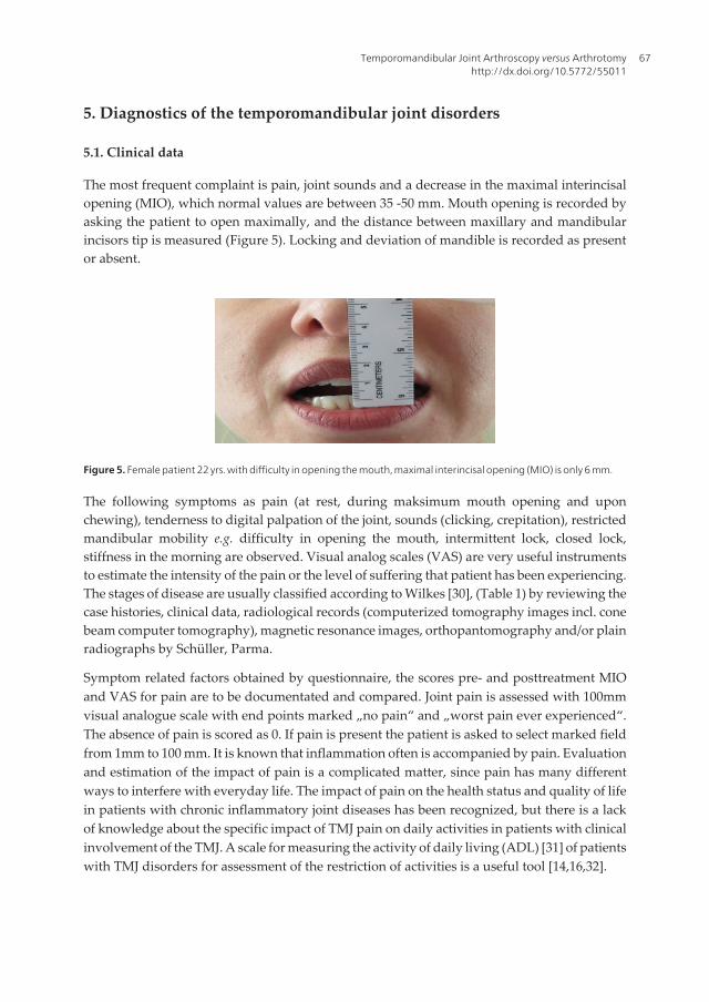
The most frequent complaint is pain, joint sounds and a decrease in the maximal interincisalopening (MIO), which normal values are between 35 -50 mm. Mouth opening is recorded byasking the patient to open maximally, and the distance between maxillary and mandibularincisors tip is measured (Figure 5). Locking and deviation of mandible is recorded as presentor absent.
Figure 5. Female patient 22 yrs. with difficulty in opening the mouth, maximal interincisal opening (MIO) is only 6 mm.
The following symptoms as pain (at rest, during maksimum mouth opening and uponchewing), tenderness to digital palpation of the joint, sounds (clicking, crepitation), restrictedmandibular mobility e.g. difficulty in opening the mouth, intermittent lock, closed lock,stiffness in the morning are observed. Visual analog scales (VAS) are very useful instrumentsto estimate the intensity of the pain or the level of suffering that patient has been experiencing.The stages of disease are usually classified according to Wilkes [30], (Table 1) by reviewing thecase histories, clinical data, radiological records (computerized tomography images incl. conebeam computer tomography), magnetic resonance images, orthopantomography and/or plainradiographs by Schüller, Parma.
Symptom related factors obtained by questionnaire, the scores pre- and posttreatment MIOand VAS for pain are to be documentated and compared. Joint pain is assessed with 100mmvisual analogue scale with end points marked „no pain“ and „worst pain ever experienced“.The absence of pain is scored as 0. If pain is present the patient is asked to select marked fieldfrom 1mm to 100 mm. It is known that inflammation often is accompanied by pain. Evaluationand estimation of the impact of pain is a complicated matter, since pain has many differentways to interfere with everyday life. The impact of pain on the health status and quality of lifein patients with chronic inflammatory joint diseases has been recognized, but there is a lackof knowledge about the specific impact of TMJ pain on daily activities in patients with clinicalinvolvement of the TMJ. A scale for measuring the activity of daily living (ADL) [31] of patientswith TMJ disorders for assessment of the restriction of activities is a useful tool [14,16,32].
Temporomandibular Joint Arthroscopy versus Arthrotomyhttp://dx.doi.org/10.5772/55011
67

I.Early stage
A. Clinical: No significant mechanical symptoms other than opening reciprocal clicking; no pain or limitation of motion
B. Radiologic: Slight forward displacement, good anatomic contour of the disc, negative tomograms, no bone
structure changes
C. Pathoanatomy: Excellent anatomic form; slight anterior displacement, passive in-coordination demonstrable
II. Early intermediate stage
A. Clinical: One or more episodes of pain: beginning major mechanical problems consisting of mid-to-late opening
loud clicking; transient catching and locking
B. Radiologic: Slight forward displacement; beginning disc deformity, slight thickening of posterior edge; negative
tomograms, no bone structure changes
C. Pathoanatomy: Anterior disk displacement; early disk deformity; good central articulating area
III. Intermediate stage
A. Clinical: Multiple episodes of pain; major mechanical symptoms consisting of locking (intermittent or fully closed):
restriction of motion, function difficulties
B. Radiologic: Anterior disc displacement with significant deformity or prolapse of disc (increased thickening of
posterior edge), negative tomograms, no bone structure changes
C. Pathoanatomy: Marked anatomic disc deformity with anterior displacement; no hard tissue changes
IV. Late intermediate stage
A. Clinical: Slight increase in severity over intermediate stage
B. Radiologic: Increase in severity over intermediate stage; positive tomograms showing early-to-moderate
degenerative changes - flattening of eminence, deformation of condylar head, erosions, sclerosis
C. Pathoanatomy: Increase in severity over intermediate stage; hard tissue degenerative remodelling of both bearing
surfaces (osteophytes), multiple adhesions in anterior and posterior recesses; no perforation of disc or attachments
V. Late stage
A. Clinical: Characterized by crepitus, variable and episodic pain, chronic restriction of motion and difficulty with
function
B. Radiologic: Disc or attachment perforation, filling defects, gross anatomic deformity of disk and hard tissues,
positive tomograms with essentially degenerative arthritic changes
C. Pathoanatomy: Degenerative changes of disc and hard tissues, perforation of posterior attachement, multiple
adhesions, osteophytes, flattening of condyle and eminence, subcortical cyst formation
Table 1. Classification for internal derangement of the TMJ by Wilkes (1989).
5.2. Radiographic investigations
Radiographic changes of the TMJ are evaluated by orthopantomography (OPTG), computedtomography (CT), magnet resonance imaging (MRI) [8,21,22,33] as well as ultrasonography[34]. OPTG is mainly used to demonstrate the structural bone changes in the TMJ and it has theadvantage of being easily available but gives limited information about the above mentionedjoint. By evaluating the OPTGs the following radiographic signs of bone structural changes can
Regional Arthroscopy68

be achieved such as presence of erosions, flattening and osteophytes of the condyle as well asof the temporal bone [35]. Erosion in condyles in the radiographs is scored as follows: score 1 -very slight erosion; score 2 - erosion on top of the condyle; score 3 - half of condyle is eroded;score 4 - condyle totally eroded [36]. The first report of TMJ CT was published by Suarez et al.[37] and this method is superior to plain transcranial or transmaxillary imaging for detectingbone changes. CT allows detailed three-dimensional examination of the TMJ and it is capableto detect even small bone changes not demonstrable by conventional tomographic proce‐dures [38, 39]. The CT sections are evaluated for presence of radiographic signs of bone changeswithin three regions (lateral, central and medial) of the mandibular and temporal part (emi‐nence) of the TMJ. The recording of the signs is made in the axial, coronal and sagittal views [22,40]. The changes are defined as follows: erosion - a local area with decreased density of thecortical joint surface including or not including adjacent subcortical bone (Figure 6), sclerosis -a local area with increased density of the cortical bony joint surface that may extend into thesubcortical bone (Figure 7), subchondral pseudocyst - a well defined, local area of bonerarefication underneath, an intact cortical outlining of the joint surface, flattening – a flat bonycontour deviating from the convex form (Figure 8). The grade of the total changes of the TMJcan be evaluated according to the scoring system [41]. Not treated properly and immediately ajuvenile trauma to the TMJ area can lead to ankylosis (Figure 9). 3-D reconstruction of themandible gives a possibility to find the fracture of the condyle not diagnosed in time (Figure 10).
Figure 6. Osteoarthritis of the TMJ, signs of erosions on the surfaces of the condyles in a coronal view of the CT.
Figure 7. Axial view of the CT from the head, sign of sclerosis in the medial and central parts of the right condyle ofthe mandible (red arrow).
Temporomandibular Joint Arthroscopy versus Arthrotomyhttp://dx.doi.org/10.5772/55011
69

Figure 8. Sagittal view of the CT from the temporomandibular joint, sign of flattening of the left mandibular condyle.
Figure 9. Coronal view of the the CT, osseous ankylosis of the left TMJ.
Figure 10. Male patient 9 yrs, trauma has been ~ 5 years ago, fracture of the left condyle is evident, displaced medially.
Regional Arthroscopy70

Foreign bodies in case of calcium pyrophosphate deposition disease (CPPD) crystals andsynovial chondromatosis granules may by diagnosed radiographically (Figure 11, 12).
Figure 11. Sagittal view of the CT, left TMJ in an open mouth position, the calcifications in the joint space are found.
Figure 12. Axial view of the CT, granules of synovial chondromatosis are in the left TMJ.
MRI has diagnostic value for internal derangements of the TMJ and rapidly surpassing CTas the imaging method of choice (Figure 13, 14). Sections in the oblique sagittal plane (i.e.perpendicular to the horizontal long axis of the mandibular condyle) and oblique coronalplane (i.e. parallel with the long axis of the condyle), and bilateral temporomandibular basesurface coils are used for obtaining the image [39]. Disc displacement without reduction is
Temporomandibular Joint Arthroscopy versus Arthrotomyhttp://dx.doi.org/10.5772/55011
71

found by using MRI in at least one of the joints in 75% of the subjects and in 54% of all thejoints imaged.
Figure 13. Sagittal view of the MRI in the closed mouth position in a patient with internalderangement of the leftTMJ. Anterior disc displacement, hypoplastic condyle, destruction of the disc. Changes of bone structures, effusion inthe anterior recess.
Figure 14. Sagittal view of the MRI in a patient with internal derangement of the left TMJ. Anterior disc displacement,destruction of the disc. Changes in the bone structures, effusion in the anterior recess.
The biting device (MEDRAD; Pittsburg) which enables dynamic imaging can be used as biteblocks during the open jaw phase of the imaging procedure [42]. Ultrasonography has been ahelpful diagnostic approach for patients with TMJ disorders, having a possibility to diagnosewith considerable reliability when compared with MRI and being a sensitive tool for assessingjoint function [43].
Regional Arthroscopy72

6. Temporomandibular joint arthroscopy
6.1. Indications for TMJ arthroscopy
The treatment decision must be based on a patient examination evaluation that integrates theimaging and clinical findings, including the history and the other diagnostic data.
Indications for arthroscopy are radiological bone changes in TMJ characteristic to osteoarthritiswith disc displacement or deformity and non effectiveness of conservative treatment withNSAIDs, intraoral splints or arthrocentesis. Arthroscopic surgery has been used to treatanteriorly displaced, nonreducing discs. Various techniques have been used as: lysis ofadhesions and joint lavage, anterior disc release, lateral capsular release, scarification of theretrodiscal region with a laser. Arthroscopic electrothermal capsulorrhaphy is performedusing a standard double puncture operative arthroscopy with a Hol: YAG laser [44]. In practice,the decision to operate and the choice of the method seems to be a matter of the individualsurgeon´s training, experience, and attitude toward the surgical management of TMJ disor‐ders. Involvement of the TMJ in patients with rheumatoid arthritis or other connective tissuediseases is rather common and arthroscopy with simultaneous biopsy is indicated in thesesituations. Posttraumatic complaints may also be an indication for arthroscopy. Arthroscopyis contraindicated in case of acute arthritis. In these situations as large medial osteophytes onthe condyle, large central cartilaginous perforations, fibrous, fibro-osseous, osseous ankylosisare better to handle via open reduction. Arthocentesis is considered as an interventingtreatment modality between nonsurgical treatment and arthroscopic surgery. All cases forarthroscopy are usually classified as advanced Wilkes [30] stages IV and V, in rare cases stageIII (Table 1).
6.2. Prearthroscopic procedures
Temporomandibular arthroscopy is usually done on an outpatient basis in the hospital. Ifdiagnostic arthroscopy is followed by arthrotomy, the patient is admitted for postoperativecare. Arthroscopy is performed under general anaesthesia with nasotracheal intubation whichmakes possible to manipulate the mandible during the operation. Both the surgeon and theassistant surgeon should have direct visibility of the monitor. First the zygomatic arch and thecondyle are palpated. The condyle is then forced in anterior position by the assistant and thepreauricular concavity is formed in the skin, marking a point for the injection. Althoughvarious arthroscopic approaches to the TMJ have been described, the one most commonly usedis the posterolateral approach to the upper joint space. After the condylar head of the TMJ hasbeen determined, a marking line and puncture points are made on the skin surface (Figure 15).
The puncture site is located by manipulating the mandible anterio-inferiorly. For distensionof the superior compartement and in order to avoid iatrogenic damage to the cartilaginoussurfaces during introduction of the trocar, 0,5 – 1,0 % lidocain solution 2,0 mL with 1: 200 000epinephrine is inserted to distend the superior compartment dilatation of capsule, in order toget hemostasis and postoperative analgesia. The solution is injected with a 27-gauge needle,which is aimed in a medial and slightly anterio-superior direction until the contact with the
Temporomandibular Joint Arthroscopy versus Arthrotomyhttp://dx.doi.org/10.5772/55011
73

glenoid fossa is achieved. The posterior recess of the superior joint space is reached when thereis a backflow into the syringe of the solution injected into the joint space (Figure 16).
Figure 16. Distension of the superior compartment with 2% lidocaine solution.
6.3. Technique for arthroscopy
Usually arthroscope KARL STORZ GmbH & Co.KG is used. Overall view of the set is givenin the Figure 17.
Figure 15. A marking line and the puncture points on the skin surface for TMJ arthroscopy.
Regional Arthroscopy74

Figure 17. Overall view of the instruments set of arthroscope KARL STORZ GmbH & Co.KG with forward oblique - tele‐scope 30° (HOPKINS® ).
Through the small skin incision 0,75 – 1,0 cm from the center of the tragus at the injection site thelateral capsule is punctured with a sharp trocar in an arthroscopic sheath inserted in the samedirection as the previous injection needle. The sharp trocar is exchanged for a blunt one and thearhroscopic sheath is advanced further into the upper joint space. Puncture with arthroscopesheath (trocar) with a blunt obturator inserted into upper posterior recess is performed anglingit medially upward ~ 2,5 cm. Another skin incision is made ~ 0,75 cm from the first skin inci‐sion in anterolateral direction for outflow cannula to be inserted into the upper joint anteriorrecess. Following insertion of the trocar (diameter 1,8 mm, length 4 cm) into the joint space, bluntobturator is removed and forward-oblique telescope 30º (HOPKINS®), diameter 1,9 mm, length6,5 cm, fiber optic light transmission incorporated is inserted (Figure 18). The inferior joint spaceis seldom entered because of the limited area makes it difficult to insert the trocar.
Figure 18. Forward-oblique telescope 30° (HOPKINS®) fiber optic light transmission incorporated and outflow cannu‐la are inserted into the upper joint space.
Temporomandibular Joint Arthroscopy versus Arthrotomyhttp://dx.doi.org/10.5772/55011
75

Initial recognition of anatomical structures as the superior surface of the disc, articularfossa, and internal aspects of the posterior and medial capsule is performed. The fluid levelin the arthroscope sheath should move with the jaw, confirming that the sheath is correctlypositioned in the joint upper space. The upper joint compartment is examined from theposterior pouch via the intermediate zone to the anterior pouch. Disc may give theimpression of being obstructed against the arthrotic surface of the temporal cartilage. Theanterior part of the disc surface looks usually smooth and collagen fibres could clearlyseen. The condylar cartilage is normally smooth, but in case of pathology e.g. in osteoarthri‐tis where irregularities of the surface as erosions, osteophyts can be seen. Sever arthroticchanges of both fossa cartilage and disc may also observed. Adhesions between the discand glenoid fossa are quite common. In rare cases the arthrotic or inflammatory changesare found in the anterior recess. Upper compartment is swept clear under constantirrigation with isotonic saline solution. This manipulation allow translation of the disc alongthe eminence, allowing the condyle to complete its natural path. After the diagnosticarthroscopy has been completed, either forceps, palpation hook or blunt probe are used tocut fibres, mainly fibers of the pterygoid muscle anterior to the disc, in order to reducepull in the anterior direction and facilitate repositioning of the disc. Cutting of adhesionsfacilitate repositioning of the disc. During arthroscopy a sweeping procedure between thedisc and fossa released the adhesions and fibrillations increasing the mobility in the joint.Release of the adhesions and fibrillations of the superior suface of the disc and shavingthe surface of articular fossa in the upper joint compartment are performed with the aidof a blunt obturator or hook and with grasping forceps, scissors or double-edged knife.Removal of the superficial layer of cortical bone induces capillar bleeding stimulatingformation of fibrocartilage on bone. Quite often a displaced disc may be found duringarthroscopy. Surgical procedure is completed by irrigating the joint space to remove smalltissue fragments. The outflowing fluid is collected and may be retained for diagnosticpurposes. Arthroscopic lysis and lavage includes also a lateral release of the upper jointcompartment performed with the aid of the blunt obturator or hook. Thus the locked disccould be mobilized sufficiently.
6.4. Analysis of arthroscopic findings
Clinical, radiographic and arthroscopic findings in patients who underwent arthroscopy aregiven in Table 2 [10].
Arthroscopic findings are as follows: irregularities of joint surfaces, foldings and synovitis –hyperaemia of the inner wall, localising also in the posterior part of the disc, intra-articularfibrous adhesions, intracapsular adhesions, fibrillations of superior surface of the disc andarthrotic lesions of temporal cartilage, pseudowalls, foreign bodies - chondromatosis (Figure19, 20, 21, 22a, 22b, 23).
Regional Arthroscopy76

Signs and
symptomsSum
%
abnRadiographic findings Sum
%
abn
Arthroscopic
findingsSum
%
abn
Pain 25 86 Flattening 10 34 Adhesions 29 100
Hypomobility 23 79 Bone cyst / Subchondral pseudocycts 9 31 Chondromatosis 5 17
Closed lock 5 17 Erosions 20 69 Fibrillations 22 76
Intermittent
lock5 17 Reduced space 10 34 Synovitis 9 31
Deviation 4 14 Sclerosis 8 27 Eburneation of fossa 15 52
Hypomobility of condyle
Osteophyts
4
5
14
17Displaced disc 23 23
Sum = total number of patients with findings; % abn = percentage of individuals with abnormal findings.
Table 2. Clinical, radiographic and arthroscopic findings in patients who underwent arthroscopy (N=29).
Figure 19. Posterior recess of the superior compartment of the right TMJ. Fibrillations and pronounced adhesionswith appearance irregularities of condylar surface, hyperaemia in the posterior capsular wall. Synovial chondromatosisgranule is in the 6 o´clock position. A greater amount of floating debris is noted.
Figure 20. Posterior recess of the superior compartment of the left TMJ. Eburneation of glenoid fossa, adhesions andfibrillations with „crab meat“ appearance, mild granulations, irregularities of condylar surface, hyperaemia of the pos‐terior attachment can be determined. Some debris is visible in the superior aspect of the field.
Temporomandibular Joint Arthroscopy versus Arthrotomyhttp://dx.doi.org/10.5772/55011
77

Figure 21. Posterior recess of the superior compartment of the right TMJ. The irregular surface of the remodeled ret‐rodiscal tissues, fibrous adhesions, fibrillations and smooth fibres seen clearly. Synovial inflammation is obvious, local‐izing in the posterior part of the disc. Some loose bodies (chondromatosis granules) are detectable.
(a)
(b)
Figure 22. a. Posterior recess of the superior compartment of the left TMJ. Debris on the posterior glenoid fossa wallcan be seen. Fibrillations, adhesions and increased vascularization in the posterior capsular wall. b. Posterior recess ofthe superior compartment of the left TMJ. Hyperaemia in the posterior capsular wall. A greater amount of floatingdebris and some granules (foreign bodies) are noted.
Regional Arthroscopy78

Figure 23. Appearance of the superior compartment of the TMJ after arthroscopic debridement. The apparent inti‐mate relationship of the glenoid fossa with the valley of the retrodiscal tissue and its junction with the disc in an es‐sentially normal TMJ. The joint space is free of debris.
The patients are to be followed up after 6 months and approximayely 5 years after theoperation. Intravenous antibiotics at the beginning of the procedure is recommended. Con‐cepts of irrigation are to maintain the capsule distended through the procedure. Continuousirrigation constantly cleanses a joint debris and blood, increases mobility, reliefing symptoms.It is also important to use of adjunctive therapy postoperatively to obtain maximum successwith arthroscopic surgery e.g. physical therapy especially in case of haemorrage, as it mayprolong healing time e.g. ultrasound with hydrocortisone ointment. A pressure dressingduring the first couple of hours after the operation is recommended.
6.5. Summary of arthroscopic findings
Arthroscopic findings included surface adhesions, stickness of the superior surface of the discto the anterior fossa portion and articular eminence, superior compartment adhesions, antero-medially displaced disc without reduction and morphologic changes in the disc. A number ofarthroscopic findings as fibrous adherences mainly between the disc and fossa, fibrillationswith „crab meat“ appearance, mild granulations, irregularities of condylar surface, foreignbodies, increased vascularisation are to be found. Synovitis in the upper joint space of the TMJhas been observed during arthroscopy and this inflamed synovium may cause pain. Thealterations in the constituents of the synovial fluid affect lubrication of the joint causingstickness and decreased mobility. Synovial chondromatosis has been found in the joint space[10, 45,46]. Synovial chondromatosis of the TMJ in both the superior and inferior joint com‐partments have found due to osteoarthritis during long period ~ 10 years [47].
6.6. Complications
Intra- and postoperative complications for arthroscopy are rare. Bleeding may be frombranches of the temporal vein during puncture. Extravasation of irrigation fluid into sur‐rounding tissues may be occur sometimes due to leakage of the irrigating fluid into thesurrounding tissues caused by accidental perforation of the TMJ capsule. This situation is easilycontroled if the surgeon always check the out-flow from out-flow cannula. From postoperativecomplications a few cases with otologic complications and nerve damage have been reported
Temporomandibular Joint Arthroscopy versus Arthrotomyhttp://dx.doi.org/10.5772/55011
79

[5,48]. Injurie of superficial branches of facial nerve resulting to paraesthesia in the preauricularregion was observed in two cases. These symptoms disappeared during one month [10].
7. Analysis of clinical data and results
It has been shown that during arthroscopy several inflammatory and pain mediators causingdestructive changes, foreign bodies as grains of chondromatosis are washed out elicitatingjoint noises [9,49]. For the patients with episodic signs and symptoms a noninvasive conser‐vative approach is indicated (Wilkies stages I-III). Procedures currently used for the TMJderangements as osteoarthritis/arthrosis (Wilkies stages IV and V) are: arthrocentesis, arthro‐scopy, arthrotomy or TMJ replacement. From arthroscopic findings fibrillation seemed to bethe most common ~76% [50]. Arthroscopic lysis and lavage has been an effective treatment forTMJ disorders refractory to nonsurgical treatments [8,12,51]. An evaluation following tem‐poromandibular joint arthroscopic surgery with lysis and lavage after 2 to 10,8 years treatmentshowed that arthroscopic surgery of the temporomandibular joint is successful in the long termfor patients with painful motion [11,52]. Assessment of symptoms reported by the patient aswell as of objective signs noted on clinical examination confirms resolution of pain onmovement and increased vertical opening.
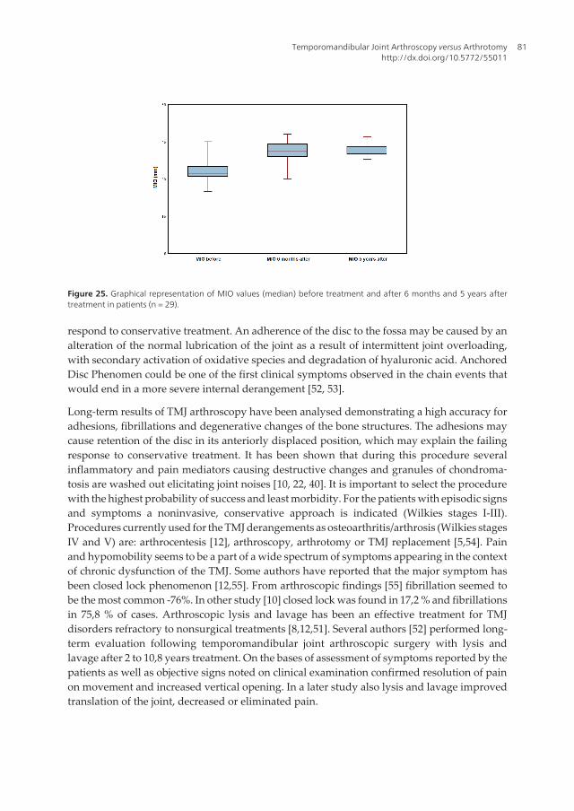
A significant and maintained improvement in MIO and VAS is also observed over the 5 yearsperiod of time (Figure 24, 25) [10].
Figure 24. Graphical representation of VAS values (median) before treatment and after 6 months and 5 years treat‐ment in patients (n = 29).
TMJ arthroscopy is especially useful when the disc has not yet been deformed. Superior jointcompartment adhesions and disc immobility can be treated during arthroscopic procedure,leading to resolution of symptoms and return of joint function [10]. The adhesions may causeretention of the disc in its anteriorly displaced position, which may explain the failure to
Regional Arthroscopy80
respond to conservative treatment. An adherence of the disc to the fossa may be caused by analteration of the normal lubrication of the joint as a result of intermittent joint overloading,with secondary activation of oxidative species and degradation of hyaluronic acid. AnchoredDisc Phenomen could be one of the first clinical symptoms observed in the chain events thatwould end in a more severe internal derangement [52, 53].
Long-term results of TMJ arthroscopy have been analysed demonstrating a high accuracy foradhesions, fibrillations and degenerative changes of the bone structures. The adhesions maycause retention of the disc in its anteriorly displaced position, which may explain the failingresponse to conservative treatment. It has been shown that during this procedure severalinflammatory and pain mediators causing destructive changes and granules of chondroma‐tosis are washed out elicitating joint noises [10, 22, 40]. It is important to select the procedurewith the highest probability of success and least morbidity. For the patients with episodic signsand symptoms a noninvasive, conservative approach is indicated (Wilkies stages I-III).Procedures currently used for the TMJ derangements as osteoarthritis/arthrosis (Wilkies stagesIV and V) are: arthrocentesis [12], arthroscopy, arthrotomy or TMJ replacement [5,54]. Painand hypomobility seems to be a part of a wide spectrum of symptoms appearing in the contextof chronic dysfunction of the TMJ. Some authors have reported that the major symptom hasbeen closed lock phenomenon [12,55]. From arthroscopic findings [55] fibrillation seemed tobe the most common -76%. In other study [10] closed lock was found in 17,2 % and fibrillationsin 75,8 % of cases. Arthroscopic lysis and lavage has been an effective treatment for TMJdisorders refractory to nonsurgical treatments [8,12,51]. Several authors [52] performed long-term evaluation following temporomandibular joint arthroscopic surgery with lysis andlavage after 2 to 10,8 years treatment. On the bases of assessment of symptoms reported by thepatients as well as objective signs noted on clinical examination confirmed resolution of painon movement and increased vertical opening. In a later study also lysis and lavage improvedtranslation of the joint, decreased or eliminated pain.
Figure 25. Graphical representation of MIO values (median) before treatment and after 6 months and 5 years aftertreatment in patients (n = 29).
Temporomandibular Joint Arthroscopy versus Arthrotomyhttp://dx.doi.org/10.5772/55011
81

The chief presenting complaint for most patients (86,2%) was pain preoperatively. A significantmaintained decrease in VAS score was achieved after 6 months and also 5 years follow-up. Asignificant and maintained improvement in MIO was also observed over the same period oftime [10]. The results are comparable to those reported in the other papers [34,52]. It isimportant to take into account that the sympathetic and sensory nerve fibres within thetemporomandibular joint are located in the anterior recess and the retrodiscal tissue of theupper compartment. Anterior disc release may reduce the number of these nerve fibres inarthroscopic procedures, thus influencing pain dynamics. The advantages of arthroscopycompared with open joint surgery using the Jaw Pain and Function Questionnaire are thatarthroscopic surgery is less invasive and associated with lower morbidity [15]. No statisticaldifferences were also observed between arthroscopic lysis and lavage and operative arthro‐scopy in relation to postoperative pain or MIO at any stage of the follow-up period [9].Arthroscopic lysis and lavage has been found effective in 84% of patients in case of osteoar‐thritis of TMJ [55]. Multiple adhesions also develop skeletal changes, with a shortened ramus.If the condition develops rapidly enough, open bite and rethrognathia may occur [40,56,57].During arthroscopic surgery nodules of TMJ synovial chondromatosis are able to pass throughthe cannula by lavage with saline solution [49].
An adherence of the disc to the fossa may be caused by an alteration of the normal lubricationof the joint as a result of intermittent joint overloading, with secondary activation of oxidativespecies and degradation of hyaluronic acid. Anchored Disc Phenomen could be one of the firstclinical symptoms observed in the chain events that would end in a more severe internalderangement [52,53].
Based on the present findings, it follows that a displaced disc, by itself, is of only limitedsignificance.This is not surprising because the majority of individuals with derangement ofthe TMJ are asymptomatic [7,57]. The intriguing question that remains is why lavage and lysisof adhesions or high-pressure irrigation of the upper joint space should be therapeutic. Theanswer is, that during this procedure several inflammatory mediators available in the synovialfluid as prostaglandins [58], cytokines [22,59], serotonin as pain mediator [28] etc. are washedout. In episodes of closed lock, the limitation in condylar movement probably originates fromchanges in the upper compartment that restrict the sliding motion of the disc; This course ofevents may explain the efficacy of lysis and lavage of only this joint space, as this manipulationallows translation of the disc along the eminence, allowing the condyle to complete its naturalpath. The data in the literature have stated that the most frequent disc displacements wereanterior and anteromedial [52].
In episodes of closed lock, the limitation in condylar movement probably originates from thechanges in the upper compartment that restrict the sliding motion of the disc. The data in theliterature have stated that the most frequent disc displacements were anterior and anterome‐dial [39,60]. Using MRI pre- and postoperatively revealed that disc position remained anteri‐orly without reduction, disc mobility increased after arthroscopic surgery[8]. Improvement injoint symptoms and function is not attributed so much as to the restoration of disc position asto possible release of the lateral capsular fibrosis during arthroscopy [52,61].
Regional Arthroscopy82
8. Arthrotomy
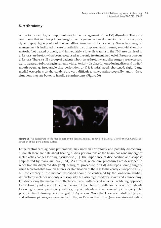
Arthrotomy can play an important role in the management of the TMJ disorders. There areconditions that require primary surgical management as developmental disturbances (con‐dylar hypo-, hyperplasia of the mandible, tumours, ankylosis etc.). Secondary surgicalmanagement is indicated in case of arthritis, disc displacements, trauma, synovial chondro‐matosis. Not treated properly and immediately a juvenile trauma to the TMJ area can lead toankylosis. Arthrotomy has been recognised as the only treatment method of fibrous or osseousankylosis.There is still a group of patients whom an arthrotomy and disc surgery are necessarye.g. to treat painful clicking in patients with anteriorly displaced, nonreducing discs and limitedmouth opening, irreparable disc perforation or if it is misshaped, shortened, rigid. Largemedial osteophyts on the condyle are very difficult to shave arthroscopically, and in thesesituations they are better to handle via arthrotomy (Figure 26).
Figure 26. An osteophyte in the medial part of the right mandibular condyle in a sagittal view of the CT. Cortical de‐struction of the glenoid fossa surface.
Large central cartilaginous perforations may need an arthrotomy and possibly discectomy,although there are data about healing of disk perforations as the bilaminar zone undergoesmetaplastic changes forming pseudodisc [61]. The importance of disc position and shape isemphasized by many authors [8, 51]. As a result, open joint procedures are developed toreposition the displaced disc [7, 9]. A surgical procedure for TMJ disc-repositioning surgeryusing bioresorbable fixation screws for stabilisation of the disc to the condyle is reported [62],but the efficacy of the method described should be confirmed by the long-term studies.Arthrotomy includes not only a discoplasty but also high condylar shave and eminectomy.For discectomy the medial disc attachment is cut with curved scissors, facilitating approachto the lower joint space. Direct comparison of the clinical results are achieved in patientsfollowing arthroscopic surgery with a group of patients who underwent open surgery. Thepostoperative follow-up period ranged 5 to 6 years and 9 months. These results following openand arthroscopic surgery measured with the Jaw Pain and Function Questionnaire a self rating
Temporomandibular Joint Arthroscopy versus Arthrotomyhttp://dx.doi.org/10.5772/55011
83
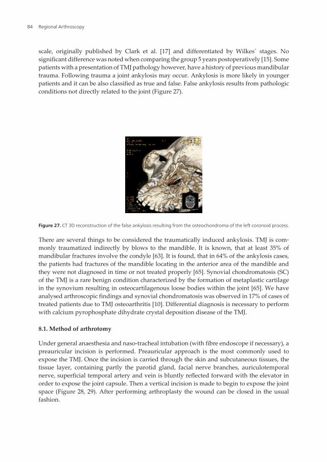
scale, originally published by Clark et al. [17] and differentiated by Wilkes´ stages. Nosignificant difference was noted when comparing the group 5 years postoperatively [15]. Somepatients with a presentation of TMJ pathology however, have a history of previous mandibulartrauma. Following trauma a joint ankylosis may occur. Ankylosis is more likely in youngerpatients and it can be also classified as true and false. False ankylosis results from pathologicconditions not directly related to the joint (Figure 27).
Figure 27. CT 3D reconstruction of the false ankylosis resulting from the osteochondroma of the left coronoid process.
There are several things to be considered the traumatically induced ankylosis. TMJ is com‐monly traumatized indirectly by blows to the mandible. It is known, that at least 35% ofmandibular fractures involve the condyle [63]. It is found, that in 64% of the ankylosis cases,the patients had fractures of the mandible locating in the anterior area of the mandible andthey were not diagnosed in time or not treated properly [65]. Synovial chondromatosis (SC)of the TMJ is a rare benign condition characterized by the formation of metaplastic cartilagein the synovium resulting in osteocartilagenous loose bodies within the joint [65]. We haveanalysed arthroscopic findings and synovial chondromatosis was observed in 17% of cases oftreated patients due to TMJ osteoarthritis [10]. Differential diagnosis is necessary to performwith calcium pyrophosphate dihydrate crystal deposition disease of the TMJ.
8.1. Method of arthrotomy
Under general anaesthesia and naso-tracheal intubation (with fibre endoscope if necessary), apreauricular incision is performed. Preauricular approach is the most commonly used toexpose the TMJ. Once the incision is carried through the skin and subcutaneous tissues, thetissue layer, containing partly the parotid gland, facial nerve branches, auriculotemporalnerve, superficial temporal artery and vein is bluntly reflected forward with the elevator inorder to expose the joint capsule. Then a vertical incision is made to begin to expose the jointspace (Figure 28, 29). After performing arthroplasty the wound can be closed in the usualfashion.
Regional Arthroscopy84

Figure 28. D image of the CT showing fibroosseous ankylosis of the right TMJ on a 15 yrs. boy. Trauma took place atthe age of 4 years.
Figure 29. Arthrotomy of the same 15 yrs boy is performed. The right ankylotic TMJ is exposed.
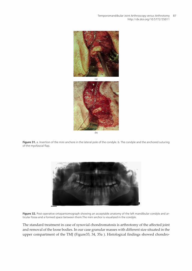
We have used [66] the suture anchor in interpositional arthroplasty in case of the temporo‐mandibular joint ankylosis traumatic origin. Ankylotic left TMJ was exposed and a gaparthroplasty was performed using different types of burs. An ipsilateral myofascial temporalpedicled flap was prepared, rotated inferiorly and interposed between the head of the condyle
Temporomandibular Joint Arthroscopy versus Arthrotomyhttp://dx.doi.org/10.5772/55011
85

and the mandibular fossa.. The mini anchor in the lateral pole of the condyle was inserted andanchored suture of the myofascial flap was performed and the capsule was sutured.(Figure30a, 30b, 31a, 31b, 32).
(a)
(b)
Figure 30. a. Ankylotic left TMJ is exposed for osteotomy. b. The osseous components of the TMJ after arthroplastyand the joint space is formed.
Regional Arthroscopy86
(a)
(b)
Figure 31. a. Insertion of the mini anchore in the lateral pole of the condyle. b. The condyle and the anchored suturingof the myofascial flap.
Figure 32. Post-operative ortopantomograph showing an acceptable anatomy of the left mandibular condyle and ar‐ticular fossa and a formed space between them.The mini anchor is visualized in the condyle.
The standard treatment in case of synovial chondromatosis is arthrotomy of the affected jointand removal of the loose bodies. In our case granular masses with different size situated in theupper compartment of the TMJ (Figure33, 34, 35a ). Histological findings showed chondro‐
Temporomandibular Joint Arthroscopy versus Arthrotomyhttp://dx.doi.org/10.5772/55011
87

metaplasia of the synovial membrane (Figure 35b) [67]. Arthroscopy is proved to be useful formanagement of synovial chondromatosis of the TMJ in case of granules smaller than 3 mmwhich are commonly removed with joint lavage [10]. OPTG and CT scans reveale usuallycalcifying lesions in the TMJ region.
Figure 33. Axial view of the CT scan of the patient (female, 44 yrs.), showing granular masses surrounding the leftcondylar head.
Figure 34. Intra-operative finding of the same patient (female, 44 yrs.) with irregular cartilaginous loose granules inthe posterior recess of the upper compartment of the TMJ.
Regional Arthroscopy88

(a)
(b)
Figure 35. a. The different size of granules are pearly white, of varying shape and ranging in the size from 3,0 to 10,0mm.b. Histologically clustered hyaline chondrocytes with synovial lining are visible. Staining with haematoxylin-eosin,magnification 40X.
9. Summary
Early diagnosis is the key to successful treatment, because it permits the use of nonsurgicalmeans or minimally invasive procedures (arthrocentesis, arthroscopy). In late stage disease isindicated arthrotomy, to attain an improved quality of life with less pain and improvedfunction. Clinical success of arthroscopy is based on several factors. Lysis and lavage removeintraarticular inflammatory and pain mediators. The release of fibrillations and adhearencesas well as improvement in discal mobility allows to distrbute the functional stresses on thearticular tissues and adverse loading on the joints is decreased. The long-term outcome of TMJarthroscopic surgery with lysis and lavage is considered to be acceptable and effective.Fibrillations and fibrous adhesions are the most usual pathological signs of arthroscopicfindings in patients with internal derangement of the TMJ. Arthroscopic releasing of theserestrictive bands improves the joint mobility and contributes to reducing pain level. The resultsof arthroscopy offered favourable long-term stable results with regard to increasing MIO andreducing pain and dysfunction. The improvement in joint mobility and disc mobility will leadto adaptive changes in the hard tissues.This may implay that the arthroscopic procedure withmechanics may stop the process of further TMJ degeneration. The advantages of arthroscopycompared with open joint surgery are that arthroscopic surgery is less invasive, procedureneeds less time and associated with lower morbidity. Arthrotomy is indicated in cases withanteriorly displaced, nonreducing discs who continue to have pain and limited mouth openingdespite the treatment by either arthrocentesis or arthroscopic surgery that has not responded
Temporomandibular Joint Arthroscopy versus Arthrotomyhttp://dx.doi.org/10.5772/55011
89
to arthrocentesis or arthroscopy. In conclusion procedures such as arthrocentesis, arthroscopicsurgery and arthrotomy can be used with reasonably good results in properly selected cases.
Acknowledgements
The publication of this chapter is supported by Ernst Jaakson Memorial Scholarship.
Author details
Edvitar Leibur1,2, Oksana Jagur1 and Ülle Voog-Oras1
1 Department of Stomatology, Tartu University, Tartu University Hospital, Estonia
2 Department of Internal Medicine, Tartu University, Tartu University Hospital, Estonia
References
[1] Sanders B, Buonocristiani R.Diagnostic and Surgical Arthroscopy of the Temporo‐mandibular Joint: Clinical Experience with 137 Procedures over a 2-year period. Jour‐nal of Craniomandibular Disorders: Facial & Oral Pain 1987;12(3) 202-213.
[2] Tag H. Arthroscope. Journal of Japanese Orthopedic Association 1939;14, 359-362.
[3] Onishi M. Arthroscopy of the temporomandibular joint (author´s transl.). Journal ofJapanese Stomatological Association 1975;(42) 207-213.
[4] Onishi M. Clinical application arthroscopy in the temporomandibular joint diseases.Bulletin Tokyo Medical Dental University 1980;(27) 141-148.
[5] McCain JP, Sanders B, Koslin MG, Quinn JH, Peters PB, Indresano AT. Temporoman‐dibular joint arthroscopy : a 6-year multicenter retrospective study of 4,831 joints.Journal of Oral and Maxillofacial Surgery 2002;50(9) 926-930.
[6] Holmlund AB, Axelsson S.Temporomandibular arthropathy: correlation betweenclinical signs and symptoms and arthroscopic findings. International Journal of Oral& Maxillofacial Surgery 1996;25(3) 266-271.
[7] Holmlund AB, Axelsson S,Gynther GW. A comparison of discectomy and arthro‐scopic lysis and lavage for the treatment of chronic closed lock of the temporoman‐dibular joint: a randomized outcome study. Journal of Oral and MaxillofacialSurgery 2001;59(9) 972-977.
[8] Ohnuki T, Fukuda M, Iino M, Takahahshi T. Magnetic resonance evaluation of thedisk before and after arthroscopic surgery for temporomandibular disorders. Oral
Regional Arthroscopy90

Surgery Oral Medicine Oral Pathology Oral Radiology Endodontics 2003;96(2)141-148.
[9] González-Garcia R, Rodriguez-Campo FJ, Monje F, Sastre-Perez J, Gil-Diez Usandi‐zaga JL. Operative versus simple arthroscopic surgery for chronic closed lock of thetemporomandibular joint: a clinical study of 344 arthroscopic procedures. Interna‐tional Journal of Oral&Maxillofacial Surery 2008;17(9) 790-796.
[10] Leibur E, Jagur O, Müürsepp P, Veede L, Voog-Oras Ü. Long-term evaluation of ar‐throscopic surgery with lysis and lavage of temporomandibular disorders. Journal ofCranio-Maxillo-Facial Surgery 2010;38(8) 615-620.
[11] Murakami K, Segami N, Okamoto I, Takahashi K,Tsuboi, Y. Outcome of arthroscopicsurgery for internal derangement of the temporomandibular joint: long – term resultscovering 10 years. Journal of Cranio-Maxillo-Facial Surgery 2000;28(3) 264 – 271.
[12] Sanroman JF. Closed lock (MRI fixed disc): a comparison of arthrocentesis and ar‐throscopy. International Journal of Oral & Maxillofacial Surgery 2004;33(4) 344-348.
[13] Mancha de la Plata M, Muñoz-Guerra M, Escorial Hernandez V, Martos Diaz P, Gil-Diez Usandizaga JL, Rodriguez-Campo FJ. Unsuccessful temporomandibular jointarthroscopy: is a second arthroscopy an acceptable alternative? Journal of Oral andMaxillofacial Surgery 2008;66(10) 2086-2092.
[14] Voog Ü, Alstergren P, Leibur E, Kallikorm R, Kopp S. Impact of temporomandibularjoint pain on activities of daily living in patients with rheumatoid arthritis. ActaOdontologica Scandinavica 2003;61(5) 278-282.
[15] Undt G, Murakami KI, Rasse M, Ewers R. Open versus arthroscopic surgery for in‐ternal derangement of the temporomandibular joint: A retrospective study compar‐ing two centers results using Jaw Pain and Function Questionnaire. Journal ofCranio-Maxillo-Facial Surgery 2006;34(4) 234-241.
[16] Jagur O, Kull M, Leibur E, Kallikorm R, Loorits D, Lember M,Voog-Oras Ü. The asso‐ciations of TMJ pain and bone characteristics on the activities of daily living. OpenJournal of Stomatology 2012 (accepted for publication).
[17] Clark GT, Seligman D, Solberg WK, Pullinger AG. Guidlines for the examination anddiagnosis of temporomandibular disorders. Journal of Craniomandibular Disorders1989;3(1) 7-14.
[18] Sommer OJ, Aigner F, Rudisch A, Gruber H, Fritsch H, Millesi W, Stiskal M. Cross-sectional and Functional Images of the Temporomandibular Joint: Radiology, Pathol‐ogy, and Basic Biomechanics of the Jaw Radiographics. Radiology 2003;23(6) 428-432.
[19] Schmelzle R. Lokalanästhesie. In: Zahnärztliche Chirurgie. 2. Auflage Urban &Schwarzenberg. München-Wien-Baltimore1989; p.19.
Temporomandibular Joint Arthroscopy versus Arthrotomyhttp://dx.doi.org/10.5772/55011
91

[20] Tallents RH, MacherDJ, Kyrkanides S, Katzberg RW, Moss M.E. Prevalence of misingposterior teeth and intraarticular temporomandibular disorders. Journal of ProstheticDentistry 2002;87(1) 45-49.
[21] Voog Ü, Alstergren P, Eliasson S, Leibur E, Kallikorm R, Kopp S.Progression of ra‐diographic changes in the temporomandibular joints of patients with rheumatoid ar‐thritis in relation to inflammatory markers and mediators in the blood. ActaOdontologica Scandinavica 2004;62(1) 7-13.
[22] Voog Ü, Alstergren P, Eliasson S, Leibur E, Kallikorm R, Kopp S. Inflammatory me‐diators and radiographic changes in temporomandibular joints in patients with rheu‐matoid arthritis. Acta Odontologica Scandinavica 2003;61(1) 57-64.
[23] Kim SJ, Park YH, Hong SP, Cho BO, Park JW, Kim SG. The presence of bacteria in thesynovial fluid of the temporomandibular joint and clinical significance: preliminarystudy. Journal of Oral and Maxillofacial Surgery 2003;61(10) 1156-1161.
[24] Paegle DI, Holmlund AB, öStlund MR, Grillner L. The occurence of Antibodiesagainst Chlamydia species in patients with monoarthritis and chronic closed lock ofthe temporomandibular joint. Journal of Oral and Maxillofacial Surgery 2004;62(4)435-439.
[25] Alstergren P, Kopp S, Theodorson E. Synovial fluid sampling from the temporoman‐dibular joint: sample quality criteria and levels of interleukin-1 beta and serotonin.Acta Odontologica Scandinavica 1999;57(1) 278-282.
[26] Kamada A, Kakudo K, Arika T, Okazaki J, Kano M, Sakaki T. Assay of synovialMMP3 in temporomandibular joint diseases. Journal of Cranio-Maxillo- Facial Sur‐gery 2000;28(3) 247-248.
[27] Alstergren P, Kopp S. Pain and synovial fluid concentration in arthritic temporoman‐dibular joints. Pain 2007;2(1-2) 137-143.
[28] Voog Ü, Alstergren P, Leibur E, Kallikorm R, Kopp S. Immediate effect of the seroto‐nin antagonist granisetron on temporomandibular joint pain in patients with system‐ic inflammatory disorders. Life Sciences 2000;68(5) 591-602.
[29] Warden SJ, Haney EM. Skeletal effects of serotonin (5-hydroxytryptamine) transport‐er inhibition: evidence from in vitro and animal-based studies. Journal of Musculos‐keletal Neuronal Interaction 2008;8(2) 121-132.
[30] Wilkes CH. Internal derangements of the temporomandibular joint. Pathological var‐iations. Archives of Otolaryngology, Head Neck Surgery 1989;115(4) 469-477.
[31] List T, Helkimo M. A scale for measuring the activities of daily living (ADL) of pa‐tients with craniomandibular disorders. Swedish Dental Journal 1995;19(1) 33-40.
Regional Arthroscopy92
[32] Kaselo E, Jagomägi T,Voog U. Malocclusion and the need for orthodontic treatmentin patients with temporomandibular dysfunction. Stomatologija. Baltic Dental andMaxillofacial Journal 2007;9(3) 79-85.
[33] Whyte AM, McNamara D, Rosenberg I,Whyte AW. Magnetic resonance imaging inthe evaluation of temporomandibular joint disc displacement. International Journalof Oral & Maxillofacial Surgery 2006;35(8) 696-703.
[34] Landes CA, Goral WA, Sader R, Mack MG. 3-D sonography for diagnosis of disc dis‐location of the temporomandibular joint compared with MRI. Ultrasound MedicalBiology 2007;32(5) 633-639.
[35] Rohlin M, Ăkerman S,Kopp S. Tomography as an aid to detect macroscopic changesof the temporomandibular joint . Acta Odontologica Scandinavica 1986;44(3) 131-140.
[36] Helenius L, Hallikainen D, Meurman J, Koskimies S, Tervahartiala P, Kivisaari L,Hietanen J, Suuronen R, Lindqvist C, Leirisalo-Repo M. HLA-DRB1* alleles and tem‐poromandibular joint erosion in patients with rheumatic disease. Scandinavian Jour‐nal of Rheumatology 2004;33(1) 24-29.
[37] Suarez FR, Bhussry BR, Neff PA, Huang HK,Vaughn D. A preliminary study of com‐puterized tomographs of the temporomandibular joint. The Compendium on con‐tinuing education in general dentistry 1980;1(3) 217-222.
[38] Raustia M, Pyhtinen J, Virtanen KK. Examination of the temporomandibular joint bydirect sagittal computed tomography. Clinical Radiology 1985;36(3) 291-296.
[39] Larheim TA,Westesson P, Sano T. Temporomandibular Joint Disk Displacement:Comparison in Asymptomatic Volunteers and Patients. Radiology 2001;218(2)428-32.
[40] Emshoff R, Brandlmaier I, Bertram S, Rudish A.Relative odds of temporomandibularjoint pain as a function of magnetic resonance imaging findings of internal derange‐ment, osteoarthrosis, effusion, and bone marrow edema. Oral Surgery Oral MedicineOral Pathology Oral Radiology Endodontics 2003;95(4) 437-445.
[41] Rohlin M, Petersson A. Rheumatoid arthritis of the temporomandibular joint: radio‐logic evaluation based on standard reference films. Oral Surgery, Oral Medicine, andOral Pathology 1989;67(5) 594-599.
[42] Gaggle A, Schults G, Santler G, Kärcher H, Simbrunner J. Clinical and magnetic reso‐nance findings in the temporomandibular joints of patients before and after ortog‐nathic surgery. The British Journal of Oral & Maxillofacial Surgery 1999;37(1) 41-45.
[43] Landes C, Walendzik H, Klein C. Sonography of the temporomandibular joint from60 examinations and comparison with MRI and axiography. Journal of Cranio-Maxil‐lo-Facial Surgery 2000;28(6) 352-361.
Temporomandibular Joint Arthroscopy versus Arthrotomyhttp://dx.doi.org/10.5772/55011
93

[44] Torres DE, McCain JP. Arthroscopic elrctrothermal capsulorrhaphy for the treatmentof recurrent temporomandibular joint dislocation. International Journal of Oral &Maxillofacial Surgery 2012;41(6) 681-689.
[45] Mercuri LG. Synovial chondromatosis of the temporomandibular joint with medialcranial fossa extension. International Journal of Oral & Maxillofacial Surgery2008;37(7) 684-685.
[46] González-Pérez LM, Concregado-Córdoba J, Salinas-Martin MV. Temporomandibu‐lar joint synovial chondromatosis with a traumatic etiology. International Journal ofOral & Maxillofacial Surgery 2011;40(3) 330-334.
[47] Sato J, Segami N, Suzuki T, Yoshitake Y, Nishikawa K.The expression of fibroblastgrowth factor receptor 1 in chondrocytes in synovial chondromatosis of the temporo‐mandibular joint. Report of two cases. International Journal of Oral & MaxillofacialSurgery 2002;31(7) 532-536.
[48] Appelbaum EL, Berg LF, Kumar A, Mafee MF. Otologic complications Followingtemporomandibular joint arthroscopy. Annals of Otology, Rhinology, Laryngology1988;97(6) 675-679.
[49] Shibuya T, Kino K, Yoshida S, Amagasa T.Arthroscopic removal of nodules of syno‐vial chondromatosis of the temporomandibular joint. Cranio 2002;20(4) 304-306.
[50] Dimitroulis G. A review of 56 cases of chronic closed lock treated with temporoman‐dibular joint arthroscopy. Journal of Oral and Maxillofacial Surgery 2002;60(5)519-524.
[51] Politi M, Sembronio S, Robiony M, Costa F, Toro C, Undt G. High condylectomy anddisc repositioning compared to arthroscopic lysis, lavage and capsular strech for thetreatment of chronic closed lock of the temporomandibular joint. Oral Surgery OralMedicine Oral Pathology Oral Radiology Endodontics 2007;103(1) 27 - 33.
[52] Sorel B, Piecuch JF. Long-term evaluation following temporomandibular joint arthro‐scopy with lysis and lavage. International Journal of Oral & Maxillofacial Surgery2000;29(4) 532-536.
[53] Krug J, Jirousek Z, Suchmova H, Germakova E. Influence of discoplasty and discec‐tomy of the temporomandibular joint on elimination of pain and restricted mouthopening. Acta Medica (Hradec Kralove) 2004;47(1) 47-53.
[54] Smolka W, Iizuka T. Arthroscopic lysis and lavage in different stages of internal de‐rangement of the temporomandibular joint: correlation of preoperative staging to ar‐throscopic findings and treatment outcome. Journal of Oral and MaxillofacialSurgery 2005;63(4) 471-478.
[55] Dimitroulis G. The prevalence of osteoarthrosis in cases of advanced internal de‐rangement of the Temporomandibular Joint: a clinical, surgical and histologicalstudy. International Journal of Oral & Maxillofacial Surgery 2005;34(2) 345-349.
Regional Arthroscopy94

[56] Emshoff R. Clinical factors affecting the outcome of arthrocentesis and hydraulic dis‐tension of the temporomandibular joint. Oral Surgery Oral Medicine Oral PathologyOral Radiology Endodontics 2005;100(4) 409-414.
[57] Hamada Y, Kondoh T, Holmlund AB, Iino M, Kobayashi K, Seto K.Influence of ar‐throscopically observed fibrous adhesions before and after joint irrigation on clinicaloutcome in patients with chronic closed lock of the temporomandibular joint. Inter‐national Journal of Oral & Maxillofacial Surgery 2005; 34(7) 727-732.
[58] Murakami KI, Shibata T, Kubota E, Maeda H. Intra-articular levels of prostaglandinE2 , hyaluronic acid, and chondroitin -4 and -6 sulfates in the temporomandibularjoint synovial fluid of patients with internal derangement. Journal of Oral and Maxil‐lofacial Surgery 1998;56(2) 199-203.
[59] Kardel R, Ulfgren AK, Reinholt FP, Holmlund A. Inflammatory cell and cytokinepatterns in patients with painful clicking and osteoarthritis in the temporomandibu‐lar joint. International Journal of Oral & Maxillofacial Surgery 2003;32(5) 390-396.
[60] Güven O. Management of chronic recurrent temporomandibular joint dislocations: Aretrospective study. Journal of Craniomaxillofacial Surgery 2009;37(1) 24-29.
[61] Moses JJ, Lo H. The treatment of internal derangement of the temporomandibularjoint – an arthroscopic approach. Oral Surgery Oral Diagnosis 1992;.3, 5-11.
[62] Sembronio S, Robiony M, Politi M. Disc-repositioning surgery of the temporoman‐dibular joint using bioresorbable screws. International Journal of Oral & Maxillofa‐cial Surgery 2006;35(10) 1149-1152.
[63] Bradley P. Injuries of the condylar region and coronal process. In Rowe NL, WilliamsJL (eds.): Maxillofacial Injuries, Vol.1. London, England, Churchill Livingstone, 1985,pp. 337-339
[64] He D, Ellis E. 3rd., Zhang Y. Etiology of temporomandibular joint ankylosis secon‐dary to condylar fractures: the role of concomitant mandibular fractures. Journal ofOral and Maxillofacial Surgery 2008;66(1).74-78
[65] Ardekian L, Faquin W, Troulis M, Kaban LB, August M. Synovial chondromatosis ofthe temporomandibular joint: report and analysisof eleven cases. Journal of Oral andMaxillofacial Surgery 2005;63,(5) 941-947.
[66] Nestal-Zibo H, Leibur E, Voog-Oras Ü, Tamme T. Use of the suture anchor in inter‐positional arthroplasty of temporomandibular joint ankylosis. Oral and MaxillofacialSurgery 2012;16(1) 157-162.
[67] Jagur O, Leibur E, Erm T. Synovial chondromatosis of the temporomandibular joint.A case report. Stomatologija. Baltic Dental and Maxillofacial Journal 2012;12(Suppl.8):29.
Temporomandibular Joint Arthroscopy versus Arthrotomyhttp://dx.doi.org/10.5772/55011
95