Embed Size (px)
Citation preview
Temporally modular gene expression during cotyledon development D. Wayne Hughes and Glenn A. Galau 1
Department of Botany, University of Georgia, Athens, Georgia 30602 USA
The regulation of cotton embryogenesis has been addressed by measuring the abundance of 47 mRNAs in cotyledons from the late cotyledon stage through early germination. There are at least 11 distinct classes of coordinately expressed mRNAs. Their expression patterns appear to result from unique combinations of five temporal abundance components. These are associated with the cotyledon stage, the endogenous concentration of free abscisic acid, maturation (reserve accumulation}, ovule abscission, and germination. This modularity suggests that only a few global regulatory factors orchestrate gene expression with many genes responding to several of them. Significant expression associated only with postabscission or free abscisic acid is restricted to that of the Lea mRNAs earlier suggested to be a component of the embryo's preparation for desiccation.
[Key Words: Gossypium hirsutum; maturation; postabscission; germination; mRNA abundance]
Received November 14, 1988; revised version accepted January 24, 1989.
Dicot cotyledons appear simple. They are usually the major embryo organ and contain mostly parenchymal cells. By the time most dicot embryos are large enough for biochemical and experimental study, cell division and morphogenesis essentially are complete at the end of the cotyledon stage of development. In the following stage of seed filling or reserve synthesis, here termed maturation, the parenchymal cells synthesize and store protein and other reserves which are degraded during germination to provide support for seedling growth. A period of little growth, and then often desiccation and quiescence, occurs between reserve synthesis and germi- nation {the term maturation is used by some to denote this period after reserve synthesis stops}. Most other cell types in cotyledons and other embryo organs also partic- ipate in these activities (for review, see Dure 1975; Raghaven 1986; Crouch 1987}. One approach to identify the regulators of embryo development has been the de- scriptive analysis of normal development combined with experimental embryo culture for testing specific hypotheses {Raghaven 1986}. There has been some prog- ress in the isolation of stage-specific mRNA markers ex- pressed in various organs of dicot and monocot embryos. Their use in this context has tended to confirm roles for the plant growth regulator abscisic acid (ABA), embryo water potential, and nutritional status {Chandler et al. 1984; Dure 1985; Quatrano 1986; Crouch 1987; Galau et al. 1987}.
Cotton cotyledons are typical of those of dicots, and qualitative study of the abundance of their individual mRNAs suggested that there are global regulatory events that occur in the later stages of cotyledon devel-
tCortesponding author.
opment (Dure et al. 1981). Using cloned representatives of three classes of coordinately expressed mRNAs, a de- cline in three storage protein mRNAs and a remarkably reciprocal increase in the two classes containing 18 Lea (Late-embryogenesis-abundant) mRNAs was noted at about the time of ovule abscission (Galau et al. 1987}. The Lea mRNAs are very abundant only between em- bryo maturation and desiccation, suggesting that their abundant hydrophilic polypeptides may function to pro- tect the embryo from desiccation damage or to repair such damage (Galau et al. 1987; Hughes and Galan 1987}. During the transition between cotyledon and maturation stages when embryo water potential de- clines, the abundance of one class of Lea mRNAs also correlates with the two periods of high concentration of free ABA which is associated in many contexts with water stress (Zeevaart and Creelman 1988), but at mRNA levels much lower than those achieved after maturation, when ABA levels are lower, or during em- bryo culture with exogenous ABA (Galau et al. 1986, 1987}.
Because a large part of the evidence for Lea function is their developmentally succinct expression, a relevant question is whether other mRNAs share any aspect of their expression. The behavior of other mRNAs is also very pertinent to the number and identity of the regu- lators of cotyledon development. The present study re- ports the accumulation kinetics of cloned representa- tives of eight additional classes of cotyledon mRNAs. Gene expression at the level of mRNA abundance is temporally modular; diverse mRNA accumulation pat- terns appear to be due to differential responses to a small number of temporally discrete events. This suggests that in this apparently simple developmental system, only a small number of global regulatory factors and responsive elements combine to regulate cotyledon gene expres-
358 GENES & DEVELOPMENT 3:358-369 �9 1989 by Cold Spring Harbor Laboratory ISSN 0890-9369/89 $1.00
Cold Spring Harbor Laboratory Press on April 27, 2019 - Published by genesdev.cshlp.orgDownloaded from
Modular gene expression
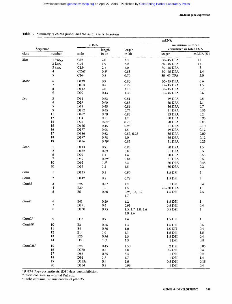
Table 1. Summary of cDNA probes and transcripts in G. h i r s u t u m
cDNA Sequence length
class n u m b e r code in kb length in kb
m R N A
m a x i m u m n u m b e r abundance in total RNA
stage a mKNA 1%J
Mat 1 VicAB C72 2.0 2.3 3 0 - 4 5 DPA 2 Leg a C94 1.9 2.0 3 0 - 4 5 DPA 3 Leg s C134 2.1 2.0 3 0 - 4 5 DPA 4 CD67 0.8 b 0.85 3 0 - 4 5 DPA 5 C164 0.8 0.70 3 0 - 4 5 DPA
MatP 6 D 129 0.9 0.90 3 0 - 4 5 DPA 7 D103 0.8 0.78 3 5 - 4 5 DPA 8 D112 2.0 2.15 3 0 - 4 5 DPA 9 D99 0.43 1.35 3 0 - 4 5 DPA
Lea 3 D11 0.62 0.81 49 DPA 4 D19 0.90 0.85 50 DPA 5 D73 0.65 0.86 56 DPA 9 D152 0.65 0.75 51 DPA
11 D102 0.70 0.83 53 DPA 12 D34 0.51 1.2 50 DPA 14 D95 0.62 c 0.75 50 DPA 15 D156 0.45 0.95 51 DPA 16 D177 0.95 1.5 49 DPA 17 D186 0.62 0.82, 0.98 56 DPA 18 D197 0.78 2.0 56 DPA 19 D176 0.79 b 0.85 51 DPA
LeaA 1 D113 0.91 0.95 50 DPA 2 D132 0.69 0.85 51 DPA 6 D29 1.1 1.4 50 DPA 7 D49 0.69 b 0.84 51 DPA
10 D92 1.3 b 2.3 50 DPA 20 D16 1.2 1.5 50 DPA
Grin 1 D125 0.5 0.90 1.5 DPI
GrmC 2 D142 0.8 0.78 1.5 DPI
GrmM 3 E26 0.37 2.2 1 DPI 4 E39 1.5 1.5 2 5 - 3 0 DPA 5 E6 0.60 0.95, 1.4, 1.7 1.5 DPI
1.7
GrmP
GrmCP
GrmMP
GrmCMP
6 E41 0.29 7 D171 0.6 8 D130 0.75
9 D38 0.9
10 E2 0.56 1.3 11 E4 0.70 1.0 12 El4 1.0 1.1 13 E25 0.96 1.5 14 D30 2.0 b 2.3
15 E28 0.45 1.50 16 D78b 0.8 2.8 17 D85 0.75 3.2 18 D91 1.7 1.7 19 D153a 0.4 2.0 20 D154 0.5 0.96
1.2 0.95 1.5, 1.7, 2.0, 2.6 2.0, 2.6
2.4
1.5 DPI 0.5 DPI 0.5 DPI
1.5 DPI
1.5 DPI 1.5 DPI 1.5 DPI 1.5 DPI
1 DPI
2 DPI 0.5 DPI
1 DPI 1 DPI
0.5 DPI 1 DPI
15 15
5 1.4 2.0
0.6 1.5 0.7 0.6
0.5 2.1 0.7 0.35 0.2 0.95 0.85 0.10 0.12 0.09 0.12 0.25
1.5 0.5 0.55 0.5 0.45 0.2
2
3
0.4 1 2.5
1 0.4 1
1
0.5 0.4 1.5 0.4 0.8
0.05 0.4 0.2 1.4 0.15 0.4
a (DPA) Days postanthesis; {DPI) days post imbibi t ion. b Insert contains an in temal PstI site. c Probe contains 125 nucleot ides of pBR325.
GENES & DEVELOPMENT 359
Cold Spring Harbor Laboratory Press on April 27, 2019 - Published by genesdev.cshlp.orgDownloaded from
Hughes and Galau
z r r
_.J
Z UJ
Z
< < z
Lea
o,o~: :~ :C, ~ . a~,--%? ...... -o-~
D 1 9 7
"- 'T-- - - I - - - - r - - - - I w "I ' OI ~1 I ' I ' 1 ' I ' I
"', LeaA
D l 1 3
' I 0 ~
"Dll
.D176
�9 D 3 4
b . D 1 5 ( , '!
12 , ','
-
I- I m
i �9 a,
D 1 5 ', D
i o - ~ . ci
" " - " 6 - ~ ,11
. - 6 D 1 8 6 ~ " .....
D 1 9 _ ~ .k -
~ I i I a I i I ,t ,
10 20 30 40 50 0 .5 10 20 30 40 50 0 5 10 20 30 40 50 0 5
I-- C O T - 4 - - - - M A T t P A ~ D t G R M }-- C O T I M A T I P A ~ D t G R M F-- C O T - b - - M A T ,', P A - 4 D I G R M
DAY DAY DAY
Figure 1. (Seep. 363 for legend.)
360 GENES & DEVELOPMENT
Cold Spring Harbor Laboratory Press on April 27, 2019 - Published by genesdev.cshlp.orgDownloaded from
Modular g e n e e x p r e s s i o n
< z r r ,._1
UJ
<
7
m <
d ~o
; #
A a,
J
C72 , ! ! i
o
' ; i '~ J J
CD67 l ,4
p,~ . . . . . -0"- # .6
I
Mat
' I ' I ' I ' I ' I '
E
D99
MatP ?"~ _~_-___ , o ~
s / o
/
i q
?
Dl12 / I 'o )
0129
,•,,, - o e~' ~&
D103 i
i i
i 1 , 1 , I i I i l i
F GrmMP
! ,
E4
"(:~ 0 !
,. o
,~ �9
D30 ~ P '
10 20 30 40 50 0 5 10 20 30 40 50 0 5 10 20 30 40 50 0 5
I-COT i MAT I-PA---tD~.GRM t-COT = MAT : PA-ID~GRM I-COT i MAT-------FPA.-*D~GRM DAY DAY DAY
Figure 1. (See p. 363 for legend.)
GENES & D E V E L O P M E N T 361
Cold Spring Harbor Laboratory Press on April 27, 2019 - Published by genesdev.cshlp.orgDownloaded from
H u g h e s a n d G a l a u
o
z
<
z <
' I ' I ' I ' 1 'I ;
G Grm
1 ore- - -~oo ....... o_6.8e~.<~, f
0 1 2 5 ~
GrmP
0171 ~ , ~ l r ~ , _ ~ _ . ~ , _ " 11
GrmC I~,
D142 ~'~',,,~ /
E6
E39
t E,26j
" I ' I ' I 'I '
GrmM
- " O . O
I
1
' I ' I ' I ' L '1 ' 4
I " ~ ~ 1
GrmCPd; ,~',
i �9 / o r I
_~ D38 p !.__o-o- . . . . . "6 ,,
D153,,,,.,,-GrmCMP.,~.J/I _]
o , o
091 Q /
'~~ ..... -o-- o,, :!
'M_ t}!t
E28 ~ ' " ~ " ' - 4 ~ I * ' ' " ____'__ ,, ,} , J
1 0 20 30 40 50 0 5 10 20 30 40 50 0 5 10 20 30 40 50 0 5
I- COT ~ MAT' 0 PA-4D{GRM I-- COT 0 MAT. '~ PA~DtGRM ~ COT 0 - MAT ; PA-4D{GRM DAY DAY DAY
Figure 1. {Seep. 363 for legend.)
3 6 2 G E N E S & D E V E L O P M E N T
Cold Spring Harbor Laboratory Press on April 27, 2019 - Published by genesdev.cshlp.orgDownloaded from
Modular gene expression
sion. Significantly, Lea mRNAs are the only ones whose major abundance is associated only with the postabscis- sion stage or the concentration of free ABA.
R e s u l t s
Isolation of 26 additional mRNA sequences
To complement the 18 Lea, 2 legumin (Leg A and LegB) , and the vicilin (VicaB) cDNA clones already described (Galau et al. 1983, 1986), 26 new eDNA clones were re- covered from cDNA libraries made from cotyledon mRNA of allotetraploid Gossypium hirsutum. These are a random selection of mRNAs that comprise at least 0.2% of cotyledon mRNA between 28 days postanthesis (DPA) and 2 days postimbibition (DPI). Each of the 47 clones recognize different mRNAs at the standard hy- bridization criterion of T m - 15~ They do not cross- hybridize with each other, and in most instances there are significant differences in either the size or expression patterns of the mRNAs to which they hybridize (Table 1; data not shown). Each probe hybridizes to different- sized restriction fragments in genomic DNAs of several cotton species, showing that each is encoded by different genes {Galau et al. 1988; data not shown). Closely re- lated genomes in diploid and allotetraploid species of Gossypium are denoted by letter and are grouped pri- marily on the basis of similar chromosome size and ability to pair during meiosis in interspecific hybrids. Per haploid genome, all Lea and Leg cDNAs recognize only single genes in A or D genorne diploid cottons and two alloallelic genes in AD genome allotetraploids such as G. hirsutum (Galau et al. 1988). This also is true of all 26 new mRNA probes (data not shown). The exception is the VicA8 probe which hybridizes to about three genes in diploids and twice that number in the allotetraploids (Chlan et al. 1987; Galau et al. 1988).
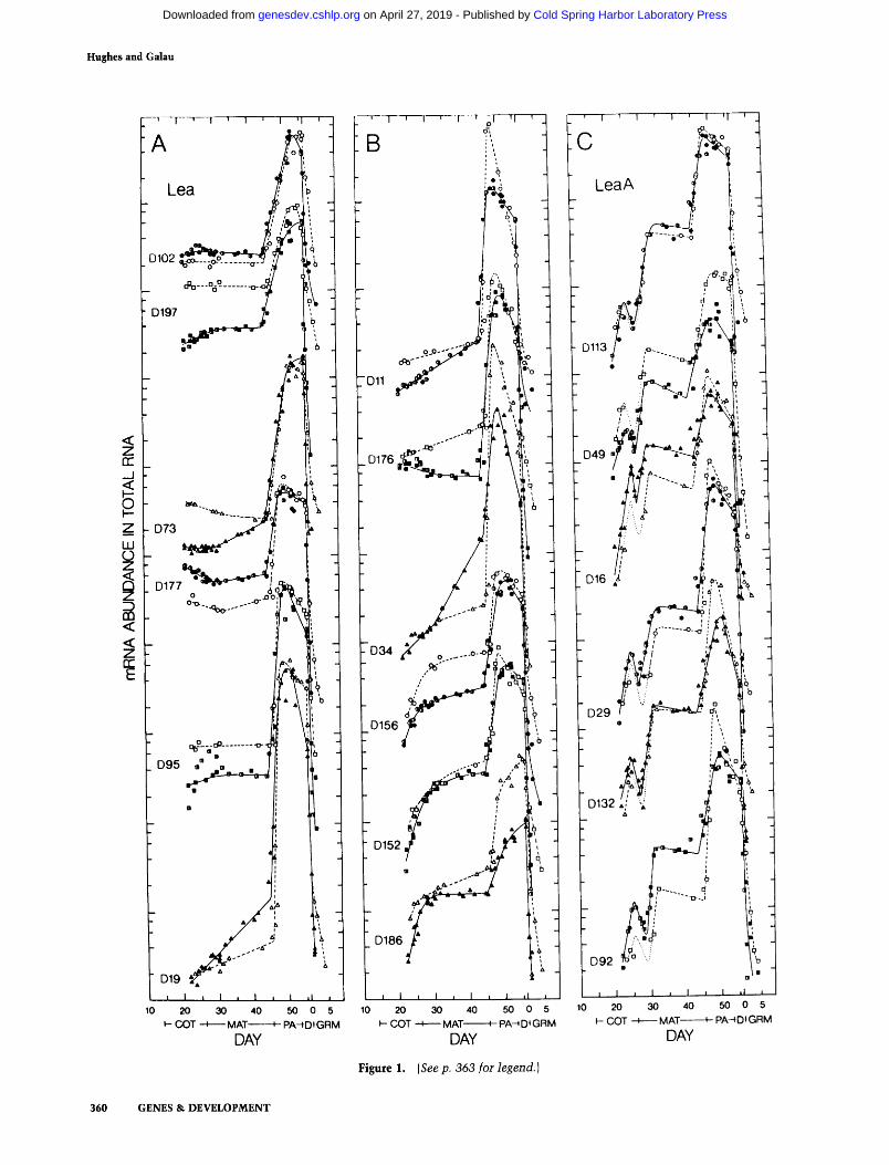
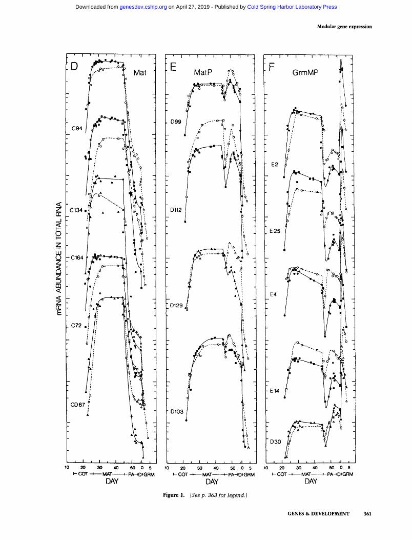
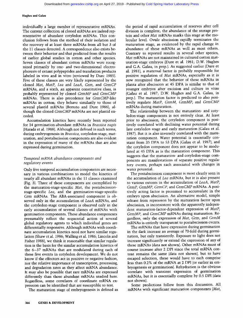
To quantify their mRNA concentrations during devel- opment, all 47 cDNA inserts were each hybridized to RNA dots (Fig. 1) and RNA gel blots (Fig. 2) containing total RNA from cotyledons of G. hirsutum embryos from the.late cotyledon stage (22 DPA, 1 mg dry weight) to the mature embryo stage (56 DPA, 67 mg dry weight), and of seedlings during 4 days of germination in the dark. The two data sets in general agree very well (see below). Table 1 presents a summary of the sizes and maximum abundance of their mRNAs as determined in this study.
Although nearly all the probes recognize single-sized mRNA bands in AD allotetraploid G. hirsutum, D186 detects two bands in G. hirsutum IFig. 2) and one band in the A genome diploid G. arboreum (data not shown). D130 detects two major and two minor bands in G. hir- sutum {Fig. 2) and only one major and one minor band in the diploid (data not shown). With both probes, the two major mRNAs in G. hirsutum have very similar expres-
sion and are almost certainly slightly different-sized mRNAs encoded by the two similarly expressed alloal- leles. The eDNA E6 detects one major and two larger- sized minor bands in the allotetraploid (Fig. 2) and the same major and but only one of the minor bands in the diploid (data not shown). The stoichiometry of the minor and major bands in both species is highly depen- dent on developmental stage (Fig. 2; data not shown). Although other interpretations are not yet excluded, the patterns of E6 expression are consistent with a pre- cursor-product relationship with the extent of pro- cessing of both alloallele transcripts being under similar developmental control. We presume, but in most in- stances have not demonstrated, that both alloalleles of the other genes are active in G. hirsuturn but have mRNAs whose lengths are not distinquished in this gel system.
Many classes of mRNAs share common expression
There are clearly temporal components common to the mRNA accumulation kinetics of many of the classes of mRNAs observed in this study. As outlined below, these are associated with the cotyledon stage, free ABA con- centration, maturation stage, abscission of the ovule, and germination (Fig. 3). Most or all of the complex ac- cumulation kinetics of these mRNAs can be modeled simply by variously combining these five components.
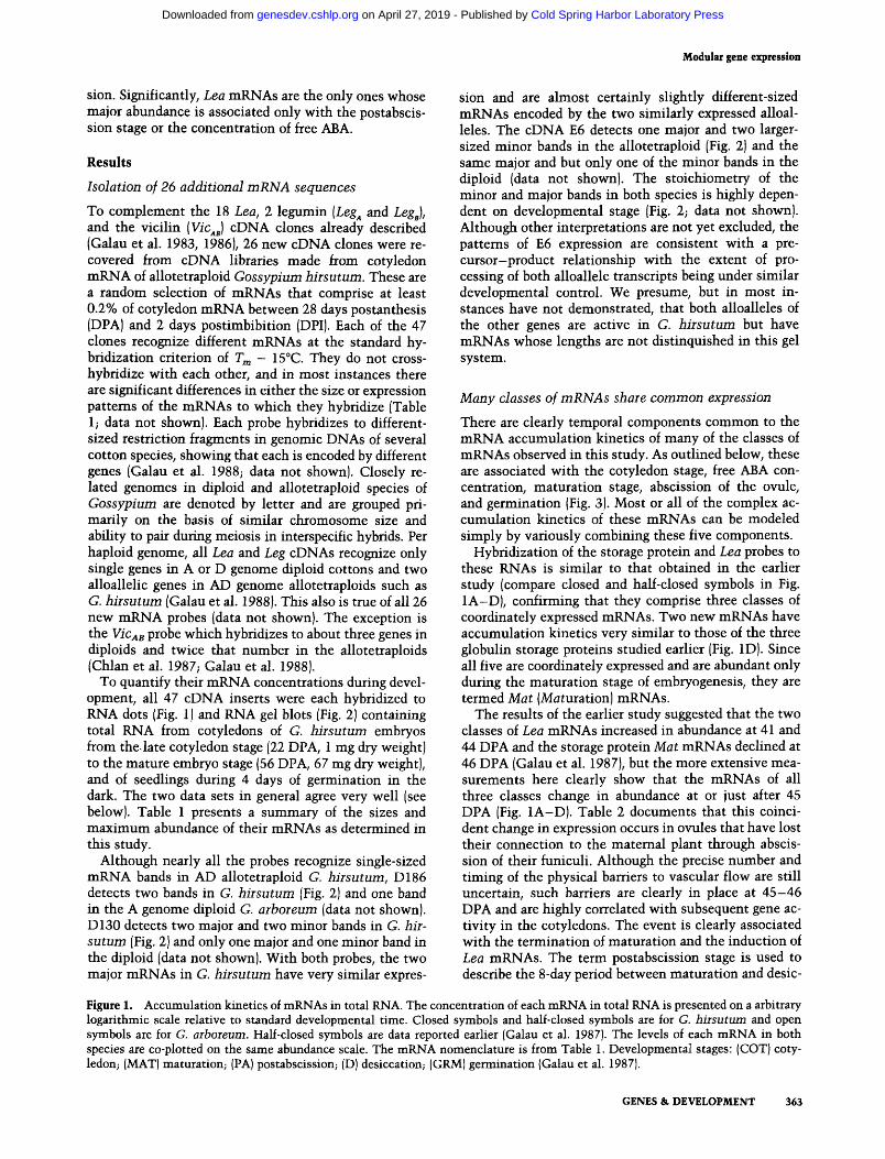
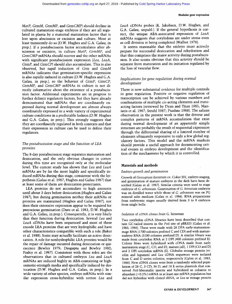
Hybridization of the storage protein and Lea probes to these RNAs is similar to that obtained in the earlier study (compare closed and half-closed symbols in Fig. 1A-D), confirming that they comprise three classes of coordinately expressed mRNAs. Two new mRNAs have accumulation kinetics very similar to those of the three globulin storage proteins studied earlier (Fig. 1D). Since all five are coordinately expressed and are abundant only during the maturation stage of embryogenesis, they are termed Mat (Maturation) mRNAs.
The results of the earlier study suggested that the two classes of Lea mRNAs increased in abundance at 41 and 44 DPA and the storage protein Mat mRNAs declined at 46 DPA (Galau et al. 1987), but the more extensive mea- surements here clearly show that the mRNAs of all three classes change in abundance at or just after 45 DPA (Fig. 1A-D). Table 2 documents that this coinci- dent change in expression occurs in ovules that have lost their connection to the maternal plant through abscis- sion of their funiculi. Although the precise number and timing of the physical barriers to vascular flow are still uncertain, such barriers are dearly in place at 45-46 DPA and are highly correlated with subsequent gene ac- tivity in the cotyledons. The event is clearly associated with the termination of maturation and the induction of Lea mRNAs. The term postabscission stage is used to describe the 8-day period between maturation and desic-
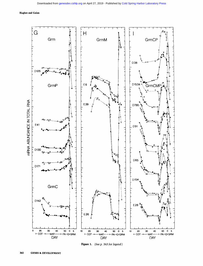
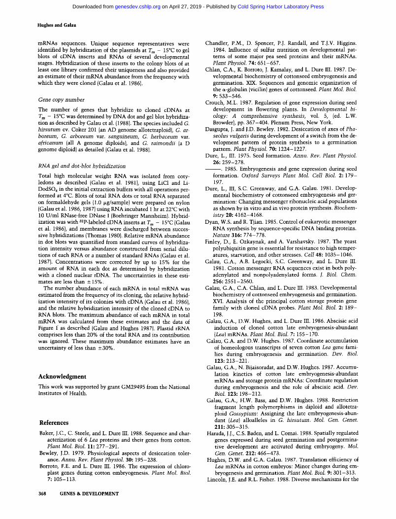
Figure 1. Accumulation kinetics of mRNAs in total RNA. The concentration of each mRNA in total RNA is presented on a arbitrary logarithmic scale relative to standard developmental time. Closed symbols and half-closed symbols are for G. hirsutum and open symbols are for G. arboreum. Half-closed symbols are data reported earlier (Galau et al. 1987). The levels of each mRNA in both species are co-plotted on the same abundance scale. The mRNA nomenclature is from Table 1. Developmental stages: (COT) coty- ledon; (MAT)maturation; (PA)postabscission; {D) desiccation; (GRM)germination {Galau et al. 1987).
GENES & DEVELOPMENT 363
Cold Spring Harbor Laboratory Press on April 27, 2019 - Published by genesdev.cshlp.orgDownloaded from
Hughes and Galau
364 GENES & D E V E L O P M E N T
t ' I ' [ i '1
PA
z "~AL-- CT- [// f 1 _J
O
z_ I11 o / ..i < I / i ! C3
Z *,
, 1 L l , I I ,1 l
10 20 30 40 50 0 5 t- COT t MAT PA-+D~GRM
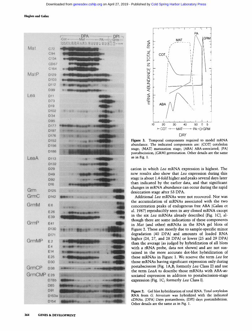
DAY Figure 3. Temporal components required to model mRNA abundance. The indicated components are: (COT) cotyledon stage; (MAT) maturation stage; (ABA)ABA-associated; (PA) postabscission; (GRM) germination. Other details are the same as in Fig. 1.
cation in which Lea mRNA expression is highest. The new results also show that Lea expression during this stage is about 1.4-fold higher and peaks several days later than indicated by the earlier data, and that significant changes in mRNA abundance can occur during the rapid desiccation stage after 53 DPA.
Additional Lea mRNAs were not recovered. Nor was the accumulation of mRNAs associated with the two concentration peaks of endogenous free ABA (Galau et al. 1987) reproducibly seen in any cloned mRNA except in the six Lea mRNAs already described (Fig. 1C), al- though there are some indications of these components in Mat (and other) mRNAs in the RNA gel blots of Figure 2. These are mostly due to sample-specific minor degradation (40 DPA) and amounts of loaded RNA higher (24, 27, and 28 DPA) or lower (25 and 29 DPA) than the average (as judged by hybridization of all blots with a rRNA probe, data not shown) and are not sus- tained in the more accurate dot-blot hybridization of these mRNAs in Figure 1. We reserve the term Lea for those mRNAs having significant expression only during postabscission (Fig. 1A, B; formerly Lea Class II) and use the term LeaA to describe those mRNAs with ABA-as- sociated expression in addition to postabscission-stage expression (Fig. 1 C; formerly Lea Class I).
Figure 2. Gel blot hybridization of total RNA. Total cotyledon RNA from G. hirsutum was hybridized with the indicated cDNAs. {DPA) Days postanthesis; (DPI) days postimbibition. Other details are the same as in Fig. 1.
Cold Spring Harbor Laboratory Press on April 27, 2019 - Published by genesdev.cshlp.orgDownloaded from
Table 2. Coordinate expression of Mat and Lea mRNAs associated with ovule abscission
Modular gene expression
Relative mRNA abundance a
Nominal Ovule Mat Lea LeaA Embryo a DPA b abscission c VicAB C67 C 164 D 11 D 73 D 113 D 132
G. arboreum s6b 42 no 0.98 0.96 0.95 0.0028 0.018 0.054 0.032 a2b 45 no 0.55 r 0.67 e =1.0 0.0033 0.020 0.052 0.049 ~ s2d 46 yes 0.80 0.63 0.66 ~ 0.0040 r 0.021 0.11 e 0.069 s6c 47 yes 0.52 0.91 0.45 0.0053 0.019 0.28 0.086 a4a 46.5 yes 0.077 0.56 0.6 0.014 0.044 ~ 0.26 0.073 a3a 48 yes 0.020 0.021 0.0053 0.80 0.13 0.89 0.72
G. hirsutum 27 40 no 0.97 0.95 1.1 0.013 0.011 0.066 0.098 T9 45 mixed 0.53 e 0.98 0.96 0.017 0.016 0.082 e 0.11 e 21 45 unknown 0.60 0.38 e 0.043 r 0.045 ~ 0.019 ~ 0.16 0.15 T10 46 yes 0.23 0.34 0.074 0.089 0.027 0.22 0.18 T12 47 yes 0.027 0.034 0.0085 0.41 0.073 0.60 0.39
a The embryo preparations are listed in the order of their apparent true age. b Standard days postanthesis as defined primarily by characteristics of the ovule and integuments (Galau et al. 1987), but not by ovule abscission. c Defined as either breakage of the funiculus or the presence of a funicular scar on the micropilar end of the ovule, or both. d Except were indicated, values are normalized to a value of 1.0 for the maximum abundance reached by the mRNA. e Earliest significant change.
Four mRNAs (Fig. 1E) share with Mat mRNAs a high abundance during maturat ion and a rapid decline upon abscission, but these recover in abundance to various extents during the postabscission stage. Since the re- c o v e r / o f these mRNAs during this period is very sim- ilar to that of the postabscission-stage-specific Lea mRNAs, these are termed MatP mRNAs, for mRNAs expressed during both maturat ion and postabscission.
The remaining 20 probes hybridize to mRNAs that are transiently present in relatively high abundance during the first 2 days of germination in the dark. These Grin (Germination) mRNAs are assigned to seven classes of mRNAs. Only the single Grm m R N A D125 has signifi- cant modulat ion only during germination, while addi- tional expression is seen during cotyledon, maturation, or postabscission stages for the single GrmC, three GrmM, and three GrmP mRNAs, respectively (Fig. 1G, H).
Finally, several mRNAs exhibit more than two abun- dance components. These include the five GrmMP mRNAs which are expressed during maturation, postab- scission, and germinat ion (Fig. IF), the single GrmCP m R N A which is expressed during cotyledon, postabscis- sion, and germination stages {Fig. lI), and six GrmCMP mRNAs which are modulated during cotyledon, matura- tion, postabscission, and germination stages (Fig. lI).
Essentially all of the complex accumulat ion kinetics of MatP, GrmM, GrmP, and GrmMP mRNAs can be modeled s imply by combining the fundamental pat tems exemplified by the accumulat ion kinetics of Mat, Lea, and Grm mRNAs. Of the 47 mRNAs examined, 23 are modulated during maturation, 37 during postabscission, and 20 during germination. Neither the ABA-associated component nor the cotyledon stage component are found isolated in the behavior of any m R N A examined; the ABA-associated component is seen only in the six
LeaA mRNAs, and the cotyledon-stage component is seen in the eight GrmC, GrmCP, and GrmCMP mRNAs. With few exceptions, upon deconvolution the kinetics of all components are remarkably similar across all mRNAs.
Individual genes have complex expression
The complex kinetics seen with many of the mRNAs in G. hirsutum could be due to the summat ion of funda- menta l ly different expression of the two alloalleles en- coding them. In this species, however, alloallele tran- scripts of two Lea and five LeaA alloallele pairs have similar expression at the m R N A level (Galau and Hughes 1987), and members of several noncloned alloal- lelic pairs have similar expression at the m R N A or pro- tein synthesis levels (Galau and Hughes 1987; Hughes and Galau 1987). Other than for D186, D130, and E6 noted above, alloallele-specific expression of additional cloned mRNAs has not been measured in the allotetra- ploid. However, the mRNAs encoded by all 46 single- copy homologs in the diploid G. arboreum (open symbols, Fig. 1) appear to have the same component ex- pression as seen in the allotetraploid (closed and half- closed symbols, Fig. 1), although the data are not suffi- cient to resolve the first peak in the ABA component in LeaA mRNAs. Expression in G. arboreum thus confirms that complex expression pat tems are intr insic to indi- vidual genes, even if this should not prove to be true for all these genes in G. hirsutum.
Discuss ion
The cloned mRNAs are typical mRNAs
A major rationale in this work was that a study of global regulatory events could be approximated by examining
GENES & DEVELOPMENT 365
Cold Spring Harbor Laboratory Press on April 27, 2019 - Published by genesdev.cshlp.orgDownloaded from
Hughes and Galau
individually a large number of representative mRNAs. The current collection of cloned mRNAs are indeed rep- resentative of abundant cotyledon mRNAs. This con- clusion follows from the method of their isolation and the recovery of at least three mRNAs from all but 3 of the 11 classes detected. A correspondence also exists be- tween their behavior and that predicted from the results of earlier global studies in cotton and other species. Seven classes of abundant cotton mRNAs were recog- nized primarily by one- and two-dimensional electro- phoresis of extant polypeptides and translation products labeled in vivo and in vitro (reviewed by Dure 1985). Five of these classes are very likely represented by the cloned Mat, MatP, Lea and LeaA, Grin, and GrmCP mRNAs, and a sixth, an apparent constitutive class, is probably represented by cloned GrmMP and GrmCMP mRNAs. There is also precedence for GrmCMP-like mRNAs in cotton; they behave similarily to those of several plastid mRNAs (Borroto and Dure 1986), al- though the cloned Grm CMP mRNAs are not plastid en- coded.
Accumulation kinetics have recently been reported for 14 germination-abundant mRNAs in Brassica napis (Harada et al. 1988). Although not defined in such terms, during embryogenesis in Brassica, cotyledon-stage, mat- uration, and postabscission components are also evident in the expression of many of the mRNAs that are also expressed during germination.
Temporal mRNA abundance components and regulatory events
Only five temporal accumulation components are neces- sary in various combinations to model the kinetics of nearly all abundant mRNAs in the 11 classes examined (Fig. 3). Three of these components are exemplified by the maturation-stage-specific Mat, the postabscission- stage-specific Lea, and the germination-stage-specific Grm mRNAs. The ABA-associated component is ob- served only in the accumulation of LeaA mRNAs, and the cotyledon-stage component is observed only in the early accumulation of several classes of mRNAs with germination components. These abundance components presumably reflect the sequential action of several global regulatory agents to which individual genes are differentially responsive. Although mRNAs with coordi- nate accumulation kinetics need not have similar regu- lation (Shaw et al. 1986; Walling et al. 1986; Lincoln and Fisher 1988), we think it reasonable that similar regula- tion is the basis for the similar accumulation kinetics of the 6 -37 mRNAs that are modulated during each of these five events in cotyledon development. We do not know if the effectors act in positive or negative fashion, nor the relative importance of transcription, processing, and degradation rates as they affect mRNA abundance. It may also be possible that rare mRNAs are expressed differently than those abundant mRNAs studied here. Regardless, some correlates of coordinate mRNA ex- pression can be identified that are susceptible to test.
The maturation stage of embryogenesis is defined as
the period of rapid accumulation of reserves after cell division is complete; the abundance of the storage pro- tein and other Mat mRNAs marks this stage at the mo- lecular level. Ovule abscission rapidly terminates the maturation stage, as evidenced by the rapid change in abundance of these mRNAs as well as most others. Contrary to reported results in several other species, Mat mRNAs are not maintained in cultured cotton mat- uration-stage embryos (Dure et al. 1981; D.W. Hughes and G.A. Galau, in prep.). As suggested earlier (Dure et al. 1981), a maternal factor is probably responsible for positive regulation of Mat mRNAs, especially as it is now recognized that the behavior of these mRNAs in planta after abscission at 45 DPA is similar to that of younger embryos after excision and culture in vitro (Galau et al. 1987; D.W. Hughes and G.A. Galau, in prep.). The maturation factor is predicted to also posi- tively regulate MatP, GrmM, GrmMP, and GrmCMP mRNAs during maturation.
The relationship between the maturation- and coty- ledon-stage components is not entirely clear. At least prior to abscission, the cotyledon component is posi- tively correlated with declining water potential during late cotyledon stage and early maturation (Galau et al. 1987). But it is also inversely correlated with the matu- ration component. Water potential is essentially con- stant from 35 DPA to 53 DPA (Galau et al. 1987), and the cotyledon component does not appear to be modu- lated at 45 DPA as is the maturation component. This suggests that the maturation- and cotyledon-stage com- ponents are manifestations of separate positive regula- tory events, perhaps each associated with changes in water potential.
The postabscission component is most clearly seen in the accumulation of Lea mRNAs, but it is also present to various extents in the accumulation of LeaA, MatP, GrmP, GrmMP, Grin CP, and Grin CMP mRNAs. A posi- tively acting factor is presumed to accumulate in the embryo upon abscission. An alternative model, simple release from repression by the maturation factor upon abscission, is inconsistent with the apparently indepen- dent maturation-factor-dependent expression of MatP, GrmMP, and Grm CMP mRNAs during maturation. Re- gardless, only the expression of Mat, Grm, and GrmM mRNAs is entirely transparent to postabscission events.
The mRNAs that have expression during germination in the dark increase an average of 70-fold during germi- nation, but only transiently. Exposure to light does not increase significantly or extend the expression of any of these mRNAs (data not shown). Other mRNAs must of course increase after 2 DPI since the total mRNA con- tent remains the same (data not shown), but to have escaped selection, these would have to each comprise less than 0.2% of the mRNA at 2 DPI (or earlier in em- bryogenesis or germination). Rehydration is the obvious correlate with transient expression of germination mRNAs, but it is essentially complete by 0.5 DPI (data not shown).
Some predictions follow from this discussion. All mRNAs with significant maturation components (Mat,
366 GENES & DEVELOPMENT
Cold Spring Harbor Laboratory Press on April 27, 2019 - Published by genesdev.cshlp.orgDownloaded from
Modular gene expression
MatP, GrmM, GrmMP, and GrmCMP) should decline in cultured maturation-stage embryos if they are all regu- lated in planta by a maternal maturation factor that is lost upon abscission or excision and culture. Most at least transiently do so (D.W. Hughes and G.A. Galau, in prep.). If a postabscission factor accumulates after ab- scission or excision, in culture MatP, GrmMP, and Grin CMP mRNAs should recover and the other mRNAs with significant postabscission expression (Lea, LeaA, GrmP, and GrmCP) should also accumulate. This is also observed, but rapid induction of Grm and GrmM mRNAs indicates that germination-specific expression is also rapidly induced in culture (D.W. Hughes and G.A. Galau, in prep.), so the behavior of GrmP, GrmCP, GrmMP, and GrmCMP mRNAs in culture is not di- rectly informative about the existence of a postabscis- sion factor. Additional experiments are in progress to further delimit regulatory factors, but they have already demonstrated that mRNAs that are coordinately ex- pressed during normal development are almost always coordinately expressed in a wide variety of experimental culture conditions in a predictable fashion (D.W. Hughes and G.A. Galau, in prep.). This strongly suggests that they are coordinately regulated in both contexts and that their expression in culture can be used to define their regulators.
The postabscission stage and the function of LEA proteins
The 8-day postabscission stage separates maturation and desiccation, and the only obvious changes in cotton during this time are recognized only at the molecular level. The current study has shown that Lea and LeaA mRNAs are by far the most highly and specifically in- duced mRNAs during this stage, consistent with the hy- pothesis (Galau et al. 1987; Hughes and Galau 1987) that at least some of them are desiccation protectants.
LEA proteins do not accumulate to high amounts until about 3 days before desiccation (Hughes and Galau 1987), but during germination neither their mRNAs or proteins are maintained (Hughes and Galau 1987), nor does their extensive expression appear to be required for precocious germination (Dure et al. 1981; D.W. Hughes and G.A. Galau, in prep.). Consequently, it is very likely that they function during desiccation. Several Lea and LeaA cDNAs have been sequenced and most of these encode LEA proteins that are very hydrophilic and have other characteristics compatible with such a role (Baker et al. 1988). Some may actually facilitate an active desic- cation. A role for nonhydrophilic LEA proteins would be the repair of damage incurred during desiccation or qui- escence (Bewley 1979; Dasgupta and Bewley 1982; Finley et al. 19871. Consistent with these roles are the observations that in cultured embryos Lea and LeaA mRNAs are induced highly in ABA-containing or high- osmotic-strength media and specifically induced by des- iccation {D.W. Hughes and G.A. Galau, in prep.). In a wide variety of other species, embryo mRNAs with sim- ilar expression cross-hybridize with cotton Lea and
LeaA cDNAs probes (K. Jakobsen, D.W. Hughes, and G.A. Galau, unpubl.). If the general hypothesis is cor- rect, the unique ABA-associated expression of LeaA mRNAs suggests that cotyledons are under stress even as cell division is being completed (Walbot 1978).
It seems reasonable that the embryo must actively prepare for successful desiccation and rehydration and that this comprises the major activity during postabscis- sion. It also seems obvious that this activity should be separate from maturation and its initiation regulated by the loss of vascular flow.
a
Implications for gene regulation during normal developm en t
There is now substantial evidence for multiple controls in gene regulation. Positive or negative regulation of transcription can be achieved by various numbers and combinations of multiple cis-acting elements and trans- acting factors (reviewed by Dyan and Tjian 1985; Man- iatis et al. 1987; Struhl 1987; Ptashne 1988). The salient observation in the present work is that the diverse and complex patterns of mRNA accumulation that exist during normal development of an apparently simple structure are probably the result of sequential expression through the differential sharing of a limited number of elements ultimately responsive to only a few global reg- ulatory factors. This model and the mRNA markers should provide a useful approach for documenting crit- ical events in embryo development and the identifica- tion of the mechanisms by which it is controlled.
Mater ia l s and m e t h o d s
Embryo growth and germination
Growth of Gossypium hirsutum cv. Coker 201, embryo staging, and germination of mature embryos in the dark have been de- scribed (Galau et al. 1987). Similar criteria were used to stage embryos of G. arboreum. Germination of G. hirsutum embryos was on distilled water while those of G. arboreum was on PG mineral salts medium (Galau et al. 1986). RNA preparations from embryonic stages usually derived from 3 to 8 embryos from single bolls.
Isolation of cDNA clones from G. hirsutum
Two cotyledon cDNA libraries have been described that con- tain GC-tailed inserts in the PstI site of pBR325 (Galau et al. 1983, 1986}. These were made with 28 DPA early-maturation- stage RNA {1700 colonies prefixed C and CD) and with mature- embryo RNA {3100 colonies prefixed D). A similar library was made from cotyledon RNA at 2 DPI (400 colonies prefixed E). Colony blots were hybridized with cDNA made from early maturation stage (C, CD, and D), mature {all), 1 DPI (CD and D) and 2 DPI cotyledon mRNA (E). Globulin storage protein {vi- cilin and legumin) and Lea cDNA sequences were isolated from C and D series colonies, respectively (Galau et al. 1983, 1986). New cDNA clones were from a randomly selected popu- lation of 28 C, 3 CD, 36 D, and 18 E series plasmids that con- tained PstI-liberatable inserts and hybridized as colonies to abundant (>0.2%) mRNA in at least one mRNA population but did not hybridize with cloned rRNA, or Lea or storage protein
GENES & DEVELOPMENT 367
Cold Spring Harbor Laboratory Press on April 27, 2019 - Published by genesdev.cshlp.orgDownloaded from
Hughes and Galau
mRNAs sequences. Unique sequence representatives were identified by hybridization of the plasmids at T m - 15~ to gel blots of cDNA inserts and RNAs of several developmental stages. Hybridization of these inserts to the colony blots of at least one library confirmed their uniqueness and also provided an estimate of their mRNA abundance from the frequency with which they were cloned (Galau et al. 19861.
Gene copy number
The number of genes that hybridize to cloned cDNAs at T~ - 15~ was determined by DNA dot and gel blot hybridiza- tion as described by Galau et al. {1988). The species included G. hirsutum cv. Coker 201 {an AD genome allotetraploid), G. ar- boreum, G. arboreum var. sanguineum, G. herbaceum var. africanum (all A genome diploidsl, and G. raimondii (a D genome diploid] as detailed {Galau et al. 19881.
RNA gel and dot-blot hybridization
Total high molecular weight RNA was isolated from coty- ledons as described (Galau et al. 1981), using LiC1 and Li- DodSO4 in the initial extraction buffers with all operations per- formed at 4~ Blots of total RNA dots or total RNA separated on formaldehyde gels (1.0 lag/sample) were prepared on nylon (Galau et al. 1986, 1987) using RNA incubated 1 hr at 22~ with 10 U/ml RNase-free DNase I {Boehringer Mannheim). Hybrid- ization was with a2p-labeled cDNA inserts at T m - 15~ (Galau et al. 1986), and membranes were discharged between succes- sive hybridizations (Thomas 1980)�9 Relative mRNA abundance in dot blots was quantified from standard curves of hybridiza- tion intensity versus abundance constructed from serial dilu- tions of each RNA or a number of standard RNAs (Galau et al. 1987). Concentrations were corrected by up to 15% for the amount of RNA in each dot as determined by hybridization with a cloned nuclear rDNA. The uncertainties in these esti- mates are less than __ 15%.
The number abundance of each mRNA in total mRNA was estimated from the frequency of its cloning, the relative hybrid- ization intensity of its colonies with cDNA (Galau et al. 1986), and the relative hybridization intensity of the cloned cDNA to RNA blots. The maximum abundance of each mRNA in total mRNA was calculated from these estimates and the data of Figure f as described (Galau and Hughes 1987). Plastid rRNA comprises less than 20% of the total RNA and its contribution was ignored. These maximum abundance estimates have an uncertainty of less than __ 30%.
A c k n o w l e d g m e n t
This work was supported by grant GM29495 from the National Institutes of Health.
References
Baker, J.C., C. Steele, and L. Dure III. 1988. Sequence and char- acterization of 6 Lea proteins and their genes from cotton�9 Plant Mol. Biol. 11: 277-291.
Bewley, J.D. 1979. Physiological aspects of desiccation toler- ance. Annu. Rev. Plant Physiol. 30: 195-238.
Borroto, F.E. and L. Dure III. 1986. The expression of chloro- plast genes during cotton embryogenesis. Plant Moi. Biol. 7: 105-113.
368 GENES & DEVELOPMENT
Chandler, P.M., D. Spencer, P.J. Randall, and T.J.V. Higgins. 1984. Influence of sulfur nutrition on developmental pat- tems of some major pea seed proteins and their mRNAs. Hant Physiol. 74: 651-657.
Chlan, C.A., K. Borroto, J. Kamalay, and L. Dure III. 1987. De- velopmental biochemistry of cottonseed embryogenesis and germination. XIX. Sequences and genomic organization of the a-globulin (vicilin) genes of cottonseed�9 Hant Mol. Biol. 9: 533-546.
Crouch, M.L. 1987. Regulation of gene expression during seed development in flowering plants. In Developmental bi- ology: A comprehensive synthesis, vol. 5, (ed. L.W. Browder), pp. 367-404. Plenum Press, New York.
Dasgupta, J. and J.D. Bewley. 1982. Desiccation of axes of Pha- seolus vulgarJs during development of a switch from the de- velopment pattern of protein synthesis to a germination pattem. Plant Physiol. 70: 1224-1227.
Dure, L., III. 1975. Seed formation. Annu. Rev. Plant Physiol. 26: 259-278.
�9 1985. Embryogenesis and gene expression during seed formation. Oxford Surveys Plant Mol. Cell Biol. 2: 179- 197.
Dure, L., III, S.C. Greenway, and G.A. Galau. 1981. Develop- mental biochemistry of cottonseed embryogenesis and ger- mination: Changing messenger ribonucleic acid populations as shown by in vitro and in vivo protein synthesis. Biochem- istry 20: 4162-4168.
Dyan, W.S. and R. Tjian. 1985. Control of eukaryotic messenger RNA synthesis by sequence-specific DNA binding proteins. Nature 316: 774-778.
Finley, D., E. Ozkaynak, and A. Varshavsky. 1987. The yeast polyubiquitin gene is essential for resistance to high temper- atures, starvation, and other stresses. Cell 48: 1035-1046.
Galau, G.A., A.B. Legocki, S.C. Greenway, and L. Dure III. 1981. Cotton messenger RNA sequences exist in both poly- adenylated and nonpolyadenylated forms�9 J. Biol. Chem. 256: 2551-2560.
Galau, G.A., C.A. Chlan, and L. Dure III. 1983. Developmental biochemistry of cottonseed embryogenesis and germination. XVI. Analysis of the principal cotton storage protein gene family with cloned eDNA probes. Plant Mol. Biol. 2: 189- 198.
Galau, G.A., D.W. Hughes, and L. Dure III. 1986. Abscisic acid induction of cloned cotton late embryogenesis-abundant {Lea) mRNAs. Plant Mol. Biol. 7: 155-170.
Galau, G.A. and D.W. Hughes. 1987. Coordinate accumulation of homeologous transcripts of seven cotton Lea gene fami- lies during embryogenesis and germination�9 Dev. Biol. 123: 213-221.
Galau, G.A., N. Bijaisoradat, and D.W. Hughes. 1987. Accumu- lation kinetics of cotton late embryogenesis-abundant mRNAs and storage protein mRNAs: Coordinate regulation during embryogenesis and the role of abscisic acid. Dev. Biol. 123: 198-212.
Galau, G.A., H.W. Bass, and D.W. Hughes�9 1988. Restriction fragment length polymorphisms in diploid and allotetra- ploid Gossypium: Assigning the late embryogenesis-abun- dant (Lea) alloalleles in G. hirsutum. Mol. Gen. Genet. 211: 305-315.
Harada, J.J., C.S. Baden, and L. Comai. 1988. Spatially regulated genes expressed during seed germination and postgermina- tive development are activated during embryogeny. Mol. Gen. Genet. 212: 466-473.
Hughes, D.W. and G.A. Galau. 1987. Translation efficiency of Lea mRNAs in cotton embryos: Minor changes during em- bryogenesis and germination. Plant Mol. Biol. 9: 301-313.
Lincoln, J.E. and R.L. Fisher�9 1988. Diverse mechanisms for the
Cold Spring Harbor Laboratory Press on April 27, 2019 - Published by genesdev.cshlp.orgDownloaded from
Modular gene expression
regulation of ethylene-inducible gene expression. Mol. Gen. Genet. 212: 71-75.
Maniatis, T., S. Goodboum, and J.A. Fischer. 1987. Regulation of inducible and tissue-specific gene expression. Science 236: 1237-1245.
Ptashne, M. 1988. How eukaryotic transcriptional activators work. Nature 335: 683-689.
Quatrano, R.S. 1986. Regulation of gene expression by abscisic acid during angiosperm embryo development. Oxford Surveys Plant Mol. Cell Biol. 3: 467-477.
Raghaven, V. 1986. Embryogenesis in angiosperms: A develop- mental and experimental study. Cambridge University Press, New York.
Shaw, P., B. Sordat, and U. Schibler. 1986. Developmental coor- dination of a-amylase and psp gene expression during mouse parotid gland differentiation is controlled posttranscription- ally. Cell 47: 107-112.
Struhl, K. 1987. Promoters, activator proteins, and the mecha- nism of transcriptional initiation in yeast. Cell 49: 295- 297.
Thomas, P.S. 1980. Hybridization of denatured RNA and small DNA fragments transferred to nitrocellulose. Proc. Natl. Acad. Sci. 77: 5201-5205.
Walbot, V. 1978. Control mechanisms for plant embryogeny. In Dormancy and developmental arrest led. M. W. Clutterl, pp. 113-166. Academic Press, New York.
Walling, L., G.N. Drews, and R.B. Goldberg. 1986. Transcrip- tional and post-transcriptional regulation of soybean seed protein mRNA levels. Proc. Natl. Acad. Sci. 83: 2123-2127.
Zeevaart, J.A.D. and R.A. Creelman. 1988. Metabolism and physiology of abscisic acid. Annu. Rev. Plant Physiol. Plant Mol. Biol. 39: 439-473.
GENES & DEVELOPMENT 369
Cold Spring Harbor Laboratory Press on April 27, 2019 - Published by genesdev.cshlp.orgDownloaded from
10.1101/gad.3.3.358Access the most recent version at doi: 3:1989, Genes Dev.
D W Hughes and G A Galau Temporally modular gene expression during cotyledon development.
References
http://genesdev.cshlp.org/content/3/3/358.full.html#ref-list-1
This article cites 28 articles, 5 of which can be accessed free at:
License
ServiceEmail Alerting
click here.right corner of the article or
Receive free email alerts when new articles cite this article - sign up in the box at the top
Copyright © Cold Spring Harbor Laboratory Press
Cold Spring Harbor Laboratory Press on April 27, 2019 - Published by genesdev.cshlp.orgDownloaded from


![Gene Discovery of Modular Diterpene Metabolism in · Gene Discovery of Modular Diterpene Metabolism in Nonmodel Systems1[W][OA] Philipp Zerbe2, Björn Hamberger2, Macaire M.S. Yuen,](https://img.dokumen.tips/doc/110x75/60532f1ece0faa29206f9da2/gene-discovery-of-modular-diterpene-metabolism-in-gene-discovery-of-modular-diterpene.jpg)