Embed Size (px)
Citation preview
TECHNIQUES OF ABSOLUTE AND RELATIVE STABILITY INCLUDING
EXTERNAL FIXATION
PRESENTER:DR.MUNENEFACILITATOR:DR.MUTISO
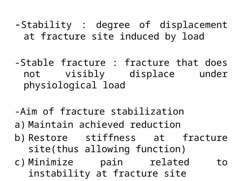
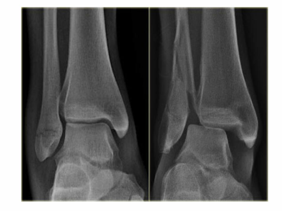
-Stability : degree of displacement at fracture site induced by load
-Stable fracture : fracture that does not visibly displace under physiological load
-Aim of fracture stabilizationa) Maintain achieved reductionb) Restore stiffness at fracture site(thus allowing
function)c) Minimize pain related to instability at fracture site

-Fracture fixation with absolute stability-there’s no micro-motion at the fracture site under physiological load
-This reduces mechanical stimulus for callus formation
-Fixation with relative stability-aims to maintain reduction and still keep mechanical stimulation for callus formation
-Displacement occurring under load is elastic(reversible)

ABSOLUTE FIXATION-It aims to provide a mechanically neutral environment for fracture healing
-Lack of micromotion results in primary bone healing mechanical stimulus for repair by callus formation
-This also reduces mechanical stimulus for repair by callus formation
-Hence implant must provide and maintain absolute stability for prolonged periods of time
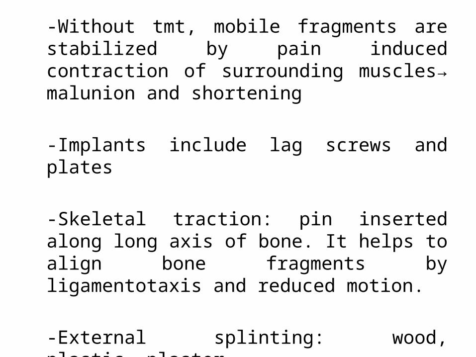
-Without tmt, mobile fragments are stabilized by pain induced contraction of surrounding muscles→ malunion and shortening
-Implants include lag screws and plates
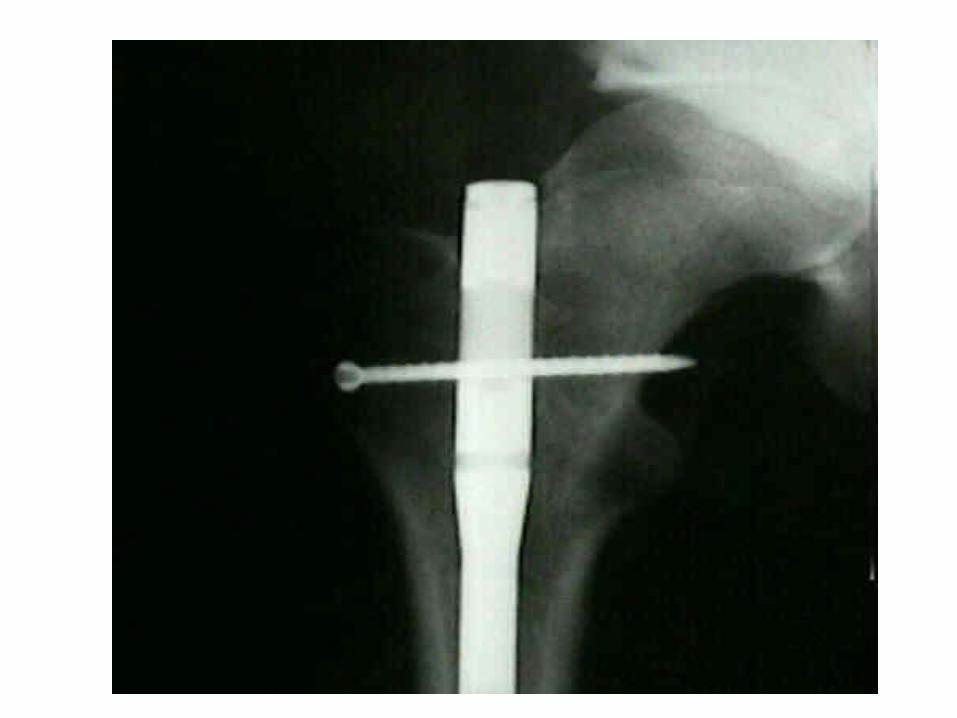
-Skeletal traction: pin inserted along long axis of bone. It helps to align bone fragments by ligamentotaxis and reduced motion.
-External splinting: wood, plastic ,plaster
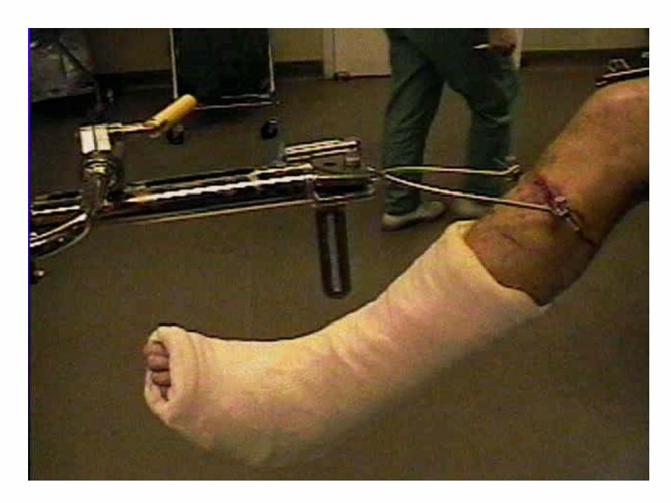
Skeletal Traction
-Traction on a bone structure by means of a pin or wire surgically inserted into the bone.
- continuous traction is desired to immobilize, position, and align a fractured bone properly during the healing process
Aim of Skeletal Traction• regain normal length and alignment of involved bone
• lessen or eliminate muscle spasms
• relieve pressure on nerves, especially spinal and
• prevent or reduce skeletal deformities or muscle contractures
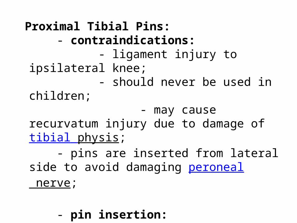
Proximal Tibial Pins: - contraindications: - ligament injury to ipsilateral knee; - should never be used in children; - may cause recurvatum injury due to damage of tibial physis; - pins are inserted from lateral side to avoid damaging peroneal nerve;
- pin insertion: proper insertion site: 2.5 cm posterior to & 2.5 cm distal to tibial tubercle; - landmark is to place pin one to two finger breaths below tibial tuberosity in the midportion of the tibia;
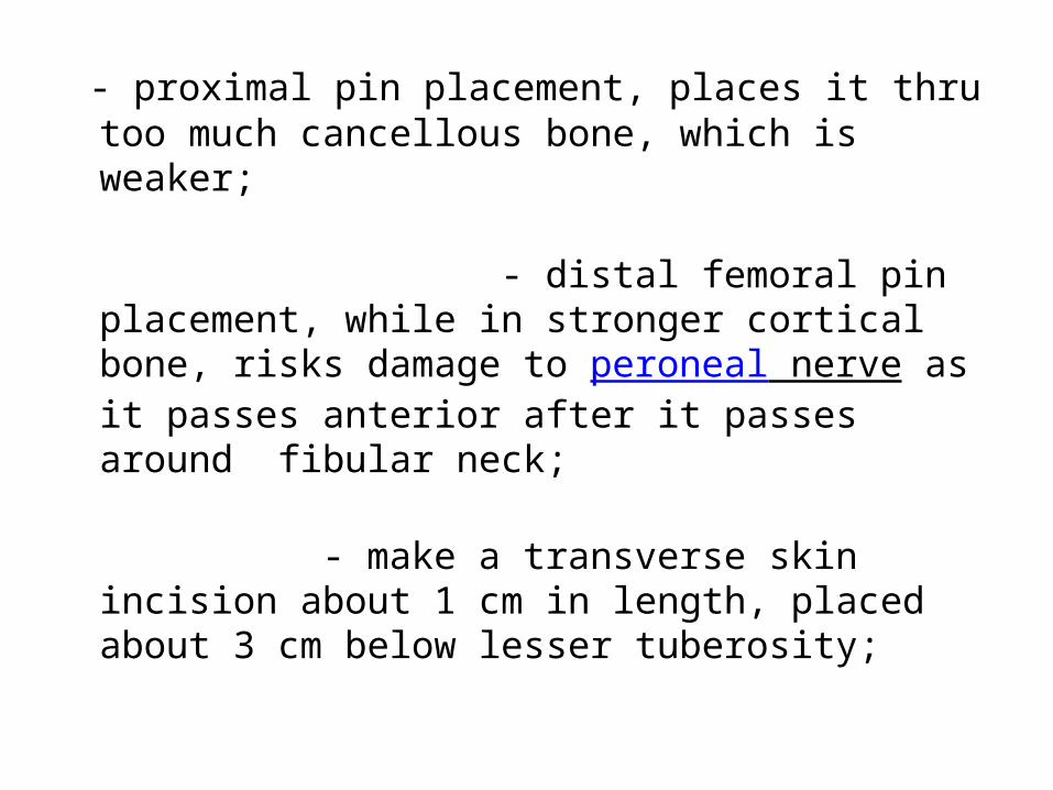
- proximal pin placement, places it thru too much cancellous bone, which is weaker;
- distal femoral pin placement, while in stronger cortical bone, risks damage to peroneal nerve as it passes anterior after it passes around fibular neck;
- make a transverse skin incision about 1 cm in length, placed about 3 cm below lesser tuberosity;
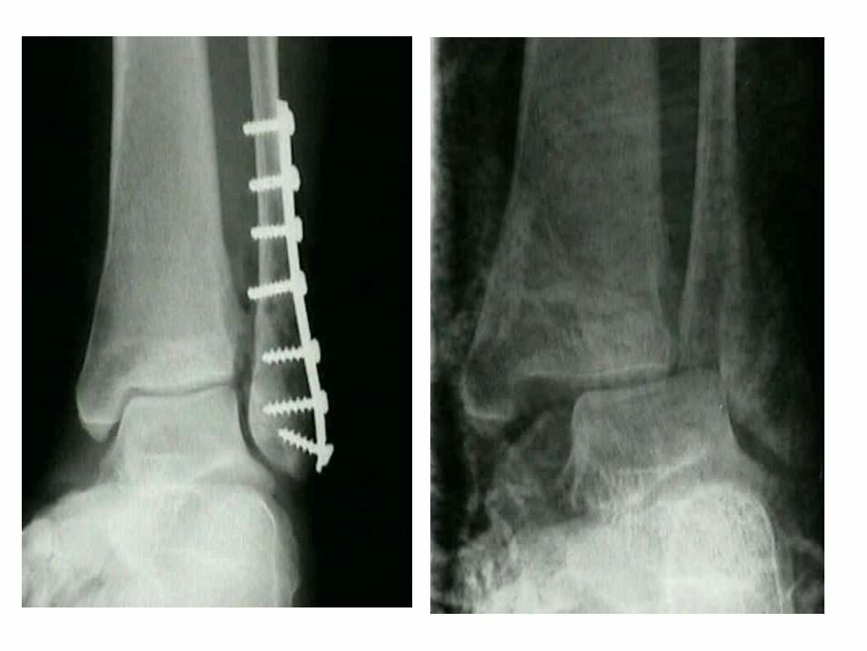
Lag Screws
-Stabilizes fracture by compression alone
- Oblique, non comminuted fractures in bones which are not osteoporotic
- Involves placement of one or more screws across an osteotomy site to achieve inter-fragmentary compression
-lag screw is best positioned at right angles to the fractures plane;
Advantages-Allow for a smaller incision-Don’t have to be removed-Don’t interfere with sydesmotic screws if needed
Disadvantages-lever arm is too small to resist functional
loading(bending/ shearing). Therefore combined with a plate to protect them from these forces
-Lack of tolerance to single overload


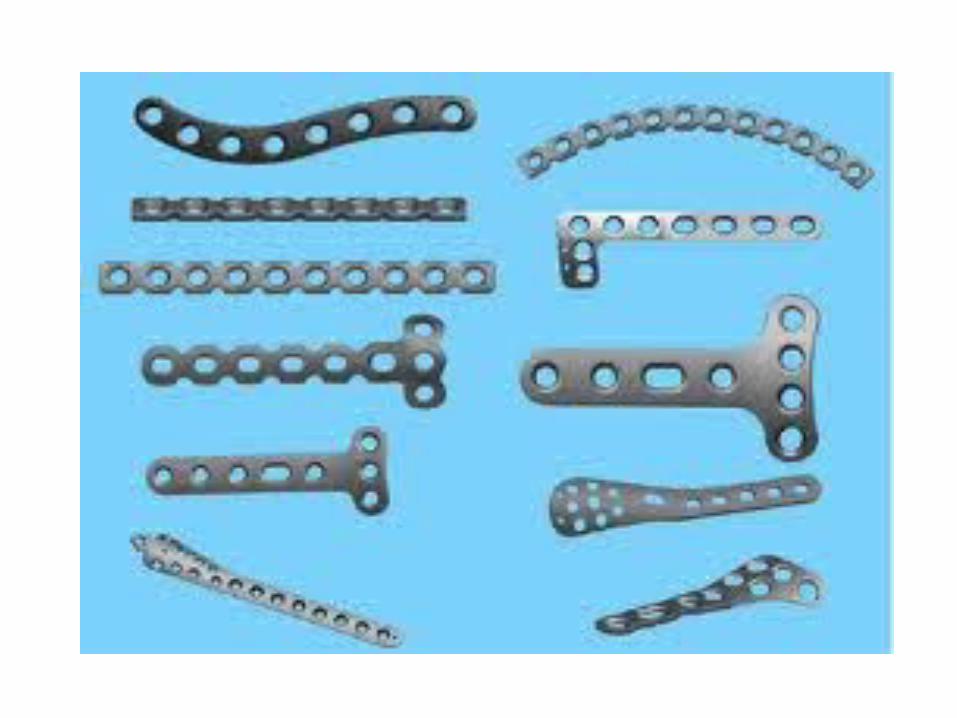
PLATES• Combined with screws, they act as splints to protect the screw by
reducing shear or bending forces( hence term protection plate/neutralization plate)
• 5 functional uses of a plate:i. Protection-of the lag screws
ii. Compression-drives ends of fracture together
iii. Tension band-plate placed on tension side of bone
iv. Bridging –used in multifragmentary fractures
v. Buttress:-used in metaphyseal areas(resists axial load by applying force at 90˚ to axis of potential deformity)


•LC-DCP has limited plate-bone contact(plate footprint), hence less impairment of capillary network of the periosteum->relative improvement of cortical perfusion
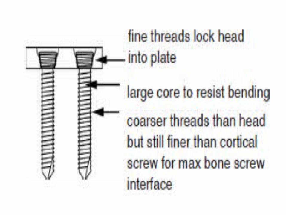
•Locking compression plates(LCP)-designed in such a way that screws effectively bolt into plate and bone, hence as screw is tightened, bone maintains its position and is not drawn to plate
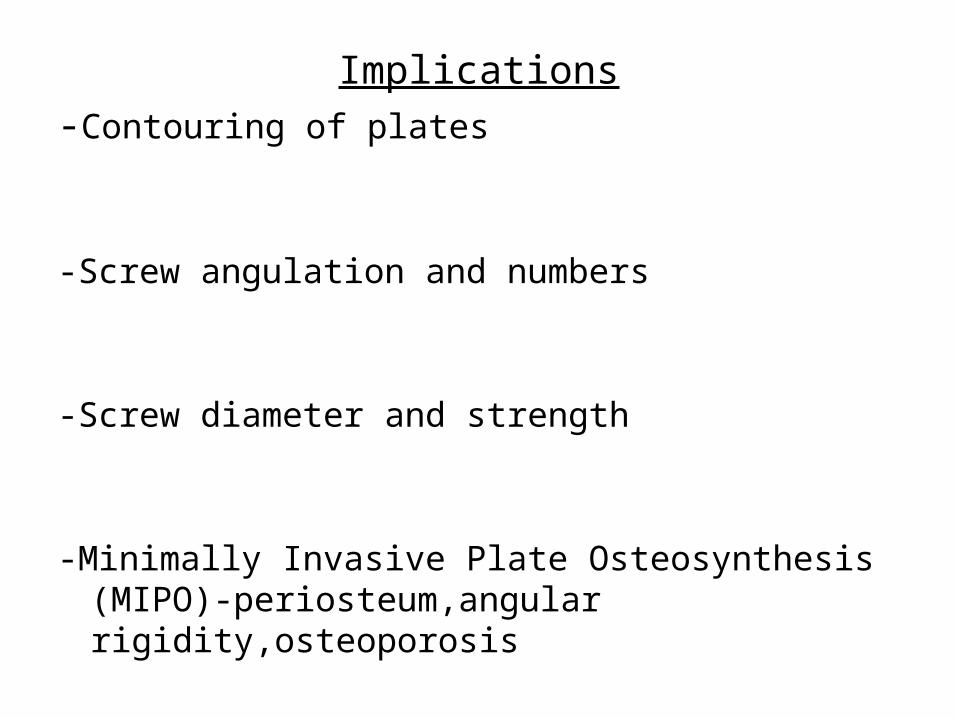
Implications-Contouring of plates
-Screw angulation and numbers
-Screw diameter and strength
-Minimally Invasive Plate Osteosynthesis (MIPO)-periosteum,angular rigidity,osteoporosis

disadv of plates-prominent lateral screws may cause symptoms or
wound necrosis
- possibility of distal intra-articular screw insertion
-inadequate fixation if distal screws are too short
-may not allow adequate fixation in osteoporotic bone
- may interfere w/ syndesmotic screw insertion (especially when two syndesmoic screws are to be used);

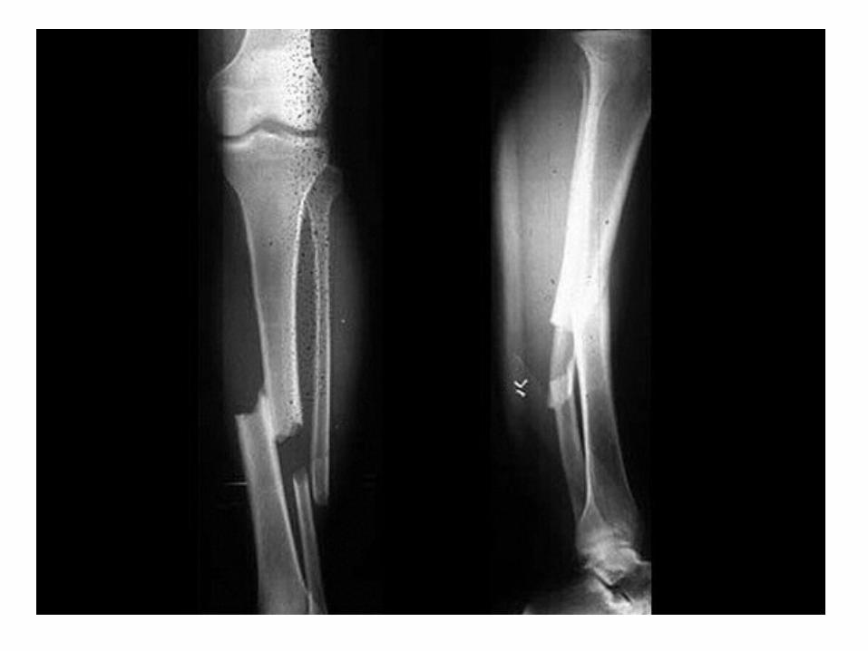
RELATIVE FIXATION-Bone fragments displace in relation to each other
when physiological load is applied across fracture.
-Implants: internal fixators,ext. fixators,IM nails -All allow inter fragmentary movement which can
stimulate callus formation
-Incorrect application leads to excess movement and inhibit bone union

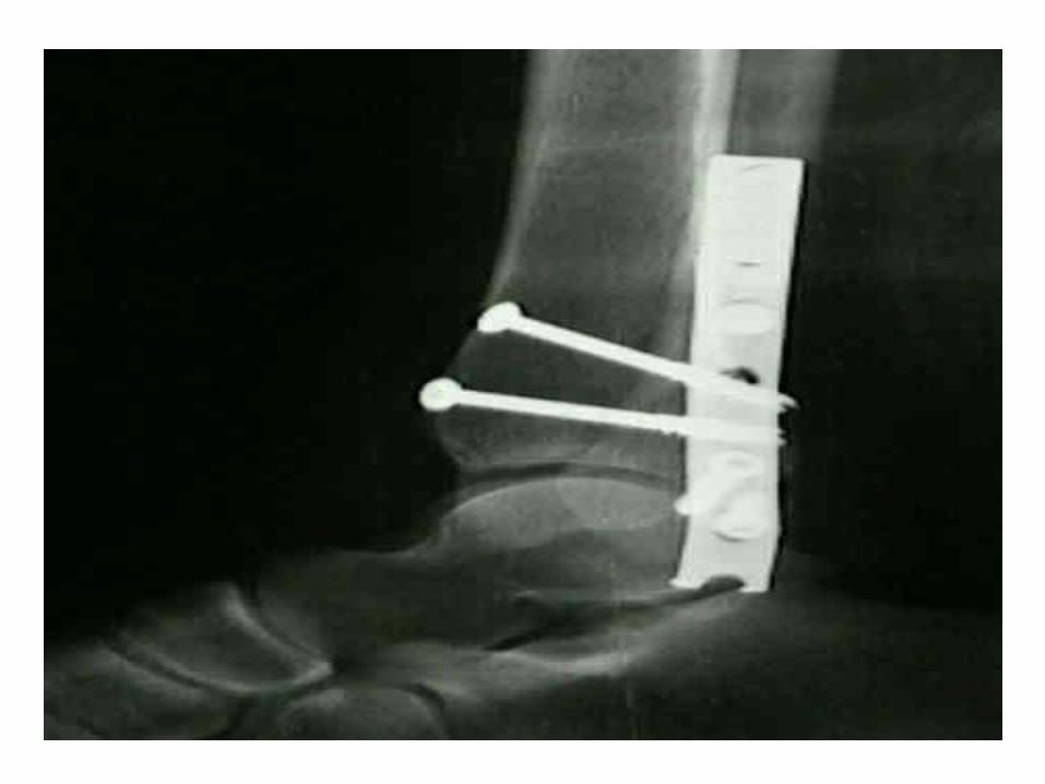
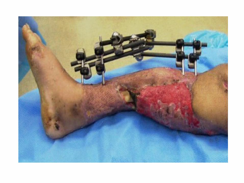
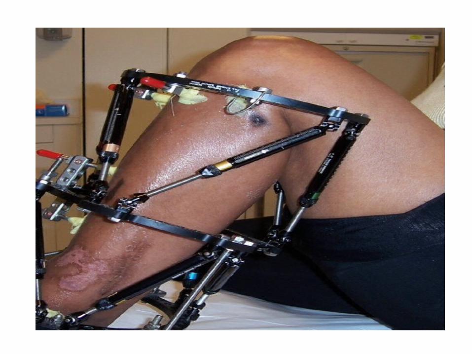
Ext. fixators- External fixation is a method of immobilizing bones to
allow a fracture to heal.
-External fixation is accomplished by placing pins or screws into the bone on both sides of the fracture
-The pins are then secured together outside the skin with clamps and rods. The clamps and rods are known as the "external frame."
Factors influencing stability of fixation: -stiffness of connecting rods
-distance between rods and bone axis
-no, spacing and diameter of schanz screws
Advantages-rigid fixation
-compression, neutralization, or fixed distraction of the fracture fragments
-direct surveillance of the limb and wound status
- associated treatment e.g dressing changes, skin grafting, bone grafting, and irrigation, is possible without disturbing the fracture alignment or fixation
-immediate motion of the proximal and distal joints is allowed
-extremity is elevated without pressure on the posterior soft tissues
-early patient mobilization
-can de done under L.A
- used in infected, acute fractures or non union

Disadvantages-pin tract infection
-expensive equipment
-cumbersome frame(aesthetic)
-fracture through pin tracts
- re fracture after ex-fix removal

-joint stiffness: over a joint e.g pilon fracture
-pin and fixator frame may be difficult to assemble
Complications
-pin tract infection
-neurovascular impairment
-muscle/tendon impairment
-compartment syndrome
-delayed union
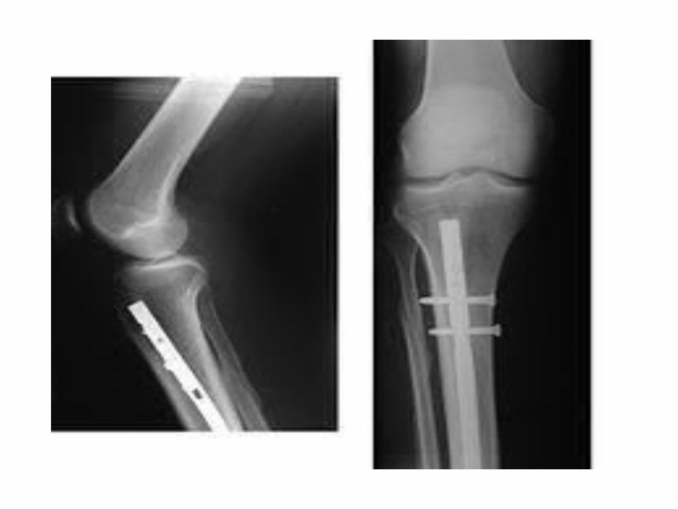
IM NAILS
-Classical kuntscher nail- stable against bending and shear forces perpendicular to its long axis. Its confined to simple transverse/oblique fractures
-IM nails are: -unstable against torsional forces -confined for simple transverse or short oblique
fractures which cannot shorten and will inter-digitate to prevent rotation
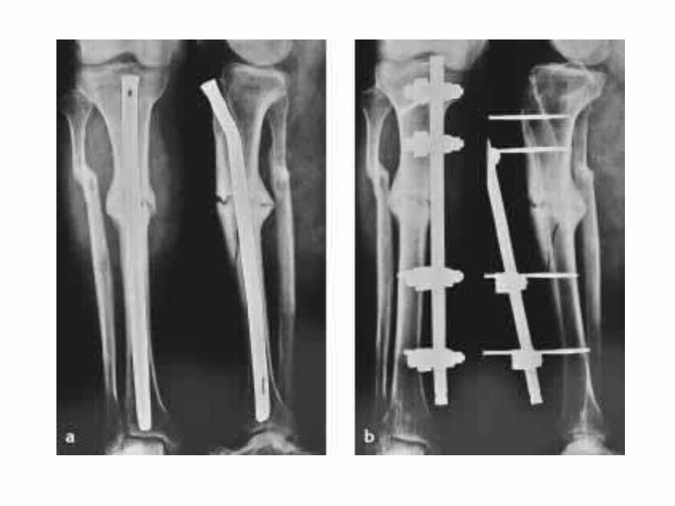
-Locked IM Nails-withstand torsional forces and axial loading
-Holes are larger than screws
-Stability dependent on diameter of the nail, geometry, number of interlocking screws, spatial arrangement

Internal fixators and bridging plates
-Plating with relative stability should only be used in multi-fragmentary fractures
-Use in simple fractures causes high incidence of delayed or nonunion
-Bridge plating uses the plate as an extramedullary splint, fixed to the two main fragments, while the intermediate fracture zone is left untouched.
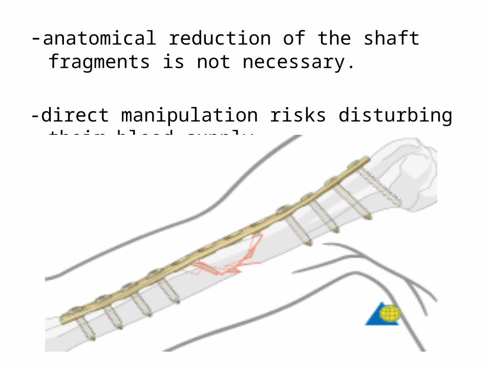
-anatomical reduction of the shaft fragments is not necessary.
-direct manipulation risks disturbing their blood supply
• Stiffness of an internal fixation method depends on: -dimensions of the implant
-number and position of screws
-quality of coupling btn screw and plate and btn screw and bone