Embed Size (px)
Citation preview

T H E I R I S H J O U R N A L M E D I C A L S C I E N C E
THE OFFICIAL JOURNAL OF THE I{OYAL ACADEMY OF MEDICINE IN I R E L A N D
OF
~/XTH SERIES. NO. 337. ol~xNu~nY, 1954.
TEAM DIACNOSIS OF CONGENITAL HEART DISEASE.*
By R. E. STEEX.
C ( ) N G E N I T A L heart disease is the Cinderella o[' pa, diatric disorders. Though the eya~mtic types (morbus c,~r~de,s, or the sky-blue disease) have been reeognised f rom the earliest times, until a few
years ago the subject a t t rac ted litt le attention. This was not surprising, since its a~tiology was without interest, its prognosis often without hope, and its t rea tment pure ly palliative. A diagnosis was made, perhaps a patent foramen ovale (which is m)w no hmger regarded as a congenital heart defect, since it is found in 10 per cent. of normal people through- out life, and indeed may be a. help in congenital disease), the case was not infrequent ly lost sight of so that seldom was one proved wrong, and if at au topsy this was so, it was not regarded as of much moment since it clearly had made little or no difference. Today, though still a. reb~- t ively restr icted speciality (only 1-2 per cent. of all organic heart disease), thanks to modern t rea tment it can produce some of the most dramat ic therapeut ic results of the whole of medicine. Helen Taussig, in Paris, in 1950 told us of two examples in her experience, both cases of cyanotic heart disease: one a boy who had been bedridden for two years and whose dream had been that one day he might walk twenty miles. This was achieved following a Blah)ck-Taussig operation. In the second instance, a young woman who had also been bedridden f(n' a similar period, mar r ied and had a child eleven months a f t e r the operat, ion.
Our experience in Dublin has, so far , been largely l imited to opera- tions for patent duetus, but, inclusive of cases operated on at St. Vincent ' s Hospital , Dublin, over 60 cases of patent ductus have been operated on without mortal i ty.
H'istoric~d. The birth of the surgical t rea tment of congenital heart disease nlay
be considered to have taken place in 1939, when Gross, of Boston, first ]igated a patent ductus arteriosus. This branch of surgery, however. received a t remendous impetus when A l f r e d Biah)ek and Helen Taussig produced their now famous Blalock-Taussig operation for eases of Falh)t 's te t ra logy and certain other r a re r types of cyanotic disease. This opera- tion, anastomosing the subclavian a r te ry to the pulmonary artery, was followed quickly by Pot t ' s operation, which is s imilar in principle hut anastomoses the aorta to the pulmonary artery. Both ot)erations act by
* From the Congenital l:[eart Unit, National Children's l~ospital, Dublin: T. C. J. O'Connell, F. J. I~enry, S. J. Douglas, R. Davys, P. N. Meenm/, E. E. Doyle, J. P. R. Rees, A. MacDonald, R. E. Steen.
Jo in t communication made to the Section of" Medicine of' the Royal Academy of Medicine in Ireland, October, 1952.
2 I R I S H JOURNAL OF MEDICA L S CIEN CE
increasing the blood supply to lungs which are " starved " of blood. It may be mentioned here that though in both cases this is equivalent ~o making an artificial ductus, the position is ra ther different f rom that in which the pa£ent ductus is overloading a normal circulation. In any event, operation is the lesser of two evils, as Fal lot 's tetralogy without operation has a mortal i ty of 97 per cent. before puberty. Potts has recently had a series of cases in which the operative mortal i ty for his operation was as low as 2 per cent. These operations for cyanotic heart disease were quickly followed by Crafoord 's operation for coaretation of the aorta, in which he divides the constriction and joins the ends together. More recently, Broek's valvulotomy for cases of pulmonary valvular stenosis is considered the operation of choice in cases where the stenosis is valvular ra ther than infundibular . Some cases of auricular, and more recently of ventricular , septal defect have been closed where it appeared that the child would not otherwise have lived. Surgery has also been a t tempted in other conditions, for example, transposition of the vessels. I t may also be lifesaving in rare vascular anomalies such as double aortic arch.
2 E t i o l o g y .
New interest has been aroused in this aspect of congenital heart disease by the unequivocal evidence that rubella in the earlier months of preg- nancy may damage the f~ tus and produce a congenital heart lesion (Gregg, 1941; Swan, 1943). Congenital heart disease is not the only congenital defect which can result from such trauma, as congenital eata- raet, deaf-mutism, and mental deficiency may also occur. The damage must take place in the first t r imester when the organs arc forming and taking shape, and in the case of the heart the damage is mainly done between the fifth and eighth weeks of f~ ta l life. One found it difficult at first to accept what seemed rather foreign to one's seientifie reasoning, but the views with regard to rubella have been accepted throughout the world, not just in pa~diatries and cardiology, but in ophthalmology and other specialties. Mr. Werner has told me that he considers it possible to tell the rubella cataract from other congenital cataracts by being of a " harder " type. 1)r. F red Kane, of Purdysburn Fever Hospital, made it easier for me to accept the view by explaining that most fevers of a more severe nature, such as smallpox and even ordinary measles, tend to produce abortion and, therefore, death of the f e tu s ra ther than its damage.
One ease does not prove anything, but when it is in one's own experi- ence, and par t icular ly striking, it may be impressive. The following is an example of this :
Dr. C. Finnegan, of Greystones, in 1947 referred to me a case of congeuital hea r t disease, congenital ca tarac ts and menta l deficiency. This was in the early days of the theories wi th regard to rubella, and I was only vaguely famil iar ~ i t h them. A year or so later, when speaking to her on the telephone, she said, " Y o u know l h a t case I referred to you , " ment ion ing the child 's name, " was a rubel la ease." I asked her how she knew abou t this, and she said tha t the mothe r had come in to see her a few days previously and had produced, w h a t so m a n y lay people now depend on for their medical advice, one of the popula r m o n t h l y Digests.] ~fhis conta ined an article cn Ge r ma n measles and congenital defects and, in this instance, was correct. The mo the r then told Dr. F innegan tha t she had t rea ted her for German measles in the second me n th of her pregnancy. Any possible subjective e lement was removed by the fact t h a t Dr. F innegan had to consult her notes to ascertain if this was true, and found tha t she had, in fact. a note t ha t a t this period of the pregnancy she had a t t ended the mothe r for an a t tack of rubella.

T E A 5 [ D I A G N O S I S OF C O N G E N I T A L H E A R T D I S E A S E 3
I t is, of course, possible that other vitals infections of a mild type, e.g., infective hepati t is and even the " common cold ", could damage the foetus in this way, and this makes tile whole problem an extremely worry- ing one. Land tman (1948) working at Univers i ty College t tospital , has produced evidence which does indeed suggest that this may sometimes be the case, but rubella, so fa r seems to be the worst offender, and one must hope that other virus infections seldom, if ever, ca r ry the same danger. Paul Wood (1952) considers that as high a percentage of cases of congenital heart disease as 5 per cent. may be due to rubella. This figure seems higher than one would have thought, but at least it emphasises that in the prophylact ic t rea tment of congenital heart disease it is impor tan t that girls should be exposed to rubella in childhood in the same way as one feels that it is desirable for a boy to be exposed to mumps. The Americans express this t raumat ic factor in the product ion of congenital heart disease by saying that, " one can have identically the same malformat ion f rom a bad egg in a good environment as f rom a good egg in a bad environment ."
Diagnosis. The diagnosis of congenital heart disease is carr ied out in two stages : 1. The diagnosis of congenital f rom acquired hearst disease. '2. The diagnosis of the type of congenital heart disease.
Diag~wsis of cow,genital hearl disease from a.~quired heart disease. All the various points which may be of help in this connection in
a r r iv ing at a decision cannot be refer red to here, but three which are of special value will be mentioned.
1. Age: Organic heart disease under o.1, years is ahnost certainly congenital. This is because the forms of acquired heart disease which produce a chronic valvular lesion are not met with below this age.
(a) Juvenile rheumatism is rare under 5, and, for practical pur- • 1 poses, unknown under 22 years.
(b) Congenital syphilis does not affect the heart, though it is pos- sible that in rare cases it may eause congenital heart disease.
(c) Arteriosclerosis is obviously not met with at this age. The very rare condition, progeria, met with in the older child would have characterist ics o£ its own.
2. Oryanic murmurs in childhood which are not rheumatic are a,!most certainly congenital. Juvenile rheumat ism for practical purposes affects only the aortic and mi t ra l valves, e.g., a harsh pulmonary systolic murmur is almost certainly congenital. Malignant endocarditis would have special characteristics of its own.
3. Cyanosis, clubbing and polycyth(tmia when present and marked are in favour of congenital heart disease Central cyanosis in childhood (see later) s t rongly suggests congenital heart disease and a venous-arterial shunt, provided puhnonary causes are exclu(ted.
Diag~wsis of the type of congenitag heart disease. The pr incipal methods of examination a re : 1. Clinical. 2. Radiological, including fluoroscopy. 3. Electrocardiographic.
4 IRISH JOURNAL OF MEDICAL SCIENCE
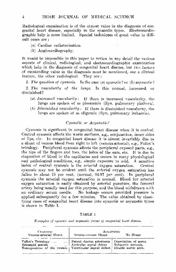
Radiological examination is of the utmost value in the diagnosis of con. genital heart disease, especially in the cyanotic types. Electrocardio- graphic help is more limited. Special techniques of great value in diffi- cult cases are:
(a) Cardiac catheterisation. (b) Angiocardiography.
It would be impossible in this paper to review in any detail the various aspects of clinical, radiological, and electrocardiographic examination which help in the diagnosis of congenital heart disease, but two factor's of outstanding value in the diagnosis must be mentioned, one a clinical feature, the other radiological.
1. The question of cyanosis.
2. The vascularity of the diminished?
(a) Increased vascular~ity :
They are:
Is the case (a) cyanotic ? or (b) acyanotie ?
lungs. Is this normal, increased, or
If there is increased vascularity, the lungs are spoken of as pleonmmic (Syn. pulmona~T plethora).
(b) Dlminished vascularity : If there is diminished vascularity, the lungs are spoken of as oligmmic (Syn. pulmonary isch~emia).
Cyanolic or Acyauotic?
Cyanosis is significant in congenital heart disease when it is central. Central cyanosis affects the warm surfaces, e.g., conjunctivae, inner sides of lips, etc. In congenital heart disease it is almost invariably due to a shunt of venous blood from right to left (venous-arterial), e.g., Fallot 's tetralogy. Peripheral cyanosis affects the peripheral exposed parts, e.g., the tips of the fingers and toes, the lobes of the ears, etc. It is due to stagnation of blood in the capillaries and occurs in many physiological and pathological conditions, e.g., simple exposure to cold. A sensitive index of central cyanosis is the arterial oxygen saturation. Central cyanosis may not be evident until the arteria~ oxygen saturation has fallen to about 75 per cent. (normal, 94-97 per cent). In peripheral cyanosis the arterial oxygen saturation is normal. Blood for arterial oxygen saturation is easily obtained by arterial puncture, the femoral artery being usually used for this purpose, and the blood withdrawn with an ordinary serum needle. No leakage occurs provided pressure is applied subsequently for a few minutes. The value obtained by classi- fying cases of congenital heart disease into cyanotic or acyanotic types is shown in Table I.
T A B L E 1
Examples of cyanotic and acyanotic ,form~ of congenital heurt disease.
CYANOTIC Venous -a r t e r i a l S h u n t
F a l l o t ' s T e t r a l o g y . . . . . . . . T r i cusp id a t r e s i a . . . . . . . . T ranspos i t i on of t he vessels
ACYANOTIC Ar te r i o -venous S h u n t No S h u n t
f P a t e n t d u c t u s a r t e r io sus Coare t a t ion of aor ta . Aur i cu l a r s ep t a l defec t Subaor t i c s tenosis . V e n t r i c u l a r s ep t a l defect Doub le ao r t i c arch.

T E A M D I A G N O S I S OF C O N G E N I T A L H E A R T D I S E A S E 5
Pleontemic or Olig~emic?
Pleonvemic lungs (pulmonary plethora) are " wet " lungs f rom increased vascular i ty due to a bigger volume of blood than normal reaching the lungs. On radiological examination, the vascular shadows are increased so that the hilar shadows appear unusual ly heavy, and the normal pul- monary vascular markings are also increased and extend out f rom the hi lum towards the per iphery of the lungs. (N.B.- -Nm~nal hi lar shadows are now regarded as being vascular r a the r than glandular in origin. This has been proved by angiocardiography.) A " hi lar dance " may be seen on screening. This is the name given to increased action of the branches of the pu lmonary artery. The points of pulsation in the hi lum can be seen to jump apar t at each systole. On clinical examination, moist sounds at the bases are very common.
Oligcernic lungs are " d r y " lungs f rom diminished vasculari ty due to a smaller volume of blood than normal reaching the lungs. The vascular shadows are unusual ly " l ight " and a " h i l a r dance " is never seen on screening. Moist sounds are uncommon on clinical examination, even in the presence of cardiac failure.
X- r ay films to i l lustrate pleon~emic and oliga~mie lungs are shown in Figs. 1, 2, and 3.
The advantages of classifying a case into whether it is pleon-emic or oliga~mic in type is shown in Table 2.
TABLE 2.
Examples of Pleonwmic and Oligcemic Form~ of Congenital Heart Disease.
PLEONzEMIC OLIGzEMIC
Arterio.venous (left.right) shunts; (a) Patent ductus ; The shunt is from aorta (higher
pressure) to pulmonary artery (lower pressure). {b) Auricular septal d~fect ; The shunt is from left auricle
(higher pressure) to right auricle (lower pressure) and thence to the lungs.
N ,B . - -When auricular septal defect is associated with mitral stenosis (congenital or acquired) very large shunts result. The pulmonary artery becomes enormous and the lungs extremely pleonaemie (Lutembacher's syndrome).
(c) Ventricular septal defect ; The shunt is from left ventricle (higher pressure) to right ventricle (lower pressure) and thence to the lungs.
N.B.--Small ventricular septal defects, e,g. (Maladie de Roger) may have a negligible shunt.
Eisenmsnger's complex ; The factors involved here are not fully understood.
Fallot' s tetralogy. Tricuspid atresia.
The value of classifying a case f rom the double point of view, whether it is cyanotic or acyanotic, and pleon~emic or oligmmic in type, is shown in Table I I I .

EX
AM
PL
ES
O
F
PL
EO
NA
EM
IC
LU
NG
S
PU
LM
ON
AR
Y
PL
ET
HO
RA
} A
ND
O
L1G
AE
MIC
L
UN
GS
(P
UL
MO
NA
RY
IS
CI-
IAE
MIA
) ~
z
Fro
. 1
.--P
ate
nt
duct
u.s
arte
rios
tts."
S
ho
win
g h
eav
y
hil
ar
shad
ow
s (p
leon
aem
ie
lung
s)
and
ca
p
of
" Z
inn
."
Fro
. 2
.--F
all
ot'
s te
tral
o;/y
" S
ho
win
g
ver
y
lig
ht
hil
ar
shad
ow
s (m
ark
ed
olig
acm
ie
lung
s)
and
ho
llow
ing
of
pu
lmo
nar
y a
rc.
lQG
. 3.
---
Ext
t'e~;
',,
I)al
mgt
~ar!
! j~
leth
ora
: T
im
rig
ht
hil
um
is
v
ery
h
eav
y
(on
scre
enin
g, t
he
rig
ht
bra
nch
of
th
e p
ulm
on
ary
ar
tery
w
a,s
ptd
sati
ng
mos
t, vi
goro
usly
), a
nd.
on t
he
left
sid
e th
ere
is e
not'
mou
s an
eury
smal
dil
atat
ion
of
the
pu
lmo
nar
y a
rter
y.
©

T E A M D I A G N ) S I S OF C O N G E N I T A L H E A R T D I S E A S E 7
T A B L E 3.
Diagram to illustrate by some c o ~ m o n examples the help in diagnosis of classifying a case into cyanotic or acyanotic and p leonwmic or oligwmic types.
C Y A N O T I C. A C Y A N O T t C.
x
J Q.
0 u
m
w tD Z W
z u l
W
£Q
LU
u)> OO Z uJZ i-uJ
n- O ~:LL Z OO
JU~ DO Q..J
u uJ ~ - r
P L E O N A E M I C . O L I G A E M I C .
Cardiac Ca.theterisation. ()wing to considerations of space, this account must necessarily be an
abbreviated one. Historical : I t is interesting to recall that though Bleichroeder passed
a catheter up his own vein in 1912, it was as late as 1929 that the first cardiac catheterisation was per formed by Forssmann. Forssmann was a German of s t rong scientific convictions. He did not choose for the pur- poses of his exper iment a pat ient dying of advanced cancer, but passed the catheter into his own heart . I t is related that when a colleague • nched, having passed the catheter 35 centimetres up F o r s s m m m ' s vein, 1he la t ter took the catheter himself and passed it on into the r ight auricle, demonstrat ing this by fluoroscopy. In the process of doing this, he walked up a flight of stairs to the X- ray Depar tment with the catheter

8 I R I S H JOURNAL OF MEDICAL SCIENCE
in his heart. When one of us (R.E.S.) was in Copenhagen, h.e met Dr. Bing, f rom Baltimore, who had just completed his 1,200th cardiac catheterisation, and was in Europe searching Germany to t ry and find Forssmann, unfor tunate ly unsuccessfully, as the 1939-45 War seemed to have eliminated this great mail. Cournand, with his modification of the ureteric catheter, made great strides.
Risks: Unlike angiocardiography, paradoxical though it may at first sight appear, cardiac catheterisation is a relatively safe procedure. Some would say (Mannheimer, 1949) that in careful hands there is no risk, and that the procedure is harmless, but there is probably a small mor- tal i ty rate of about I in 1,000. This is, however, no higher than that for blood transfusion, gastroscopy, and even lumbar puncture (Editorial , Lancet, 1950). Guy's Hospital (Holling and Zak, 1950) has laid stress on the risk of thrombotic complications, especially in the grossly cyanotic group, and in one series, they placed this as high as 6 in 70. F o r this reason, and on account of the possible very small risk of a fatali ty, our practice at the National Children's Hospital has been only to carry out cardiac catheterisation on cases where operation was being considered, and where a decision could not be reached by the ordinary methods of diagnosis.
Purposes : The two main purposes of cardiac catheterisation are for the taking of :
1. Specimens of blood for oxygen analysis. This will often demon- strate the addition of arterial blood to venous blood which may take place in (a) Patent ductus arteHosus : The left-to-right shunt of arterial blood from the aorta to the pulmonary a r te ry tends to cause the oxygen saturation in the pulmonary a r te ry to be higher than in the superior vena cava, right auricle and right ventricle. (b) Auricular septal defect : the left-to-right shunt similarly tends to cause the oxygen saturation to be higher in the r ight auricle, r ight ventricle and pulmonary ar tery than in the superior vena cava. (c) Ventricul.ar septa.l defect : the left-to-right shunt tends to cause the oxygen saturation to be higher in the right ventricle and pulmonary ar tery than in the right auricle and superior vena eava.
2. Pressures, e.g. (a) Patent ductus arteriosus : the pressure is usually raised in the pulmonary a~%ery; (b) Fcdlot's tetralogy: the pressure is raised in the right ventricle and very low in the pulmonary a r t e ry ; (c) Eise~menger's complex : the pressure is raised in the right ventricle and raised in the pulmonary ar tery.
Equipment : American nylon catheters are used. Nos. 6, 7, 8, and 9 are the s tandard sizes, but No. 9 is very big for a child, even for an older child, and No. 6 is on the small side for taking pressures, though it is quite possible to work satisfactori ly with this size, and it is pre- ferable to use it than to fail altogether, which happened in one well- known clinic which apparent ly never uses the No. 6. Nos. 7 and 8 are the most generally useful size. The method of sterilisation of these is extremely important, and especially the cleaning of the catheters af ter use. A most rigorous technique is necessary to free them of pyrogens, which are liable to give such severe reactions that Paul Wood at one time would use a catheter only once. (The cost of a catheter is approximately £3.)

TEAM I) IAGNOSIS OF CONGENITAL H E A R T D I S E A S E i)
The manometer we have used up to the present has been a saline mano- meter, graduated in centimetres (136 cms. H~O=100 rams. Hg), the zero mark being placed at the level of Ludwig's angle. This type of mano- meter, however, only records mean pressures and we have now obtained the services of an electrically-contr,)lied manometer which has the great advantage of recording both systolic and diastolic pressures. The blood is taken in syringes under a paraffin seal and kept on ice and in the dark. Oxygen saturations are estimated by the Haldane apparatus. The use of the x-ray fluoroscope must not exceed a total t ime of 15 minutes. The screening time is careful ly recorded by an assistant with a stopwatch, and until experience is obtained, it is ext raordinary how quickly the minutes are recorded. No gloves are worn, but the operator and all assistants wear lead aprons.
Prepc~ration of the child: A day or two in hospital is desirable in order to get the child accustomed to its surroundings. A small dose of quinidine is given before catheterisation in order to diminish the tend- ency to cardiac irregularities. Almost every form of cardiac arrhythmia ha~s been described. These tend to occur more par t icular ly when the tip of the catheter is in the region of the tr icuspid valve or the pulmonary conus. I t is advisable not to leave the tip of the catheter in these situa- tions for any length of time. On the whole, however, cardiac arrhythmias are usually more alarming than dangerous, and otherwise there seems to be very little risk from the: actual presence of the catheter in the heart. In one recorded instance it was left in the heart for 4~ days. An " umbrella " of penicillin is also given and heparin is given through the catheter at the beginning of catheterisation and also in t h e " drip ". The question of pre-medication with a sedative is often a difficult decision. In Scandinavia, they seem to do most of the children without premedi- tation, whereas in London the major i ty seem to receive it. We find that many of the older children are best done without heavy premedica£ion, which is liable to make them go off to sleep, and when they wake up they (fften tend to struggle. On the other hand, in the case of small children, it is usually impossible to do the catheterisation without its help, and a suitable dose of barbi turate is given for this purpose. General anaesthe- tics cannot be employed, as they affect the oxygen saturations.
Technique: The catheter is introduced into the heart via a median antecubital vein, a lateral vein being unsatisfactory for the purpose. The left arm is usually chosen, as the curve of the catheter into the heaI~ is more uniform, and its introduction through the tr icuspid valve and into the pulmonary a r te ry may be possibly more easy by this route. The right arm may also be u~d , or the femoral vein in a small child, but the passage through the tr icuspid valve is probably not quite so easy from this direction. The external jugular vein has also been employed. Care with regard to asepsis is important. I t is very easy to overlook this; the screen should be protected with a sterile towel, as it is very easy to touch it with the hands. P lenty of local anaesthetic should be used, as this is probably one of the best protections against venous spasm. The tendency to the la t ter is also diminished by seeing that the catheter is entirely f ree of antiseptic, and that it is not too big.
A difficulty in the passage of the catheter is often experienced in the shoulder region. Taking a big breath seems to be one of the best, methods

10 I R I S t t JOURNAL ()F MEI)ICAL SCIENCE
CARDIAC C A T I ~ E T E R I S A T I O N .
(X- rays 4.10 are from the same case : A patent (luctus artcriosus)
Fro. 4 . - -The tip of the catheter is lying in the super ior vena caca.
FI(:. 5 . - -The tip of the catheter has bee~l passed on in to the right auricle.
FIG. 6 . - -The t ip of the catheter has been passed down into the ireferior vena
cava.
FIc. 7 . - -The tip of the catheter has been passed th rough the t r icuspid valve into the right ventricle and is lying in the infundibular region near
the pu lmonary valve.

T E A M I)IAGN()SIS ()F CONGENITAL HEART DISEASE 11
CARDIAC CATKETERISATION
FI(~. S. -The tip o[" the catheter has been passed on into the r ight branch of the pulmo~tary artery and is lying out in the
r ight lung field.
FIG. 9 . - -The tip of the catheter has beets w i thd rawn into the main pulrr, onary artery and is lying close to the pulmon-
ary valve.
FIG. 10. -The lip of the catheter has been ~)assed forward again into the ~ft bra~tch of the jmhr, v~ ~ry ~rtery.
FIG. l l . - - T h e tip of the catheter has not pierced the h e a r t ! I t has passed f rom the r ight auricle t h rough a pa ten t foramen ovale into the left auricle and on into a pulmonary vein. A sample of blood taken f rom here ,was 99%
sa tu ra ted wi th oxygen.
12 I R I S H JOURNAL OF MEDICAL SCIEN CE
of overcoming the obstruction. In one case, the tip of the catheter kept going up into the internal jugular in an i r r i ta t ing way (as every second seems valuable), but soon with a little manipulat ion one had the. pleasant experience of seeing it passing on into the auricle. Another possible " snag " is to go into the coronary sinus. We have not, so far, had this experience, but when this happens the tip of the catheter stops inside the shadow of the heart and a specimen of blood shows it to be ahnost black, as venous blood from such an energetic organ as the heart is only 20-30 per cent. saturated.
Control of the cath, eter is by three methods--(a) Pushing and with- dra,wing, (b) twisting, and (c) gravity. In passing from the right auricle to the r ight ventricle, it is often useful to put the child into the right oblique position, as this helps the onward passage of the catheter and its tip is also more easily seen when it is clear of the spine. The best plan is to go as f a r .as is possible at the outset, if possible into the pulmonary artery, where one takes the first pressure reading and speci- men of blood for saturation, and withdraws the tip of the catheter, taking recordings and samples from the right ventricle (preferably from both the upper and lower portions), the right auricle and superior vena cava. I t is bet ter to work on this plan than by taking recordings and samples as one advances, because unless they are taken reasonably close together they are not so comparable. I t is an advantage also, if possible, to get a specimen from the inferior vena cava. This is because inferior vena caval blood, possibly as a result of the contribution of the renal vein, is usually, though not invariably, slightly higher than superior vena caval blood. The mean of the two, therefore, best represents what one would expect to find in the right auricle. In the diagnosis of auricular septal defect, r ight aur icular blood should be at least 2 volumes per cent. higher than superior vena caval blood to be significant. In taking pres- sures with the saline manometer, we have found it valuable for one person alone (J.P.R.) to concentrate on the management of the mano- meter, as it is valuable to get constant information with regard to this. In passing from a low pressure chamber, like the right auricle, to a high pressure one, like the right ventricle, blood tends to regurgi tate into the catheter. Flushing of the catheter at this time is important, h i the same way when taking specimens for oxygen saturation, one of us (E.E.D.) has concentrated on this and handled the paraffined syringes. I t is important when separating the catheter f rom the " drip " in order to take a specimen of blood that the junct ion should be kept as low as possible, though the hmmodynamic factor makes the sucking of air into the heart a very unlikely occurrence. I t is not usually necessary to tie the vein at the upper end, a pressure pad being usually sufficient. The vein may be used again, in many cases a for tnight later. In Figs. 1 to 8, the t ip of the catheter is shown in various situations in the heart.
Angiocard iography . This is not nearly so safe a procedure as cardiac catheterisation, but
for tunate ly it is essential for the diagnosis of only a very small number of difficult cases. I t is well, however, that it should be ful ly appreciated that there is a definite fatal i ty rate. Guy 's Hospital reported four deaths in 172 cases. Two of these were in their first 30 cases, which shows that, with experience the risk can be reduced. In their series, many of the

TEA~[ D I A G N O S I S OF C O N G E N I T A L H E A R T 1) ]SEASE 1'~
eases were bad cyanotic ones, and there is no doubt that this type of case,. or one which is associated with anoxmmic attacks, carries a much highel ~ risk. In America, Sussman has published a series of 1,500 cases without fatal i ty, and Robb and Steinberg have done the same, but in Amer ican series there is often a very high preponderance of acyanotic cases which ca r ry a very much smaller risk. Unfor tuna te ly it is jus t in the type of case which carr ies the greatest risk that the procedure, is often the most valuable. Anybody with experience, however, knows that anglo- card iography is sufficiently dangerous and is, therefore, only desirable if very decided advantages are to. be obtained. While it is difficult to give any definite mor ta l i ty rate for angiocardiography, it is probably about 1 : 100 in a mixed group of bad and mild risks such as one gets in an average unit which deals with cases of congenital heart disease, and this figure is possibly a generous one as regards its dangers. Gibson, working with Ports in Chicago, told me recently (personM communica- tion) tha t he had done only 40 cases, most of which were in infants where the risk is, curiously enough, ra ther less than in older children or the adult. Taussig, who works with Blalock, also uses the procedure very sparingly. In the National Children's Hospital , Harcour t Street, we have done only one case of angiocardiography, and that was on a mongol, and our intention is only to use it i f considered absolutely essential.
I t is not clear wherein lies the danger. Paul Wood even considers that it is not due to the diotrast which is used, but to some other factor, and he only uses a " p i l o t " dose for medico-legal purposes. The major i ty of observer's are inclined to think tha t it is some reaction f rom the material , and this seems to han, e gained ground more strongly in recent years, Certainly, it is associated with a considerable amount of shock and fall of blood pressure, and t ransient conduction changes are of ten noted in the electrocardiogram. Adul ts in whom angiocardio- g raphy is done without an anmsthetic experience very unpleasant sensa- tions. In our case, though the child was anmsthetised, our anmsthetist was impressed with the way he gave what resembled a shudder when the dye was introduced.
T e c h n i q u . e : 5 to 30 ml., depending on the age of the child, of the opaque medium (70 per cent. diodone) is injected i n t o a n arm vein af ter a small " pilot " dose has been given to test for sensitivity. The child has previously had premedicat ion with a barb i tura te and atropine. Some clinics use no anmsthesia at all. In our case, following the advice of Sussman, we used " closed ether " with a small amount of cvclopropane at the induction. At the end of the investigation the ehilcl should be put in an oxygen tent and t rea ted as if recovering f rom a major operation.
X- r ay films are taken at a rate of about one per second for eight to ten seconds, but a fas te r ra te of two per second would be preferable. Selected films f rom the one series are shown in Figs. 12-15. I t is absolutely essential that the mater ial is injected rap id ly in order to get a " bolus " effect, as it is sometimes in the pu lmonary arte~T in half a second, and through the child's heart in about 5 seconds. No clear pictures will be obtained i f the mater ia l is injected slowly over 2 or 3 seconds. The rap id i ty with which the dye travels is well i l lus t ra ted ' in Fig. 12. Though iV is said to have been taken at approximate ly

14 , IRISH JOURNAL OF MEDICAL SCIENCE
A N G I O C A R D I O G R A P H Y .
( F o u r fihns f r o m a series taken in left obliquc posit ion : a case of pa t en t duc tus arteriosus.)
FIG. l ' 2 . - - F i l m talcen at approxi.m,'*tely { sec. (from admin is t ra t ion of diodone). The " head " of the con t ras t medium is in the super ior vena cava enter ing the
r ight auricle.
F i e . 1 3 . - - F i l m t a k e n at 12 secs. The con t ras t med ium fills the r ight ventricle, p u h n o n a r y a r t e ry and its branches.
Fie,. 1 4 . - - F i l m t a k e n at 3½ secs. The dye fills the left auricle (circular shadow in centre); some remains in the r ight ventricle and p u l m o n a r y branches.
FIG. 1 5 . - - F i l m ta£en at 5 secs. The cont ras t med ium is in the left ventricle and aorta , which is well outlined; some con t ras t is still present in the pu l ruonary
branches.

TEAM DIAGNOSIS OF CONGENITAL H E A R T D I S E A S E 15
half a second from the administrat ion of the diodone, in point of faet the operator (R.E.S.) ar ranged with the radiologist (S.J.D.) that he would count four slowly, and on the word " four " one would inject the (lye and simultaneously the other press the button to take the x-ray film, Though these were synehronised as nearly as possible, and one would have expected that this filet fihn of the heart would have been a blank one, actually the head of the contrast medium can be seen in the superior vena eava entering the right auricle. The syringe employed is a Twomey bladder syringe with a large opening, which overcomes the difficulty that most Record syringes have too small an opening for the: size of the ean- nula employed. A Luer-Lok mounting helps to make the at tachment of the eannula to the syringe quite firm. The bracket (Fig. 16) in which the syringe is fixed, allows the eannula, syringe, and the child's arm to be fixed in such a way that one can press with the flat of one's hand, and
FIG. 16. -Bracket for angiocardiogl:aphic syringe,
the whole weight of one's body against the plunger without any of t hese parts moving. I t enables one to deliver the material mueh more rapidly than would be the ease if one squeezed the syringe with one's fingers. This apparatus was designed by one of u s (R.E.S,) in assoeiation with Mr. Jones, Fannin and Co., Ltd., and is similar to a piece of apparatus seen in Stoekhohn costing £70. I t seemed to work satisfaetorily in our hands, and the angioeardiographie department at the Postgraduate School, Hammersmith, asked us for a photograph of its design. A photo- graph of the x-ray apparatus for changing the easettes rapidly (designed by S.J.D.) is shown in Fig. 17. This apparatus, which takes approxi- mately one film per second in one position, has worked as a ver~" good substitute for the more ideal Swedish apparatus, which takes simultaneous pictures in two positions, but which costs approximately £10,000. A beautiful apparatus designed by Lind at Nortul l 's Hospital, Stockholm,

16 I R I S H ..JOURNAL OF MEDICAL SCIENCE
is at present only possible for research purposes. I t takes 180 films (90 in two positions) in about ten seconds, and one can see systole and diastole beautiful ly in following one fihn to another. This astonishing feat is (tone with extremely rapidly changing casettes, and does not work on the cinematograph principle, which, so far, does not give sufficiently clear pictures, though it would, otherwise, be ideal for the purpose.
In conclusion, for obvious reasons it has been only possible in this paper to touch on the fringe of the diagnosis of congenital heart disease. Prognosis and t reatment have been barely mentioned. The pre-operative and post-operative care of these cases is part icularly important. Though
F~c~. 17.--Casette-changer for angiocardiography.
the major credit must go to the surgeon who, working in such difficult circumstances, is able to produce such spectacular results, at the same time, special[seal work of this kind is essentially team work, and it is hoped that the number of such teams will not be multiplied to an extent tha t would diminish efficiency. Looking to the fu tu re of the surgery of congenital heart disease, though it is t rue that at the moment there is much virgin material which when tapped must lead to a fall ing off in the number of cases, nevel~heless, there is bound to be an irreducible mini- mum. Fur thermore , for some time to come there is likely to be advance ra ther than regression. Cardiac surgery is at the moment of all branches of surgery the most dynamic and least static. Who is to say what boundary will be set to its march?
Summary : The subject of congenital heart disease is discussed mainly with reference to some aspects of its diagnosis.
Acknowledgment.
We wish to thank the Medical Research Council for a generous grant to one of us (J.P.R.R.), for laboratory work, especially in connection with the arterial oxygen saturations. Professor W. J. E. gessop very kindly supervised this.
References.
Gregg, N. R. (1941). Trans. Ophth. Soc. Australia, 3, 35. Swan, C., et al. (1943). Med. J. Australia, 2, 201. Landtman, B. (1948). Arch. Dis. Child., 23, 116. Wood, Paul (1952). Diseases of the Heart and Circulation. Eyre and Spottlswoode,
London. Mannheimer, E. {1949). Morbus ca~ruleus, S. Karger. Editorial. Lancet (1950). i, 863. I-Iolling, H. E., and Zak, G. A. (1950). Brit. Heart Jo., xii, No. 2.