Embed Size (px)
Citation preview

TEAD mediates YAP-dependent geneinduction and growth controlBin Zhao,1,2 Xin Ye,3 Jindan Yu,4 Li Li,1,2 Weiquan Li,5 Siming Li,5 Jianjun Yu,4 Jiandie D. Lin,5
Cun-Yu Wang,6 Arul M. Chinnaiyan,4 Zhi-Chun Lai,3 and Kun-Liang Guan1,7
1Department of Pharmacology and Moores Cancer Center, University of California at San Diego, La Jolla, California 92093,USA; 2Department of Biological Chemistry, University of Michigan, Ann Arbor, Michigan 48109, USA; 3Department ofBiology and Intercollege Graduate Program in Genetics, The Pennsylvania State University, University Park, Pennsylvania16802, USA; 4Department of Pathology, University of Michigan, Ann Arbor, Michigan 48109, USA; 5Life Sciences Institute,University of Michigan, Ann Arbor, Michigan 48109, USA; 6Laboratory of Molecular Signaling, Division of Oral Biology andMedicine, University of California at Los Angeles School of Dentistry, Los Angeles, California 90095, USA
The YAP transcription coactivator has been implicated as an oncogene and is amplified in human cancers.Recent studies have established that YAP is phosphorylated and inhibited by the Hippo tumor suppressorpathway. Here we demonstrate that the TEAD family transcription factors are essential in mediatingYAP-dependent gene expression. TEAD is also required for YAP-induced cell growth, oncogenictransformation, and epithelial–mesenchymal transition. CTGF is identified as a direct YAP target geneimportant for cell growth. Moreover, the functional relationship between YAP and TEAD is conserved inDrosophila Yki (the YAP homolog) and Scalloped (the TEAD homolog). Our study reveals TEAD as a newcomponent in the Hippo pathway playing essential roles in mediating biological functions of YAP.
[Keywords: TEAD; YAP; CTGF; Hippo; transcription, cancer]
Supplemental material is available at http://www.genesdev.org.
Received February 19, 2008; revised version accepted May 16, 2008.
Recent genetic studies in Drosophila have identified anovel tumor suppressor pathway, the Hippo pathway(Harvey et al. 2003; Wu et al. 2003; Lai et al. 2005; Edgar2006; Hariharan and Bilder 2006; Harvey and Tapon2007). Genetic experiments demonstrated that the Ykitranscription coactivator is inhibited by the Hippo path-way (Huang et al. 2005). Consistently, biochemical stud-ies showed that Yki is directly phosphorylated and in-hibited by the Wts protein kinase, which is phosphory-lated and activated by the Hippo (Hpo) protein kinase(Dong et al. 2007). Yki induces expression of genes likecyclin E and Diap1, and therefore promotes proliferationand inhibits apoptosis (Udan et al. 2003; Huang et al.2005). However, Yki does not have a DNA-binding do-main, and therefore must interact with a DNA-bindingtranscription factor(s) to regulate gene expression. Scal-loped (Sd), a transcription factor in Drosophila, has beenreported recently to act downstream from Yki (Goulev etal. 2008; Wu et al. 2008; Zhang et al. 2008).
Components of the Hippo pathway are highly con-served, and recent studies from us and other groups havedemonstrated the function of the Hippo pathway inmammalian cell growth (Hao et al. 2007; Zhao et al.
2007). YAP, the human homolog of Yki, is phosphory-lated by the Lats tumor suppressor, which is a homologof Drosophila Wts. Phosphorylation of YAP by Lats re-sults in cytoplasmic translocation and, therefore, inacti-vation of YAP. This mechanism of YAP regulation isinvolved in cell contact inhibition and tissue growthcontrol (Zhao et al. 2007).
The importance of the Hippo pathway in human can-cer was gradually uncovered. Mutation of the Hippopathway components, such as the NF2 tumor suppres-sor, is known to contribute to human tumorigenesis(McClatchey and Giovannini 2005). More importantly,YAP is the candidate oncogene in the human chromo-some 11q22 amplicon, which is evident in several hu-man cancers (Overholtzer et al. 2006; Zender et al. 2006).YAP overexpression stimulates proliferation and in-creases saturation cell density in monolayer culture ofNIH-3T3 cells (Zhao et al. 2007). Furthermore, YAPoverexpression in MCF10A cells induces epithelial–mesenchymal transition (EMT), which is a hallmark of tu-morigenic transformation (Overholtzer et al. 2006).Moreover, elevated YAP protein levels and increasednuclear localization have been observed in multiple hu-man cancer tissues (Zhao et al. 2007). Interestingly, YAPoverexpression causes a dramatic increase in liver sizeand eventually leads to tumor growth (Camargo et al.2007; Dong et al. 2007). These observations have estab-
7Corresponding author.E-MAIL [email protected]; FAX (858) 534-7628.Article published online ahead of print. Article and publication date areonline at http://www.genesdev.org/cgi/doi/10.1101/gad.1664408.
1962 GENES & DEVELOPMENT 22:1962–1971 © 2008 by Cold Spring Harbor Laboratory Press ISSN 0890-9369/08; www.genesdev.org
Cold Spring Harbor Laboratory Press on June 5, 2020 - Published by genesdev.cshlp.orgDownloaded from

lished the importance of the Hippo pathway in humancancer.
Several transcription factors, including ErbB4, Runx2,TEAD, and p73, have been reported to interact with YAP(Yagi et al. 1999; Vassilev et al. 2001; Basu et al. 2003;Komuro et al. 2003). However, the significance of thesetranscription factors in mediating the biological func-tions of YAP, especially in promoting cell growth, hasnot been demonstrated. In this study, we identifiedTEAD as the most potent YAP target from a transcrip-tion activity-based screen. By means of dominant-nega-tive or RNAi, we further showed that TEAD is requiredfor YAP to stimulate gene expression, cell growth, an-chorage-independent growth, and EMT. We identifiedthe connective tissue growth factor (CTGF) as a directtarget gene of YAP and TEAD. Interestingly, knockdownof CTGF blocks YAP-stimulated cell growth and signifi-cantly reduces YAP-induced colony formation in softagar. Furthermore, experiments in Drosophila demon-strated that Sd and Yki genetically interact to enhancetissue growth and organ size. Together, our observationsestablish TEAD as the key transcription factor in theHippo pathway acting downstream from YAP.
Results
TEAD mediates YAP-dependent gene induction
To identify YAP target transcription factors, we screeneda human transcription factor library in which the knownor putative transcription factors were fused to Gal4DNA-binding domain. Clones of the Gal4-TF library (atotal of 1100) (J.D. Lin, unpubl.) were individually co-transfected with a 5× UAS-luciferase reporter, which isdriven by five Gal4-binding elements, in the presence orabsence of YAP cotransfection. This unbiased strategyidentified TEAD2, TEAD3, and TEAD4 as the strongestpositives based on the transcription reporter assay. Thehuman genome contains four TEAD transcription fac-tors. TEAD1 was not present in our Gal4-TF library, butit could also be potently activated by YAP (Fig. 1A). Sev-eral other transcription factors, including ErbB4 andRUNX2, have been reported to interact with YAP (Yagiet al. 1999; Komuro et al. 2003). However, the activationof ErbB4 by YAP is much weaker than that of TEAD (Fig.1A). Furthermore, YAP showed a strong physical inter-action with TEAD but little interaction with RUNX2(data not shown). These data indicate that the TEADsmay represent the major target transcription factors ofYAP.
By point mutation scanning, we found that the YAPSer 94 to alanine (S94A) mutant was defective in TEAD4activation (Fig. 1B) as well as other TEADs activation(data not shown). However, YAP-S94A retains full po-tential to activate RUNX2 (Fig. 1B) and ErbB4 (data notshown). This indicates that mutation of YAP S94 selec-tively abolishes its ability to activate TEAD but does notimpair its general transcriptional activity. Consistently,we observed that YAP-S94A lost its ability to physicallyinteract with TEAD4 (Fig. 1C) and other TEADs (datanot shown).
To assess the importance of TEAD interaction in YAP-induced gene expression, we established MCF10A stablepools with expression of YAP, constitutively activeYAP-5SA (Zhao et al. 2007), and YAP-S94A. Gene ex-pression profiles were determined by microarray (Supple-mental Table S1). Our data showed that YAP-5SA causeda stronger induction of YAP-inducible genes than thewild-type YAP (Fig. 1D). Interestingly, YAP-S94A wasseverely compromised in gene regulation (both induc-tion and repression) (Fig. 1D; Supplemental Table S1).We reported previously that YAP regulates gene expres-sion in NIH-3T3 cells (Zhao et al. 2007). Comparing thedata from NIH-3T3 and MCF10A cells by Gene Set En-richment Analysis (GSEA) (Subramanian et al. 2005), wefound a significant overlap of gene profiles between thetwo cell lines (Supplemental Fig. S1A). The majority ofgenes that are affected by YAP expression are similarlyregulated (either up or down) in both NIH-3T3 andMCF10A cells (Supplemental Table S2), while a subset ofgenes is oppositely regulated in NIH-3T3 and MCF10Acells (Supplemental Table S2).
Among the confirmed YAP-inducible genes inMCF10A were CTGF and ITGB2 (integrin � 2). Theywere strongly induced by YAP-5SA but not by YAP-S94A(Supplemental Fig. S1B). Furthermore, coexpression ofthe dominant-negative TEAD1-�C, which has a deletionof the C-terminal YAP-binding domain, blocked the in-duction of both CTGF and ITGB2 (Supplemental Fig.S1B). The four TEAD family members are all expressedin MCF10A cells, while TEAD1 has the highest expres-sion (data not shown). We generated lentiviral constructswith shRNAs designed in a region identical in TEAD1,TEAD3, and TEAD4. Indeed, these shRNAs were able toknock down TEAD1, TEAD3, and TEAD4 concurrentlybut not TEAD2 (Supplemental Fig. S1C). Nevertheless,these TEAD1/3/4 shRNAs strongly blocked the induc-tion of CTGF and ITGB2 by YAP-5SA expression (Fig.1E). These data demonstrate that in MCF10A cells, theTEAD1/3/4 transcription factors play a critical role inthe expression of YAP-dependent genes.
If TEAD plays a major role in YAP-regulated gene ex-pression, they should occupy a similar set of gene pro-moters. We performed genome-wide location analysis ofYAP and TEAD1 occupancy in MCF10A cells by chro-matin immunoprecipitation (ChIP)-on-chip experi-ments. Interestingly, our results demonstrated that YAPand TEAD1 co-occupy >80% of the promoters pulleddown by either of them (Fig. 1F; raw data in Supplemen-tal Table S3). The Androgen Receptor (AR)-associatedgenes were included as a control, which showed a muchlesser degree of overlap with those occupied by YAPcompared with TEAD1 (odds ratio = 34.6, P < 0.00001).This observation further supports that the overlap be-tween YAP and TEAD1 targets is not a random event.Gene Set Enrichment Analysis (GSEA) demonstratedthat a significant (P < 0.001) portion of YAP-bound genesare differentially expressed upon YAP overexpression inMCF10A cells. Since YAP does not have DNA-binding ac-tivity, these data strongly indicate that TEAD plays a ma-jor role in mediating the binding of YAP to gene promoters.
TEAD in the Hippo pathway
GENES & DEVELOPMENT 1963
Cold Spring Harbor Laboratory Press on June 5, 2020 - Published by genesdev.cshlp.orgDownloaded from
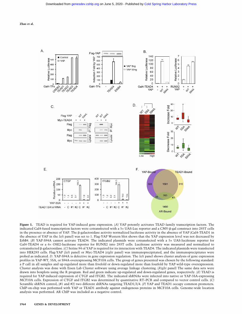
Figure 1. TEAD is required for YAP-induced gene expression. (A) YAP potently activates TEAD family transcription factors. Theindicated Gal4-fused transcription factors were cotransfected with a 5× UAS-Luc reporter and a CMV-�-gal construct into 293T cellsin the presence or absence of YAP. The �-galactosidase activity normalized luciferase activity in the absence of YAP (Gal4-TEAD1 inthe absence of YAP in the left panel) was set to 1. Flag-YAP Western blot shows that the YAP expression level was not decreased byErbB4. (B) YAP-S94A cannot activate TEAD4. The indicated plasmids were cotransfected with a 5× UAS-luciferase reporter forGal4-TEAD4 or a 6× OSE2-luciferase reporter for RUNX2 into 293T cells. Luciferase activity was measured and normalized tocotransfected �-galactosidase. (C) Serine 94 of YAP is required for its interaction with TEAD4. The indicated plasmids were transfectedinto HEK293 cells. Flag-YAP (left panel) or Myc-TEAD4 (right panel) was immunoprecipitated, and the immunoprecipitates wereprobed as indicated. D. YAP-S94A is defective in gene expression regulation. The left panel shows cluster analysis of gene expressionprofiles in YAP-WT, 5SA, or S94A-overexpressing MCF10A cells. The group of genes presented was chosen by the following standard:a P call in all samples and up-regulated more than fivefold or down-regulated more than fourfold by YAP-wild-type overexpression.Cluster analysis was done with Eisen Lab Cluster software using average linkage clustering. (Right panel) The same data sets weredrawn into boxplots using the R program. Red and green indicate up-regulated and down-regulated genes, respectively. (E) TEAD isrequired for YAP-induced expression of CTGF and ITGB2. The indicated shRNAs were infected into native or YAP-5SA-expressingMCF10A cells. Expression of CTGF and ITGB2 was determined by quantitative RT–PCR and compared to vector control cells. (C)Scramble shRNA control; (#1 and #2) two different shRNAs targeting TEAD1/3/4. (F) YAP and TEAD1 occupy common promoters.ChIP-on-chip was performed with YAP or TEAD1 antibody against endogenous proteins in MCF10A cells. Genome-wide locationanalysis was performed. AR ChIP was included as a negative control.
Zhao et al.
1964 GENES & DEVELOPMENT
Cold Spring Harbor Laboratory Press on June 5, 2020 - Published by genesdev.cshlp.orgDownloaded from

TEAD binding is required for YAP-induced cell growthand EMT
We reported that YAP expression in NIH-3T3 cells en-hances cell growth (Zhao et al. 2007). NIH-3T3 stablepools with expression of YAP and YAP-S94A were estab-lished, and cell growth was determined. We found thatYAP-S94A was much less potent than the wild-type YAPto stimulate NIH-3T3 cell growth (Fig. 2A). Further-more, in MCF10A cells, wild-type YAP induced cell pro-liferation even when cells reached confluency, while theYAP-S94A mutant was largely inactive as determined bythe staining of proliferation marker Ki67 (SupplementalFig. S2A). To confirm that the loss of growth-promotingactivity in YAP-S94A is due to the loss of its interactionwith TEAD, we generated a TEAD1-YAP-S94A fusionprotein. Interestingly, this fusion protein stimulatedNIH-3T3 cell growth as effectively as the wild-type YAP,while neither TEAD1 nor YAP-S94A stimulated cellgrowth (Fig. 2A). Furthermore, the TEAD1-YAP-S94Afusion also rescued the expression of Ctgf and Inhba, twoYAP target genes, in NIH-3T3 cells (Fig. 2B). We alsoexamined the effect of S94A mutation in the constitu-tively active YAP-5SA background in MCF10A cells. Ex-pression of YAP-5SA resulted in the formation of muchlarger acini in three-dimensional (3D) culture comparedwith vector control. Importantly, this effect was largelyreduced if an S94A mutation was introduced into YAP-5SA (Fig. 2C). These results indicate that S94, henceTEAD binding, is required for YAP-induced cell prolif-eration.
It has been reported that YAP induces EMT inMCF10A cells (Overholtzer et al. 2006). Indeed, expres-sion of the active YAP-5SA induced EMT-like morpho-logical change in monolayer culture (Fig. 2C). However,YAP-5SA-S94A was not effective in eliciting EMT mor-phology. Furthermore, in 3D culture, YAP-5SA-S94Afailed to induce complex-shaped large acini with spike-like projections and rough surface, which were obviousin YAP-5SA-expressing cultures (Fig. 2C). As anotherhallmark of EMT, YAP-5SA-expressing cells also dis-played disorganized adherens junctions, as shown by theloss of cell–cell junction localized E-cadherin, and theswitch from cortical actin to stress fibers (Fig. 2D). How-ever, these phenotypes were not seen in YAP-5SA-S94A-expressing cells. YAP-5SA expression also changed theexpression pattern of epithelial and mesenchymal mark-ers, which was not induced by YAP-5SA-S94A expres-sion (Fig. 2E). These results indicate that S94 of YAP,presumably by mediating TEAD interaction, is at leastpartially responsible for YAP function in inducing EMT.
To further confirm the function of TEAD, we usedshRNAs to knock down TEAD1/3/4 in YAP-5SA-ex-pressing cells. TEAD1/3/4 knockdown not only reversedthe EMT-like morphology in monolayer and 3D cul-tures, but also rescued the expression of epithelial mark-ers (Fig. 2F; Supplemental Fig. S2B). Knockdown ofTEAD1/3/4 also significantly shrank the aberrantly en-larged acini caused by YAP-5SA expression, further sup-porting a role of TEAD in YAP-induced growth. A YAP-
dependent function of TEAD in cell growth is also im-plicated in Sveinsson’s chorioretinal atrophy, a raregenetic disease caused by TEAD1 mutation and charac-terized by atrophic lesions involving retina and choroids(Fossdal et al. 2004; Kitagawa 2007). The mutated tyro-sine Y406 is highly conserved in TEAD family members(Supplemental Fig. S2C), and is located within the YAP-binding domain (Supplemental Fig. S2D). Interestingly,mutation of this tyrosine residue in TEADs abolishedtheir interaction with and their activation by YAP(Supplemental Fig. S2E–G), which may explain the atro-phic phenotype caused by this mutation.
Anchorage-independent growth is a hallmark of onco-genic transformation. YAP overexpression is reported toinduce anchorage-independent growth of MCF10A cells(Overholtzer et al. 2006). We observed that YAP-5SA po-tently induced MCF10A colony formation in soft agar. Incontrast, YAP-5SA-S94A was unable to induce anchor-age-independent growth of MCF10A cells (Fig. 2G;Supplemental Fig. S2H). Similarly, almost no colony wasformed if TEAD1/3/4 were down-regulated in the YAP-5SA expressing cells (Fig. 2G; Supplemental Fig. S2H).These data indicate the requirement of at least one ofTEAD1/3/4 for the YAP-induced anchorage-independentgrowth. Together, the above observations support amodel in which TEAD is essential for the function ofYAP in cell proliferation, EMT, and oncogenic transfor-mation.
CTGF is a direct YAP-TEAD target gene requiredfor cell growth
YAP expression affected many cell proliferation-relatedgenes (Supplemental Table S1). However, cyclin E andIAP, the key Yki-inducible genes in Drosophila, were notsignificantly induced by YAP in either NIH-3T3 orMCF10A cells (Supplemental Table S1). This indicatesthat there might be different genes in mammalian cellsto mediate YAP function. CTGF is highly induced byYAP expression in both NIH-3T3 and MCF10A cells,and its promoter is co-occupied by YAP and TEAD1, asshown by ChIP (Fig. 3A); therefore, it might be a directYAP target gene. We cloned the CTGF promoter into abasic luciferase reporter and found that it was potentlyactivated by YAP but not by YAP-S94A, and the activa-tion was further enhanced by TEAD1 coexpression (Fig.3B). Expression of the dominant-negative TEAD1-�C,but not the TEAD1-�C-AD (in which the C-terminalYAP-binding domain was replaced by the YAP transac-tivation domain), blocked the activation of CTGF re-porter by YAP (Fig. 3C). These results indicate that YAPactivates the CTGF promoter through TEAD. Examina-tion of the CTGF promoter region revealed three puta-tive TEAD-binding sites (Fig. 3D; Anbanandam et al.2006). Individual or combinatory mutation of the puta-tive TEAD-binding sites indicated that TB2 and TB3were more important for CTGF promoter activity whileTB1 was also involved (Fig. 3E).
The function of endogenous YAP and TEAD in CTGFexpression was examined by YAP or TEAD1/3/4 knock-
TEAD in the Hippo pathway
GENES & DEVELOPMENT 1965
Cold Spring Harbor Laboratory Press on June 5, 2020 - Published by genesdev.cshlp.orgDownloaded from
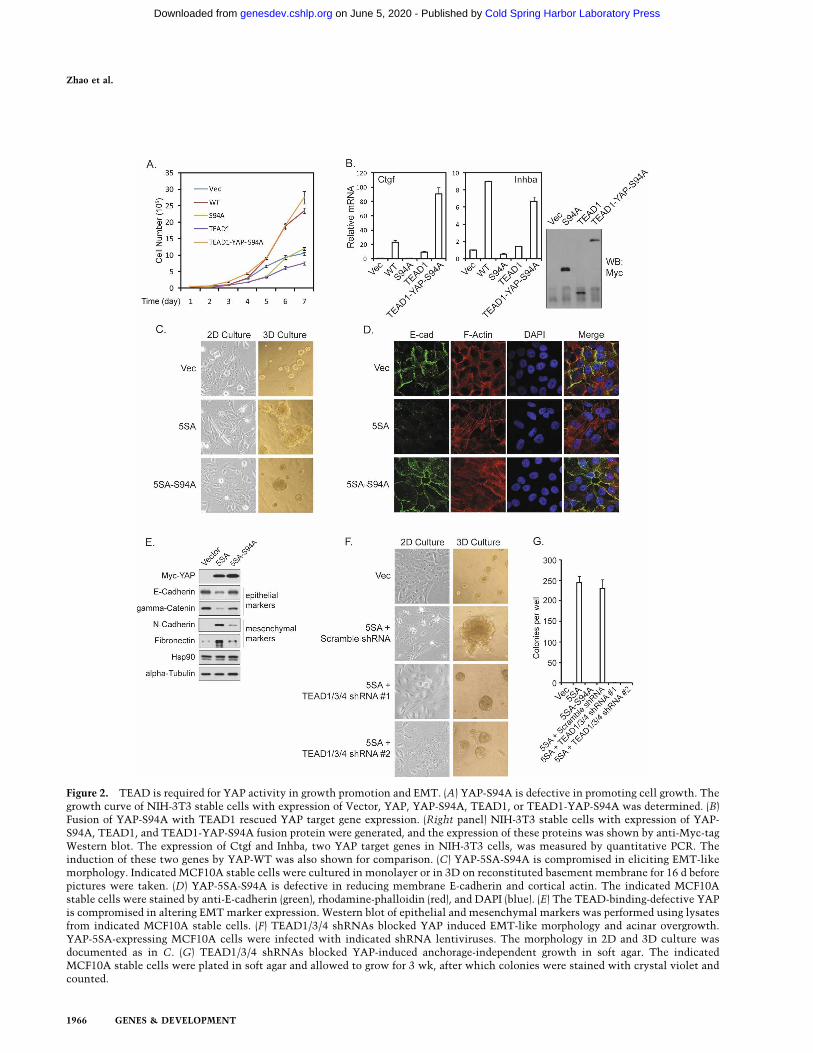
Figure 2. TEAD is required for YAP activity in growth promotion and EMT. (A) YAP-S94A is defective in promoting cell growth. Thegrowth curve of NIH-3T3 stable cells with expression of Vector, YAP, YAP-S94A, TEAD1, or TEAD1-YAP-S94A was determined. (B)Fusion of YAP-S94A with TEAD1 rescued YAP target gene expression. (Right panel) NIH-3T3 stable cells with expression of YAP-S94A, TEAD1, and TEAD1-YAP-S94A fusion protein were generated, and the expression of these proteins was shown by anti-Myc-tagWestern blot. The expression of Ctgf and Inhba, two YAP target genes in NIH-3T3 cells, was measured by quantitative PCR. Theinduction of these two genes by YAP-WT was also shown for comparison. (C) YAP-5SA-S94A is compromised in eliciting EMT-likemorphology. Indicated MCF10A stable cells were cultured in monolayer or in 3D on reconstituted basement membrane for 16 d beforepictures were taken. (D) YAP-5SA-S94A is defective in reducing membrane E-cadherin and cortical actin. The indicated MCF10Astable cells were stained by anti-E-cadherin (green), rhodamine-phalloidin (red), and DAPI (blue). (E) The TEAD-binding-defective YAPis compromised in altering EMT marker expression. Western blot of epithelial and mesenchymal markers was performed using lysatesfrom indicated MCF10A stable cells. (F) TEAD1/3/4 shRNAs blocked YAP induced EMT-like morphology and acinar overgrowth.YAP-5SA-expressing MCF10A cells were infected with indicated shRNA lentiviruses. The morphology in 2D and 3D culture wasdocumented as in C. (G) TEAD1/3/4 shRNAs blocked YAP-induced anchorage-independent growth in soft agar. The indicatedMCF10A stable cells were plated in soft agar and allowed to grow for 3 wk, after which colonies were stained with crystal violet andcounted.
Zhao et al.
1966 GENES & DEVELOPMENT
Cold Spring Harbor Laboratory Press on June 5, 2020 - Published by genesdev.cshlp.orgDownloaded from

Figure 3. CTGF is a direct target of YAP and TEAD. (A) Both YAP and TEAD1 bind to the CTGF promoter. ChIP from MCF10A cellswas performed with control IgG, YAP, or TEAD1 antibody as indicated. The presence of CTGF promoter was detected by PCR. (B)Activation of CTGF reporter by YAP and TEAD1. A luciferase reporter driven by CTGF promoter was cotransfected with YAP wildtype or S94A mutant as indicated with or without TEAD1 cotransfection. Luciferase activity was measured and normalized tocotransfected �-galactosidase. (C) Dominant-negative TEAD1 blocks the YAP stimulation of the CTGF reporter. The indicatedplasmids were cotransfected, and luciferase activity was determined as in B. (D) The human CTGF promoter region contains threeputative TEAD-binding sites. The putative TEAD-binding sites (TB1–TB3) are shown in red. (E) The putative TEAD-binding sites areimportant for CTGF promoter activity. The putative TEAD-binding sites (TB) were mutated individually or in combination. Theluciferase activity of each reporter was measured in the presence or absence of YAP and TEAD1. The activation folds by YAP andTEAD1 are shown. (F) YAP and TEAD are required for CTGF expression. ACHN cells were infected with the indicated shRNAlentiviruses, and CTGF mRNA levels were determined by quantitative RT–PCR. (G) Knockdown of YAP or TEAD1/3/4 decreasesCTGF protein levels. Experiments were similar to F except Western blotting was performed with the indicated antibodies. (H) YAP,TEAD, and CTGF are important for the AHCN cell growth. YAP, TEAD1/3/4, and CTGF were knocked down by shRNAs. Cell growthrate was determined. (I) CTGF is required for YAP-induced growth and morphological change in 3D culture. MCF10A cells expressingYAP-5SA were infected with indicated shRNA lentiviruses. The morphology in 2D and 3D culture was documented as in Figure 2C.(J) CTGF knockdown attenuates YAP induced anchorage-independent growth in soft agar. The indicated MCF10A stable cells wereplated in soft agar and allowed to grow for 3 wk, after which colonies were stained with crystal violet and counted. Pictures of thestained colonies were presented in higher magnification to show the colony size reduction by CTGF shRNAs.
TEAD in the Hippo pathway
GENES & DEVELOPMENT 1967
Cold Spring Harbor Laboratory Press on June 5, 2020 - Published by genesdev.cshlp.orgDownloaded from
down in ACHN cells, which have elevated YAP activitydue to a mutation of Sav, a key component of the Hippopathway (Tapon et al. 2002). RNAi specificity and effi-ciency were confirmed by quantitative RT–PCR (Supple-mental Fig. S3A) and Western blot (Fig. 3G). We foundthat knockdown of either YAP or TEAD1/3/4 caused adramatic reduction of both CTGF mRNA (Fig. 3F) andprotein (Fig. 3G). We next examined the function ofCTGF in mediating the cellular function of YAP. Similarto the knockdown of YAP and TEAD1/3/4, knockdownof CTGF significantly inhibited ACHN cell growth (Fig.3H). These data further demonstrate the functional sig-nificance of TEAD1/3/4 and CTGF as important down-stream targets of YAP in the Hippo pathway in cellgrowth regulation. Furthermore, knockdown of CTGF inthe YAP-5SA-expressing MCF10A cells decreased theacini growth and reversed the complex-shaped and roughsurface morphology in 3D culture (Fig. 3I). However,CTGF knockdown did not reverse the EMT-like mor-phology in monolayer culture. These results indicatethat CTGF plays an important role in the growth-pro-moting function but may not be required for the EMT-inducing activity of YAP.
We also tested the effect of CTGF knockdown in theanchorage-independent growth potential of YAP-5SA-overexpressing MCF10A cells. Although CTGF knock-down did not completely block the anchorage-indepen-dent growth of YAP-5SA-overexpressing MCF10A cells,it significantly decreased the number of colonies formed(Fig. 3J; Supplemental Fig. S3B) and dramatically reducedthe colony size (Fig. 3J). However, expression of CTGFalone did not phenocopy the effects of YAP overexpres-sion in MCF10A cells (data not shown). Therefore, wespeculate that CTGF works with other YAP target genesto mediate the oncogenic transformation potential ofYAP.
YAP/Yki and TEAD/Sd genetically interact to promotetissue growth in Drosophila
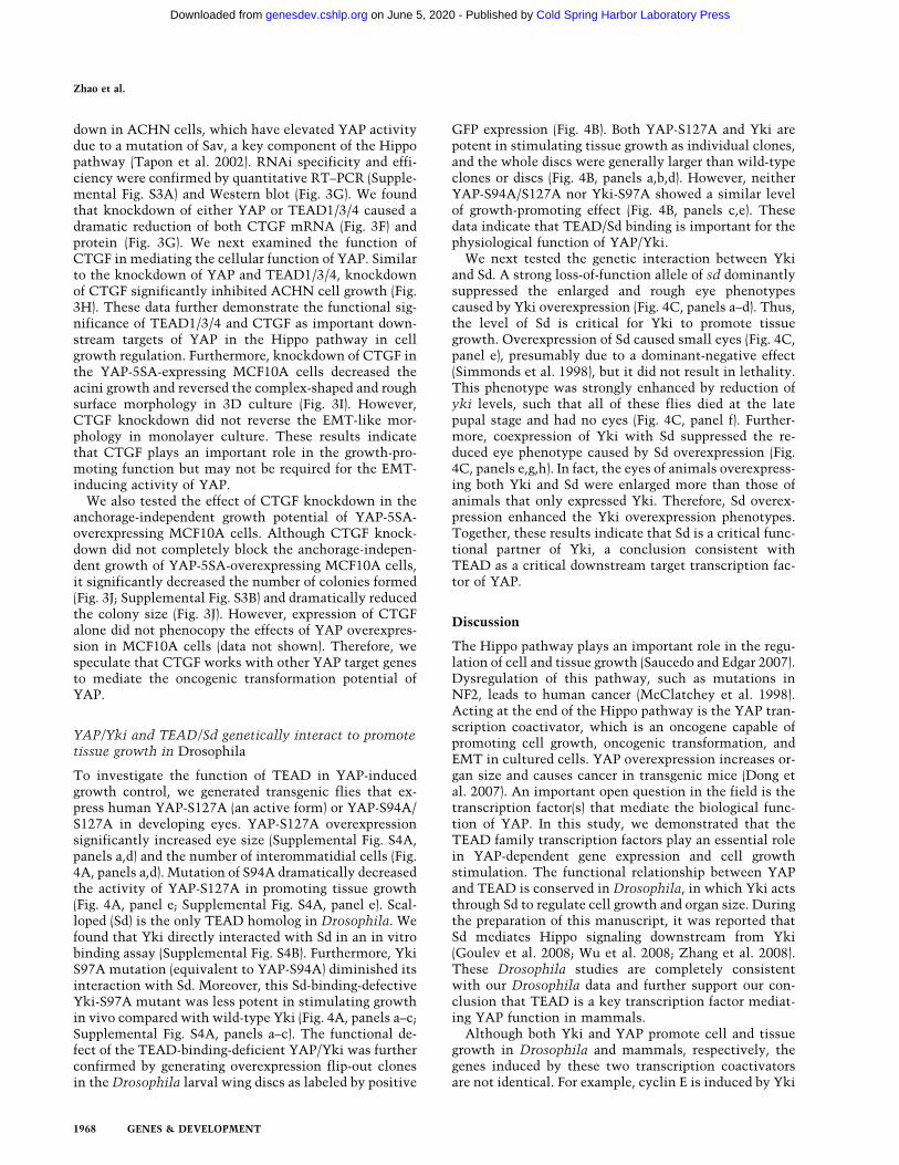
To investigate the function of TEAD in YAP-inducedgrowth control, we generated transgenic flies that ex-press human YAP-S127A (an active form) or YAP-S94A/S127A in developing eyes. YAP-S127A overexpressionsignificantly increased eye size (Supplemental Fig. S4A,panels a,d) and the number of interommatidial cells (Fig.4A, panels a,d). Mutation of S94A dramatically decreasedthe activity of YAP-S127A in promoting tissue growth(Fig. 4A, panel e; Supplemental Fig. S4A, panel e). Scal-loped (Sd) is the only TEAD homolog in Drosophila. Wefound that Yki directly interacted with Sd in an in vitrobinding assay (Supplemental Fig. S4B). Furthermore, YkiS97A mutation (equivalent to YAP-S94A) diminished itsinteraction with Sd. Moreover, this Sd-binding-defectiveYki-S97A mutant was less potent in stimulating growthin vivo compared with wild-type Yki (Fig. 4A, panels a–c;Supplemental Fig. S4A, panels a–c). The functional de-fect of the TEAD-binding-deficient YAP/Yki was furtherconfirmed by generating overexpression flip-out clonesin the Drosophila larval wing discs as labeled by positive
GFP expression (Fig. 4B). Both YAP-S127A and Yki arepotent in stimulating tissue growth as individual clones,and the whole discs were generally larger than wild-typeclones or discs (Fig. 4B, panels a,b,d). However, neitherYAP-S94A/S127A nor Yki-S97A showed a similar levelof growth-promoting effect (Fig. 4B, panels c,e). Thesedata indicate that TEAD/Sd binding is important for thephysiological function of YAP/Yki.
We next tested the genetic interaction between Ykiand Sd. A strong loss-of-function allele of sd dominantlysuppressed the enlarged and rough eye phenotypescaused by Yki overexpression (Fig. 4C, panels a–d). Thus,the level of Sd is critical for Yki to promote tissuegrowth. Overexpression of Sd caused small eyes (Fig. 4C,panel e), presumably due to a dominant-negative effect(Simmonds et al. 1998), but it did not result in lethality.This phenotype was strongly enhanced by reduction ofyki levels, such that all of these flies died at the latepupal stage and had no eyes (Fig. 4C, panel f). Further-more, coexpression of Yki with Sd suppressed the re-duced eye phenotype caused by Sd overexpression (Fig.4C, panels e,g,h). In fact, the eyes of animals overexpress-ing both Yki and Sd were enlarged more than those ofanimals that only expressed Yki. Therefore, Sd overex-pression enhanced the Yki overexpression phenotypes.Together, these results indicate that Sd is a critical func-tional partner of Yki, a conclusion consistent withTEAD as a critical downstream target transcription fac-tor of YAP.
Discussion
The Hippo pathway plays an important role in the regu-lation of cell and tissue growth (Saucedo and Edgar 2007).Dysregulation of this pathway, such as mutations inNF2, leads to human cancer (McClatchey et al. 1998).Acting at the end of the Hippo pathway is the YAP tran-scription coactivator, which is an oncogene capable ofpromoting cell growth, oncogenic transformation, andEMT in cultured cells. YAP overexpression increases or-gan size and causes cancer in transgenic mice (Dong etal. 2007). An important open question in the field is thetranscription factor(s) that mediate the biological func-tion of YAP. In this study, we demonstrated that theTEAD family transcription factors play an essential rolein YAP-dependent gene expression and cell growthstimulation. The functional relationship between YAPand TEAD is conserved in Drosophila, in which Yki actsthrough Sd to regulate cell growth and organ size. Duringthe preparation of this manuscript, it was reported thatSd mediates Hippo signaling downstream from Yki(Goulev et al. 2008; Wu et al. 2008; Zhang et al. 2008).These Drosophila studies are completely consistentwith our Drosophila data and further support our con-clusion that TEAD is a key transcription factor mediat-ing YAP function in mammals.
Although both Yki and YAP promote cell and tissuegrowth in Drosophila and mammals, respectively, thegenes induced by these two transcription coactivatorsare not identical. For example, cyclin E is induced by Yki
Zhao et al.
1968 GENES & DEVELOPMENT
Cold Spring Harbor Laboratory Press on June 5, 2020 - Published by genesdev.cshlp.orgDownloaded from

overexpression in Drosophila but not by YAP overex-pression in mammalian cells (Dong et al. 2007). We iden-tified CTGF as a direct target gene of YAP-TEAD inmammalian cells. Interestingly, elevated CTGF levelshave been detected in human cancers (Xie et al. 2001),and anti-CTGF antibody inhibited tumor growth andmetastasis (Dornhofer et al. 2006). This supports a pos-sible role of CTGF in mediating the growth-stimulatingand oncogenic function of YAP-TEAD. Although CTGFappears to play an important role in YAP-induced cellgrowth, it may not be required for YAP-induced EMT.This indicates that other genes may be involved in thebiological function of YAP. Consistently, the TEAD-binding-defective YAP-S94A mutant can still induce ex-
pression of a fraction of the YAP-regulated genes. Fur-thermore, overexpression of the Sd-binding-defectiveYki-S97A elicits a significantly reduced but still obviousovergrowth in Drosophila eyes and wings. These obser-vations indicate that additional transcription factorsmay be used by YAP/Yki to regulate cell and tissuegrowth.
Materials and methods
Cell culture, transfection, and retroviral infection
HEK293 cells, HEK293-T cells, NIH-3T3 cells, and ACHN cellswere cultured in DMEM (Invitrogen) containing 10% FBS (In-
Figure 4. yki and scalloped genetically interact to control tissue growth and organ size. (A) The TEAD/Sd-binding-defective YAP andYki are compromised in inducing extra interommatidial cells. Mid-pupal eye discs were stained with Discs large (Dlg) antibody tooutline cells. The genotypes of the fly tissues are Wild-type (Canton S) (panel a), GMR-Gal4/UAS-yki-V5 (panel b), GMR-Gal4/UAS-ykiS97A-V5 (panel c), GMR-Gal4/UAS-Flag-YAPS127A (panel d), and GMR-Gal4/UAS-Flag-YAPS94A/S127A (panel e). (B) The TEAD/Sd-binding-defective YAP and Yki are compromised in inducing clone expansion. Wing imaginal discs containing 72-h-old control (panela) or various YAP/Yki-overexpressing clones (panels b–e) were generated by flip-out and positively marked by GFP. The genotypes ofthe fly tissues are hsFLP/+; act>y+>Gal4, UAS-GFPS65T/+ (panel a), hsFLP/+; act>y+>Gal4, UAS-GFPS65T/UAS-yki-V5 (panel b),hsFLP/+; act>y+>Gal4, UAS-GFPS65T/UAS-ykiS97A-V5 (panel c), hsFLP/+; act>y+>Gal4, UAS-GFPS65T/UAS-Flag-YAPS127A (panel d),and hsFLP/+; act>y+>Gal4, UAS-GFPS65T/UAS-Flag-YAPS94A/S127A (panel e). (C) yki and scalloped genetically interact to control tissuegrowth and organ size. Genotypes of the fly tissues are indicated. SEM (scanning electron microscopy) images of adult eyes are shownin panels a–e, g, and h. A late pupal head is shown in panel f. The arrow in panel f indicates where a retina is normally expected togrow.
TEAD in the Hippo pathway
GENES & DEVELOPMENT 1969
Cold Spring Harbor Laboratory Press on June 5, 2020 - Published by genesdev.cshlp.orgDownloaded from

vitrogen) and 50 µg/mL penicillin/streptomycin (P/S). MCF10Acells were cultured in DMEM/F12 (Invitrogen) supplementedwith 5% horse serum (Invitrogen), 20 ng/mL EGF, 0.5 µg/mLhydrocortisone, 10 µg/mL insulin, 100 ng/mL cholera toxin, and50 µg/mL P/S. Transfection with lipofectamine was performedaccording to the manufacturer’s instructions.
To generate wild-type or mutant YAP-expressing stable cells,retrovirus infection was performed by transfecting 293 Phoenixretrovirus packaging cells with empty vector or pQCXIH-YAPconstructs. Forty-eight hours after transfection, retroviral su-pernatant was supplemented with 5 µg/mL polybrene, filteredthrough a 0.45-µm filter, and used to infect MCF10A or NIH-3T3 cells. Thirty-six hours after infection, cells were selectedwith 200 µg/mL hygromycin (Roche) in culture medium.
Lentiviral shRNA cloning, production, and infection
To generate YAP, TEAD1/3/4, or CTGF knockdown cells, oli-gonucleotides were cloned into pLKO.1 with the AgeI/EcoRIsites (Moffat et al. 2006). TEAD1/3/4 shRNAs were designed ina region identical in TEAD1, 3, and 4. The sequences of theoligonucleotides are as follows: YAP #1-sense, 5�-CCGGCTGGTCAGAGATACTTCTTAACTCGAGTTAAGAAGTATCTCTGACCAGTTTTTC-3�; YAP #1-antisense, 5�-AATTGAAAAACTGGTCAGAGATACTTCTTAACTCGAGTTAAGAAGTATCTCTGACCAG-3�; YAP #2-sense, 5�-CCGGAAGCTTTGAGTTCTGACATCCCTCGAGGGATGTCAGAACTCAAAGCTTTTTTTC-3�; YAP #2-antisense, 5�-AATTGAAAAAAAGCTTTGAGTTCTGACATCCCTCGAGGGATGTCAGAACTCAAAGCTT-3�; TEAD1/3/4 #1-sense, 5�-CCGGATGATCAACTTCATCCACAAGCTCGAGCTTGTGGATGAAGTTGATCATTTTTTC-3�; TEAD1/3/4 #1-antisense, 5�-AATTGAAAAAATGATCAACTTCATCCACAAGCTCGAGCTTGTGGATGAAGTTGATCAT-3�; TEAD1/3/4 #2-sense, 5�-CCGGGATCAACTTCATCCACAAGCTCTCGAGAGCTTGTGGATGAAGTTGATCTTTTTC-3�; TEAD1/3/4 #2-antisense, 5�-AATTGAAAAAGATCAACTTCATCCACAAGCTCTCGAGAGCTTGTGGATGAAGTTGATC-3�; CTGF #1-sense, 5�-CCGGAAATCTCCAAGCCTATCAAGTCTCGAGACTTGATAGGCTTGGAGATTTTTTTTC-3�; CTGF #1-antisense, 5�-AATTGAAAAAAAATCTCCAAGCCTATCAAGTCTCGAGACTTGATAGGCTTGGAGATTT-3�; CTGF #2-sense, 5�-CCGGCTGCACCAGCATGAAGACATACTCGAGTATGTCTTCATGCTGGTGCAGTTTTTC-3�; CTGF #2-antisense, 5�-AATTGAAAAACTGCACCAGCATGAAGACATACTCGAGTATGTCTTCATGCTGGTGCAG-3�.
Plasmids were propagated in and purified from Stbl2 compe-tent cells (Invitrogen). The infection process was similar to thatof retroviral infection except that the lentiviral packaging plas-mids psPAX2 and pMD2.G were cotransfected into HEK293-Tcells for virus production. Cells were selected in 5 µg/mL pu-romycin in culture medium.
ChIP and ChIP-on-chip
ChIP-on-chip and genome-wide location analysis were per-formed as previously described (Yu et al. 2007). Briefly, cellswere cross-linked, lysed, and sonicated to generate DNA frag-ments with an average size of 0.5 kb. ChIP was performed using5 µg of antibodies against YAP, TEAD1, AR, or control IgG.ChIP-enriched DNA, along with input whole lysate DNA, weresubjected to a ligation-mediated PCR step to generate enoughDNA materials, which were then labeled with fluorescent dyesand hybridized to a promoter microarray according to the manu-
facturer’s protocols (Agilent Technologies). The hybridizationintensity was extracted using the Agilent Feature ExtractionSoftware. The bound probes were determined at a cut-off P-value of XDEV, which is a scaled log-ratio value generated fromsingle-gene error model, <0.001.
Three-dimensional culture of MCF10A cells
The 3D culture of MCF10A cells was done as described (Deb-nath et al. 2003). Briefly, Growth Factor Reduced Matrigel waslayered onto eight-well glass chamber slides to make a recon-stituted basement membrane. MCF10A cells were seeded ontop of that at a concentration of 5000 cells/well in assay me-dium containing 2% Matrigel and 5 ng/mL EGF. Cells werecultured in a 5% CO2 humidified incubator at 37°C. The me-dium was replaced every 4 d.
Acknowledgments
We thank Drs. Marius Sudol for pCMV-Flag-YAP2 and pM-ErbB4-CTF�K constructs, David M. Sabatini for the pLKO.1vector, Sean Carroll for pGST-sd, Duojia Pan for pGal4-yki,Hongjiao Ouyang for RUNX2 and 6× OSE2-luc reporter, JingYang for EMT marker antibodies, and Taocong Jin for assistanceon gene expression microarray. We thank Dr. Georg Halder forhsFLP; act>y+>GFP-S65T, the Bloomington Drosophila StockCenter for sd12 and UAS-sd fly strains, and the DevelopmentalStudies Hybridoma Bank at the University of Iowa for Dlg an-tibody. This work is supported by grants from NIH (to K.L.G.),NIH (to C.Y.W.), NSF (to Z.C.L.), and Rackham GraduateSchool, University of Michigan (to B.Z.).
References
Anbanandam, A., Albarado, D.C., Nguyen, C.T., Halder, G.,Gao, X., and Veeraraghavan, S. 2006. Insights into transcrip-tion enhancer factor 1 (TEF-1) activity from the solutionstructure of the TEA domain. Proc. Natl. Acad. Sci. 103:17225–17230.
Basu, S., Totty, N.F., Irwin, M.S., Sudol, M., and Downward, J.2003. Akt phosphorylates the Yes-associated protein, YAP,to induce interaction with 14–3–3 and attenuation of p73-mediated apoptosis. Mol. Cell 11: 11–23.
Camargo, F.D., Gokhale, S., Johnnidis, J.B., Fu, D., Bell, G.W.,Jaenisch, R., and Brummelkamp, T.R. 2007. YAP1 increasesorgan size and expands undifferentiated progenitor cells.Curr. Biol. 17: 2054–2060.
Debnath, J., Muthuswamy, S.K., and Brugge, J.S. 2003. Morpho-genesis and oncogenesis of MCF-10A mammary epithelialacini grown in three-dimensional basement membrane cul-tures. Methods 30: 256–268.
Dong, J., Feldmann, G., Huang, J., Wu, S., Zhang, N., Comer-ford, S.A., Gayyed, M.F., Anders, R.A., Maitra, A., and Pan,D. 2007. Elucidation of a universal size-control mechanismin Drosophila and mammals. Cell 130: 1120–1133.
Dornhofer, N., Spong, S., Bennewith, K., Salim, A., Klaus, S.,Kambham, N., Wong, C., Kaper, F., Sutphin, P., Nacamuli,R., et al. 2006. Connective tissue growth factor-specificmonoclonal antibody therapy inhibits pancreatic tumorgrowth and metastasis. Cancer Res. 66: 5816–5827.
Edgar, B.A. 2006. From cell structure to transcription: Hippoforges a new path. Cell 124: 267–273.
Fossdal, R., Jonasson, F., Kristjansdottir, G.T., Kong, A., Stefans-son, H., Gosh, S., Gulcher, J.R., and Stefansson, K. 2004. Anovel TEAD1 mutation is the causative allele in Sveinsson’s
Zhao et al.
1970 GENES & DEVELOPMENT
Cold Spring Harbor Laboratory Press on June 5, 2020 - Published by genesdev.cshlp.orgDownloaded from

chorioretinal atrophy (helicoid peripapillary chorioretinaldegeneration). Hum. Mol. Genet. 13: 975–981.
Goulev, Y., Fauny, J.D., Gonzalez-Marti, B., Flagiello, D., Silber,J., and Zider, A. 2008. SCALLOPED interacts with YORKIE,the nuclear effector of the Hippo tumor-suppressor pathwayin Drosophila. Curr. Biol. 18: 435–441.
Hao, Y., Chun, A., Cheung, K., Rashidi, B., and Yang, X. 2007.Tumor suppressor LATS1 is a negative regulator of oncogeneYAP. J. Biol. Chem. 283: 5496–5509.
Hariharan, I.K. and Bilder, D. 2006. Regulation of imaginal discgrowth by tumor-suppressor genes in Drosophila. Annu.Rev. Genet. 40: 335–361.
Harvey, K. and Tapon, N. 2007. The Salvador-Warts-Hippopathway—An emerging tumour-suppressor network. Nat.Rev. Cancer 7: 182–191.
Harvey, K.F., Pfleger, C.M., and Hariharan, I.K. 2003. The Dro-sophila Mst ortholog, Hippo, restricts growth and cell pro-liferation and promotes apoptosis. Cell 114: 457–467.
Huang, J., Wu, S., Barrera, J., Matthews, K., and Pan, D. 2005.The Hippo signaling pathway coordinately regulates cell pro-liferation and apoptosis by inactivating Yorkie, the Dro-sophila homolog of YAP. Cell 122: 421–434.
Kitagawa, M. 2007. A Sveinsson’s chorioretinal atrophy-associ-ated missense mutation in mouse Tead1 affects its interac-tion with the co-factors YAP and TAZ. Biochem. Biophys.Res. Commun. 361: 1022–1026.
Komuro, A., Nagai, M., Navin, N.E., and Sudol, M. 2003. WWdomain-containing protein YAP associates with ErbB-4 andacts as a co-transcriptional activator for the carboxyl-termi-nal fragment of ErbB-4 that translocates to the nucleus. J.Biol. Chem. 278: 33334–33341.
Lai, Z.C., Wei, X., Shimizu, T., Ramos, E., Rohrbaugh, M.,Nikolaidis, N., Ho, L.L., and Li, Y. 2005. Control of cellproliferation and apoptosis by mob as tumor suppressor,mats. Cell 120: 675–685.
McClatchey, A.I. and Giovannini, M. 2005. Membrane organi-zation and tumorigenesis–the NF2 tumor suppressor, Mer-lin. Genes & Dev. 19: 2265–2277.
McClatchey, A.I., Saotome, I., Mercer, K., Crowley, D., Gusella,J.F., Bronson, R.T., and Jacks, T. 1998. Mice heterozygous fora mutation at the Nf2 tumor suppressor locus develop arange of highly metastatic tumors. Genes & Dev. 12: 1121–1133.
Moffat, J., Grueneberg, D.A., Yang, X., Kim, S.Y., Kloepfer,A.M., Hinkle, G., Piqani, B., Eisenhaure, T.M., Luo, B., Gre-nier, J.K., et al. 2006. A lentiviral RNAi library for humanand mouse genes applied to an arrayed viral high-contentscreen. Cell 124: 1283–1298.
Overholtzer, M., Zhang, J., Smolen, G.A., Muir, B., Li, W., Sgroi,D.C., Deng, C.X., Brugge, J.S., and Haber, D.A. 2006. Trans-forming properties of YAP, a candidate oncogene on thechromosome 11q22 amplicon. Proc. Natl. Acad. Sci. 103:12405–12410.
Saucedo, L.J. and Edgar, B.A. 2007. Filling out the Hippo path-way. Nat. Rev. Mol. Cell Biol. 8: 613–621.
Simmonds, A.J., Liu, X., Soanes, K.H., Krause, H.M., Irvine,K.D., and Bell, J.B. 1998. Molecular interactions betweenVestigial and Scalloped promote wing formation in Dro-sophila. Genes & Dev. 12: 3815–3820.
Subramanian, A., Tamayo, P., Mootha, V.K., Mukherjee, S., Eb-ert, B.L., Gillette, M.A., Paulovich, A., Pomeroy, S.L., Golub,T.R., Lander, E.S., et al. 2005. Gene set enrichment analysis:A knowledge-based approach for interpreting genome-wideexpression profiles. Proc. Natl. Acad. Sci. 102: 15545–15550.
Tapon, N., Harvey, K.F., Bell, D.W., Wahrer, D.C., Schiripo,T.A., Haber, D.A., and Hariharan, I.K. 2002. salvador pro-
motes both cell cycle exit and apoptosis in Drosophila and ismutated in human cancer cell lines. Cell 110: 467–478.
Udan, R.S., Kango-Singh, M., Nolo, R., Tao, C., and Halder, G.2003. Hippo promotes proliferation arrest and apoptosis inthe Salvador/Warts pathway. Nat. Cell Biol. 5: 914–920.
Vassilev, A., Kaneko, K.J., Shu, H., Zhao, Y., and DePamphilis,M.L. 2001. TEAD/TEF transcription factors utilize the acti-vation domain of YAP65, a Src/Yes-associated protein local-ized in the cytoplasm. Genes & Dev. 15: 1229–1241.
Wu, S., Huang, J., Dong, J., and Pan, D. 2003. hippo encodes aSte-20 family protein kinase that restricts cell proliferationand promotes apoptosis in conjunction with salvador andwarts. Cell 114: 445–456.
Wu, S., Liu, Y., Zheng, Y., Dong, J., and Pan, D. 2008. TheTEAD/TEF family protein Scalloped mediates transcrip-tional output of the Hippo growth-regulatory pathway. Dev.Cell 14: 388–398.
Xie, D., Nakachi, K., Wang, H., Elashoff, R., and Koeffler, H.P.2001. Elevated levels of connective tissue growth factor,WISP-1, and CYR61 in primary breast cancers associatedwith more advanced features. Cancer Res. 61: 8917–8923.
Yagi, R., Chen, L.F., Shigesada, K., Murakami, Y., and Ito, Y.1999. A WW domain-containing yes-associated protein(YAP) is a novel transcriptional co-activator. EMBO J. 18:2551–2562.
Yu, J., Rhodes, D.R., Tomlins, S.A., Cao, X., Chen, G., Mehra,R., Wang, X., Ghosh, D., Shah, R.B., Varambally, S., et al.2007. A polycomb repression signature in metastatic pros-tate cancer predicts cancer outcome. Cancer Res. 67: 10657–10663.
Zender, L., Spector, M.S., Xue, W., Flemming, P., Cordon-Cardo, C., Silke, J., Fan, S.T., Luk, J.M., Wigler, M., Hannon,G.J., et al. 2006. Identification and validation of oncogenes inliver cancer using an integrative oncogenomic approach.Cell 125: 1253–1267.
Zhang, L., Ren, F., Zhang, Q., Chen, Y., Wang, B., and Jiang, J.2008. The TEAD/TEF family of transcription factor Scal-loped mediates Hippo signaling in organ size control. Dev.Cell 14: 377–387.
Zhao, B., Wei, X., Li, W., Udan, R.S., Yang, Q., Kim, J., Xie, J.,Ikenoue, T., Yu, J., Li, L., et al. 2007. Inactivation of YAPoncoprotein by the Hippo pathway is involved in cell contactinhibition and tissue growth control. Genes & Dev. 21:2747–2761.
TEAD in the Hippo pathway
GENES & DEVELOPMENT 1971
Cold Spring Harbor Laboratory Press on June 5, 2020 - Published by genesdev.cshlp.orgDownloaded from

10.1101/gad.1664408Access the most recent version at doi: originally published online June 25, 200822:2008, Genes Dev.
Bin Zhao, Xin Ye, Jindan Yu, et al. TEAD mediates YAP-dependent gene induction and growth control
Material
Supplemental
http://genesdev.cshlp.org/content/suppl/2008/07/01/gad.1664408.DC1
References
http://genesdev.cshlp.org/content/22/14/1962.full.html#ref-list-1
This article cites 35 articles, 13 of which can be accessed free at:
License
ServiceEmail Alerting
click here.right corner of the article or
Receive free email alerts when new articles cite this article - sign up in the box at the top
Copyright © 2008, Cold Spring Harbor Laboratory Press
Cold Spring Harbor Laboratory Press on June 5, 2020 - Published by genesdev.cshlp.orgDownloaded from