Embed Size (px)
Citation preview
1/10/2013
1
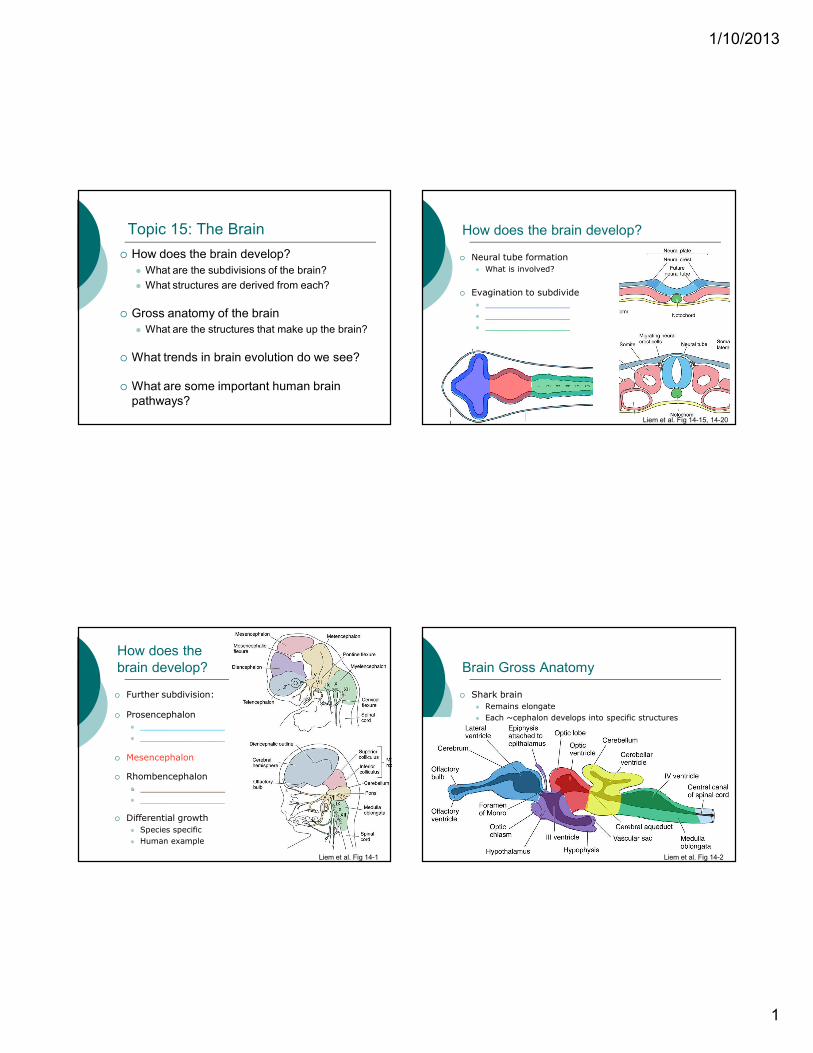
Topic 15: The Brain
� How does the brain develop?
� What are the subdivisions of the brain?
� What structures are derived from each?
� Gross anatomy of the brain
� What are the structures that make up the brain?
� What trends in brain evolution do we see?
� What are some important human brain
pathways?
How does the brain develop?
� Neural tube formation
� What is involved?
� Evagination to subdivide
� _________________
� _________________
� _________________
Liem et al. Fig 14-15, 14-20
How does the
brain develop?
� Further subdivision:
� Prosencephalon
� _________________
� _________________
� Mesencephalon
� Rhombencephalon
� _________________
� _________________
� Differential growth
� Species specific
� Human example
Liem et al. Fig 14-1
Brain Gross Anatomy
� Shark brain
� Remains elongate
� Each ~cephalon develops into specific structures
Liem et al. Fig 14-2
1/10/2013
2
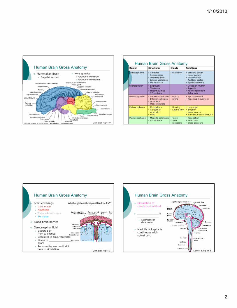
Human Brain Gross Anatomy
� Mammalian Brain
� Saggital section
Liem et al. Fig 14-11
� More spherical
� Growth of cerebrum
� Growth of cerebellum
Human Brain Gross AnatomyRegion Structures Inputs Functions
Telencephalon • Cerebral hemispheres
• Olfactory bulb• Lateral ventricles• Hypocampus
• Olfactory • Sensory cortex• Motor cortex• Visual cortex• Auditory cortex• Spatial memory
Diencephalon • Epiphysis• Thalamus• Hypothalamus• 3rd ventricle
• Circadian rhythm• Appetite• Hormonal control• Relay info
Mesencephalon • Superior colliculus• Inferior colliculus• Optic lobe• Optic ventricle
• Optic / retina
• Eye movement• Reaching movement
Metencephalon • Cerebellum• Cerebellarventricle
• Pons
• Hearing• Lateral line
• Language• Emotion• Motor control• Equilibrium/coordination
Myelencephalon • Medulla oblongata• 4th ventricle
• Taste• Skin receptors
• Respiration• Heart rate• Blood pressure
Human Brain Gross Anatomy
� Brain coverings
� Dura mater
� Arachnoid
� Subarachnoid space
� Pia mater
� Blood-brain barrier
� Cerebrospinal fluid
� Secreted by ____________
from capillaries
� Circulates in brain ventricles
� Moves to _______________
space
� Removed by arachnoid villi
back to circulationLiem et al. Fig 14-3
What might cerebrospinal fluid be for?
Human Brain Gross Anatomy
� Circulation of cerebrospinal fluid
� _____________ & _____________� Extensions of dura mater
� Medulla oblogata is continuous with spinal cord
Liem et al. Fig 14-3
1/10/2013
1
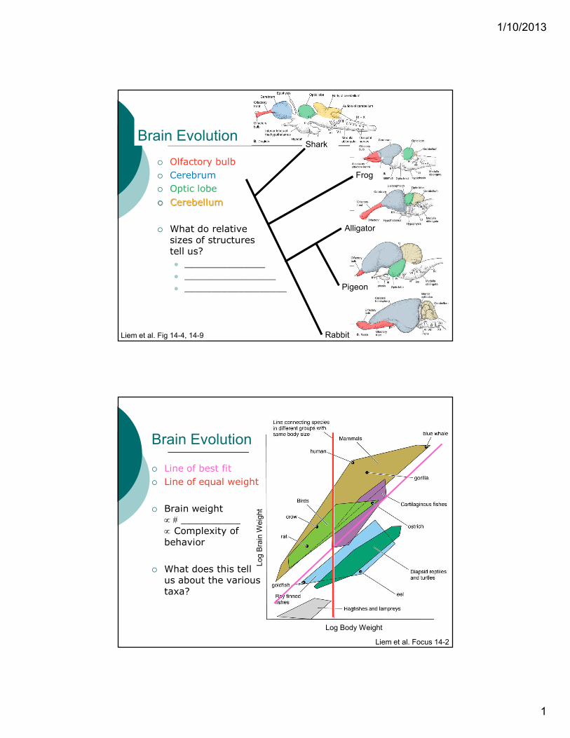
Brain Evolution
� Olfactory bulb
� Cerebrum
� Optic lobe
� Cerebellum
� What do relative sizes of structures tell us?
� _______________
� _________________
� ___________________
Liem et al. Fig 14-4, 14-9
Shark
Frog
Alligator
Pigeon
Rabbit
Brain Evolution
Liem et al. Focus 14-2
Log Body Weight
Log Brain Weight
� Line of best fit
� Line of equal weight
� Brain weight ∝ # __________ ∝ Complexity of
behavior
� What does this tell us about the various taxa?
1/10/2013
1
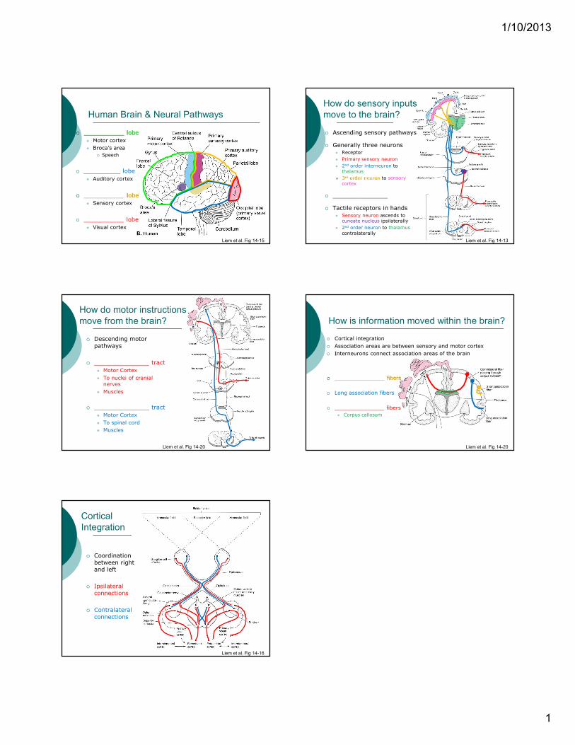
Human Brain & Neural Pathways
� ___________ lobe
� Motor cortex
� Broca’s area
� Speech
� __________ lobe
� Auditory cortex
� ___________ lobe
� Sensory cortex
� ___________ lobe
� Visual cortex
Liem et al. Fig 14-15
How do sensory inputs
move to the brain?
� Ascending sensory pathways
� Generally three neurons� Receptor
� Primary sensory neuron
� 2nd order interneuron to thalamus
� 3rd order neuron to sensory
cortex
� _______________
� Tactile receptors in hands
� Sensory neuron ascends to cuneate nucleus ipsilaterally
� 2nd order neuron to thalamus
contralaterally
Liem et al. Fig 14-13
How do motor instructions
move from the brain?
� Descending motor
pathways
� _______________ tract
� Motor Cortex
� To nuclei of cranial nerves
� Muscles
� _______________ tract
� Motor Cortex
� To spinal cord
� Muscles
Liem et al. Fig 14-20
How is information moved within the brain?
� Cortical integration
� Association areas are between sensory and motor cortex
� Interneurons connect association areas of the brain
� _______________ fibers
� Long association fibers
� _______________ fibers
� Corpus callosum
Liem et al. Fig 14-20
Cortical
Integration
� Coordination
between right and left
� Ipsilateral
connections
� Contralateral
connections
Liem et al. Fig 14-16