Embed Size (px)
Citation preview
0041·133; /89/4706·0978$02.00/0 TRANSPLANTATION
Copyright~. 1989 by The Williams & Wilkins Co.
Vol. 47. 9~S-984. No.6. June 1989 Printed in U.S.A.
SYSTEMIC EFFECTS OF TISSUE PLASMINOGEN ACTIVATORASSOCIATED FIBRINOLYSIS AND ITS RELATION TO
THROMBIN GENERATION IN ORTHOTOPIC LIVER TRANSPLANT ATION1
ROBERT J. PORTE,2.3.4 FRANKLIN A. BONTEMPO! EDUARD A. R. KNOT/ JESSICA H. LEWIS!
Yoo Goo KANG,5 AND THOMAS E. STARZL6
Departments of MediciM. AMsthesialogy, arui Surgery, Unil'ersity of Pittsburgh; Department of Internal Medicine II, University Hospital Dijkzigt, Rotterdam, The Netherlands; arui Central Blood Bank, Pittsburgh, Pennsyluania
Orthotopic liver transplantation is frequently associated with hyperfibrinolysis. the origin and clinical relevance of which is largely unknown. In 20 orthotopic liver transplantations, we studied the occurrence and systemic effects of hyperfibrinolysis. Severe fibrinolysis was defined to be present when the euglobulin-clot lysis time and the whole-blood-clot lysis time, as measured by thrombelastography, were shorter than 60 and 90 min, respectively, at some time during the operation. Based on these criteria, 7 patients had minimal fibrinolysis (group I), and 13 patients had severe fibrinolysis (group IT). In group II a gradual increase of tissue-type plasminogen activator (t-PA) activity was seen during the an hepatic stage, followed by an "explosive" increase immediately after graft reperfusion (P=0.0004, compared with group I), and a reduction of plasminogen activator inhibitor (PAl) activity. Plasma degradation products of fibrinogen and fibrin increased parallel to toP A activity, and levels were significantly higher at 45 min after graft reperfusion in group II compared with group I (P<0.04). Thrombin-antithrombin III complexes showed an identical steady increase in both groups, indicating that increased t-PA activity was not related to thrombin formation. A combination of increased endothelial release and reduced hepatic clearance may have caused the increased t-PA activity. The top A-associated destruction of fibrinogen and fibrin after graft reperfusion is consistent with the clinical signs of severe oozing often seen in this period. These observations may have important clinical implications for the treatment of bleeding in patients undergoing orthotopic liver transplantation.
Orthotopic liver transplantation has become an accepted and clinically useful treatment for patients with a variety of irre· versible liver disease (1). The gradual improvements of the surgical technique, anesthesiologic management, and immu·
I This study was sponsored by the Netherlands Digestive Diseases Foundation and the A. A. Van Beek Fund. Assay kits were gifts from Behringwerke (Marburg, FRG), Kabi Vitrum Haematology (Stock· holm, Sweden), and Organon Teknika (Turnhout, Belgium).
2 Address correspondence to: Robert J. Porte, Ph.D., Department of Internal Medicine II, Room L·441, University Hospital Dijkzigt, Dr. Molewaterplein 40, 3015 GD Rotterdam, The Netherlands.
3 Department of Internal Medicine II, University Hospital Dijkzigt, Rotterdam, The Netherlands.
• Department of Medicine. University of Pittsburgh. 'Department of Anesthesiology, University of Pittsburgh. • Department of Surgery, University of Pittsburgh.
978
nosuppressive therapy have contributed to an increase in the success rate and long-term survival (1,2). The surgical operation however is an extensive procedure, which may be frequently associated with serious bleeding, requiring massive blood transfusions (3). Maintenance of surgical hemostasis may be seriously complicated by disturbances in the hemostatic system. Previous studies have suggested an important role of hyperfibrinolysis in the origin of bleeding complications during orthotopic liver transplantation (4, 5).
Recently, studies involving only a few subjects have shown that the hyperfibrinolysis during orthotopic liver transplanta· tion may be related to increased plasma levels of tissue-type plasminogen activator (t·PAl* (6, 7), a key enzyme of the fibrinolytic system (8). Under normal physiologic conditions, t·PA activity in the circulation is low, due to its rapid inacti· vation by formation of complexes with PA-inhibitors (PAl), but t·PA activity may increase several fold after specific stimuli (8). Both t-PA and its major inhibitor (PAI·l) are produced and secreted by endothelial cells, whereas hepatocytes and blood platelets are additional sources of PAl·! (8, 9). Elimination of t·PA from the blood is mainly regulated by the liver with a tl/2 of 3-5 minutes (10). Increased levels of t·PA during orthotopic liver transplantation probably result from a combi· nation of increased endothelial release and decreased hepatic clearance during the anhepatic stage (6). The mechanisms underlying the increased release of t-PA and the role of PAl in the regulation of t·PA activity during liver transplantation however are still unknown.
Some investigators have suggested that hyperfibrinolysis in orthotopic liver transplantation may be secondary to dissemi· nated intravascular coagulation (DIe) (7, 11, 12), Differentiation between secondary and primary fibrinolytic activity how· ever has been difficult, mainly due to lack of appropriate laboratory tests. Lack of specific parameters has also hampered the assessment of the role of increased fibrinolytic activity in the actual breakdown of coagulation factors and the develop· ment of a bleeding diathesis. Especially under strongly hyper· fibrinolytic conditions, fibrinogenolysis may also occur (13).
Although increased serum levels of fibrin(ogen) degradation
• Abbreviations: AU, arbitrary units: DIe, disseminated intravascu· lar coagulation: EL T, euglobin clot lysis time: FbDP, fibrin degradation products; FgDP, fibrinogen degradation products; PAl, plasminogen activator inhibitor; TAT, thrombin· antithrombin 11\; TEG. thrombelastography; t·PA. tissue·type plasminogen activator; WBLT, whole blood cIot lysis time.
Jun.
~r0< ~ht
de"· I·
relt tio: ~it
of hy; lyt me for
tra: anr seL dUE
caT dii: 1. (
nie; pre (sl
wi! co:hf· Sll:
of pa lr.
A·
or
\"
a:
W·
""----_._------
June 1989 PORTE ET AL. 979
products have been found during liver transplantation (4, 11), whether these were the result of fibrin breakdown or plasmin destruction of fibrinogen could not be determined.
In the present study, we examined the origin and clinical relevance of hyperfibrinolysis in orthotopic liver transplanta· tion. Measurement of t-PA and PAl activity in combination with the separate quantitation of plasma degradation products of fibrinogen and fibrin enabled us to study the origin of hyperfibrinolysis and its role in the development of a systemic lytic state. Thrombin-antithrombin III (TAT) complexes were measured to study the role of clotting activation and thrombin formation in the origin of hyperfibrinolysis.
MATERIALS AND METHODS
patil!nts. Twenty·three adult patients who underwent their first liver transplantation at Presbyterian University Hospital between June 1 and 2;,1988 were observed prospectively. In an otherwise consecutive series, 3 patients, of whom more than 2 blood samples were missing due to technical errors, were omitted. The remaining 20 patients were categorized by pathological diagnosis as described previously (14). Five different diagnostic groups could be distinguished, as shown in Table 1. Orthotopic liver transplantation was performed by a standard tech· nique, using a venovenous bypass in all patients (I5). The surgical procedure can be divided into 3 stages. During the preanhepatic stage (stage I), the host Liver is isolated. The anhepatic stage (stage II) begins with the clamping of the vessels of the native liver and ends with the completion of the vascular anastomosis of the graft Liver. The postanhepatic stage (stage III) lasts from graft reperfusion to the end of surgery. Intraoperative blood loss was compensated by the transfusion of modified whole blood (from which platelets have been removed) or packed RBC and fresh frozen plasma in an approximate ratio of 1:1. In case of massive blood loss, a rapid infusion system was used, by which a mixture of packed RBC, fresh-frozen plasma, and Plasmalyte A was infused in a ratio of 1 U:l U:250 mL Platelets and cryoprecipitate were usually not given before stage Ill. All patients gave their informed consent for blood sampling during the operation, as part of the intraoperative patient care.
Intraoperative blood samples for hemostasis monitoring were collected from an arterial line. Blood (9 mil was coJIected in 1 ml 0.13 moUL trisodium citrate and immediately centrifuged at 2800xg for 10 min. Plasrna was either directly used for testing or frozen at -70°('. Whole blood (0.36 rnl) was used for thrornbelastographic rnonitoring within 2 rnin after sampling. Blood samples were taken according to the following schedule: immediately after inductIOn of anesthesia (BASEl; 30 min before removing the liver (11-301: 5 min in the anhepatic stage (11+5'; 5 min before graft reperfusion (III-5,; 5 min after graft r£'perfusion 011+5); 45 min after reperfusion !II1+45,; 150 min after reperfusion (1I1+150), and at the end of the operation (E]'I;DI.
Assays Standard hemostasis tests were performed using previously described methods (16, 17). Thrombelastographic monitoring of whole blood coagulation and fibrinolysis was performed using a Thromb Elastograph-D (Haemoscope Corporation, Morton Grove, IL). The whole blood clot lysis time (WBL T) was defined as the time between
TABLE 1. Diagnosis and characteristics in 20 patients undergoing orthothopic liver transplantation
Diagnosis No. F M Age range
Post necrotic cirrhosis 9 3 6 27-54 Primary biliary cirrhosis 5 5 27-60 Sclerosing cholangitis 3 2 29-41 Carcinoma 1 1 63 Miscellaneous· 2 1 1 22,37
Total 20 10 10 22-63
• Two patients with Wilson's disease.
the maximum amplitude and the registration of complete lysis on the thrombelastographic recording (normal >150 min) (18).
Levels of t-PA activity (normal range, 0-1 IU/ml) and PAl activity were measured using chromogenic substrate methods (Coasets t-PA and PAl, Kabi Vitrum Hematology, Stockholm, Sweden). For the measurement of t-PA activity, 100 III of plasma was acidified (pH 4.0-4_1) with 100 III of acetate buffer and 20 III of 20% acetic acid, both supplied in the assay kit. T-PA activity was determined by measuring the amidolytic activity of plasmin onto the chromogenic substrate S-2251, after incubation in the presence of plasminogen and human fibrin(ogen) fragments (19,20). The fibrinolytic activity of t-PA was expressed in International Units assessed by calibration against the international standard of t-PA from human melanoma cells (\ot 83/ 517, National Institute for Biological Standards and Control, London, UK). PAl activity was measured by adding 40 IU/ml t-PA to an equal volume of plasma. After incubation for 10 min at room temperature, samples were diluted with sterile water (1:80), and residual t-PA activity was determined as described above. PAl activity was expressed in arbitrary units (AU), defined as the amount tbat inhibits 1 IV of t-PA in 10 min (21) (normal, 0-40 AU/mI).
Two different sandwich-type enzyme-linked immunosorbent assays (Fibrinoatika, Organon Teknika, Tumhout, Belgium) were used for the quantitation of plasma levels of fibrinogen degradation products (FgDP; normal <0.5 Ilg/ml) and fibrin degradation products (FbDP; normal <0.5 Ilg/ml). In both ELISAs, a monoclonal antibody, which reacts exclusively with FgDP and FbDP and not with intact fihrinogen or fibrin, is used 88 catching antibody. The FgDP ELISA contains a monoclonal tagging antibody that is specific for covalently bound fibrinopeptide A_ Since fibrinopeptide A is split off during the activation of fibrinogen by thrombin, this ELISA tags only FgDPs tbat result from the plasmin-mediated destruction of fibrinogen (22). The FbDP ELISA gets its specificity for FbDP by using a monoclonal antibody that is elicited with D-dimer as immunogen (23).
TAT complexes (normal range, 1.0-4.1 Ilg/L) were measured by an ELISA (Behringwerke. ~larburg. FRG), based on rabbit antibodies to human thrombin and antithrombin III, respectively (24). Samples with TAT levels exceeding the highest standard contained in the assay kit (60 Ilg/L), were diluted (1:2 or 1:4) in normal pooled plasma, which had been shown to have a TAT concentration of 1.2 .. g/ml.
Statistical analysis Statistical analysis was performed using the NPARl WA Y computer program of the Statistical Analysis System (SAS Institute Inc .. Cary. :"IiC). The significance of differences within and between groups were tested using the Wilcoxon rank-sum test and two-sample test. respectively. Values for P<0.05 were considered to be significant.
In all but 1 patient, slightly to severely increased fibrinoh1ic activity, as measured by shonening of the euglobulin clot l~'sis time (ELT; normal >120 min) or WBLT (normal >150 ~in), was found in at least 1 blood sample during the operation. Signs of hyperfibrinolysis were most frequent at the end of the anhepatic stage and early after graft reperfusion of the donor liver. Fibrinolysis was defined as minimal if the EL T was longer than 60 min or when the WBL T was longer than 90 min in all blood samples_ Severe fibrinolysis was defined as being present when the EL T and WBL T were shorter than 60 and 90 min, respectively, in at least one ofthe intraoperative blood samples. According to these criteria, the patients were divided into 2 groups. Group I was formed by 7 patients with minimal fibrinplysis. Group II consisted of 13 patients with severe fibrinolysis. Comparison of the preoperative hemostasis parameters showed no significant differences between the 2 groups (Table 2). Both groups included patients with different diagnoses,

980 TRANSPLANTATION Vol. 47, NO.6
TABLE 2. Comparison of preoperative hemostasis profile in patients with minimal (group J) and severe (group II) intraoperative fibrinolysis
Variables Reference values
Coagulation: PT (sec) 10.S-13.0 aPTT(~) 26-34 ThT(~) 13-18 Fibrinogen (mg/dl) 150-450 Factor II (%)" 50-150 Factor VIII (%)" 50-150 TAT complex (I'g/ml) 1.0-4.1 Platelets (1 OS IL) 150-450
Fibrinolysis: ELT (min) >120 WBLT (min) >150 t-PA act. (IU/ml) 0-1.0 PAl act. (IU/mI) 0-40.0 FgDP (llI/mI) <0.5 FbDP (I'g/ml) <0.5
• Percentage of pooled normal pluma. • For ail patients.
without an accumulation of any diagnostic group in either of the 2 groups.
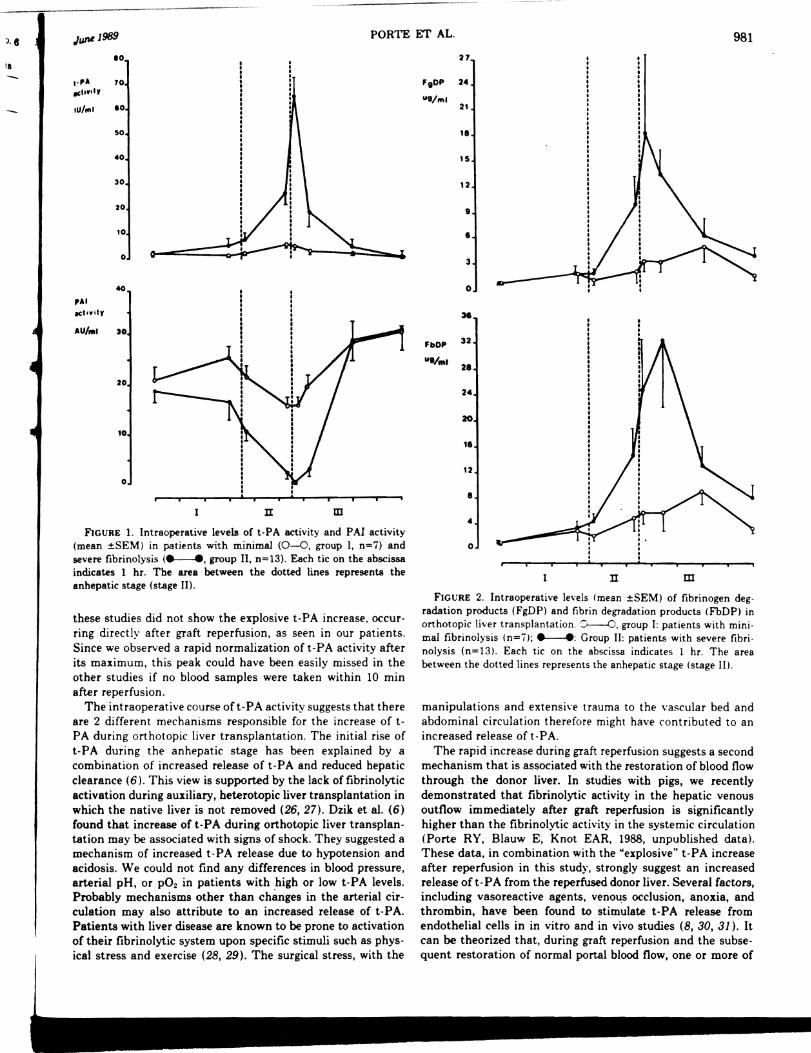
Mean intraoperative levels oft-PA activity and PAl activity of group I and II are depicted in Figure 1. There were no significant changes in t-PA and PAl activity during the preanhepatic stage in both groups. In group I t-P A levels remained below 12 IV /ml during the rest of the operation in all patients. In group II t-P A activity increased after clamping of the vessels of the native liver. and levels were significantly higher at the end of the anhepatic stage (II1-5), compared with group I (P=O.OO2). During reperfusion of the graft. t-PA levels increased sharply. resulting in a more than doubling at 5 min after reperfusion. compared with the values at 5 min before reperfusion (P<O.OO7). At this time t-PA activity in group II (65.1±8.5 IV/mi. mean ±SEM) was about 30 times higher than the preoperative value and more than 10 times higher than tPA activity in group I (P=0.OOO4). Later in the postanhepatic stage, a rapid disappearance of t-PA activity was seen. and levels fell into the normal range (0-1 IU/m!) at the end of the operation in all but 2 patients. In these 2 patients t-PA levels were still moderately increased (3.4 and 10 IU/mi). In group II free PAl activity showed a pattern inverse that of t- PA activity. and only minimal PAl activity (1.l±0.7 AU/ml, was left at the peak of fibrinolytic activity. but levels increased during the later postanhepatic period in both groups.
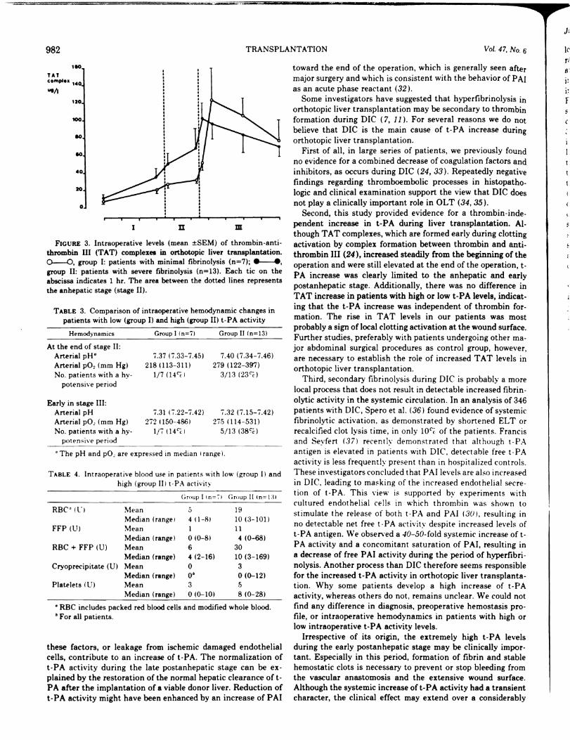
Mean plasma levels of FbDP and FgDP in groups I and II are shown in Figure 2. Although an increase of FbDP and FgDP was seen in both groups. levels in group II were significantly higher in the postanhepatic period at 45 min after graft reperfusion when compared with group I (P<O.04l. In group II the highest FgDP level (lS.4±7.9 I'g/ml) coincided with the peak in t-PA activity (11+5), whereas the maximum in FbDP (32.5± 11.2 ~g/ml) occurred somewhat later in the postanhepatic stage (III+45).
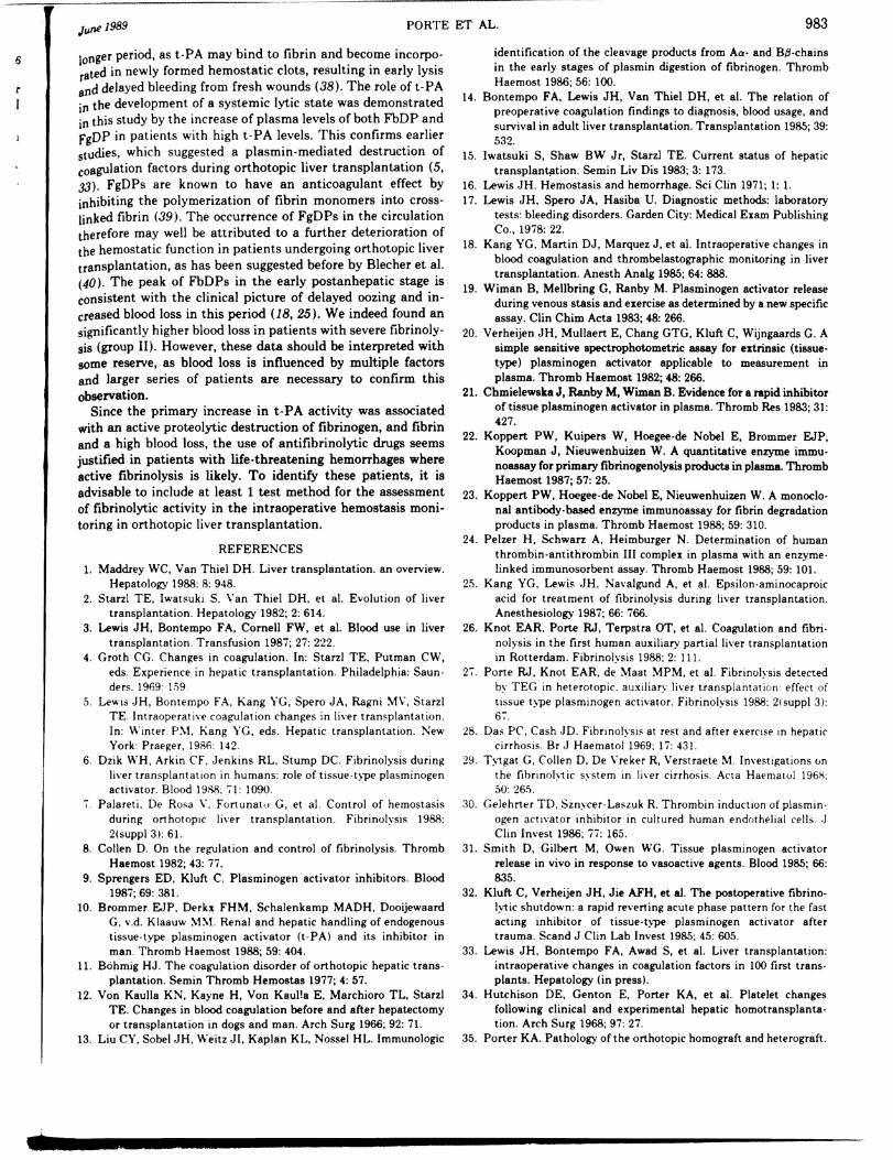
Mean levels of TAT complexes in group I and II are shown in Figure 3. An identical increasing pattern was seen in both groups. and at none of the time points were TAT levels significantly cliffe rent between the 2 groups. Highest TAT levels were found during the postanhepatic stage. and levels were still
Median (range) -Group I Group II ---
11.3 (10.4-21.2) 12.2 (9.7-21.2) 36.0 (28.9-51.6) 42.9 (29.1-127) 17.1 (15.3-32.9) 22.0 (14.3-47.7)
285 (159-460) 140 (85-350) 66 (32-130) 38 (15-135)
185 (130-300) 120 (82-280) 8.0 (2.0-16.0) 4.3 (1.6-60.0)
81 (56-510) 118 (39-336)
>120 (105->120) 60 (15->120) >150· >150·
2.4 (0-6.0) 1.4 (0-8.0) 18.5 (14.0-36.0) 18.8 (3.0-37.5) 0.28 (0.20-3.0) 0.50 (0.20-2.6) 0.30 (0.26-4.5) 0.84 (0.22-6.0)
above the normal upper limit (>4.1 "gIL) at the end of the operation in ail patients.
Comparison of changes in arterial pH, p02, and blood pressure in group I and II are shown in Table 3. There was no evidence for a relationship between intraoperative changes in hemodynamics and the increase in t-PA activity. Periods of shock, as determined by a drop in blood pressure and pH. were found among both patients with high and low t-PA activities.
The intraoperative use of blood products is shown in Table 4. There were no differences in the use of cryoprecipitate and platelets between the 2 groups. However. intraoperative blood loss, as reflected by the total use of modified whole blood or packed RBC and fresh-frozen plasma. was significantly higher in group II than in group I (P<0.02).
DISCL'SSIO:\
In earlier studies we have found that increased fibrinolytic activity, as measured by the EL T or thrombelastography lTEG), occurs in about 80~ of the patients undergoing orthotopic liver transplantation (18, 25). Fibrinolytic activity may mcrease during the anhepatic stage and is most often severe early after graft recirculation. A simultaneous decrease of plasminogen and a"-antiplasmin, the main inhibitor of plasmin, has been found during this period, which supports the view of an active fibrinolytic process (5). However, the use of EL T and TEG in these studies did not allow an exact characterization of the fibrinolytic defect. and the origin and clinical relevance of the increased fibrinolytic activity have remained largely unclear.
In this study we found an extreme increase of t-PA activity, and concomitant decrease of PAl activity during the anhepatic and early postanhepatic period in patients with severe fibrinolysis, as measured by the ELT and TEG. Reduction in PAl activity can be explained by the formation of complexes with t-PA. After saturation of free PAl, a further increase of t-PA will result in the increase of free t-PA activity in the circulation (9, 21). Recently. Dzik et al. (6) and Palareti et al. (7) have reported a similar increase of t-PA during the anhepatic stage in a limited number of patients undergoing OL T. However.
-------'", ----~--------.-
J. e JUflLl989 PORTE ET AL. 981
II 10
t·,A 70 "IiYit,
lu/",1 10
50
.0
30
20
to
0
-0
,At ICl.vitr
AUt .. 1 )0
20
10
o
n m
FiGURE 1. Intraoperative levels of t-PA activity and PAL activity (mean ±SEM) in patients with minimal (0-0, group I, n=7) and severe fibrinolysis (~, group II, n=13). Each tic on the abscissa indicates 1 hr_ The area between the dotted lines represents the anhepatic stage (stage 11).
these studies did not show the explosive t-PA increase. occurring directly after graft reperfusion, as seen in our patients. Since we observed a rapid normalization of t-PA activity after its maximum, this peak could have been easily missed in the other studies if no blood samples were taken within 10 min after reperfusion,
The intraoperative course of t- PA activity suggests that there are 2 different mechanisms responsible for the increase of tPA during orthotopic liver transplantation. The initial rise of t-PA during the anhepatic stage has been explained by a combination of increased release of t-PA and reduced hepatic clearance (6). This view is supported by the lack of fibrinolytic activation during auxiliary, heterotopic liver transplantation in which the native liver is not removed (26, 27). Dzik et aL (6) found that increase of t-PA during orthotopic liver transplantation may be associated with signs of shock, They suggested a mechanism of increased t-PA release due to hypotension and acidosis. We could not find any differences in blood pressure, arterial pH, or p02 in patients with high or low t-PA levels. Probably mechanisms other than changes in the arterial circulation may also attribute to an increased release of t-PA_ Patients with liver disease are known to be prone to activation of their fibrinolytic system upon specific stimuli such as physical stress and exercise (28, 29). The surgical stress, with the
27
F,OP 24
ut/",1 21
I' 15
12
9
• 3
0
36
FbDP 32
Uti ... 2. 24
20
1.
12
• • 0
n m FIGURE 2. Intraoperative levels (mean ±SEM) of fibrinogen deg
radation products (FgDP) and fibrin degradation products (FbDP) in orthotopic liver transplantation .. J....---O, group I: patients with minimal fibrinolysis (n=71;~: Group II: patients with severe fibrinolysis (n=13). Each tic on the abscissa indicates 1 hr. The area between the dotted lines represents the anhepatic stage (stage II),
manipulations and extensive trauma to the vascular bed and abdominal circulation therefore might have contributed to an increased release oft-PA,
The rapid increase during graft reperfusion suggests a second mechanism that is associated with the restoration of blood flow through the donor liver. In studies with pigs, we recently demonstrated that fibrinolytic activity in the hepatic venous outflow immediately after graft reperfusion is significantly higher than the fibrinolytic activity in the systemic circulation (Porte RY, Blauw E, Knot EAR, 1988, unpublished data). These data, in combination with the "explosive" t-PA increase after reperfusion in this study, strongly suggest an increased release of t-PA from the reperfused donor liver. Several factors, including vasoreactive agents, venous occlusion, anoxia, and thrombin, have been found to stimulate t-PA release from endothelial cells in in vitro and in vivo studies (8, 30, 31). It can be theorized that, during graft reperfusion and the subsequent restoration of normal portal blood flow, one or more of
982 TRANSPLANTATION Vol. 47, No.6
110
TAT cOlnpte .. I.
"'/1 120
100
10
10
.0
20
0
n m FIGURE 3. Intraoperative levels (mean ±SEM) of thrombin-anti
thrombin III (TAT) complexes in orthotopic liver transplantation. 0--0, group I: patients with minimal fibrinolysis (n=7); _____ , group II: patients with severe fibrinolysis (n=13). Each tic on the abscissa indicates 1 hr. The area between the dotted lines represents the anhepatic stage (stage II).
TABLE 3. Comparison of intraoperative hemodynamic changes in patients with low (group I) and high (group II) t-PA activity
Hemodynamics
At the end of stage ,,: Arterial pH" Arterial pO, (mm Hg) No. patients with a hy-
potensive period
Early in stage III: Arterial pH Arterial pO, (mm Hg) No. patients with a hy-
potensive period
Group 1 In=i)
7.37 (7.33-7.45) 218 (113-311)
1/7(14~1
7.31 (7.22-7.42) 272 (150-486)
Group II (n=13)
7.40 (7.34-7.46) 279 (122-397)
3/13 (23~)
7.32 (7.15-7.42) 275 (114-531)
5/13 (38se)
" The pH and pO 2 are expressed in median' range).
TABLE 4. Intraoperative blood use in patients with low (group Il and high (group II) t-PA activity
RBC" (n Mean 5 19 Median (rangel 4 (1-8) 10 (3-1011
FFP (V) Mean 1 11 Median (range) 0(0-8) 4 (0-68)
RBC + FFP (0) Mean 6 30 Median (range) 4 (2-16) 10 (3-169)
Cryoprecipitate (U) Mean 0 3 Median (range) O· 0(0-12)
Platelets (U) Mean 3 5 Median (range) 0(0-10) 8 <0-28)
" RBC includes packed red blood cells and modified whole blood. b For all patients.
these factors, or leakage from ischemic damaged endothelial cells, contribute to an increase of t-PA. The normalization of t-PA activity during the late postanhepatic stage can be explained by the restoration of the normal hepatic clearance of tPA after the implantation of a viable donor liver. Reduction of t-PA activity might have been enhanced by an increase of PAl
toward the end of the operation, which is generally seen after major surgery and which is consistent with the behavior of PAl as an acute phase reactant (32).
Some investigators have suggested that hyperfibrinolysis in orthotopic liver transplantation may be secondary to thrombin formation during DIe (7, 11). For several reasons we do not believe that DIe is the main cause of t-PA increase during orthotopic liver transplantation.
First of all, in large series of patients, we previously found no evidence for a combined decrease of coagulation factors and inhibitors, as occurs during DIe (24,33). Repeatedly negative findings regarding thromboembolic processes in histopathologic and clinical examination support the view that DIC does not playa clinically important role in OLT (34,35).
Second, this study provided evidence for a thrombin-independent increase in t-PA during liver transplantation. Although TAT complexes, which are formed early during clotting activation by complex formation between thrombin and antithrombin III (24), increased steadily from the beginning of the operation and were still elevated at the end of the operation, tPA increase was clearly limited to the an hepatic and early postanhepatic stage. Additionally, there was no difference in TAT increase in patients with high or low t-PA levels, indicating that the t-PA increase was independent of thrombin formation. The rise in TAT levels in our patients was most probably a sign of local clotting activation at the wound surface. Further studies, preferably with patients undergoing other major abdominal surgical procedures as control group, however, are necessary to establish the role of increased TAT levels in orthotopic liver transplantation.
Third, secondary fibrinolysis during DIe is probably a more local process that does not result in detectable increased fibrinolytic activity in the systemic circulation. In an analysis of 346 patients with DIe, Spero et al. (36) found evidence of systemic fibrinolytic activation. as demonstrated by shortened EL T or recalcified clot lysis time, in only lOe; of the patients. Francis and Seyfert (37) recently demonstrated that although t-PA antigen is elevated in patients with DIe, detectable free t-PA activity is less frequently present than in hospitalized controls. These investigators concluded that PAl levels are also increased in Die, leading to masking of the increased endothelial secretion of t-PA. This \'iew is supported by experiments with cultured endothelial cells in which thrombin was shown to stimulate the release of both t-PA and PAl (30), resulting in no detectable net free t-PA activity despite increased levels of t-PA antigen. We observed a 40-50-fold systemic increase of tP A activity and a concomitant saturation of PAl, resulting in a decrease of free PAl activity during the period of hyperfibrinolysis. Another process than DIC therefore seems responsible for the increased t-PA activity in orthotopic liver transplantation. Why some patients develop a high increase of t-PA activity, whereas others do not. remains unclear. We could not find any difference in diagnosis, preoperative hemostasis profile, or intraoperative hemodynamics in patients with high or low intraoperative t-PA activity levels.
Irrespective of its origin, the extremely high t-PA levels during the early postanhepatic stage may be clinically important. Especially in this period, formation of fibrin and stable hemostatic clots is necessary to prevent or stop bleeding from the vascular anastomosis and the extensive wound surface. Although the systemic increase of t-PA activity had a transient character, the clinical effect may extend over a considerably
Jr.
Ie r; 8'
ir ir F
6
f
I
-----------------------Ju.~1989 PORTE ET AL. 983
longer period, as t-PA may bind.to fibrin and ?ec~me incorp~rated in newly formed hemostatiC clots, resultmg m early lYSIS and delayed bleeding from fresh wounds (38). The role of t-PA in the development of a systemic lytic state was demonstrated in this study by the increase of plasma levels of both FbDP and f'gDP in patients with high t-PA levels. This confirms earlier studies, which suggested a plasmin-mediated destruction of coagulation factors during orthotopic liver transplantation (5, 33). FgDPs are known to have an anticoagulant effect by inhibiting the polymerization of fibrin monomers into crosslinked fibrin (39). The occurrence of FgDPs in the circulation therefore may well be attributed to a further deterioration of the hemostatic function in patients undergoing orthotopic liver transplantation, as has been suggested before by Blecher et a1. (40). The peak of FbDPs in the early postanhepatic stage is consistent with the clinical picture of delayed oozing and increased blood loss in this period (I8, 25). We indeed found an significantly higher blood loss in patients with severe fibrinolysis (group II). However, these data should be interpreted with some reserve, as blood loss is influenced by multiple factors and larger series of patients are necessary to confirm this observation.
Since the primary increase in t-PA activity was associated with an active proteolytic destruction of fibrinogen, and fibrin and a high blood loss, the use of antifibrinolytic drugs seems justified in patients with life-threatening hemorrhages where active fibrinolysis is likely. To identify these patients, it is advisable to include at least 1 test method for the assessment of fibrinolytic activity in the intraoperative hemostasis monitoring in orthotopic liver transplantation.
REFERENCES
1. Maddrey WC, Van Thiel DH. Liver transplantation. an overview. Hepatology 1988: 8: 948.
2. Stanl TE. Iwatsuki S. Van Thiel DH. et al. Evolution of liver transplantation. Hepatology 1982; 2: 614.
3. Lewis JH. Bontempo FA. Cornell FW. et al. Blood use in liver transplantation. Transfusion 1987; 27: 222.
4. Groth CG. Changes in coagulation. In: Starzl TE. Putman CWo eds. Experience in hepatic transplantation. Philadelphia: Saunders. 1969: 1.')9.
5. Le ..... is JH. Bontempo FA. Kang YG, Spero JA. Ragni ~V. Starzl TE. Intraoperative coagulation changes in liver transplantation. In: Winter P:\1. Kang YG. eds. Hepatic transplantation. New York: Praeger. 1986: 142.
6. Dzik WHo Arkin CF. Jenkins RL. Stump DC. Fibrinolysis during liver transplantation in humans: role of tissue-type plasminogen activator. Blood 19S8: ';'1: 1090.
i. Palareti. De Rosa V. Fortunat._, G. et al. Control of hemostasis during orthotopic liver transplantation. Fibrinolysis 1988: 2(suppl 3): 6l.
8. Collen D. On the regulation and control of fibrinolysis. Thromb Haemost 1982; 43: 77.
9. Sprengers ED. Kluft C. Plasminogen activator inhibitors. Blood 1987; 69: 381.
10. Brommer EJP. Derkx FHM. Schalenkamp MADH. Dooijewaard G. v.d. Klaauw :\1~1. Renal and hepatic handling of endogenous tissue-type plasminogen activator (t-PA) and its inhibitor in man. Thromb Haemost 1988; 59: 404.
11. Bohmig HJ. The coagulation disorder of orthotopic hepatic transplantation. Semin Thromb Hemostas 1977; 4: 57.
12. Von Kaulla KN. Kayne H. Von Kaul!a E. Marchioro TL. Starzl TE. Changes in blood coagulation before and after hepatectomy or transplantation in dogs and man. Arch Surg 1966; 92: 71.
13. Liu CY. Sobel JH. Weitz JI. Kaplan KL. Nossel HL. Immunologic
identification of the cleavage products from Aa- and Bt/-chains in the early stages of plasmin digestion of fibrinogen. Thromb Haemost 1986; 56: 100.
14. Bontempo FA. Lewis JH. Van Thiel DH, et al. The relation of preoperative coagulation findings to diagnosis, blood usage, and survival in adult liver transplantation. Transplantation 1985; 39: 532.
15. Iwatsuki S, Shaw BW Jr, Starzl TE. Current status of hepatic transplant/ltion. Semin Liv Dis 1983; 3: 173.
16. Lewis JH. Hemostasis and hemorrhage. Sci Clin 1971; 1: 1. 17. Lewis JH. Spero JA, Hasiba U. Diagnostic methods: laboratory
tests: bleeding disorders. Garden City: Medical Exam Publishing Co., 1978: 22.
18. Kang YG. Martin DJ, Marquez J, et al. Intraoperative changes in blood coagulation and thrombelastographic monitoring in liver transplantation. Anesth Analg 1985; 64: 888.
19. Wiman B, Mellbring G, Ranby M. Plasminogen activator release during venous stasis and exercise as determined by a new specific assay. Clin Chim Acta 1983; 48: 266.
20. Verheijen JH, Mullaert E, Chang GTG, Kluft C, Wijngaards G_ A simple sensitive spectrophotometric aaaay for extrinsic (tissuetype) plasminogen activator applicable to measurement in plasma. Thromb Haemost 1982; 48: 266.
21. Chmielewska J, Ranby M, Wiman B. Evidence for a rapid inhibitor of tissue plasminogen activator in plasma_ Thromb Res 1983; 31: 427.
22. Koppert PW, Kuipers W, Hoegee-de Nobel E, Brommer EJP, Koopman J, Nieuwenhuizen W. A quantitative enzyme immunoassay for primary fibrinogenolyais producta in plasma. Thromb Haemost 1987; 57: 25.
23. Koppert PW, Hoegee-de Nobel E, Nieuwenhuizen W. A monoclonal antibody-based enzyme immunoassay for fibrin degradation products in plasma. Thromb Haemost 1988; 59: 310.
24. Pelzer H, Schwan A. Heimburger N. Determination of human thrombin-antithrombin III complex in plasma with an enzymelinked immunosorbent assay. Thromb Haemost 1988; 59: 101.
25. Kang YG. Lewis JH. Navalgund A. et al. Epsilon·aminocaproic acid for treatment of fibrinolysis during liver transplantation. Anesthesiology 1987; 66: 766.
26. Knot EAR. Porte RJ, Terpstra OTt et al. Coagulation and fibrinolysis in the first human auxiliary partial liver transplantation in Rotterdam. Fibrinolysis 1988; 2: Ill.
27. Porte RJ. Knot EAR. de Maat MPM. et al. Fibrinolysis detected by TEG in heterotopic. auxiliary liver transplantation: effect of tissue type plasminogen activator. Fibrinolysis 1988; 2(suppl 3): 67.
28. Das PC. Cash .JD. Fibrinolysis at rest and after exercise in hepatic cirrhosis. Br J Haematol 1969; 1 i: 431.
29. Tytgat G. Collen D. De Vreker R. Verstraete M. Investigations on the fihrinol:'1ic system in liver cirrhosis. Acta Haematol 196R: 50: 265.
30. Gelehrter TD. Sznycer-Laszuk R. Thrombin induction of plasminogen activator inhibitor in cultured human endothelial cells .. J Clin Invest 1986; 77: 165.
31. Smith D, Gilbert M. Owen WG. Tissue plasminogen activator release in vivo in response to vasoactive agents. Blood 1985; 66: 835.
32_ Kluft C, Verheijen JH, Jie AFH, et aI. The postoperative fibrinolytic shutdown: a rapid reverting acute phase pattern for the fast acting inhibitor of tissue·type plasminogen activator after trauma. Scand J Clin Lab Invest 1985; 45: 605.
33. Lewis JH. Bontempo FA. Awad S, et al. Liver transplantation: intraoperative changes in coagulation factors in 100 first transplants. Hepatology (in press).
34. Hutchison DE. Genton E. Porter KA, et aI. Platelet changes following clinical and experimental hepatic homotransplanta· tion. Arch Surg 1968; 97: 27.
35. Porter KA. Pathology of the orthotopic homograft and heterograft.
984 TRANSPLANTATION Vol. 47, NO. 6
In: Starzl TE. Putman CW, eds. Experience in hepatic transplantation. Philadelphia: Saunders, 1969: 422.
36. Spero JA. Lewis JH, Hasiba U. Disseminated intravascular coagulation: findings in 346 patients. Thromb Haemost 1980; 43: 28.
37. Francis RB. Seyfert U. Tissue plasminogen activator antigen and activity in disseminated intravascular coagulation: clincopathologie correlations. J Lab Clin Med 1987; 110: 541.
38. Brommer EJP. The level of extrinsic plasminogen activator (t-PA) during clotting as a determinant of the rate of fibrinolysis:
inefficiency oft-PA added afterwards. Thromb Res 1984; 34: 109 39. Kowalski E. Fibrinogen derived inhibitors of coagulation. Thro b
Diath Haemorrh 1960; 4(supp!): 211. Il'l
40. Blecher TE, Terblanche J, Peacock JH. Orthotopic liver tran plantation: coagulation and hematologic changes in the pig. Arc8~ Surg 1968; 96: 331.
Received 6 October 1988. Accepted 13 December 1988.
------------------------------------------------------------------------------------------------------
0041·1337/89/4706-0984$02.00/0 TRANSPLANTATION Copyright~. 1989 by The Williams & Wilkins Co.
Vol. 47, 984-988. No.6. June 1989 Printed in U.S.A.
T CELL DEPLETION OF HUMAN BONE MARROW
COMPARISON OF CAMPATH-l PLUS COMPLEMENT, ANTI-T CELL RICIN A CHAIN IMMUNOTOXIN, AND
SOYBEAN AGGLUTININ ALONE OR IN COMBINATION WITH SHEEP ERYTHROCYTES OR IMMUNOMAGNETIC BEADS!
JAMES N. FRAME,2 NANCY H. COLLINS, TERESA CARTAGENA, HERMAN WALDMANN,3
RICHARD J. O'REILLY, Bo DUPONT, AND NANCY A. KERNAN4
Human Immunogenetics Laboratory and Bone Marrow Transplantation Service, Memorial Sloan-Kettering Cancer Center, New York, New York. and Department of PatMlogy, University of Cambridge. Cambridge. United Kingdom
The aim of this study was to compare the extent of in vitro T cell depletion and recovery of hematopoietic progenitor cells achieved with five methods of T cell depletion. Bone marrow samples from the same source were treated with monoclonal antibody Campath-l (CPl) and human complement, XomaZyme-H65 (antiT cell ricin A chain immunotoxin), or soybean agglutinin (SBA) alone or in combination with sheep erythrocytes (EUT) or a cocktail of immunomagnetic beads (B) directly coated with anti-CD2. anti-CD3, or anti-CDS monoclonal antibodies. Residual T cells were enumerated by limiting dilution analysis, EAET rosetting. and proliferative responses to phytohemagglutinin. The results of this study demonstrated the following reductions in BM T cells as detected by limiting dilution analysis (mean o/c contro\): SBA+B (99.9%), SBA+EM:T (99.S'7c), CPt +C' (99.4 '7d , anti-T cell ricin A chain immunotoxin (99.00/c), and SBA alone (94.2%). Neither PHA response nor enumeration of residual EAET rosettes provided discriminating differences in the degree of T cell depletion
I This work was supported by National Institutes of Health Grants CA 22507, CA 23766, CA 08748, as well as by the XOMA Corporation. Berkeley. CA. the Charles A. Dana Foundation. the Zelda Weintraub Cancer Foundation. the Robert J. Kleberg and Helen C. Kleberg Foundation. the Lila Acheson Wallace Foundation, the Andrew Gaffney Foundation. and the Frito Lay IN ew York Yankees Challenge Fund.
'J. F. is supported by postdoctoral National Institutes of Health Immunology Training Grant CA 09148-11.
) University of Cambridge. • Address correspondence to: Nancy A. Kernan. M.D., Memorial
Sloan-Kettering Cancer Center. 1275 York Avenue. New York, NY 10021.
2£
by treatment method when T cell reductions exceeded' 99.0% by LDA. These results demonstrate the ability of CPl+C', XomaZyme-H65. and SBA plus sheep erythrocyte or magnetic bead depletion to achieve a greater than 99% reduction of BM T cells and the importance of limiting dilution analysis in defining differences in T cell numbers when depletion exceeded 99%.
Pretransplantation depletion of donor T cells from rodent ( 1. 2) or human bone marrow grafts has produced significant reduction in the incidence and severity of acute graft -versushost disease (3-8). A number of methods for in vitro T cell depletion (TeD)" have been evaluated, including the use of monoclonal antibodyls) (MoAb) + complement (4-7). soybean agglutination (SBA) plus sheep erythrocJ1e deplet ion (.'i. 9), elutriation (10), anti-T cell ricin (]]) and ricin A chain immunotoxins (12 J, and immunomagnetic beads coated with anti-T cell MoAb (13). Methods for enumerating the extent of TCD have included E-rosette analysis, proliferative responses to phytohemagglutinin, and immunofluorescence analysis with anti-T cell MoAbs. However, quantitating residual BM T cells by E-rosette analysis, PHA response, and immunofluorescence with anti-T cell MoAb has not provided a consistent correlation between the number of residual T cells transplanted and the subsequent development or severity of GVHD (14-16).
• Abbreviations: B, beads; BFU-E, burst-forming unit-erythroid; CD. cluster designation; CPl. Campath-1; EAI'T. 2-amino-ethyliosothiouronium-treated erythrocytes; HSA, human serum albumin; SSA, soybean agglutinin; TCD, T cell depletion.
Ju(li' J.
We ~J1111~'~
~J1rl n .-ell,. ( p}-iA. ct'll ,.
"f clc (li }-i' !I,,~lc'
(iVH tJ:,ed difie7 SIl!11~
resid !11ar~
i!11 !11
settt agne' ent r to as byez
Ir. Zy!11'
bear netic em hurr. rOSE'
glu' lyrr me: celi
I
IS c.
he g<
ai ir. Sf
('
0-
a:
a;
S<
C


![Administering Intravenous Alteplase (Tissue …Administering Intravenous Alteplase (Tissue Plasminogen Activator [tPA]) Step 1: Eligibility---The eligibility criteria for patients](https://img.dokumen.tips/doc/110x75/5e44a7adc59a354aef0b8cf8/administering-intravenous-alteplase-tissue-administering-intravenous-alteplase.jpg)

![Tissue-Type Plasminogen Activator-Mediated Activation of ... · TISSUE PLASMINOGEN ACTIVATOR IN STREPTOCOCCAL BINDING 197 sodium phosphate, 0.14 Msodium chloride [pH 7.4]) con- taining0.02%(wt/vol)](https://img.dokumen.tips/doc/110x75/5f46a6d9df5f79688c496b2a/tissue-type-plasminogen-activator-mediated-activation-of-tissue-plasminogen.jpg)