Embed Size (px)
Citation preview
Stark et al., Sci. Adv. 2018; 4 : eaat5107 1 August 2018
S C I E N C E A D V A N C E S | R E S E A R C H A R T I C L E
1 of 10
S Y N T H E T I C B I O L O G Y
BioBits™ Bright: A fluorescent synthetic biology education kitJessica C. Stark1,2,3*, Ally Huang4,5,6*, Peter Q. Nguyen6,7*, Rachel S. Dubner8, Karen J. Hsu9, Thomas C. Ferrante6, Mary Anderson10, Ada Kanapskyte11, Quinn Mucha10, Jessica S. Packett11, Palak Patel10, Richa Patel10, Deema Qaq10, Tyler Zondor10, Julie Burke12, Thomas Martinez10, Ashlee Miller-Berry13, Aparna Puppala14, Kara Reichert15, Miriam Schmid16, Lance Brand17, Lander R. Hill18†, Jemima F. Chellaswamy19, Nuhie Faheem20, Suzanne Fetherling21, Elissa Gong22, Eddie Marie Gonzalzles23, Teresa Granito13, Jenna Koritsaris13, Binh Nguyen24, Sujud Ottman25, Christina Palffy26, Angela Patel27, Sheila Skweres13, Adriane Slaton13, TaRhonda Woods13, Nina Donghia6, Keith Pardee28, James J. Collins4,5,6,29,30,31‡, Michael C. Jewett1,2,3,32,33‡
Synthetic biology offers opportunities for experiential educational activities at the intersection of the life sciences, engineering, and design. However, implementation of hands-on biology activities in classrooms is challenging be-cause of the need for specialized equipment and expertise to grow living cells. We present BioBits™ Bright, a shelf- stable, just-add-water synthetic biology education kit with easy visual outputs enabled by expression of fluorescent proteins in freeze-dried, cell-free reactions. We introduce activities and supporting curricula for teaching the central dogma, tunable protein expression, and design-build-test cycles and report data generated by K-12 teachers and students. We also develop inexpensive incubators and imagers, resulting in a comprehensive kit costing <US$100 per 30-person classroom. The user-friendly resources of this kit promise to enhance biology education both inside and outside the classroom.
INTRODUCTIONSynthetic biology aims to program biological systems to carry out useful functions. As a field, synthetic biology has made meaningful progress toward biomanufacturing of medicines (1, 2), sustainable chemicals (3, 4), and advanced fuels (5), as well as cellular diagnos-tics and therapies (6–9). At the core of these advances is the ability to control and tune the processes of transcription and translation, offering a point of entry for teaching fundamental biology topics through cutting-edge biological technologies. Synthetic biology also offers rich educational opportunities, as it requires students to con-front real-world, interdisciplinary problems at the intersection of di-verse disciplines including chemistry, biology, engineering, computer science, design, policy, and ethics. Such cross-cutting educational ac-tivities align closely with the objectives of K-12 STEAM (science, tech-
nology, engineering, the arts, and mathematics) education and pri-orities identified by the National Academy of Engineering to enable students to apply, adapt, and connect fundamental principles across multiple disciplines (10).
Synthetic biology–based educational efforts such as the BioBuilder Educational Foundation (11–14) and the International Genetically Engineered Machines competition (15, 16) have made great strides toward incorporating synthetic biology into high school and university education. These programs have resulted in student-reported academic gains, high student engagement, and increased self-identification as biological engineers (17–19). However, efforts to incorporate a hands-on molecular or synthetic biology curriculum have been limited by (i) the number of robust systems that can be converted into teaching materials; (ii) the need for expensive, specialized equipment to store,
1Department of Chemical and Biological Engineering, Northwestern University, 2145 Sheridan Road, Technological Institute E136, Evanston, IL 60208–3120, USA. 2Chem-istry of Life Processes Institute, Northwestern University, 2170 Campus Drive, Evanston, IL 60208–3120, USA. 3Center for Synthetic Biology, Northwestern University, 2145 Sheridan Road, Technological Institute E136, Evanston, IL 60208–3120, USA. 4Department of Biological Engineering, Massachusetts Institute of Technology (MIT), Cambridge, MA 02139, USA. 5Institute for Medical Engineering and Science, MIT, Cambridge, MA 02139, USA. 6Wyss Institute for Biologically Inspired Engineering, Harvard University, Boston, MA 02115, USA. 7School of Engineering and Applied Sciences, Harvard University, Cambridge, MA 02138, USA. 8Department of Biological Sciences, Northwestern University, 2205 Tech Drive, Hogan Hall 2144, Evanston, IL 60208, USA. 9Department of Mechanical Engineering, Northwestern University, 2145 Sheridan Road, Technological Institute B224, Evanston, IL 60208–3120, USA. 10Glenbard East High School, 1014 South Main Street, Lombard, IL 60148–3938, USA. 11Amos Alonzo Stagg High School, 8015 West 111th Street, Palos Hills, IL 60465–2203, USA. 12Grover Cleveland Elementary School, 3121 West Byron Street, Chicago, IL 60618–3403, USA. 13Evanston Township High School, 1600 Dodge Avenue, Evanston, IL 60201–3449, USA. 14Glenbrook South High School, 4000 West Lake Avenue, Glenview, IL 60026–1239, USA. 15Jones College Prep High School, 700 South State Street, Chicago, IL 60605–2109, USA. 16Gwendolyn Brooks College Preparatory Academy, 250 East 111th Street, Chicago, IL 60628–4324, USA. 17Delta High School, 3400 East SR 28, Muncie, IN 47303, USA. 18ASPIRA Business and Finance High School, 2989 North Milwaukee Avenue, Chicago, IL 60618–7347, USA. 19Aptakisic-Tripp School District 102, 850 Highland Grove Drive, Buffalo Grove, IL 60089, USA. 20Oak Lawn Hometown Middle School, 5345 West 99th Street, Oak Lawn, IL 60453–3815, USA. 21Hoffman Estates High School, 1100 West Higgins Road, Hoffman Estates, IL 60169–4050, USA. 22Vernon Hills High School, 145 Lakeview Parkway, Vernon Hills, IL 60061–1566, USA. 23Morgan Park High School, 1744 West Pryor Avenue, Chicago, IL 60643–3497, USA. 24Northside College Prep High School, 5501 North Kedzie Avenue, Chicago, IL 60625–3923, USA. 25Aqsa School, 7361 West 92nd Street, Bridgeview, IL 60455–2133, USA. 26Adlai E. Stevenson High School, 1 Stevenson Drive, Lincolnshire, IL 60069–2824, USA. 27Lyons Township High School, 100 South Brainard Avenue, La Grange, IL 60525–2101, USA. 28Leslie Dan Faculty of Pharmacy, University of Toronto, 144 College Street, Toronto, Ontario M5S 3M2, Canada. 29Synthetic Biology Center, MIT, Cambridge, MA 02139, USA. 30Harvard-MIT Program in Health Sciences and Technology, Cambridge, MA 02139, USA. 31Broad Institute of MIT and Harvard, Cambridge, MA 02142, USA. 32Robert H. Lurie Comprehensive Cancer Center, Northwestern University, 676 North Saint Clair Street, Suite 1200, Chicago, IL 60611–3068, USA. 33Simpson Querrey Institute, Northwestern University, 303 East Superior Street, Suite 11-131, Chicago, IL 60611–2875, USA.*These authors contributed equally to this work.†Present address: Ombudsman Chicago South CPS Options School, 6057 South Western Avenue, Chicago, IL 60636–2019, USA.‡Corresponding author. Email: [email protected] (J.J.C.); [email protected] (M.C.J.)
Copyright © 2018 The Authors, some rights reserved; exclusive licensee American Association for the Advancement of Science. No claim to original U.S. Government Works. Distributed under a Creative Commons Attribution NonCommercial License 4.0 (CC BY-NC).
on May 21, 2020
http://advances.sciencemag.org/
Dow
nloaded from
Stark et al., Sci. Adv. 2018; 4 : eaat5107 1 August 2018
S C I E N C E A D V A N C E S | R E S E A R C H A R T I C L E
2 of 10
grow, and transport cells; and (iii) biosafety considerations that limit the ability to work with cells outside of a laboratory setting (20). Addressing these limitations would help expand educational oppor-tunities for students in classrooms, as well as inform and promote public engagement in synthetic biology.
Freeze-dried, cell-free (FD-CF) systems represent an emerging tech-nology with exciting potential as a chassis for educational tools. FD-CF systems harness an ensemble of catalytic components [for example, RNA polymerases, ribosomes, aminoacyl–transfer RNA (tRNA) syn-thetases, translation initiation, and elongation factors, etc.] from cell lysates to synthesize proteins in vitro (21). Hence, FD-CF reactions do not use intact organisms; thus, they circumvent many of the bio-safety and biocontainment regulations that exist for living cells. Further,
FD-CF systems are stable at room temperature for more than 1 year (22) and can be run simply by adding water and DNA template to a freeze- dried pellet of reagents, eliminating the need for specialized equip-ment or expertise to run reactions. Finally, FD-CF systems are robust, with demonstrated utility for point-of-use biosynthesis of sophisticated diagnostics, protein therapeutics, vaccines, small mole-cules, and molecular biology reagents (22–29). If FD-CF technology could be used to develop safe, portable, and easy-to-use educational tools, it would significantly lower the barrier to entry for teaching synthetic biology.
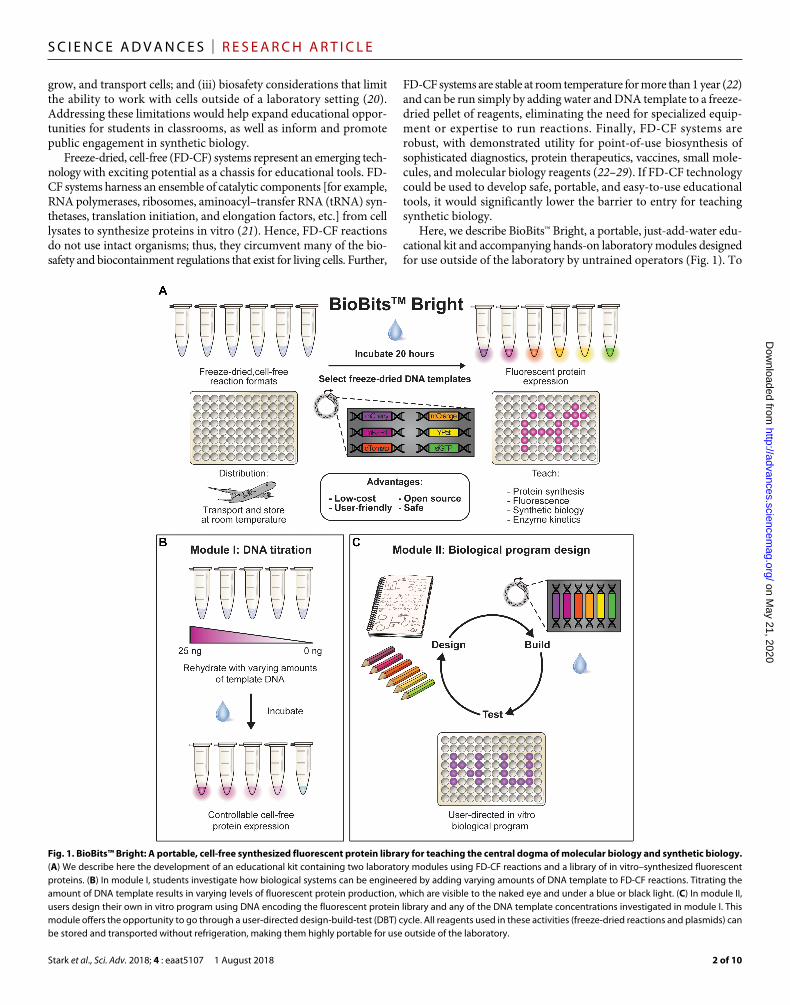
Here, we describe BioBits™ Bright, a portable, just-add-water edu-cational kit and accompanying hands-on laboratory modules designed for use outside of the laboratory by untrained operators (Fig. 1). To
Fig. 1. BioBits™ Bright: A portable, cell-free synthesized fluorescent protein library for teaching the central dogma of molecular biology and synthetic biology. (A) We describe here the development of an educational kit containing two laboratory modules using FD-CF reactions and a library of in vitro–synthesized fluorescent proteins. (B) In module I, students investigate how biological systems can be engineered by adding varying amounts of DNA template to FD-CF reactions. Titrating the amount of DNA template results in varying levels of fluorescent protein production, which are visible to the naked eye and under a blue or black light. (C) In module II, users design their own in vitro program using DNA encoding the fluorescent protein library and any of the DNA template concentrations investigated in module I. This module offers the opportunity to go through a user-directed design-build-test (DBT) cycle. All reagents used in these activities (freeze-dried reactions and plasmids) can be stored and transported without refrigeration, making them highly portable for use outside of the laboratory.
on May 21, 2020
http://advances.sciencemag.org/
Dow
nloaded from
Stark et al., Sci. Adv. 2018; 4 : eaat5107 1 August 2018
S C I E N C E A D V A N C E S | R E S E A R C H A R T I C L E
3 of 10
facilitate kit construction, we developed a library of fluorescent proteins that express at high yields (≥600 g ml−1) in FD-CF reactions. We report data for each module from workshops with Chicago K-12 students and teachers to demonstrate robustness and ease of use. Lab-oratory modules are designed to (i) synergize with fundamental biol-ogy education, as evidenced by the supporting curriculum developed by Chicago middle and high school teachers (curricula S1 to S5); (ii) be run independently or in sequence; and (iii) be adapted for use with students at various educational levels. Notably, to make BioBits™ Bright laboratory activities accessible to resource-limited classrooms, we have also developed low-cost incubators and imagers. Separately, we describe BioBits™ Explorer [see companion article by Huang et al. (30)], a next-generation BioBits™ kit developed to illustrate an even wider range of biological concepts (for example, enzymatic catalysis and genetic circuits). We anticipate that the availability of our BioBits™ kits and the data reported here will encourage teaching and broaden participation in the field of synthetic biology.
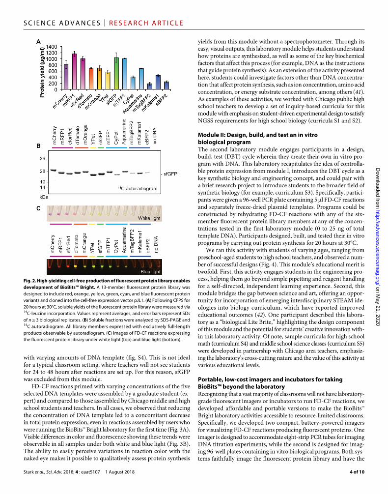
RESULTSHigh-yielding in vitro expression of a diverse fluorescent protein libraryBased on the success of colorimetric chemistry kits, we sought to create synthetic biology classroom modules for BioBits™ Bright with simple, visual readouts. We reasoned that the ability to link a visual output to abstract concepts such as the central dogma of molecular biology would increase student engagement and understanding. Fluo-rescent proteins are routinely used as reporters in synthetic biology and represent an attractive readout for an educational kit for two main reasons. First, a wide variety of fluorescent protein variants have been discovered or engineered (31–36), which produce an array of colors visible to the naked eye. Second, these variants are well studied and
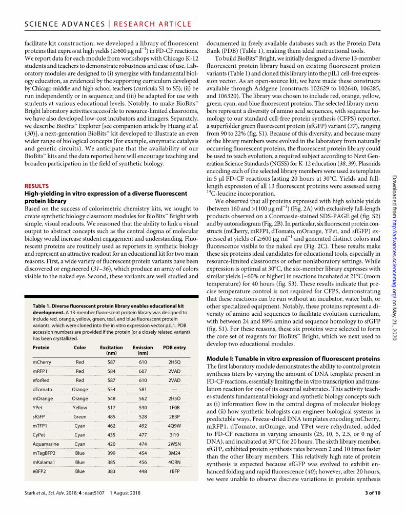
documented in freely available databases such as the Protein Data Bank (PDB) (Table 1), making them ideal instructional tools.
To build BioBits™ Bright, we initially designed a diverse 13-member fluorescent protein library based on existing fluorescent protein variants (Table 1) and cloned this library into the pJL1 cell-free expres-sion vector. As an open-source kit, we have made these constructs available through Addgene (constructs 102629 to 102640, 106285, and 106320). The library was chosen to include red, orange, yellow, green, cyan, and blue fluorescent proteins. The selected library mem-bers represent a diversity of amino acid sequences, with sequence ho-mology to our standard cell-free protein synthesis (CFPS) reporter, a superfolder green fluorescent protein (sfGFP) variant (37), ranging from 90 to 22% (fig. S1). Because of this diversity, and because many of the library members were evolved in the laboratory from naturally occurring fluorescent proteins, the fluorescent protein library could be used to teach evolution, a required subject according to Next Gen-eration Science Standards (NGSS) for K-12 education (38, 39). Plasmids encoding each of the selected library members were used as templates in 5 l FD-CF reactions lasting 20 hours at 30°C. Yields and full-length expression of all 13 fluorescent proteins were assessed using 14C-leucine incorporation.
We observed that all proteins expressed with high soluble yields (between 160 and >1100 g ml−1) (Fig. 2A) with exclusively full-length products observed on a Coomassie-stained SDS-PAGE gel (fig. S2) and by autoradiogram (Fig. 2B). In particular, six fluorescent protein con-structs (mCherry, mRFP1, dTomato, mOrange, YPet, and sfGFP) ex-pressed at yields of ≥600 g ml−1 and generated distinct colors and fluorescence visible to the naked eye (Fig. 2C). These results make these six proteins ideal candidates for educational tools, especially in resource-limited classrooms or other nonlaboratory settings. While expression is optimal at 30°C, the six-member library expresses with similar yields (~60% or higher) in reactions incubated at 21°C (room temperature) for 40 hours (fig. S3). These results indicate that pre-cise temperature control is not required for CFPS, demonstrating that these reactions can be run without an incubator, water bath, or other specialized equipment. Notably, these proteins represent a di-versity of amino acid sequences to facilitate evolution curriculum, with between 24 and 89% amino acid sequence homology to sfGFP (fig. S1). For these reasons, these six proteins were selected to form the core set of reagents for BioBits™ Bright, which we next used to develop two educational modules.
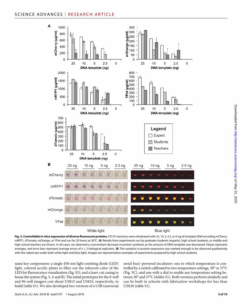
Module I: Tunable in vitro expression of fluorescent proteinsThe first laboratory module demonstrates the ability to control protein synthesis titers by varying the amount of DNA template present in FD-CF reactions, essentially limiting the in vitro transcription and trans-lation reaction for one of its essential substrates. This activity teach-es students fundamental biology and synthetic biology concepts such as (i) information flow in the central dogma of molecular biology and (ii) how synthetic biologists can engineer biological systems in predictable ways. Freeze-dried DNA templates encoding mCherry, mRFP1, dTomato, mOrange, and YPet were rehydrated, added to FD-CF reactions in varying amounts (25, 10, 5, 2.5, or 0 ng of DNA), and incubated at 30°C for 20 hours. The sixth library member, sfGFP, exhibited protein synthesis rates between 2 and 10 times faster than the other library members. This relatively high rate of protein synthesis is expected because sfGFP was evolved to exhibit en-hanced folding and rapid fluorescence (40); however, after 20 hours, we were unable to observe discrete variations in protein synthesis
Table 1. Diverse fluorescent protein library enables educational kit development. A 13-member fluorescent protein library was designed to include red, orange, yellow, green, teal, and blue fluorescent protein variants, which were cloned into the in vitro expression vector pJL1. PDB accession numbers are provided if the protein (or a closely related variant) has been crystallized.
Protein Color Excitation (nm)
Emission (nm)
PDB entry
mCherry Red 587 610 2H5Q
mRFP1 Red 584 607 2VAD
eforRed Red 587 610 2VAD
dTomato Orange 554 581 —
mOrange Orange 548 562 2H5O
YPet Yellow 517 530 1F0B
sfGFP Green 485 528 2B3P
mTFP1 Cyan 462 492 4Q9W
CyPet Cyan 435 477 3I19
Aquamarine Cyan 420 474 2WSN
mTagBFP2 Blue 399 454 3M24
mKalama1 Blue 385 456 4ORN
eBFP2 Blue 383 448 1BFP
on May 21, 2020
http://advances.sciencemag.org/
Dow
nloaded from
Stark et al., Sci. Adv. 2018; 4 : eaat5107 1 August 2018
S C I E N C E A D V A N C E S | R E S E A R C H A R T I C L E
4 of 10
with varying amounts of DNA template (fig. S4). This is not ideal for a typical classroom setting, where teachers will not see students for 24 to 48 hours after reactions are set up. For this reason, sfGFP was excluded from this module.
FD-CF reactions primed with varying concentrations of the five selected DNA templates were assembled by a graduate student (ex-pert) and compared to those assembled by Chicago middle and high school students and teachers. In all cases, we observed that reducing the concentration of DNA template led to a concomitant decrease in total protein expression, even in reactions assembled by users who were running the BioBits™ Bright laboratory for the first time (Fig. 3A). Visible differences in color and fluorescence showing these trends were observable in all samples under both white and blue light (Fig. 3B). The ability to easily perceive variations in reaction color with the naked eye makes it possible to qualitatively assess protein synthesis
yields from this module without a spectrophotometer. Through its easy, visual outputs, this laboratory module helps students understand how proteins are synthesized, as well as some of the key biochemical factors that affect this process (for example, DNA as the instructions that guide protein synthesis). As an extension of the activity presented here, students could investigate factors other than DNA concentra-tion that affect protein synthesis, such as ion concentration, amino acid concentration, or energy substrate concentration, among others (41). As examples of these activities, we worked with Chicago public high school teachers to develop a set of inquiry-based curricula for this module with emphasis on student-driven experimental design to satisfy NGSS requirements for high school biology (curricula S1 and S2).
Module II: Design, build, and test an in vitro biological programThe second laboratory module engages participants in a design, build, test (DBT) cycle wherein they create their own in vitro pro-gram with DNA. This laboratory recapitulates the idea of controlla-ble protein expression from module I, introduces the DBT cycle as a key synthetic bio logy and engineering concept, and could pair with a brief research project to introduce students to the broader field of synthetic biology (for example, curriculum S3). Specifically, partici-pants were given a 96-well PCR plate containing 5 l FD-CF reactions and separately freeze-dried plasmid templates. Programs could be constructed by rehydrating FD-CF reactions with any of the six- member fluorescent protein library members at any of the concen-trations tested in the first laboratory module (0 to 25 ng of total template DNA). Participants designed, built, and tested their in vitro programs by carrying out protein synthesis for 20 hours at 30°C.
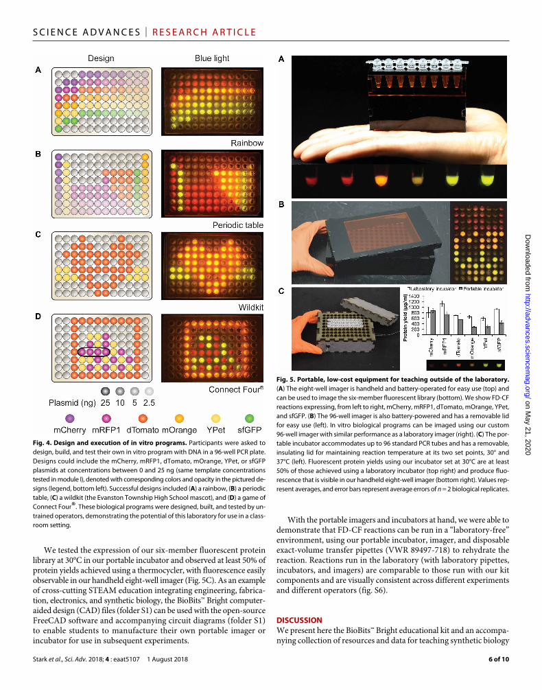
We ran this activity with students of varying ages, ranging from preschool-aged students to high school teachers, and observed a num-ber of successful designs (Fig. 4). This module’s educational merit is twofold. First, this activity engages students in the engineering pro-cess, helping them go beyond simple pipetting and reagent handling for a self-directed, independent learning experience. Second, this module bridges the gap between science and art, offering an oppor-tunity for incorporation of emerging interdisciplinary STEAM ide-ologies into biology curriculum, which have reported improved educational outcomes (42). One participant described this labora-tory as a “biological Lite Brite,” highlighting the design component of this module and the potential for students’ creative innovation with-in this laboratory activity. Of note, sample curricula for high school math (curriculum S4) and middle school science classes (curriculum S5) were developed in partnership with Chicago area teachers, emphasiz-ing the laboratory’s cross-cutting nature and the value of this activity at various educational levels.
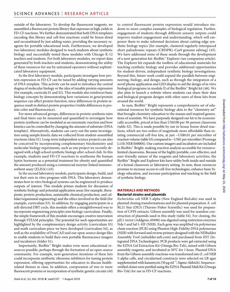
Portable, low-cost imagers and incubators for taking BioBits™ beyond the laboratoryRecognizing that a vast majority of classrooms will not have laboratory- grade fluorescent imagers or incubators to run FD-CF reactions, we developed affordable and portable versions to make the BioBits™ Bright laboratory activities accessible to resource-limited classrooms. Specifically, we developed two compact, battery-powered imagers for visualizing FD-CF reactions producing fluorescent proteins. One imager is designed to accommodate eight-strip PCR tubes for imaging DNA titration experiments, while the second is designed for imag-ing 96-well plates containing in vitro biological programs. Both sys-tems faithfully image the fluorescent protein library and have the
Fig. 2. High-yielding cell-free production of fluorescent protein library enables development of BioBits™ Bright. A 13-member fluorescent protein library was designed to include red, orange, yellow, green, cyan, and blue fluorescent protein variants and cloned into the cell-free expression vector pJL1. (A) Following CFPS for 20 hours at 30°C, soluble yields of the fluorescent protein library were measured via 14C-leucine incorporation. Values represent averages, and error bars represent SDs of n ≥ 3 biological replicates. (B) Soluble fractions were analyzed by SDS-PAGE and 14C autoradiogram. All library members expressed with exclusively full-length products observable by autoradiogram. (C) Images of FD-CF reactions expressing the fluorescent protein library under white light (top) and blue light (bottom).
on May 21, 2020
http://advances.sciencemag.org/
Dow
nloaded from
Stark et al., Sci. Adv. 2018; 4 : eaat5107 1 August 2018
S C I E N C E A D V A N C E S | R E S E A R C H A R T I C L E
5 of 10
same key components: a single 450-nm light-emitting diode (LED) light, colored acrylic plates to filter out the inherent color of the LED for fluorescence visualization (fig. S5), and a laser-cut casing to house the system (Fig. 5, A and B). The initial prototypes for the 8-well and 96-well imagers cost about US$15 and US$32, respectively, to build (table S1). We also developed two versions of a USB (universal
serial bus)–powered incubator: one in which temperature is con-trolled by a switch calibrated to two temperature settings, 30° or 37°C (Fig. 5C), and one with a dial to enable any temperature setting be-tween 30° and 37°C (folder S1). Both versions perform similarly and can be built in schools with fabrication workshops for less than US$20 (table S1).
Fig. 3. Controllable in vitro expression of diverse fluorescent proteins. FD-CF reactions were rehydrated with 25, 10, 5, 2.5, or 0 ng of template DNA encoding mCherry, mRFP1, dTomato, mOrange, or YPet and run for 20 hours at 30°C. (A) Results from experiments run by graduate students (experts), high school students, or middle and high school teachers are shown. In all cases, we observed a concomitant decrease in protein synthesis as the amount of DNA template was decreased. Values represent averages, and error bars represent average errors of n ≥ 2 biological replicates. (B) The variation in protein expression was marked enough to be observed qualitatively with the naked eye under both white light and blue light. Images are representative examples of experiments prepared by high school students.
on May 21, 2020
http://advances.sciencemag.org/
Dow
nloaded from
Stark et al., Sci. Adv. 2018; 4 : eaat5107 1 August 2018
S C I E N C E A D V A N C E S | R E S E A R C H A R T I C L E
6 of 10
We tested the expression of our six-member fluorescent protein library at 30°C in our portable incubator and observed at least 50% of protein yields achieved using a thermocycler, with fluorescence easily observable in our handheld eight-well imager (Fig. 5C). As an example of cross-cutting STEAM education integrating engineering, fabrica-tion, electronics, and synthetic biology, the BioBits™ Bright computer- aided design (CAD) files (folder S1) can be used with the open-source FreeCAD software and accompanying circuit diagrams (folder S1) to enable students to manufacture their own portable imager or incubator for use in subsequent experiments.
With the portable imagers and incubators at hand, we were able to demonstrate that FD-CF reactions can be run in a “laboratory-free” environment, using our portable incubator, imager, and disposable exact-volume transfer pipettes (VWR 89497-718) to rehydrate the reaction. Reactions run in the laboratory (with laboratory pipettes, incubators, and imagers) are comparable to those run with our kit components and are visually consistent across different experiments and different operators (fig. S6).
DISCUSSIONWe present here the BioBits™ Bright educational kit and an accompa-nying collection of resources and data for teaching synthetic biology
Fig. 4. Design and execution of in vitro programs. Participants were asked to design, build, and test their own in vitro program with DNA in a 96-well PCR plate. Designs could include the mCherry, mRFP1, dTomato, mOrange, YPet, or sfGFP plasmids at concentrations between 0 and 25 ng (same template concentrations tested in module I), denoted with corresponding colors and opacity in the pictured de-signs (legend, bottom left). Successful designs included (A) a rainbow, (B) a periodic table, (C) a wildkit (the Evanston Township High School mascot), and (D) a game of Connect Four®. These biological programs were designed, built, and tested by un-trained operators, demonstrating the potential of this laboratory for use in a class-room setting.
Fig. 5. Portable, low-cost equipment for teaching outside of the laboratory. (A) The eight-well imager is handheld and battery-operated for easy use (top) and can be used to image the six-member fluorescent library (bottom). We show FD-CF reactions expressing, from left to right, mCherry, mRFP1, dTomato, mOrange, YPet, and sfGFP. (B) The 96-well imager is also battery-powered and has a removable lid for easy use (left). In vitro biological programs can be imaged using our custom 96-well imager with similar performance as a laboratory imager (right). (C) The por-table incubator accommodates up to 96 standard PCR tubes and has a removable, insulating lid for maintaining reaction temperature at its two set points, 30° and 37°C (left). Fluorescent protein yields using our incubator set at 30°C are at least 50% of those achieved using a laboratory incubator (top right) and produce fluo-rescence that is visible in our handheld eight-well imager (bottom right). Values rep-resent averages, and error bars represent average errors of n = 2 biological replicates.
on May 21, 2020
http://advances.sciencemag.org/
Dow
nloaded from
Stark et al., Sci. Adv. 2018; 4 : eaat5107 1 August 2018
S C I E N C E A D V A N C E S | R E S E A R C H A R T I C L E
7 of 10
outside of the laboratory. To develop the fluorescent reagents, we assembled a fluorescent protein library that expresses at high yields in FD-CF reactions. We further demonstrated that both DNA templates encoding this library and cell-free reactions could be freeze-dried and reconstituted by just adding water, providing the necessary re-agents for portable educational tools. Furthermore, we developed two laboratory modules designed to teach students about synthetic biology and successfully tested these modules with Chicago K-12 teachers and students. For both laboratory modules, we report data generated by both teachers and students, demonstrating the utility of these resources for use by untrained operators without sophisti-cated laboratory equipment.
In the first laboratory module, participants investigate how pro-tein expression in FD-CF can be tuned by adding varying amounts of DNA template. This activity can be used to introduce the central dogma of molecular biology or the idea of tunable protein expression (for example, curricula S1 and S2). This module also reinforces basic biology concepts by demonstrating how variations in gene/protein sequence can affect protein function, since differences in protein se-quence result in distinct protein properties (visible differences in pro-tein color and fluorescence).
For more advanced groups, differences in protein synthesis rates and final titers can be measured and quantified to investigate how protein synthesis can be modeled as an enzymatic reaction and how kinetics can be controlled by changing the amount of substrate (DNA template). Alternatively, students can carry out the same investiga-tion using sample kinetic data we collected from student-assembled reactions (data S1). Long-term independent science projects can also be conceived by incorporating complementary biochemistry and molecular biology experiments, such as one project we recently de-signed with a high school synthetic biology after-school club. In this example, students used FD-CF reactions to synthesize the human leptin hormone as a potential treatment for obesity and quantified the amount produced using a commercial enzyme-linked immuno-sorbent assay (ELISA) (43).
In the second laboratory module, participants design, build, and test their own in vitro program with DNA. This laboratory demon-strates how in vitro biological systems can be engineered to produce outputs of interest. This module primes students for discussion of synthetic biology and potential application areas (for example, thera-peutic protein production, sustainable chemical production, and cel-lular/organismal engineering) and the ethics involved in the field (for example, curriculum S3). In addition, by engaging participants in a self-directed DBT cycle, this module offers a straightforward way to incorporate engineering principles into biology curriculum. Finally, the simple framework of this module encourages creative innovation through STEAM principles. The potential for such opportunities are highlighted by the complementary design activity (curriculum S5) and math curriculum piece we have developed (curriculum S4), as well as the availability of FreeCAD and our open-source design files to enable students to build their own portable fluorescence imagers and incubators (folder S1).
Importantly, BioBits™ Bright makes even more educational re-sources possible, perhaps through the formation of an open-source community. For example, next-generation iterations of these kits could incorporate antibiotic ribosome inhibitors for tuning protein expression, offering opportunities for educators to discuss health- related themes in class. In addition, coexpression of two or more fluorescent proteins or incorporation of synthetic genetic circuits (44)
to control fluorescent protein expression would introduce stu-dents to more complex examples of biological regulation. Further, engagement of students through different sensory outputs could improve student engagement and understanding, which will em-power them to make informed decisions about cutting-edge syn-thetic biology topics [for example, clustered regularly interspaced short palindromic repeats (CRISPR)–Cas9 genome editing] (45). We have addressed some of these needs through the development of a next-generation kit: BioBits™ Explorer (see companion article). The Explorer kit expands the toolbox of educational materials for teaching synthetic biology and provides additional opportunities for student-driven, independent synthetic biology investigations. Beyond this, future work could expand the parallels between engi-neering, biology, and design, such as through the integration of a novel phone application and LED display to aid the design of in vitro biological programs in module II of the BioBits™ Bright kit (46). We also plan to launch a website where students can share their data and biological program designs with other users of these kits from around the world.
In sum, BioBits™ Bright represents a comprehensive set of edu-cational resources for synthetic biology akin to the “chemistry set” that brought chemistry education to the masses and inspired genera-tions of scientists. We have purposely designed our kit to be economi-cally accessible, priced at less than US$100 per 30-person classroom (table S2). This is made possible by our in-house freeze-dried reac-tions, which are two orders of magnitude more affordable than ex-isting commercial cell-free kits, at just ~US$0.01 per microliter of reaction volume (table S3) compared to ~US$1 per microliter (Promega L110; NEB E6800S). Our custom imagers and incubators are included in BioBits™ Bright, making reaction analysis accessible for resource- limited classrooms. Because of the highly portable, cost-effective, and user-friendly nature of the reagents and laboratory activities, the BioBits™ Bright and Explorer kits have utility both inside and outside of a formal classroom or laboratory setting. In sum, these resources promise to increase access to cell-free technologies, enhance basic bi-ology education, and increase participation and teaching in the field of synthetic biology.
MATERIALS AND METHODSBacterial strains and plasmidsEscherichia coli NEB 5-alpha (New England BioLabs) was used in plasmid cloning transformations and for plasmid preparation. E. coli BL21 Star (DE3) (Thermo Fisher Scientific) was used for prepara-tion of CFPS extracts. Gibson assembly was used for seamless con-struction of plasmids used in this study (table S4). For cloning, the pJL1 vector (Addgene, 69496) was digested using restriction enzymes Nde I and Sal I–HF (NEB). Each gene was amplified via polymerase chain reaction (PCR) using Phusion High-Fidelity DNA polymerase (NEB) with forward and reverse primers designed with the NEBuilder Assembly Tool (nebuilder.neb.com) and purchased from IDT (In-tegrated DNA Technologies). PCR products were gel-extracted using the EZNA Gel Extraction Kit (Omega Bio-Tek), mixed with Gibson assembly reagents, and incubated at 50°C for 1 hour. Plasmid DNA from the Gibson assembly reactions was transformed into E. coli NEB 5-alpha cells, and circularized constructs were selected on LB agar supplemented with kanamycin (50 g ml−1; Sigma-Aldrich). Sequence- verified clones were purified using the EZNA Plasmid Midi Kit (Omega Bio-Tek) for use in FD-CF reactions.
on May 21, 2020
http://advances.sciencemag.org/
Dow
nloaded from
Stark et al., Sci. Adv. 2018; 4 : eaat5107 1 August 2018
S C I E N C E A D V A N C E S | R E S E A R C H A R T I C L E
8 of 10
CFPS extract preparationCFPS extract was prepared by sonication, as previously reported (47). Briefly, E. coli BL21 Star (DE3) (Thermo Fisher Scientific) was grown in 2× YTPG media at 37°C. T7 polymerase expression was induced at an OD600 (optical density at 600 nm) of 0.6 to 0.8 with 1 mM isopropyl- -d-1-thiogalactopyranoside. Cells were grown at 30°C to a final OD600 of 3.0, at which point cells were pelleted by centrifugation at 5000g for 15 min at 4°C. Cell pellets were then washed three times with cold S30 buffer [10 mM tris-acetate (pH 8.2), 14 mM magne-sium acetate, and 60 mM potassium acetate] and pelleted at 5000g for 10 min at 4°C. After the final wash, cells were pelleted at 7000g for 10 min at 4°C, weighed, flash-frozen in liquid nitrogen, and stored at −80°C. For lysis, cell pellets were suspended in 1 ml of S30 buffer per 1 g of wet cell mass, and cells were transferred into 1.5-ml micro-centrifuge tubes and placed in an ice-water bath to minimize heat damage during sonication. The cells were lysed using a Q125 Sonicator (Qsonica) with a 3.175-mm-diameter probe at 20 kHz and 50% am-plitude. The input energy was monitored, with 640 J used to lyse 1 ml of suspended cells. The lysate was then centrifuged once at 12,000g at 4°C for 10 min. Cell extract was aliquoted, flash-frozen on liquid nitrogen, and stored at −80°C. Alternatively, for classroom settings where it is not practical to generate or obtain FD-CF reactions, simi-lar cell-free systems are available commercially from companies such as Promega (L1130).
Cell-free protein synthesisFD-CF reactions were carried out in PCR tubes or plates (5 l reac-tions). The CFPS reaction mixture consisted of the following compo-nents: 1.2 mM adenosine 5′-triphosphate; 0.85 mM each of guanosine 5′-triphosphate, uridine 5′-triphosphate, and cytidine 5′-triphosphate; l-5-formyl-5,6,7,8-tetrahydrofolic acid (34.0 g ml−1; folinic acid); E. coli tRNA mixture (170.0 g ml−1); 130 mM potassium glutamate; 10 mM ammonium glutamate; 8 mM magnesium glutamate; 2 mM each of 20 amino acids; 0.4 mM nicotinamide adenine dinucleotide; 0.27 mM coenzyme A; 1.5 mM spermidine; 1 mM putrescine; 4 mM sodium oxalate; 33 mM phosphoenolpyruvate; 57 mM HEPES; plas-mid (13.3 g ml−1; unless otherwise noted); and 27% (v/v) of cell extract (48). For quantification of fluorescent protein yields via radio-active leucine incorporation, 10 M l-14C-leucine (11.1 gigabecquerel mmol−1, PerkinElmer) was added to the CFPS mixture.
Lyophilization of cell-free reactionsFD-CF reactions were prepared according to the recipe above, but without plasmid added. CFPS reactions and plasmids were separately lyophilized using a VirTis BenchTop Pro lyophilizer (SP Scientific) at 100 mtorr and −80°C overnight or until fully freeze-dried. Following lyophilization, plasmids were rehydrated with nuclease-free water (Ambion) and added to FD-CF reaction pellets at a final concentra-tion of 13.3 g mL−1, unless otherwise noted. CFPS reactions were carried out at 30°C for 20 hours after rehydration, unless otherwise noted. In a classroom setting, reactions can be incubated in our portable incubator at 30°C or in a 30°C water bath in an insulated container (Styrofoam, plastic cooler, etc.) for 20 hours. Alternatively, reactions can be run in a room temperature water bath or on a tabletop for 40 hours.
Quantification of in vitro–synthesized proteinActive full-length protein synthesis was measured continuously via fluorescence using the CFX96 Touch Real-Time PCR Detection
System (Bio-Rad). If fluorescence saturated the real-time PCR de-tector, then endpoint fluorescence was measured in 96-well half-area black plates (CoStar 3694; Corning Incorporated) using a Synergy2 plate reader (BioTek). Excitation (ex) and emission (em) wavelengths used to measure fluorescence of each protein construct were as fol-lows: mCherry, eforRed, mRFP1, and dTomato: ex, 560 to 590 nm; em, 610 to 650 nm; mOrange: ex, 515 to 535 nm; em, 560 to 580 nm; YPet, sfGFP, mTFP1, CyPet, Aquamarine, mTagBFP2, mKalama1, and eBFP2: ex, 450 to 490; em, 510 to 530 nm. Following CFPS, re-actions were centrifuged at 20,000g for 10 min to remove insoluble or aggregated protein products before further analysis. To quantify the amount of protein synthesized, two approaches were used. For assessing yields of the full 13-member library, reaction samples were analyzed directly by incorporation of 14C-leucine into trichlo-roacetic acid–precipitable radioactivity using a liquid scintillation counter, as described previously (49). These reactions were also run on a Coomassie-stained SDS-PAGE gel and exposed by auto-radiography. Autoradiographs were imaged with Typhoon 7000 (GE Healthcare Life Sciences). Following selection of the smaller six-member library, standard curves were generated for mCherry, mRFP1, dTomato, mOrange, and YPet constructs via serial dilution of CFPS reactions containing 14C-leucine and correlating protein yields with measured fluorescence (fig. S7). Fluorescence units of sfGFP were converted to concentrations using a standard curve, as previously described (50).
For quantification without a spectrophotometer, reactions can be semiquantitatively analyzed via imaging using one of our portable, low-cost imagers and subsequent fluorescence analysis in ImageJ, a free image-processing program (imagej.nih.gov/ij). Images of FD-CF reactions were taken with a digital single-lens reflex (DSLR) camera and arranged in Adobe Illustrator. Protein production can also be qualitatively assessed with the naked eye under white light or blue or black light using our portable blue light imagers (Fig. 5) or others [for example, Bio-Rad ultraviolet (UV) pen lights #1660530EDU, Walmart black light bulb with fixture #552707607, Home Science Tools portable UV black light #OP-BLKLITE, and miniPCR blue-Box transilluminator #QP-1700-01].
Construction of portable imagers and incubatorsTo design our portable laboratory equipment, we used the open-source three-dimensional CAD modeling software FreeCAD. Open- source tutorials for FreeCAD are also available on their website (freecadweb.org). Designed acrylic or wood components were laser- cut to desired specifications (folder S1) and assembled using adhesive (SCIGRIP Weld-On 16 for acrylics or Gorilla Wood Glue for wood components). Individual acrylic or wood parts were gently pressed together by hand for about a minute and left to cure overnight. Electronic components were soldered, and heat shrink was applied as necessary. Once the incubator circuit was assembled (folder S1), it was mounted onto the incubator with 0.25-inch screws through laser-cut and/or predrilled pilot holes.
After the incubator was assembled, the set temperature was cali-brated. For the switch version of the incubator, various resistors or re-sistor combinations were tested to achieve the two desired temperature set points (30° and 37°C). For the dial version of the incubator, the potentiometer position was adjusted to reach the desired set points. In both cases, the temperature was monitored using an Arduino and, once determined, the set positions were labeled and temperatures were verified through additional temperature monitoring.
on May 21, 2020
http://advances.sciencemag.org/
Dow
nloaded from
Stark et al., Sci. Adv. 2018; 4 : eaat5107 1 August 2018
S C I E N C E A D V A N C E S | R E S E A R C H A R T I C L E
9 of 10
Statistical analysisStatistical parameters including the definitions and values of n, SDs, and/or SEs are reported in the figures and corresponding figure legends.
SUPPLEMENTARY MATERIALSSupplementary material for this article is available at http://advances.sciencemag.org/cgi/content/full/4/8/eaat5107/DC1Fig. S1. Diversity of the fluorescent protein library facilitates evolution curriculum.Fig. S2. Fluorescent protein library expresses with soluble, full-length products observed by SDS-PAGE and autoradiogram.Fig. S3. FD-CF reactions tolerate a range of incubation temperatures.Fig. S4. DNA template is not limiting for in vitro sfGFP synthesis due to relatively high initial rates of protein synthesis.Fig. S5. Orange and yellow filters enable imaging of diverse fluorescent proteins in portable imagers.Fig. S6. FD-CF reactions can be run in a laboratory-free environment using low-cost, portable imagers and incubators.Fig. S7. Standard curves for converting fluorescence to protein concentrations.Table S1. Cost analysis of portable imagers and incubators.Table S2. Cost analysis for BioBits™ Bright.Table S3. Cost analysis of FD-CF reactions.Table S4. Plasmids used in this study.Curriculum S1. Let it glow!Curriculum S2. What factors affect CFPS yields?Curriculum S3. Synthetic biology: Looking to nature to engineer new designs.Curriculum S4. How fast is it really?Curriculum S5. Super power protein!Data S1. This file contains example student-generated fluorescence data from the tunable protein expression laboratory activity (Fig. 3) and includes time-course data for modeling protein synthesis as an enzymatic reaction with varying amounts of substrate (DNA template).Folder S1. This folder contains FreeCAD files and circuit diagrams to enable user construction of portable imagers and incubators.
REFERENCES AND NOTES 1. C. J. Paddon, P. J. Westfall, D. J. Pitera, K. Benjamin, K. Fisher, D. McPhee, M. D. Leavell,
A. Tai, A. Main, D. Eng, D. R. Polichuk, K. H. Teoh, D. W. Reed, T. Treynor, J. Lenihan, H. Jiang, M. Fleck, S. Bajad, G. Dang, D. Dengrove, D. Diola, G. Dorin, K. W. Ellens, S. Fickes, J. Galazzo, S. P. Gaucher, T. Geistlinger, R. Henry, M. Hepp, T. Horning, T. Iqbal, L. Kizer, B. Lieu, D. Melis, N. Moss, R. Regentin, S. Secrest, H. Tsuruta, R. Vazquez, L. F. Westblade, L. Xu, M. Yu, Y. Zhang, L. Zhao, J. Lievense, P. S. Covello, J. D. Keasling, K. K. Reiling, N. S. Renninger, J. D. Newman, High-level semi-synthetic production of the potent antimalarial artemisinin. Nature 496, 528–532 (2013).
2. S. Galanie, K. Thodey, I. J. Trenchard, M. F. Interrante, C. D. Smolke, Complete biosynthesis of opioids in yeast. Science 349, 1095–1100 (2015).
3. J. Nielsen, J. D. Keasling, Engineering cellular metabolism. Cell 164, 1185–1197 (2016). 4. H. Yim, R. Haselbeck, W. Niu, C. Pujol-Baxley, A. Burgard, J. Boldt, J. Khandurina,
J. D. Trawick, R. E. Osterhout, R. Stephen, J. Estadilla, S. Teisan, H. B. Schreyer, S. Andrae, T. H. Yang, S. Y. Lee, M. J. Burk, S. Van Dien, Metabolic engineering of Escherichia coli for direct production of 1,4-butanediol. Nat. Chem. Biol. 7, 445–452 (2011).
5. C. Liu, B. C. Colón, M. Ziesack, P. A. Silver, D. G. Nocera, Water splitting–biosynthetic system with CO2 reduction efficiencies exceeding photosynthesis. Science 352, 1210–1213 (2016).
6. T. Danino, A. Prindle, G. A. Kwong, M. Skalak, H. Li, K. Allen, J. Hasty, S. N. Bhatia, Programmable probiotics for detection of cancer in urine. Sci. Transl. Med. 7, 289ra84 (2015).
7. D. L. Porter, B. L. Levine, M. Kalos, A. Bagg, C. H. June, Chimeric antigen receptor–modified T cells in chronic lymphoid leukemia. N. Engl. J. Med. 365, 725–733 (2011).
8. D. T. Riglar, T. W. Giessen, M. Baym, S. J. Kerns, M. J. Niederhuber, R. T. Bronson, J. W. Kotula, G. K. Gerber, J. C. Way, P. A. Silver, Engineered bacteria can function in the mammalian gut long-term as live diagnostics of inflammation. Nat. Biotechnol. 35, 653–658 (2017).
9. K. T. Roybal, J. Z. Williams, L. Morsut, L. J. Rupp, I. Kolinko, J. H. Choe, W. J. Walker, K. A. McNally, W. A. Lim, Engineering T cells with customized therapeutic response programs using synthetic notch receptors. Cell 167, 419–432.e16 (2016).
10. National Academy of Engineering, Educating the Engineer of 2020: Adapting Engineering Education to the New Century (National Academies Press, 2005).
11. D. Chandran, F. T. Bergmann, H. M. Sauro, TinkerCell: Modular CAD tool for synthetic biology. J. Biol. Eng. 3, 19 (2009).
12. J. Dixon, N. Kuldell, BioBuilding: Using banana-scented bacteria to teach synthetic biology. Methods Enzymol. 497, 255–271 (2011).
13. J. Dixon, N. Kuldell, Mendel’s modern legacy: How to incorporate engineering in the biology classroom. Sci. Teach. 79, 52–57 (2012).
14. A. Levskaya, A. A. Chevalier, J. J. Tabor, Z. B. Simpson, L. A. Lavery, M. Levy, E. A. Davidson, A. Scouras, A. D. Ellington, E. M. Marcotte, C. A. Voigt, Synthetic biology: Engineering Escherichia coli to see light. Nature 438, 441–442 (2005).
15. iGEM Team EPFL 2017, Educational Cell-Free Mini Kit (ECFK) (2017); 2017.igem.org/Team:EPFL/HP/Mini-Kit.
16. R. Kelwick, L. Bowater, K. H. Yeoman, R. P. Bowater, Promoting microbiology education through the iGEM synthetic biology competition. FEMS Microbiol. Lett. 362, fnv129 (2015).
17. A. M. Campbell, Meeting report: Synthetic biology Jamboree for undergraduates. Cell Biol. Educ. 4, 19–23 (2005).
18. R. Mitchell, N. Kuldell, MIT’s introduction to biological engineering: A longitudinal study of a freshman inquiry-based class, in Inquiry-Based Learning for Science, Technology, Engineering, and Math (STEM) Programs: A Conceptual and Practical Resource for Educators. (Emerald Group Publishing Limited, 2015), pp. 75–92.
19. R. Mitchell, Y. J. Dori, N. H. Kuldell, Experiential engineering through iGEM—An undergraduate summer competition in synthetic biology. J. Sci. Educ. Technol. 20, 156–160 (2011).
20. N. Kuldell, Authentic teaching and learning through synthetic biology. J. Biol. Eng. 1, 8 (2007).
21. E. D. Carlson, R. Gan, C. E. Hodgman, M. C. Jewett, Cell-free protein synthesis: Applications come of age. Biotechnol. Adv. 30, 1185–1194 (2012).
22. K. Pardee, A. A. Green, T. Ferrante, D. E. Cameron, A. DaleyKeyser, P. Yin, J. J. Collins, Paper-based synthetic gene networks. Cell 159, 940–954 (2014).
23. K. Pardee, S. Slomovic, P. Q. Nguyen, J. W. Lee, N. Donghia, D. Burrill, T. Ferrante, F. R. McSorley, Y. Furuta, A. Vernet, M. Lewandowski, C. N. Boddy, N. S. Joshi, J. J. Collins, Portable, on-demand biomolecular manufacturing. Cell 167, 248–259.e12 (2016).
24. K. Pardee, A. A. Green, M. K. Takahashi, D. Braff, G. Lambert, J. W. Lee, T. Ferrante, D. Ma, N. Donghia, M. Fan, N. M. Daringer, I. Bosch, D. M. Dudley, D. H. O’Connor, L. Gehrke, J. J. Collins, Rapid, low-cost detection of Zika virus using programmable biomolecular components. Cell 165, 1255–1266 (2016).
25. J. S. Gootenberg, O. O. Abudayyeh, J. W. Lee, P. Essletzbichler, A. J. Dy, J. Joung, V. Verdine, N. Donghia, N. M. Daringer, C. A. Freije, C. Myhrvold, R. P. Bhattacharyya, J. Livny, A. Regev, E. V. Koonin, D. T. Hung, P. C. Sabeti, J. J. Collins, F. Zhang, Nucleic acid detection with CRISPR-Cas13a/C2c2. Science 356, 438–442 (2017).
26. A. S. M. Salehi, M. T. Smith, A. M. Bennett, J. B. Williams, W. G. Pitt, B. C. Bundy, Cell-free protein synthesis of a cytotoxic cancer therapeutic: Onconase production and a just-add-water cell-free system. Biotechnol. J. 11, 274–281 (2016).
27. M. T. Smith, S. D. Berkheimer, C. J. Werner, B. C. Bundy, Lyophilized Escherichia coli-based cell-free systems for robust, high-density, long-term storage. Biotechniques 56, 186–193 (2014).
28. Q. M. Dudley, K. C. Anderson, M. C. Jewett, Cell-free mixing of Escherichia coli crude extracts to prototype and rationally engineer high-titer mevalonate synthesis. ACS Synth. Biol. 5, 1578–1588 (2016).
29. T. Jaroentomeechai, J. C. Stark, A. Natarajan, C. J. Glasscock, L. E. Yates, K. J. Hsu, M. Mrksich, M. C. Jewett, M. P. DeLisa, Single-pot glycoprotein biosynthesis using a cell-free transcription-translation system enriched with glycosylation machinery. Nat. Commun. 9, 2686 (2018).
30. A. Huang, P. Q. Nguyen, J. C. Stark, M. K. Takahashi, N. Donghia, T. Ferrante, A. J. Dy, K. J. Hsu, R. S. Dubner, K. Pardee, M. C. Jewett, J. J. Collins, BioBits™ Explorer: A modular synthetic biology education kit. Sci. Adv. 4, eaat5105 (2018).
31. N. C. Shaner, R. E. Campbell, P. A. Steinbach, B. N. G. Giepmans, A. E. Palmer, R. Y. Tsien, Improved monomeric red, orange and yellow fluorescent proteins derived from Discosoma sp. red fluorescent protein. Nat. Biotechnol. 22, 1567–1572 (2004).
32. A. W. Nguyen, P. S. Daugherty, Evolutionary optimization of fluorescent proteins for intracellular FRET. Nat. Biotechnol. 23, 355–360 (2005).
33. H.-w. Ai, J. N. Henderson, S. J. Remington, R. E. Campbell, Directed evolution of a monomeric, bright and photostable version of Clavularia cyan fluorescent protein: Structural characterization and applications in fluorescence imaging. Biochem. J. 400, 531–540 (2006).
34. R. Heim, R. Y. Tsien, Engineering green fluorescent protein for improved brightness, longer wavelengths and fluorescence resonance energy transfer. Curr. Biol. 6, 178–182 (1996).
35. O. M. Subach, P. J. Cranfill, M. W. Davidson, V. V. Verkhusha, An enhanced monomeric blue fluorescent protein with the high chemical stability of the chromophore. PLOS ONE 6, e28674 (2011).
36. B. N. G. Giepmans, S. R. Adams, M. H. Ellisman, R. Y. Tsien, The fluorescent toolbox for assessing protein location and function. Science 312, 217–224 (2006).
37. B. C. Bundy, J. R. Swartz, Site-specific incorporation of p-propargyloxyphenylalanine in a cell-free environment for direct protein–protein click conjugation. Bioconjug. Chem. 21, 255–263 (2010).
on May 21, 2020
http://advances.sciencemag.org/
Dow
nloaded from
Stark et al., Sci. Adv. 2018; 4 : eaat5107 1 August 2018
S C I E N C E A D V A N C E S | R E S E A R C H A R T I C L E
10 of 10
38. Next Generation Science Standards, Middle School Life Science Standards (Next Generation Science Standards, 2013); nextgenscience.org/sites/default/files/ MS%20LS%20DCI%20combinedf.pdf.
39. Next Generation Science Standards, High School Natural Selection and Evolution Standards (Next Generation Science Standards, 2017); nextgenscience.org/topic-arrangement/hsnatural-selection-and-evolution.
40. J. D. Pédelacq, S. Cabantous, T. Tran, T. C. Terwilliger, G. S. Waldo, Engineering and characterization of a superfolder green fluorescent protein. Nat. Biotechnol. 24, 79–88 (2006).
41. C. Albayrak, K. C. Jones, J. R. Swartz, Broadening horizons and teaching basic biology through cell-free synthesis of green fluorescent protein in a high school laboratory course. J. Sci. Educ. Technol. 22, 963–973 (2013).
42. K. Peppler, STEAM-powered computing education: Using e-textiles to integrate the arts and STEM. Computer 46, 38–43 (2013).
43. M. Anderson, Q. Mucha, R. Patel, P. Patel, D. Qaq, T. Zondor, The production of human leptin using cell-free protein synthesis. Biotreks 2, e201705 (2017).
44. R. M. Murray, Build a DNA circuit (2017); txtlbio.org/waitwhat/. 45. J. A. Doudna, E. Charpentier, The new frontier of genome engineering with CRISPR-Cas9.
Science 346, 1258096 (2014). 46. H. Steiner, T. Meany, bi.xels (2017); designbio.co.uk/blog/bi-xels. 47. Y.-C. Kwon, M. C. Jewett, High-throughput preparation methods of crude extract for
robust cell-free protein synthesis. Sci. Rep. 5, 8663 (2015). 48. M. C. Jewett, J. R. Swartz, Mimicking the Escherichia coli cytoplasmic environment
activates long-lived and efficient protein synthesis. Biotechnol. Bioeng. 86, 19–26 (2004).
49. M. C. Jewett, K. A. Calhoun, A. Voloshin, J. J. Wuu, J. R. Swartz, An integrated cell-free metabolic platform for protein production and synthetic biology. Mol. Syst. Biol. 4, 220 (2008).
50. S. H. Hong, I. Ntai, A. D. Haimovich, N. L. Kelleher, F. J. Isaacs, M. C. Jewett, Cell-free protein synthesis from a release factor 1 deficient Escherichia coli activates efficient and multiple site-specific nonstandard amino acid incorporation. ACS Synth. Biol. 3, 398–409 (2014).
Acknowledgments: We acknowledge M. Barbier, R. Campbell, P. Daugherty, M. Davidson, J. Leonard, and R. Tsien for the gift of plasmids encoding fluorescent protein genes. We also acknowledge A. d’Aquino and M. Takahashi for help in editing the manuscript, J. Dietch for help with the Python Environment for Tree Exploration (ETE) toolkit, L. Durbin for help with portable laboratory equipment assembly and the laboratory-free experiments, and M. Beltran, Christopher Jewett, Colin Jewett, and E. Jewett for help in the design and execution of the in vitro Connect Four® game. Funding: This work was supported by the Army Research Office grant W911NF-16-1-0372 (to M.C.J.), NSF grants MCB-1413563 and MCB-1716766 (to M.C.J.),
the Air Force Research Laboratory Center of Excellence grant FA8650-15-2-5518 (to M.C.J.), the Defense Threat Reduction Agency grant HDTRA1-15-10052/P00001 (to M.C.J.), the David and Lucile Packard Foundation (to M.C.J.), the Camille Dreyfus Teacher-Scholar Program (to M.C.J.), and the U.S. Department of Energy BER (Biological and Environmental Research) grant DE-SC0018249 (to M.C.J.). We also acknowledge support from the Wyss Institute (to J.J.C.), the Paul G. Allen Frontiers Group (to J.J.C.), the Air Force Office of Scientific Research (to J.J.C.), and the Natural Sciences and Engineering Council of Canada (RGPIN-2016-06352 to K.P.). J.C.S. was supported by an NSF Graduate Research Fellowship. A.H. was supported by the Paul G. Allen Frontiers Group. P.Q.N. was supported by a Wyss Technology Development Fellowship. R.S.D. was funded, in part, by the Northwestern University Chemistry of Life Processes Summer Scholars program. The U.S. government is authorized to reproduce and distribute reprints for governmental purposes notwithstanding any copyright notation thereon. The views and conclusions contained herein are those of the authors and should not be interpreted as necessarily representing the official policies or endorsements, either expressed or implied, of the Air Force Research Laboratory, the Air Force Office of Scientific Research, the Defense Threat Reduction Agency, or the U.S. government. Author contributions: J.C.S., A.H., and P.Q.N. designed research, performed research, analyzed data, and wrote the paper. J.B., T.M., A.M.-B., A.P., K.R., M.S., and L.B. performed research and designed the curriculum. T.C.F., R.S.D., K.J.H., M.A., A.K., Q.M., J.S.P., R.P., P.P., D.Q., T.Z., L.R.H., J.F.C., N.F., S.F., E.G., E.M.G., T.G., J.K., B.N., S.O., C.P., A.P., S.S., A.S., and T.W. performed research. N.D. and K.P. aided in research design. M.C.J. and J.J.C. directed research, analyzed data, and wrote the paper. Competing interests: The authors declare that they have no competing interests. Data and materials availability: All plasmid constructs used in this study are deposited on Addgene (constructs 102629 to 102640, 104776, and 104777), and student-generated fluorescence data from Fig. 3 are included as part of the Supplementary Material (data S1). Reagents are available by request from M.C.J. All data needed to evaluate the conclusions in the paper are present in the paper and/or the Supplementary Materials. Additional data related to this paper may be requested from the authors.
Submitted 6 March 2018Accepted 4 July 2018Published 1 August 201810.1126/sciadv.aat5107
Citation: J. C. Stark, A. Huang, P. Q. Nguyen, R. S. Dubner, K. J. Hsu, T. C. Ferrante, M. Anderson, A. Kanapskyte, Q. Mucha, J. S. Packett, P. Patel, R. Patel, D. Qaq, T. Zondor, J. Burke, T. Martinez, A. Miller-Berry, A. Puppala, K. Reichert, M. Schmid, L. Brand, L. R. Hill, J. F. Chellaswamy, N. Faheem, S. Fetherling, E. Gong, E. M. Gonzalzles, T. Granito, J. Koritsaris, B. Nguyen, S. Ottman, C. Palffy, A. Patel, S. Skweres, A. Slaton, T. Woods, N. Donghia, K. Pardee, J. J. Collins, M. C. Jewett, BioBits™ Bright: A fluorescent synthetic biology education kit. Sci. Adv. 4, eaat5107 (2018).
on May 21, 2020
http://advances.sciencemag.org/
Dow
nloaded from
Bright: A fluorescent synthetic biology education kitBioBits
Keith Pardee, James J. Collins and Michael C. JewettBinh Nguyen, Sujud Ottman, Christina Palffy, Angela Patel, Sheila Skweres, Adriane Slaton, TaRhonda Woods, Nina Donghia,Chellaswamy, Nuhie Faheem, Suzanne Fetherling, Elissa Gong, Eddie Marie Gonzalzles, Teresa Granito, Jenna Koritsaris, Martinez, Ashlee Miller-Berry, Aparna Puppala, Kara Reichert, Miriam Schmid, Lance Brand, Lander R. Hill, Jemima F.Kanapskyte, Quinn Mucha, Jessica S. Packett, Palak Patel, Richa Patel, Deema Qaq, Tyler Zondor, Julie Burke, Thomas Jessica C. Stark, Ally Huang, Peter Q. Nguyen, Rachel S. Dubner, Karen J. Hsu, Thomas C. Ferrante, Mary Anderson, Ada
DOI: 10.1126/sciadv.aat5107 (8), eaat5107.4Sci Adv
ARTICLE TOOLS http://advances.sciencemag.org/content/4/8/eaat5107
MATERIALSSUPPLEMENTARY http://advances.sciencemag.org/content/suppl/2018/07/30/4.8.eaat5107.DC1
REFERENCES
http://advances.sciencemag.org/content/4/8/eaat5107#BIBLThis article cites 43 articles, 9 of which you can access for free
PERMISSIONS http://www.sciencemag.org/help/reprints-and-permissions
Terms of ServiceUse of this article is subject to the
is a registered trademark of AAAS.Science AdvancesYork Avenue NW, Washington, DC 20005. The title (ISSN 2375-2548) is published by the American Association for the Advancement of Science, 1200 NewScience Advances
License 4.0 (CC BY-NC).Science. No claim to original U.S. Government Works. Distributed under a Creative Commons Attribution NonCommercial Copyright © 2018 The Authors, some rights reserved; exclusive licensee American Association for the Advancement of
on May 21, 2020
http://advances.sciencemag.org/
Dow
nloaded from