Embed Size (px)
Citation preview

ORIGINAL ARTICLE
Synergistic effects of light and temperature on anthocyaninbiosynthesis in callus cultures of red-fleshed apple(Malus sieversii f. niedzwetzkyana)
Nan Wang1,2 • Zongying Zhang1,2 • Shenghui Jiang1,2 • Haifeng Xu1,2 •
Yicheng Wang1,2 • Shouqian Feng1,2 • Xuesen Chen1,2
Received: 24 February 2016 / Accepted: 6 July 2016 / Published online: 13 August 2016
� The Author(s) 2016. This article is published with open access at Springerlink.com
Abstract We established a red callus from the leaves of a
red-fleshed apple individual, which was a hybrid offspring
of the cross between Malus sieversii f. niedzwetzkyana and
Malus domestica cv. ‘Fuji’. We analyzed callus growth and
anthocyanin biosynthesis/metabolism under different
combinations of temperature and light conditions. Incuba-
tion in darkness resulted in decreased anthocyanin accu-
mulation, while it promoted callus growth. Exposure to
light and low temperature (16 �C) induced the expression
of MYB10 and bHLH3/33, which are responsible for
coordinating the regulation of anthocyanin biosynthesis, as
well as the expression of other structural genes. Treatments
with light and high temperature (32 �C) induced MYB16
expression, which repressed anthocyanin biosynthesis.
Additionally, low temperature (16 �C) inhibited the
expression of MYB111. We analyzed the expression pat-
terns of MYB and bHLH transcription factor genes by
quantitative real-time polymerase chain reaction. Our data
suggest that light-induced regulation of anthocyanin
biosynthesis is primarily caused by altered MYB10 tran-
script levels, while temperature-induced regulation is the
result of changes to the expression of bHLH3/33, MYB16,
MYB17, MYB111, and other repressors. In conclusion, we
investigated the reciprocal effects of light and temperature
on anthocyanin biosynthesis in red-fleshed apple calli. Our
findings may provide a theoretical basis for breeding red-
fleshed apple varieties with high anthocyanin contents.
Keywords Malus sieversii f. niedzwetzkyana � Callus �Anthocyanin � Light � Temperature
Introduction
Anthocyanins are water-soluble natural pigments that
provide color to various plant tissues and organs (Hor-
bowicz et al. 2008). They also function as natural antiox-
idants that provide protection from free radicals and other
harmful substances. Additionally, anthocyanins enhance
vascular elasticity, prevent cardiovascular disease, and
protect the liver from damage (Winkel-Shirley 2001;
Regan et al. 2001; Schaefer et al. 2008; Butelli et al. 2008).
In apple (Malus domestica Borkh.), the health benefits of
flavonoids and anthocyanins have been investigated
(Szankowski et al. 2009; Balasuriya and Rupasinghe 2012).
The anthocyanin content in the pulp of most cultivated
apples is very low and unstable (Nie et al. 2010). Malus
sieversii f. niedzwetzkyana is a red-fleshed variant of M.
sieversii. Its branches, leaves, flowers, fruit skin, and pulp
are all red, with extremely high anthocyanin concentrations
and considerable health benefits (Wang et al. 2010).
Therefore, investigating the mechanism regulating antho-
cyanin biosynthesis in M. sieversii f. niedzwetzkyana is
warranted, and may promote the breeding of red-fleshed
apple varieties with high anthocyanin contents. Additional
studies may also provide novel information regarding the
genetics and diversity of cultivated apple species.
Nan Wang and Zongying Zhang have contributed equally to this
work.
Electronic supplementary material The online version of thisarticle (doi:10.1007/s11240-016-1044-z) contains supplementarymaterial, which is available to authorized users.
& Xuesen Chen
1 State Key Laboratory of Crop Biology, Shandong
Agricultural University, Tai’an, Shandong, China
2 College of Horticulture Sciences, Shandong Agricultural
University, Tai’an, Shandong, China
123
Plant Cell Tiss Organ Cult (2016) 127:217–227
DOI 10.1007/s11240-016-1044-z
Anthocyanin biosynthesis is a process involving the
coordinated expression of transcription factors (TFs) and
structural genes (Dixon and Steele 1999; Takos et al.
2006). The MYB (ZmC1) and bHLH (ZmR and ZmB) TFs
regulating anthocyanin biosynthesis were first detected in
maize, and were subsequently isolated from petunia and
Arabidopsis thaliana (Paz-Ares et al. 1988; Chandler et al.
1989; Quattrocchio et al. 1993; Borevitz et al. 2000).
Investigations focused on anthocyanin biosynthesis in
apple species occurred after these initial studies. The
MdMYB1 and MdMYBA TFs were first isolated from apple
skin, and were observed to regulate anthocyanin biosyn-
thesis (Takos et al. 2006; Ban et al. 2007). The expression
of MdMYB1 and MdMYBA is strongly induced by light.
The MdMYB10 TF, which regulates color development in
red apples, was isolated and identified from a red-fleshed
apple cultivar (i.e., ‘Red Field’). Its regulatory activities
require MdbHLH3 and MdbHLH33 (Espley et al. 2007).
In addition to genotype effects, environmental factors
(e.g., light and temperature) play a crucial role in the
accumulation of anthocyanins and endogenous hormones
(Chandler et al. 1989; Quattrocchio et al. 1993; Borevitz
et al. 2000; Solfanelli et al. 2006). In A. thaliana, the
expression of MYB (PAP1 and PAP2) and bHLH (TT8,
EGL3, and GL3) TF genes involved in anthocyanin
biosynthesis is induced by light (Cominelli et al. 2008). In
contrast, high temperature can inhibit the expression of
EGL3, TTG1, and TT8, thus inhibiting the biosynthesis and
accumulation of anthocyanins (Rowan et al. 2009). Low
temperature and light intensity have a synergistic effect on
the expression of genes in the flavonoid biosynthesis
pathway in grape berry skin (Azuma et al. 2012). In apple
fruit skin, high intensity light can induce the expression of
MdMYB1 and promote the accumulation of anthocyanins
(Takos et al. 2006). Low temperatures and UV-B irradia-
tion can significantly increase the expression of related
genes to promote the accumulation of anthocyanins in
apple fruit skin (Ubi et al. 2006). Results of a previous
study indicated that high temperatures inhibit MYB10
expression, resulting in a decrease in the biosynthesis and
accumulation of anthocyanins (Lin-Wang et al. 2011).
To date, the light- and temperature-regulated mecha-
nisms controlling anthocyanin biosynthesis have been
partially characterized. However, because of variabilities in
growth position, developmental phase, and nutritional sta-
tus, it is difficult to accurately analyze the effects of
environmental factors on anthocyanin biosynthesis in
fruits. Furthermore, light, temperature, and other environ-
mental conditions cannot be controlled during field
experiments, and exactly which TFs are regulated by
environmental conditions is currently unclear. Conse-
quently, there have been few reports describing the rela-
tionship between light and temperature effects on
anthocyanin accumulation inM. sieversii f. niedzwetzkyana
red callus tissue. How structural genes and the MYB and
bHLH TFs respond to various combinations of light and
temperature to regulate anthocyanin biosynthesis has not
been comprehensively investigated.
We used M. sieversii f. niedzwetzkyana germplasm
available from the Luntai National Fruit Germplasm
Resources Garden (Xinjiang Academy of Agricultural
Science) as the parents to generate hybrids in 2006. Wang
et al. (2010) studied a red-fleshed apple individual (‘Zihong
1’) obtained from segregating populations, and determined
that its flesh contains extremely high anthocyanin contents.
Ji et al. (2015) induced callus formation using a red-fleshed
apple strain, and observed that increasing auxin concen-
trations can significantly inhibit anthocyanin biosynthesis.
In this study, we generated a red callus using the leaves of
‘Zihong 1’ apple trees. The red callus responded to envi-
ronmental changes. This callus tissue represented an
uncommon material for studying the regulation of antho-
cyanin biosynthesis in red-fleshed apples. We investigated
the effects of different temperature and light conditions on
anthocyanin metabolism in red callus tissue, as well as the
expression of related genes. Our objective was to generate
novel molecular information regarding anthocyanin
biosynthesis regulated by light and temperature conditions,
which may be useful for the breeding of new red-fleshed
apple cultivars.
Materials and methods
Plant materials and callus induction
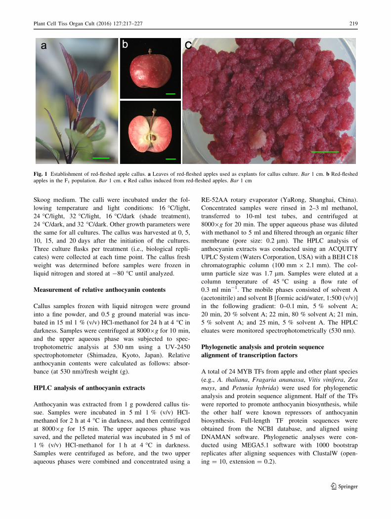
The F1 hybrid population of the cross between M. sieversii
f. niedzwetzkyana and M. domestica cv. ‘Fuji’ was grown
at the Tai’an Hengling Fruit Tree Breeding Base of
Shandong Agricultural University (36� 260 N, 117� 290
E). We used young ‘Zihong 1’ leaves as explants for callus
cultures (Fig. 1a). The red callus culturing method was
based on a published procedure (Ji et al. 2015). The callus
induction medium consisted of Murashige and Skoog
medium supplemented with 0.6 mg l-1 2,4-dichlorophe-
noxyacetic acid, 0.5 mg l-1 thidiazuron, 30 g l-1 sugar,
and 7 g l-1 agar. The pH was adjusted to 5.8 ± 0.1. Sur-
face-sterilized leaves were first cultured at 24 �C for
15 days in darkness, and then cultured under light (16-h
light/8-h dark; intensity: 1000–2000 lx). The red callus was
finally induced and subcultured every 20 days (Fig. 1c).
Determination of callus growth
The callus/cell cultures (fresh weight: 0.15 g) were trans-
ferred to new culture flasks containing Murashige and
218 Plant Cell Tiss Organ Cult (2016) 127:217–227
123
Skoog medium. The calli were incubated under the fol-
lowing temperature and light conditions: 16 �C/light,24 �C/light, 32 �C/light, 16 �C/dark (shade treatment),
24 �C/dark, and 32 �C/dark. Other growth parameters were
the same for all cultures. The callus was harvested at 0, 5,
10, 15, and 20 days after the initiation of the cultures.
Three culture flasks per treatment (i.e., biological repli-
cates) were collected at each time point. The callus fresh
weight was determined before samples were frozen in
liquid nitrogen and stored at -80 �C until analyzed.
Measurement of relative anthocyanin contents
Callus samples frozen with liquid nitrogen were ground
into a fine powder, and 0.5 g ground material was incu-
bated in 15 ml 1 % (v/v) HCl-methanol for 24 h at 4 �C in
darkness. Samples were centrifuged at 80009g for 10 min,
and the upper aqueous phase was subjected to spec-
trophotometric analysis at 530 nm using a UV-2450
spectrophotometer (Shimadzu, Kyoto, Japan). Relative
anthocyanin contents were calculated as follows: absor-
bance (at 530 nm)/fresh weight (g).
HPLC analysis of anthocyanin extracts
Anthocyanin was extracted from 1 g powdered callus tis-
sue. Samples were incubated in 5 ml 1 % (v/v) HCl-
methanol for 2 h at 4 �C in darkness, and then centrifuged
at 80009g for 15 min. The upper aqueous phase was
saved, and the pelleted material was incubated in 5 ml of
1 % (v/v) HCl-methanol for 1 h at 4 �C in darkness.
Samples were centrifuged as before, and the two upper
aqueous phases were combined and concentrated using a
RE-52AA rotary evaporator (YaRong, Shanghai, China).
Concentrated samples were rinsed in 2–3 ml methanol,
transferred to 10-ml test tubes, and centrifuged at
80009g for 20 min. The upper aqueous phase was diluted
with methanol to 5 ml and filtered through an organic filter
membrane (pore size: 0.2 lm). The HPLC analysis of
anthocyanin extracts was conducted using an ACQUITY
UPLC System (Waters Corporation, USA) with a BEH C18
chromatographic column (100 mm 9 2.1 mm). The col-
umn particle size was 1.7 lm. Samples were eluted at a
column temperature of 45 �C using a flow rate of
0.3 ml min-1. The mobile phases consisted of solvent A
(acetonitrile) and solvent B [formic acid/water, 1:500 (v/v)]
in the following gradient: 0–0.1 min, 5 % solvent A;
20 min, 20 % solvent A; 22 min, 80 % solvent A; 21 min,
5 % solvent A; and 25 min, 5 % solvent A. The HPLC
eluates were monitored spectrophotometrically (530 nm).
Phylogenetic analysis and protein sequence
alignment of transcription factors
A total of 24 MYB TFs from apple and other plant species
(e.g., A. thaliana, Fragaria ananassa, Vitis vinifera, Zea
mays, and Petunia hybrida) were used for phylogenetic
analysis and protein sequence alignment. Half of the TFs
were reported to promote anthocyanin biosynthesis, while
the other half were known repressors of anthocyanin
biosynthesis. Full-length TF protein sequences were
obtained from the NCBI database, and aligned using
DNAMAN software. Phylogenetic analyses were con-
ducted using MEGA5.1 software with 1000 bootstrap
replicates after aligning sequences with ClustalW (open-
ing = 10, extension = 0.2).
Fig. 1 Establishment of red-fleshed apple callus. a Leaves of red-fleshed apples used as explants for callus culture. Bar 1 cm. b Red-fleshed
apples in the F1 population. Bar 1 cm. c Red callus induced from red-fleshed apples. Bar 1 cm
Plant Cell Tiss Organ Cult (2016) 127:217–227 219
123
RNA isolation and quantitative real-time
polymerase chain reaction
Total RNA was isolated from callus tissues using an
RNAprep Pure Plant kit (Tiangen, Beijing, China). The
concentration and quality of the purified RNA were
determined using a NanoDrop 2000 spectrophotometer
(Thermo Fisher Scientific, USA). First-strand cDNA was
synthesized from 1 lg total RNA using the RevertAid First
Strand cDNA Synthesis kit (Fermentas, Hanover, MD,
USA). The cDNA samples were stored at -20 �C until
used.
The quantitative real-time polymerase chain reaction
(qRT-PCR) primers were designed using the Beacon
Designer 7 program (Table S1). Primers were synthesized
by Sangon Biotech (Shanghai, China) and purified by
polyacrylamide gel electrophoresis. The qRT-PCR was
conducted using tenfold diluted cDNA samples as tem-
plates, the SYBR Green PCR Master Mix (TransGen
Biotech, Beijing, China), and the iCycler iQ5 system (Bio-
Rad, Hercules, CA). The MdActin gene served as an
internal control, and the relative quantities of mRNA were
calculated using the 2-DDCt method of the IQ5 2.0 program
(Livak and Schmittgen 2001).
Results
Comparative analysis of callus growth
under various conditions
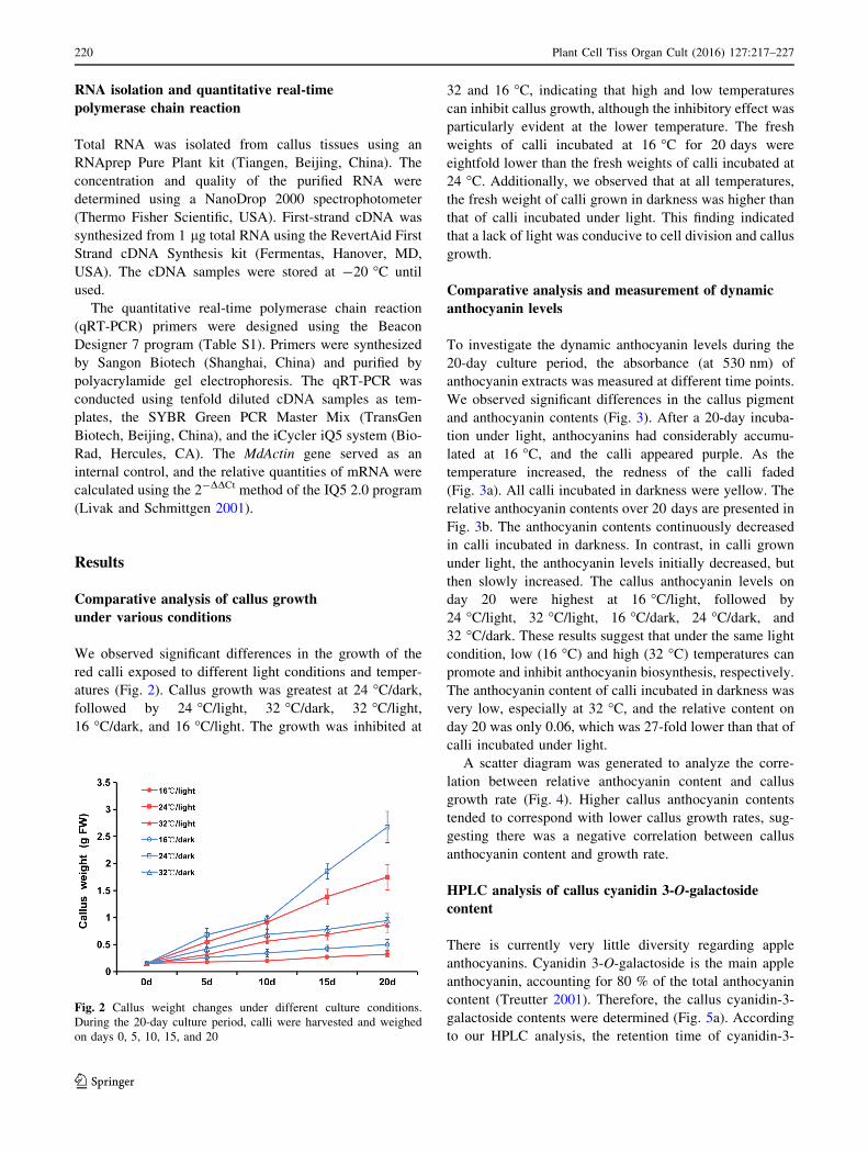
We observed significant differences in the growth of the
red calli exposed to different light conditions and temper-
atures (Fig. 2). Callus growth was greatest at 24 �C/dark,followed by 24 �C/light, 32 �C/dark, 32 �C/light,16 �C/dark, and 16 �C/light. The growth was inhibited at
32 and 16 �C, indicating that high and low temperatures
can inhibit callus growth, although the inhibitory effect was
particularly evident at the lower temperature. The fresh
weights of calli incubated at 16 �C for 20 days were
eightfold lower than the fresh weights of calli incubated at
24 �C. Additionally, we observed that at all temperatures,
the fresh weight of calli grown in darkness was higher than
that of calli incubated under light. This finding indicated
that a lack of light was conducive to cell division and callus
growth.
Comparative analysis and measurement of dynamic
anthocyanin levels
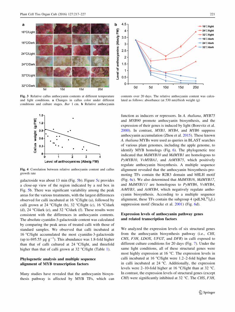
To investigate the dynamic anthocyanin levels during the
20-day culture period, the absorbance (at 530 nm) of
anthocyanin extracts was measured at different time points.
We observed significant differences in the callus pigment
and anthocyanin contents (Fig. 3). After a 20-day incuba-
tion under light, anthocyanins had considerably accumu-
lated at 16 �C, and the calli appeared purple. As the
temperature increased, the redness of the calli faded
(Fig. 3a). All calli incubated in darkness were yellow. The
relative anthocyanin contents over 20 days are presented in
Fig. 3b. The anthocyanin contents continuously decreased
in calli incubated in darkness. In contrast, in calli grown
under light, the anthocyanin levels initially decreased, but
then slowly increased. The callus anthocyanin levels on
day 20 were highest at 16 �C/light, followed by
24 �C/light, 32 �C/light, 16 �C/dark, 24 �C/dark, and
32 �C/dark. These results suggest that under the same light
condition, low (16 �C) and high (32 �C) temperatures can
promote and inhibit anthocyanin biosynthesis, respectively.
The anthocyanin content of calli incubated in darkness was
very low, especially at 32 �C, and the relative content on
day 20 was only 0.06, which was 27-fold lower than that of
calli incubated under light.
A scatter diagram was generated to analyze the corre-
lation between relative anthocyanin content and callus
growth rate (Fig. 4). Higher callus anthocyanin contents
tended to correspond with lower callus growth rates, sug-
gesting there was a negative correlation between callus
anthocyanin content and growth rate.
HPLC analysis of callus cyanidin 3-O-galactoside
content
There is currently very little diversity regarding apple
anthocyanins. Cyanidin 3-O-galactoside is the main apple
anthocyanin, accounting for 80 % of the total anthocyanin
content (Treutter 2001). Therefore, the callus cyanidin-3-
galactoside contents were determined (Fig. 5a). According
to our HPLC analysis, the retention time of cyanidin-3-
Fig. 2 Callus weight changes under different culture conditions.
During the 20-day culture period, calli were harvested and weighed
on days 0, 5, 10, 15, and 20
220 Plant Cell Tiss Organ Cult (2016) 127:217–227
123
galactoside was about 13 min (Fig. 5b). Figure 5c provides
a close-up view of the region indicated by a red box in
Fig. 5b. There was significant variability among the peak
areas for the various treatments, with the largest differences
observed for calli incubated at 16 �C/light (a), followed by
calli grown at 24 �C/light (b), 32 �C/light (c), 16 �C/dark(d), 24 �C/dark (e), and 32 �C/dark (f). These results were
consistent with the differences in anthocyanin contents.
The absolute cyanidin-3-galactoside content was calculated
by comparing the peak areas of treated calli with those of
standard samples. We observed that calli incubated at
16 �C/light accumulated the most cyanidin-3-galactoside
(up to 695.55 lg g-1). This abundance was 1.8-fold higher
than that of calli cultured at 24 �C/light, and threefold
higher than that of calli grown at 32 �C/light (Table 1).
Phylogenetic analysis and multiple sequence
alignment of MYB transcription factors
Many studies have revealed that the anthocyanin biosyn-
thesis pathway is affected by MYB TFs, which can
function as inducers or repressors. In A. thaliana, MYB75
and MYB90 promote anthocyanin biosynthesis, and the
expression of their genes is induced by light (Borevitz et al.
2000). In contrast, MYB3, MYB4, and MYB6 suppress
anthocyanin accumulation (Zhou et al. 2015). These known
A. thalianaMYBs were used as queries in BLAST searches
of various plant genomes, including the apple genome, to
identify MYB homologs (Fig. 6). The phylogenetic tree
indicated that MdMYB10 and MdMYB1 are homologous to
PyMYB10, VvMYBA1, and AtMYB75, which positively
regulate anthocyanin biosynthesis. A multiple sequence
alignment revealed that the anthocyanin biosynthesis-pro-
moting TFs contain the R2R3 domain and bHLH motif
(Fig. 6c). We also determined that MdMYB16, MdMYB17,
and MdMYB111 are homologous to PyMYB6, VvMYB4,
AtMYB3, and AtMYB4, which negatively regulate antho-
cyanin biosynthesis. According to a multiple sequence
alignment, these TFs contain the subgroup 4 (pdLNLD/EL)
suppression motif (Stracke et al. 2001) (Fig. 6d).
Expression levels of anthocyanin pathway genes
and related transcription factors
We analyzed the expression levels of six structural genes
from the anthocyanin biosynthesis pathway (i.e., CHI,
CHS, F3H, LDOX, UFGT, and DFR) in calli exposed to
different culture conditions for 20 days (Fig. 7). Under the
same light conditions, all of these structural genes were
most highly expression at 16 �C. The expression levels in
calli incubated at 16 �C/light were 1.2–2-fold higher than
in calli incubated at 24 �C. Additionally, the expression
levels were 2–10-fold higher at 16 �C/light than at 32 �C.In contrast, the expression levels of structural genes (except
CHI) were significantly inhibited at 32 �C. The CHS, F3H,
Fig. 3 Relative callus anthocyanin contents at different temperature
and light conditions. a Changes in callus color under different
conditions and culture stages. Bar 1 cm. b Relative anthocyanin
contents over 20 days. The relative anthocyanin content was calcu-
lated as follows: absorbance (at 530 nm)/fresh weight (g)
Fig. 4 Correlation between relative anthocyanin content and callus
growth rate
Plant Cell Tiss Organ Cult (2016) 127:217–227 221
123
LDOX, UFGT, and DFR expression levels at 32 �C were
approximately 7-, 2-, 3-, 3-, and 2-fold lower than at 24 �C,and 11-, 3-, 5-, 5-, and 3-fold lower than at 16 �C,respectively. In calli incubated in darkness, the expression
levels of all of the analyzed structural genes were relatively
low.
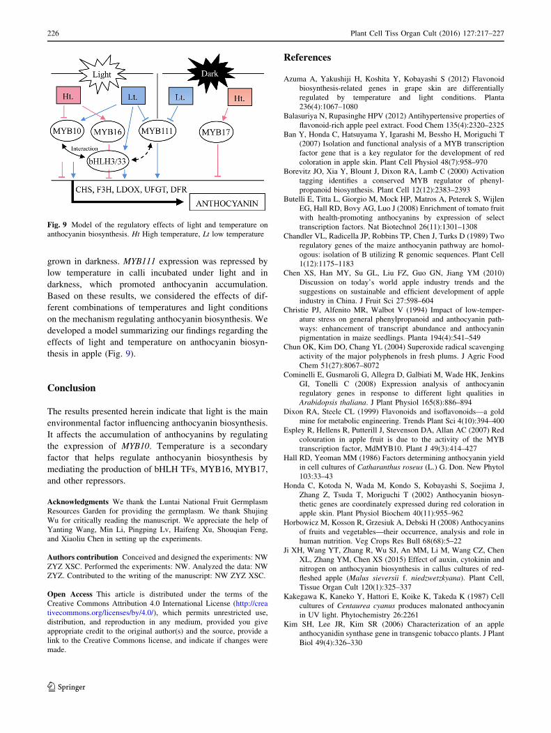
To validate the regulatory mechanism of TFs during
anthocyanin biosynthesis under different light and tem-
perature conditions, the expression levels of MYB TF
(MYB10, MYB16, MYB17, and MYB111) and bHLH TF
(bHLH3 and bHLH33) genes were analyzed by qRT-
PCR (Fig. 8). The MYB10 transcript levels were higher
in calli incubated under light than in calli grown in
darkness (i.e., approximately 17-, 6-, and 13-fold higher
at 16, 24, and 32 �C, respectively). Additionally, tem-
perature also affected the MYB10 expression levels
under light, with 16 �C inducing the highest expression
levels and 32 �C inhibiting expression. MYB16, MYB17,
and MYB111 were speculated to be anthocyanin
biosynthesis repressors. The highest MYB16 expression
level was observed at 32 �C/light, and was about 2–3-
fold higher than at other temperature and light condi-
tions. MYB111 expression was inhibited by low tem-
perature under light and dark conditions, which
promoted the accumulation of anthocyanins. The MYB17
expression levels were higher in calli incubated in
darkness, with 32 �C inducing the highest expression
levels. BHLH is another important TF family involved
in regulating anthocyanin biosynthesis. The bHLH3 and
bHLH33 expression levels were highest at 16 �C, and
were approximately twofold higher than at other tem-
peratures (Fig. 8).
Fig. 5 HPLC chromatograms of anthocyanins from calli grown
under different culture conditions. a Comparison of the colors of calli
treated with different conditions for 20 days. b HPLC graph of the
cyanidin-3-galactoside content of calli exposed to different
treatments. c Close-up view of the region in the red box in (B),
with (a)–(f) indicating the following treatments: (a) 16 �C/light,(b) 24 �C/light, (c) 32 �C/light, (d) 16 �C/dark, (e) 24 �C/dark, and(f) 32 �C/dark
Table 1 HPLC datas and cyanidin 3-O-galactoside content of each callus
Sample Retention
time (min)
Peak area Concentration
(ug/ml)
Cyanidin 3-O-galactoside
content (ug/g)
Standard samplea 13.444 3,464,065 100.00 –
16 �C/light 13.566 4,818,866 139.11 695.55
24 �C/light 13.573 2,618,632 75.59 377.97
32 �C/light 13.158 1,602,026 46.25 231.23
16 �C/dark 13.213 413,458 11.94 59.68
24 �C/dark 12.866 55,156 1.59 7.96
32 �C/dark 12.912 14,398 0.42 2.08
a Cyaniding 3-O-galactoside standards (Sigma chemical, St, Louis, USA)
222 Plant Cell Tiss Organ Cult (2016) 127:217–227
123

Discussion
Effect of light and temperature on callus growth
and anthocyanin biosynthesis
Apple is one of the most widely cultivated fruit trees in the
world, with many cultivars producing fruits with a red peel
(Chen et al. 2010). Fruit color is an important apple trait,
and largely determines the market value of apple crops.
Anthocyanins are the main pigments affecting the color
and nutritional value of apple fruits, and there has been
considerable interest in their synthesis and regulation
among apple researchers (Takos et al. 2006; Ubi et al.
2006; Espley et al. 2007; Lin-Wang et al. 2011). As a type
of antioxidant, anthocyanins have important implications
for human health (Smith et al. 2000; Wang and Mazza
2002; Chun et al. 2004). However, anthocyanins accumu-
late only in the red peel of cultivated apple fruits, with very
little (or none at all) detected in the white fruit flesh (Nie
et al. 2010). Most studies on anthocyanins have concen-
trated on the apple peel, which is not the main part of
consumers to eat. Meng et al. (2016) studied anthocyanin
biosynthesis in red-skinned cultivars at different fruit
development stages, and suggested that the expression of
the responsible genes is induced by light. Wang et al.
(2006) observed that anthocyanin contents of apple peels
increase significantly following exposure to continuous
light. Other researchers determined that UV-B irradiation
can induce anthocyanin biosynthesis in apple peels, and the
regulatory effect of UV-B is enhanced at low temperatures
(Ubi et al. 2006).
In this study, we developed a red callus culture system
using red-fleshed apple ‘Zihong1’. The generated calli
exhibited consistent growth and development, making
them appropriate for studies on anthocyanin biosynthesis
regulated by light, temperature, and other environmental
factors. We observed that incubations in darkness signifi-
cantly inhibited the accumulation of anthocyanins. The
anthocyanin content of calli grown in darkness was only
4–10 % of that of calli exposed to light. Low temperatures
are conducive to anthocyanin biosynthesis. The antho-
cyanin content was significantly higher at 16 �C than at 24
or 32 �C. These findings are consistent with the results of
previous studies, indicating that temperature and light
regulate anthocyanin biosynthesis not only in red-skinned
cultivated apples, but also in red-fleshed apples and red
callus tissue. Thus, anthocyanin metabolism may be rela-
tively stable in response to different temperatures and light
Fig. 6 Phylogenetic analysis and multiple sequence alignment of
MYB transcription factors from various plants. a Phylogenetic
analysis of MYB transcription factors reported to promote antho-
cyanin biosynthesis. b Phylogenetic analysis of MYB transcription
factors reported to inhibit anthocyanin biosynthesis. c Multiple
sequence alignment of MYB transcription factors. All of the
anthocyanin biosynthesis-promoting transcription factors contained the
R2R3 domain and bHLH motif. d Multiple sequence alignment of
transcription factors that negatively regulate anthocyanin biosynthesis.
These transcription factors contained the subgroup 4 (pdLNL-D/EL)
suppression motif
Plant Cell Tiss Organ Cult (2016) 127:217–227 223
123
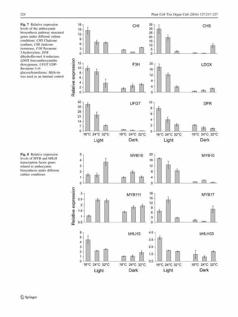
Fig. 7 Relative expression
levels of the anthocyanin
biosynthesis pathway structural
genes under different culture
conditions. CHS Chalcone
synthase, CHI chalcone
isomerase, F3H flavanone
3-hydroxylase, DFR
dihydroflavonol 4-reductase,
LDOX leucoanthocyanidin
dioxygenase, UFGT UDP-
flavanone-3-O-
glucosyltransferase. MdActin
was used as an internal control
Fig. 8 Relative expression
levels of MYB and bHLH
transcription factor genes
related to anthocyanin
biosynthesis under different
culture conditions
224 Plant Cell Tiss Organ Cult (2016) 127:217–227
123

conditions, and is not influenced by different genotypes or
tissues.
Interestingly, Ji et al. (2015) studied anthocyanin accu-
mulation in the same red callus at different concentration
of auxins and/or cytokinins, and suggested that antho-
cyanin accumulation decreased with increased auxin (NAA
and 2,4-D) concentration, while nitrogen deficiency could
reverse the inhibition of anthocyanin synthesis by auxins.
Both growth regulators and nitrogen affected the primary
metabolism of the plant. Growth accumulation and sec-
ondary metabolite require different conditions to induce a
shift from the growth state to the metabolite production
state (Simoes et al. 2012). Our results confirmed this
statement, and found that callus growth is influenced by
temperature and light treatments, and that callus growth
and anthocyanin content are negatively correlated to a
certain extent. Therefore, there may be a competitive
relationship between the growth state and the metabolite
production state in plant. This is consistent with the apple
fruit developmental process in which the fruit first increa-
ses in size during the primary growth stage, and then sec-
ondary metabolic activities lead to the accumulation of
anthocyanins.
Regulatory effects of light and temperature
on related transcription factors and structural genes
Light and temperature can induce the expression of related
TF and structural genes to regulate anthocyanin metabo-
lism. In apple skin, CHS, ANS, F3H, DFR, and UFGT have
been isolated and identified (Honda et al. 2002). Among
these genes, ANS and UFGT have important functions
during anthocyanin biosynthesis. Their expression is
induced by UV-B irradiation and low temperatures in apple
fruit skin (Kondo et al. 2002; Kim et al. 2006). In in vitro
culture systems, some studies have suggested that light
irradiation can induce anthocyanin production in Catha-
ranthus roseus (Hall and Yeoman 1986), Centaurea cyanus
(Kakegawa et al. 1987), Perilla frutescens (Zhong et al.
1993), Strawberry (Sato et al. 1996) and other plant spe-
cies. In contrast, the effect of temperature on anthocyanin
synthesis was more variable. The highest anthocyanin
accumulation was at 30 �C when compared to lower tem-
peratures in callus cultures of Daucus carota (Narayan
et al. 2005), while low temperature enhanced both antho-
cyanin accumulation and CHS expression in petunia flower
(Shvarts et al. 1997). In this study, we focused on the
expression of TF and structural genes in calli treated with
different combinations of temperatures and light condi-
tions. The ANS, F3H, CHS, DFR, and UFGT expression
patterns based on our data are consistent with those of
previous studies. The expression level of the five genes was
higher under light than in darkness, and increasing
temperatures resulted in decreasing expression levels. We
observed that the expression of CHI was induced by low
temperature. Additionally, its expression was not inhibited
by high temperature, unlike the expression of CHS and
F3H. The expression levels of LDOX, DFR, and UFGT
decreased with increasing temperature.
A previous study on TFs concluded that light is an
essential factor influencing the color of apple fruit skin
(Takos et al. 2006). Li et al. confirmed that MdMYB1
protein stability is enhanced by exposure to light, leading
to increased anthocyanin biosynthesis in apple skin (Li
et al. 2012). Azuma et al. (2012) reported that low tem-
perature and light have a synergistic effect on the expres-
sion of the flavonoid biosynthesis pathway genes in grape
berry skin. Tian et al. (2015) found that low temperatures
induced the expression of McMYB10, McbHLH3/33 and
McTTG1, leading to anthocyanin accumulation in crabap-
ple leaves. In this study, we observed that MdMYB10
expression was up-regulated in calli cultured under light,
while the expression was very low in calli grown in
darkness, which is in agreement with earlier findings
regarding anthocyanin biosynthesis. Furthermore, we
revealed that temperature can also regulate MdMYB10
expression, as indicated by the fact its expression level
decreased with increasing temperatures.
The bHLH TFs and the negative regulatory factors,
MYB16, MYB17, and MYB111, were unaffected by light,
but they were sensitive to temperature. The mechanism
underlying the regulation of anthocyanin biosynthesis by
temperature is relatively complex. Low temperatures
increase the expression level of anthocyanin biosynthesis
genes in Z. mays, V. vinifera, Citrus sinensis, and other
plants (Christie et al. 1994; Lo Piero et al. 2005; Mori et al.
2005). Additionally, MdbHLH3 promotes anthocyanin
accumulation and fruit coloration in apples at low tem-
peratures (Xie et al. 2012). In this study, bHLH3 and
bHLH33 were highly expressed in calli grown at
16 �C/light, implying that the expression of bHLH TF
genes may be induced by low temperature, but not light, to
regulate anthocyanin accumulation. Heating apple fruits
rapidly decreases the expression levels of MYB10, which is
responsible for coordinating the development of red fruit
skin color (Lin-Wang et al. 2011). We observed that high
temperatures inhibited MYB10 expression. Additionally,
some transcription repressors regulating anthocyanin
biosynthesis were also affected by temperature. For
example, MYB16 was highly expressed at 32 �C/light,indicating that high temperatures up-regulate MYB16
expression, leading to inhibited accumulation of antho-
cyanins. However, this regulatory mechanism did not apply
to calli incubated in darkness. In contrast, the high tem-
perature-induced expression of MYB17, and subsequent
inhibition of anthocyanin accumulation, occurred in calli
Plant Cell Tiss Organ Cult (2016) 127:217–227 225
123
grown in darkness. MYB111 expression was repressed by
low temperature in calli incubated under light and in
darkness, which promoted anthocyanin accumulation.
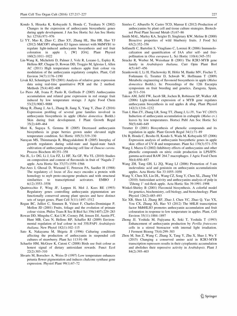
Based on these results, we considered the effects of dif-
ferent combinations of temperatures and light conditions
on the mechanism regulating anthocyanin biosynthesis. We
developed a model summarizing our findings regarding the
effects of light and temperature on anthocyanin biosyn-
thesis in apple (Fig. 9).
Conclusion
The results presented herein indicate that light is the main
environmental factor influencing anthocyanin biosynthesis.
It affects the accumulation of anthocyanins by regulating
the expression of MYB10. Temperature is a secondary
factor that helps regulate anthocyanin biosynthesis by
mediating the production of bHLH TFs, MYB16, MYB17,
and other repressors.
Acknowledgments We thank the Luntai National Fruit Germplasm
Resources Garden for providing the germplasm. We thank Shujing
Wu for critically reading the manuscript. We appreciate the help of
Yanting Wang, Min Li, Pingping Lv, Haifeng Xu, Shouqian Feng,
and Xiaoliu Chen in setting up the experiments.
Authors contribution Conceived and designed the experiments: NW
ZYZ XSC. Performed the experiments: NW. Analyzed the data: NW
ZYZ. Contributed to the writing of the manuscript: NW ZYZ XSC.
Open Access This article is distributed under the terms of the
Creative Commons Attribution 4.0 International License (http://crea
tivecommons.org/licenses/by/4.0/), which permits unrestricted use,
distribution, and reproduction in any medium, provided you give
appropriate credit to the original author(s) and the source, provide a
link to the Creative Commons license, and indicate if changes were
made.
References
Azuma A, Yakushiji H, Koshita Y, Kobayashi S (2012) Flavonoid
biosynthesis-related genes in grape skin are differentially
regulated by temperature and light conditions. Planta
236(4):1067–1080
Balasuriya N, Rupasinghe HPV (2012) Antihypertensive properties of
flavonoid-rich apple peel extract. Food Chem 135(4):2320–2325
Ban Y, Honda C, Hatsuyama Y, Igarashi M, Bessho H, Moriguchi T
(2007) Isolation and functional analysis of a MYB transcription
factor gene that is a key regulator for the development of red
coloration in apple skin. Plant Cell Physiol 48(7):958–970
Borevitz JO, Xia Y, Blount J, Dixon RA, Lamb C (2000) Activation
tagging identifies a conserved MYB regulator of phenyl-
propanoid biosynthesis. Plant Cell 12(12):2383–2393
Butelli E, Titta L, Giorgio M, Mock HP, Matros A, Peterek S, Wijlen
EG, Hall RD, Bovy AG, Luo J (2008) Enrichment of tomato fruit
with health-promoting anthocyanins by expression of select
transcription factors. Nat Biotechnol 26(11):1301–1308
Chandler VL, Radicella JP, Robbins TP, Chen J, Turks D (1989) Two
regulatory genes of the maize anthocyanin pathway are homol-
ogous: isolation of B utilizing R genomic sequences. Plant Cell
1(12):1175–1183
Chen XS, Han MY, Su GL, Liu FZ, Guo GN, Jiang YM (2010)
Discussion on today’s world apple industry trends and the
suggestions on sustainable and efficient development of apple
industry in China. J Fruit Sci 27:598–604
Christie PJ, Alfenito MR, Walbot V (1994) Impact of low-temper-
ature stress on general phenylpropanoid and anthocyanin path-
ways: enhancement of transcript abundance and anthocyanin
pigmentation in maize seedlings. Planta 194(4):541–549
Chun OK, Kim DO, Chang YL (2004) Superoxide radical scavenging
activity of the major polyphenols in fresh plums. J Agric Food
Chem 51(27):8067–8072
Cominelli E, Gusmaroli G, Allegra D, Galbiati M, Wade HK, Jenkins
GI, Tonelli C (2008) Expression analysis of anthocyanin
regulatory genes in response to different light qualities in
Arabidopsis thaliana. J Plant Physiol 165(8):886–894
Dixon RA, Steele CL (1999) Flavonoids and isoflavonoids—a gold
mine for metabolic engineering. Trends Plant Sci 4(10):394–400
Espley R, Hellens R, Putterill J, Stevenson DA, Allan AC (2007) Red
colouration in apple fruit is due to the activity of the MYB
transcription factor, MdMYB10. Plant J 49(3):414–427
Hall RD, Yeoman MM (1986) Factors determining anthocyanin yield
in cell cultures of Catharanthus roseus (L.) G. Don. New Phytol
103:33–43
Honda C, Kotoda N, Wada M, Kondo S, Kobayashi S, Soejima J,
Zhang Z, Tsuda T, Moriguchi T (2002) Anthocyanin biosyn-
thetic genes are coordinately expressed during red coloration in
apple skin. Plant Physiol Biochem 40(11):955–962
Horbowicz M, Kosson R, Grzesiuk A, Debski H (2008) Anthocyanins
of fruits and vegetables—their occurrence, analysis and role in
human nutrition. Veg Crops Res Bull 68(68):5–22
Ji XH, Wang YT, Zhang R, Wu SJ, An MM, Li M, Wang CZ, Chen
XL, Zhang YM, Chen XS (2015) Effect of auxin, cytokinin and
nitrogen on anthocyanin biosynthesis in callus cultures of red-
fleshed apple (Malus sieversii f. niedzwetzkyana). Plant Cell,
Tissue Organ Cult 120(1):325–337
Kakegawa K, Kaneko Y, Hattori E, Koike K, Takeda K (1987) Cell
cultures of Centaurea cyanus produces malonated anthocyanin
in UV light. Phytochemistry 26:2261
Kim SH, Lee JR, Kim SR (2006) Characterization of an apple
anthocyanidin synthase gene in transgenic tobacco plants. J Plant
Biol 49(4):326–330
Fig. 9 Model of the regulatory effects of light and temperature on
anthocyanin biosynthesis. Ht High temperature, Lt low temperature
226 Plant Cell Tiss Organ Cult (2016) 127:217–227
123
Kondo S, Hiraoka K, Kobayashi S, Honda C, Terahara N (2002)
Changes in the expression of anthocyanin biosynthetic genes
during apple development. J Am Soc Hortic Sci Am Soc Hortic
Sci 127(6):971–976
Li YY, Mao K, Zhao C, Zhao XY, Zhang HL, Shu HR, Hao YJ
(2012) MdCOP1 ubiquitin E3 ligases interact with MdMYB1 to
regulate light-induced anthocyanin biosynthesis and red fruit
coloration in apple 1, [W] [OA]. Plant Physiol
160(2):1011–1022
Lin-Wang K, Micheletti D, Palmer J, Volz R, Lozano L, Espley R,
Hellens RP, Chagne D, Rowan DD, Troggio M, Iglesias I, Allen
AC (2011) High temperature reduces apple fruit colour via
modulation of the anthocyanin regulatory complex. Plant, Cell
Environ 34(7):1176–1190
Livak KJ, Schmittgen TD (2001) Analysis of relative gene expression
data using real-time quantitative PCR and the 2 method.
Methods 25(4):402–408
Lo Piero AR, Ivana P, Paolo R, Goffredo P (2005) Anthocyanins
accumulation and related gene expression in red orange fruit
induced by low temperature storage. J Agric Food Chem
53(23):9083–9088
Meng R, Zhang J, An L, Zhang B, Jiang X, Yang Y, Zhao Z (2016)
Expression profiling of several gene families involved in
anthocyanin biosynthesis in apple (Malus domestica. Borkh.)
Skin during fruit development. J Plant Growth Regul
35(2):449–464
Mori K, Sugaya S, Gemma H (2005) Decreased anthocyanin
biosynthesis in grape berries grown under elevated night
temperature condition. Sci Hortic 105(3):319–330
Narayan MS, Thimmaraju R, Bhagyalakshmi B (2005) Interplay of
growth regulators during solid-state and liquid-state batch
cultivation of anthocyanin producing cell line of Daucus carota.
Process Biochem 40:351–358
Nie JL, Lv DG, Li J, Liu FZ, Li HF, Xu GF, Wu YL (2010) Studies
on composition and content of flavonoids in fruit of ‘Nagafu 2’
apple. Acta Hortic Sin 37(37):1559–1566
Paz-Ares J, Ghosal D, Wienand U, Peterson PA, Saedler H (1988)
The regulatory c1 locus of Zea mays encodes a protein with
homology to myb proto-oncogene products and with structural
similarities to transcriptional activators. EMBO J
6(12):3553–3558
Quattrocchio F, Wing JF, Leppen H, Mol J, Koes RE (1993)
Regulatory genes controlling anthocyanin pigmentation are
functionally conserved among plant species and have distinct
sets of target genes. Plant Cell 5(11):1497–1512
Regan BC, Julliot C, Simmen B, Vienot F, Charles-Dominique P,
Mollon JD (2001) Fruits, foliage and the evolution of primate
colour vision. Philos Trans R Soc B Biol Sci 356(1407):229–283
Rowan DD, Mingshu C, Kui LW, Cooney JM, Jensen DJ, Austin PT,
Hunt MB, Cara N, Hellens RP, Schaffer RJ (2009) Environ-
mental regulation of leaf colour in red 35S:PAP1 Arabidopsis
thaliana. New Phytol 182(1):102–115
Sato K, Nakayama M, Shigeta JI (1996) Culturing conditions
affecting the production of anthocyanin in suspended cell
cultures of strawberry. Plant Sci 113:91–98
Schaefer HM, McGraw K, Catoni C (2008) Birds use fruit colour as
honest signal of dietary antioxidant rewards. Funct Ecol
22(2):303–310
Shvarts M, Borochov A, Weiss D (1997) Low temperature enhances
petunia flower pigmentation and induces chalcone synthase gene
expression. Physiol Plant 99(1):67–72
Simoes C, Albarello N, Castro TCD, Mansur E (2012) Production of
anthocyanins by plant cell and tissue culture strategies. Biotech-
nol Prod Plant Second Metab (5):67–86
Smith MAL, Marley KA, Seigler D, Singletary KW, Meline B (2000)
Bioactive properties of wild blueberry fruits. J Food Sci
65(2):352–356
Solfanelli C, Bartolini S, Vitagliano C, Lorenzi R (2006) Immunolo-
calization and quantification of IAA after self- and free-
pollination in Olea europaea L. Sci Hortic 110(4):345–351
Stracke R, Werber M, Weisshaar B (2001) The R2R3-MYB gene
family in Arabidopsis thaliana. Curr Opin Plant Biol
4(5):447–456
Szankowski I, Li H, Flachowsky H, Hofer M, Hanke MV, Fischer T,
Forkmann G, Treutter D, Schwab W, Hoffmann T (2009)
Metabolic engineering of flavonoid biosynthesis in apple (Malus
domestica Borkh.). In: Proceedings of the 12th Eucarpia
symposium on fruit breeding and genetics, Zaragoza, Spain,
pp 511–516
Takos AM, Jaffe FW, Jacob SR, Jochen B, Robinson SP, Walker AR
(2006) Light-induced expression of a MYB gene regulates
anthocyanin biosynthesis in red apples & nbsp. Plant Physiol
142(3):1216–1232
Tian J, Han ZY, Zhang LR, Song TT, Zhang J, Li JY, Yao YC (2015)
Induction of anthocyanin accumulation in crabapple (Malus cv.)
leaves by low temperatures. Hortsci Publ Am Soc Hortic Sci
50(5):640–649
Treutter D (2001) Biosynthesis of phenolic compounds and itsregulation in apple. Plant Growth Regul 34(1):71–89
Ubi B, Honda C, Bessho H, Kondo S, Wada M, Kobayashi ST (2006)
Expression analysis of anthocyanin biosynthetic genes in apple
skin: effect of UV-B and temperature. Plant Sci 170(3):571–578
Wang J, Mazza G (2002) Inhibitory effects of anthocyanins and other
phenolic compounds on nitric oxide production in LPS/IFN-
gamma-activated RAW 264.7 macrophages. J Agric Food Chem
50(4):850–857
Wang ZH, Tang GH, Li ZQ, Wang LJ (2006) Promotion of 5-am
inolevulinic acid and genistein on anthocyanin accumulationin
apples. Acta Hortic Sin 33:1055–1058
Wang Y, Chen XS, Liu DL, Wang CZ, Song Y, Chen XL, Zhang YM
(2010) Antioxidant activity and anthocyanins analysis of pulp in
‘Zihong 1’ red-flesh apple. Acta Hortic Sin 39:1991–1998
Winkel-Shirley B (2001) Flavonoid biosynthesis. A colorful model
for genetics, biochemistry, cell biology, and biotechnology. Plant
Physiol 126(2):485–493
Xie XB, Shen LI, Zhang RF, Zhao J, Chen YC, Zhao Q, Yao YX,
You CX, Zhang XS, Hao YJ (2012) The bHLH transcription
factor MdbHLH3 promotes anthocyanin accumulation and fruit
colouration in response to low temperature in apples. Plant, Cell
Environ 35(11):1884–1897
Zhong JJ, Yoshida M, Fujiyama K, Seki T, Yoshida T (1993)
Enhancement of anthocyanin production by Perilla frutescens
cells in a stirred bioreactor with internal light irradiation.
J Ferment Bioeng 75(4):299–303
Zhou M, Sun Z, Wang C, Zhang X, Tang Y, Zhu X, Shao J, Wu Y
(2015) Changing a conserved amino acid in R2R3-MYB
transcription repressors results in their cytoplasmic accumulation
and abolishes their repressive activity in Arabidopsis. Plant J
84(2):395–403
Plant Cell Tiss Organ Cult (2016) 127:217–227 227
123


![Genetic Dissection of a Major Anthocyanin QTL Contributing ... · anthocyanin (pink) pigment was estimated as [(R + B)/2] 2 G. QTL affecting anthocyanin concentration in the backcross](https://img.dokumen.tips/doc/110x75/5e6421962a91715ff42dfa60/genetic-dissection-of-a-major-anthocyanin-qtl-contributing-anthocyanin-pink.jpg)