Embed Size (px)
Citation preview
Symbiotic Relationship between Streptococcus mutans and Candidaalbicans Synergizes Virulence of Plaque Biofilms In Vivo
Megan L. Falsetta,a Marlise I. Klein,a Punsiri M. Colonne,a Kathleen Scott-Anne,a Stacy Gregoire,a Chia-Hua Pai,a
Mireya Gonzalez-Begne,a Gene Watson,a Damian J. Krysan,b,c William H. Bowen,a,b Hyun Kooa,b,d
Center for Oral Biology,a Department of Microbiology and Immunology,b and Department of Pediatrics,c University of Rochester Medical Center, Rochester, New York,USA; Biofilm Research Laboratory, Levy Center for Oral Health, Department of Orthodontics, School of Dental Medicine, University of Pennsylvania, Philadelphia,Pennsylvania, USAd
Streptococcus mutans is often cited as the main bacterial pathogen in dental caries, particularly in early-childhood caries (ECC).S. mutans may not act alone; Candida albicans cells are frequently detected along with heavy infection by S. mutans in plaquebiofilms from ECC-affected children. It remains to be elucidated whether this association is involved in the enhancement of bio-film virulence. We showed that the ability of these organisms together to form biofilms is enhanced in vitro and in vivo. Thepresence of C. albicans augments the production of exopolysaccharides (EPS), such that cospecies biofilms accrue more biomassand harbor more viable S. mutans cells than single-species biofilms. The resulting 3-dimensional biofilm architecture displayssizeable S. mutans microcolonies surrounded by fungal cells, which are enmeshed in a dense EPS-rich matrix. Using a rodentmodel, we explored the implications of this cross-kingdom interaction for the pathogenesis of dental caries. Coinfected animalsdisplayed higher levels of infection and microbial carriage within plaque biofilms than animals infected with either speciesalone. Furthermore, coinfection synergistically enhanced biofilm virulence, leading to aggressive onset of the disease with ram-pant carious lesions. Our in vitro data also revealed that glucosyltransferase-derived EPS is a key mediator of cospecies biofilmdevelopment and that coexistence with C. albicans induces the expression of virulence genes in S. mutans (e.g., gtfB, fabM). Wealso found that Candida-derived �1,3-glucans contribute to the EPS matrix structure, while fungal mannan and �-glucan pro-vide sites for GtfB binding and activity. Altogether, we demonstrate a novel mutualistic bacterium-fungus relationship that oc-curs at a clinically relevant site to amplify the severity of a ubiquitous infectious disease.
Biofilms often contribute to and/or cause disease in humans(1). In the United States and worldwide, dental caries is the
single-most common and costly biofilm-dependent oral infec-tious disease, which continues to compromise the health and well-being of children and adults alike (2). Furthermore, the preva-lence of dental caries, particularly early-childhood caries (ECC), isincreasing among preschool children (2). ECC is a hypervirulentform of the disease that is characterized by a heavy Streptococcusmutans burden (often exceeding 30% of the cultivable plaque bio-film flora) (3, 4), accompanied by protracted feeding of dietarysugars, especially sucrose (5). The child is often allowed to con-sume sugary beverages almost constantly from a nursing bottle.The adverse effects of sugars are enhanced by the mechanical ef-fects of the nipple on the bottle, which restricts the access of buff-ering saliva to the tooth surfaces (6, 7).
Streptococcus mutans has often been regarded as one of the keyetiologic agents of ECC (3, 4, 8, 9), although other organisms mayalso contribute to its pathogenesis (9–11). S. mutans cells can rap-idly orchestrate the formation of cariogenic plaque biofilms onsusceptible tooth surfaces when they are exposed frequently todietary sucrose. Sucrose is utilized by S. mutans-derived enzymes(e.g., glucosyltransferases [Gtfs]) to produce exopolysaccharides(EPS), the prime building blocks of cariogenic biofilms (12). TheGtfs are secreted into the extracellular milieu, become constitu-ents of the pellicle that covers teeth, and are also adsorbed tobacterial surfaces while retaining enzymatic activity (12–14). Glu-can synthesis on the pellicle provides additional nonmammalianbacterial binding sites (e.g., through membrane-associated glu-can-binding proteins in S. mutans), while the polymers on thesurfaces of resident microorganisms increase the cohesion be-
tween organisms (12–14). As a consequence, a structured com-munity forms, which is enmeshed in an EPS-rich matrix that isdiffusion limiting (12, 15). At the same time, S. mutans and otheracidogenic/aciduric organisms produce acids as by-products ofsugar metabolism, creating acidic microenvironments within thebiofilm that further select for the growth of these organisms (12,15–19). Low pH values present at the biofilm-tooth interface pro-mote the dissolution of adjacent tooth enamel, leading to the clin-ical onset of cavitation.
The onset and progression of carious lesions in children withECC is rapid and aggressive, resulting in rampant destruction ofthe smooth surfaces of the teeth (3, 4, 8, 20, 21). The underlyingbiological reasons for the development of ECC remain unclear.Microbiological studies of plaque biofilms from children withECC reveal that in addition to high levels of S. mutans, the com-mon opportunistic fungal pathogen Candida albicans is also fre-quently detected; in contrast, it is detected sporadically, if at all, inthe plaque of ECC-free children (22–24). Why C. albicans is found
Received 17 January 2014 Returned for modification 13 February 2014Accepted 20 February 2014
Published ahead of print 24 February 2014
Editor: G. S. Deepe, Jr.
Address correspondence to Hyun Koo, [email protected].
Supplemental material for this article may be found at http://dx.doi.org/10.1128/IAI.00087-14.
Copyright © 2014, American Society for Microbiology. All Rights Reserved.
doi:10.1128/IAI.00087-14
1968 iai.asm.org Infection and Immunity p. 1968 –1981 May 2014 Volume 82 Number 5
on July 6, 2020 by guesthttp://iai.asm
.org/D
ownloaded from
together with high levels of S. mutans in plaque biofilms andwhether this bacterium-fungus association at sites of ECC infec-tion plays a significant role in the pathogenesis of ECC remain tobe elucidated.
Bacterium-fungus interactions occur commonly in humansand may influence the transition from a healthy to a diseased statewithin a specific host niche (25, 26). C. albicans is by far the mostcommonly detected fungal organism on human mucosal surfaces,and it often participates in the formation of polymicrobial bio-films on soft tissue and acrylic surfaces (26, 27). Candida coad-heres with many oral commensal species, namely, viridans groupstreptococci (e.g., Streptococcus gordonii, Streptococcus oralis), invitro (28–30) and enhances fungal carriage and infectivity in mu-cosal diseases in vivo (31). Yet Candida was initially regarded ashaving little to no physical adhesion with S. mutans in the absenceof sucrose (30). However, when sucrose is present, the adhesiveinteraction between these two organisms is enhanced (32–34).Images derived from electron microscopy revealed extracellularmaterial that had formed between cocci and yeast cells, suggestingthat locally produced glucans play a role in mediating their coad-herence (32, 34). We have determined that all three S. mutans Gtfexoenzymes bind to the surfaces of C. albicans cells in vitro; GtfBshows the greatest affinity (12, 35). When sucrose is available, theGtfs adsorbed onto C. albicans cells produce large amounts ofglucan on the fungal surface. These glucans formed in situ provideenhanced binding sites for S. mutans while simultaneously en-hancing fungal adhesion to saliva-coated hydroxyapatite surfaces(35). Here we explore whether this sucrose-dependent cross-king-dom interaction modulates cospecies biofilm developmentand/or influences the infectivity and the pathogenesis of dentalcaries in vivo.
We hypothesize that S. mutans-C. albicans associations mayenhance S. mutans infection and modulate the development ofhypervirulent biofilms on tooth surfaces, which will, in turn, in-fluence the onset and severity of dental caries in vivo. We demon-strate that the presence of C. albicans enhances the assembly of theEPS-rich matrix, such that cospecies biofilms accrue more bio-mass and more viable S. mutans cells than single-species biofilmsin vitro. More importantly, coinfection of rats with S. mutans andC. albicans enhances the colonization and carriage of both organ-isms in vivo and dramatically amplifies the virulence of plaquebiofilms formed on rodent dentition, leading to the developmentof rampant carious lesions. Furthermore, our in vitro data revealplausible explanations for the enhanced ability of these organismsto form virulent cospecies biofilms. Altogether, we demonstratethat a novel synergistic interaction occurs between an opportunis-tic fungus and a bacterial pathogen on a clinically relevant site(teeth) in the mouth. Our data offer new insight into the micro-biological and clinical features of ECC and may have relevance forother bacterium-fungus interactions associated with polymicro-bial infections in humans (25, 27).
(Part of this paper was presented at the 91st General Session ofthe International Association for Dental Research, Seattle, WA, 20to 23 March 2013 [36]).
MATERIALS AND METHODSIn vitro biofilm model. Streptococcus mutans strain UA159 serotype c (aproven virulent cariogenic bacterial pathogen selected for genome se-quencing) and Candida albicans SC5314 (a well-characterized strainwhose genome has been sequenced) were used to generate single or co-
species biofilms. Biofilms were formed using our saliva-coated hydroxy-apatite (sHA) disc model as described previously (15, 37). The hydroxy-apatite discs (surface area, 2.7 � 0.2 cm2; Clarkson ChromatographyProducts, Inc., South Williamsport, PA) coated with filter-sterilized, clar-ified whole saliva (15) were vertically suspended in 24-well plates using acustom-made wire disc holder, which was designed to mimic the freesmooth surfaces of the teeth (15, 37). For single-species biofilms, each discwas inoculated with approximately 2 � 106 CFU of S. mutans/ml in ul-trafiltered (10-kDa cutoff; Millipore, Billerica, MA) tryptone-yeast extract(UFYTE) broth containing 1% (30 mM) sucrose at 37°C under 5% CO2.For cospecies biofilms, approximately 2 � 104 CFU of C. albicans/ml(containing predominantly yeast cell forms [35]) was also added to theinoculum; the proportion of the microorganisms in the inoculum is sim-ilar to that found in saliva samples from children with ECC. During thefirst 18 h, the organisms were grown undisturbed so as to allow initialbiofilm formation; the culture medium was then changed twice daily at 8a.m. and 6 p.m. until the end of the experimental period (42 h). The pH ofthe culture medium was measured daily at each medium change.
Quantitative biofilm analysis. The development of each of the bio-films was assessed at 18 and 42 h postinoculation using our well-estab-lished protocols optimized for biofilm imaging and quantification (15, 37,38). The sequential assembly of the matrix was followed by incorporatingan Alexa Fluor 647-labeled dextran conjugate (10 kDa; absorbance/fluo-rescence emission maxima, 647/668 nm; Molecular Probes, InvitrogenCorp., Carlsbad, CA) into the glucans synthesized during the assembly ofthe EPS matrix (37, 38). The total microbial biomass was stained with Syto9 (485/498 nm; Molecular Probes) (15, 37). Imaging was performed usingan Olympus FV 1000 two-photon laser scanning microscope (Olympus,Tokyo, Japan) equipped with a 10� (numerical aperture, 0.45) waterimmersion objective lens. The excitation wavelength was 810 nm, and theemission wavelength filter for Syto 9 was a 495/540 OlyMPFC1 filter,while the filter for Alexa Fluor 647 was an HQ655/40M-2P filter (37). Eachbiofilm was scanned at 5 positions randomly selected on the microscopestage (39), and confocal image series (512- by 512-pixel resolution) weregenerated by optical sectioning at each of these positions. At least 3 inde-pendent biofilm experiments were performed. The confocal images wereanalyzed using software for the quantitation of EPS and microbial cellswithin intact biofilms (15, 37). COMSTAT (available at http://www.imageanalysis.dk) was used to calculate the biomass, as well as the num-ber and size of microcolonies (15). Furthermore, a separate set of biofilmswas used for standard microbiological analysis. The biofilms were homog-enized by sonication, and the number of viable cells (total number of CFUper biofilm) was determined as described elsewhere (40). It should benoted that CFU data for C. albicans have limitations given the morphol-ogy, since the cells exist in both the yeast and hyphal forms; hyphae areessentially multicellular structures that, when plated, form a single CFU,despite having a larger biomass than yeast forms.
Visualization of 3D biofilm architecture. We examined the spatialdistribution of individual microbial species and the EPS matrix withinintact biofilms at 4, 6, 8, 18, and 42 h (15, 37, 38). Because Syto 9 (as wellas other commercially available nucleic acid stains) labels both S. mutansand C. albicans, we used a green fluorescent protein (GFP)-expressingstrain of S. mutans (constructed from S. mutans strain UA159) to resolvethese species in cospecies biofilms. The GFP-expressing strain was a giftfrom Jose Lemos (Center for Oral Biology, University of Rochester Med-ical Center, Rochester, NY). Based on work from other groups (41, 42), weelected to stain the yeast using concanavalin A (ConA) lectin conjugatedwith tetramethylrhodamine (absorbance/fluorescence emission maxima,555/580 nm; Molecular Probes). We used a concentration of 40 �g/mlwith an incubation time of 30 min to reduce the possibility that ConAmight bind S. mutans, since ConA is capable of agglutinating several se-rotypes of S. mutans, but not serotype c strains (43). Prior to three-colorimaging, we determined that our concentration and incubation time weresufficient to label the C. albicans cells within the biofilm without cross-reacting with S. mutans (44). The EPS matrix was labeled with Alexa Fluor
Cross-Kingdom Interactions Enhance Biofilm Virulence
May 2014 Volume 82 Number 5 iai.asm.org 1969
on July 6, 2020 by guesthttp://iai.asm
.org/D
ownloaded from
647, as described above. Imaging was again performed using the OlympusFV 1000 laser scanning microscope equipped with a 25� LPlan N (nu-merical aperture, 1.05) water immersion objective lens. The excitationwavelength was 780 nm, and the emission wavelength filter for GFP was a495/540 OlyMPFC1 filter, while an HQ655/40M-2P and a 598/28OlyMPFC2 filter were used for Alexa Fluor 647 and rhodamine, respec-tively. In order to visualize all three fluorophores, the channels werescanned in the following pairs: (i) GFP and rhodamine and (ii) rhodamineand Alexa Fluor 647. Images at a 1,024- by 1,024-pixel resolution werecollected and analyzed using software for the simultaneous visualizationof EPS and each of the microbial cells within intact biofilms (15). Amirasoftware (version 5.4.1; Visage Imaging, San Diego, CA) was used to create3-dimensional (3D) renderings of each biofilm structural component(EPS and microorganisms) by combining the GFP and rhodamine chan-nels from scan 1 with the Alexa Fluor 647 channel from scan 2.
Labeling of �-glucan in biofilms. Cospecies biofilms were formedusing S. mutans UA159 and C. albicans SC5314 as described above. Ourprotocols are optimized and limited to three-color confocal imaging.Thus, we examined the presence of C. albicans-derived �-glucan in cospe-cies biofilms in two ways. First, we investigated the spatial distribution ofthe EPS matrix, C. albicans cells, and �-glucan. The EPS matrix was la-beled with Alexa Fluor 647, while C. albicans was stained with ConAconjugated with rhodamine. �-Glucan produced by C. albicans wasstained using a commercially available mouse monoclonal IgG antibodyto 1,3-�-glucan (Biosupplies Australia Pty. Ltd., Victoria, Australia)paired with a fluorescently labeled secondary antibody. All antibody stain-ing steps were performed in the dark at 4°C. The primary antibody wasdiluted 1:20 in phosphate-buffered saline (PBS; pH 7.0) and was incu-bated with the biofilm for 60 min. The biofilm was then washed in freshPBS and was blocked with 3% bovine serum albumin (Sigma-Aldrich, St.Louis, MO) for 15 min. The biofilm was again washed in fresh PBS andwas incubated for 30 min with the Fab= fragment of a goat-anti-mouse IgGantibody conjugated to Alexa Fluor 488 (absorbance/fluorescence emis-sion maxima, 488/519 nm; Molecular Probes) at a concentration of 4mg/ml. The biofilm was finally washed in 0.89% NaCl and was then im-aged using the Olympus FV 1000 microscope equipped with a 25� LPlanN (numerical aperture, 1.05) objective as described in the preceding sec-tion. In a separate set of experiments, we determined the spatial distribu-tion of S. mutans, C. albicans, and �-glucan within biofilms. We used ourGFP-expressing strain of S. mutans, while C. albicans was labeled withConA-tetramethylrhodamine (Molecular Probes). �-Glucan was labeledvia the anti-�-glucan antibody with a secondary antibody conjugated toAlexa Fluor 647 (Molecular Probes). Images at a 1,024- by 1,024-pixelresolution were collected and were analyzed using Amira software, ver-sion 5.4.1.
Cospecies biofilm formation using gtf-defective S. mutans strains.Knockout mutant strains of S. mutans with insertions in gtfB, gtfC, or both(�gtfB::kan, �gtfC::kan, and �gtfBC::kan) (15, 37) were cultured with C.albicans SC5314 to form cospecies biofilms. The gtfB-, gtfC-, and gtfBC-null mutants, constructed from the parental S. mutans UA159 strain, werekindly provided by Robert A. Burne (Department of Oral Biology, Uni-versity of Florida, Gainesville, FL). The mutant strains were generated bystandard allelic replacement with a nonpolar kanamycin resistancemarker and were verified by DNA sequence and biochemical analysis (forGtf production); the lack of polarity of the mutations was also verified byreverse transcription– quantitative PCR (RT-qPCR). The mutant strainswere also devoid of any growth or growth rate defects (relative to thegrowth of UA159). Biofilms formed with the parental S. mutans strain,UA159, were compared to those formed with the �gtfB::kan, �gtfC::kan,or �gtfBC::kan mutant at 42 h postinoculation. The 3D architecture ofcospecies biofilms was determined using confocal microscopy as de-scribed above. We used an anti-S. mutans antibody conjugated to AlexaFluor 488 (Molecular Probes) to label the mutant strain (which does notexpress GFP) within biofilms as described previously (45). The antibodywas provided courtesy of Robert Palmer and John Cisar at the National
Institutes for Health, Bethesda, MD. In addition, we also determined (in aseparate experiment) the total number of viable microbial cells in each ofthe cospecies biofilms (40).
Purification of biofilm RNAs. RNA was extracted and purified usingprotocols optimized for biofilms formed in vitro (46). Briefly, disc setswere incubated in RNALater (Applied Biosystems/Ambion, Austin, TX),and the biofilm material was removed from the sHA discs. The RNAs werepurified and were treated with DNase on a column using the QiagenRNeasy Micro kit (Qiagen, Valencia, CA). The RNAs were then subjectedto a second DNase I treatment with Turbo DNase (Applied Biosystems/Ambion) and were purified using the Qiagen RNeasy MinElutecleanup kit (Qiagen). The RNAs were quantified using the NanoDropND-1000 spectrophotometer (Thermo Scientific/NanoDrop, Wil-mington, DE). RNA quality was evaluated using an Agilent 2100 bio-analyzer (Agilent Technologies Inc., Santa Clara, CA), and all RNAsused to prepare cDNAs were determined to have RNA integrity num-bers (RIN) of 9.6 and above.
RT-qPCR analysis for selected genes. We performed RT-qPCR tomeasure the expression profiles of specific genes directly associated withextracellular polysaccharide matrix development and acid stress survival,including gtfB, gtfC, gtfD, fruA, dexA, fabM, and atpD. Briefly, cDNAswere synthesized using 0.5 �g of purified RNA and the Bio-Rad iScriptcDNA synthesis kit (Bio-Rad Laboratories, Inc., Hercules, CA). To checkfor DNA contamination, purified total RNA without reverse transcriptaseserved as a negative control. The resulting cDNAs were amplified with aBio-Rad CFX96 system (Bio-Rad Laboratories, Inc., Hercules, CA) usingpreviously published specific primers and TaqMan probes (15, 47). Astandard curve was plotted for each primer set, as described elsewhere(48). The standard curves were used to transform the critical thresholdcycle (CT) values to relative numbers of cDNA molecules. Comparativeexpression was calculated by normalizing each gene of interest to the 16SrRNA signal (48).
In vivo model of dental caries. Animal experiments were performedas described previously with some modifications (7, 49, 50). Six litters of 8female Sprague Dawley rats aged 15 days were purchased with their damsfrom Harlan Laboratories (Madison, WI). Upon arrival, animals werescreened for S. mutans and C. albicans, and were determined not to beinfected with either organism, by plating oral swabs on selective media:ChromAgar (VWR International LLC, Radnor, PA) for C. albicans andMitis Salivarius Agar plus Bacitracin (MSB) for S. mutans. Half of thedams, while still nursing, were infected by mouth with an actively growingculture of S. mutans UA159, which is transmitted to the pups (49); thesewere maintained separately from uninfected animals. Pups in cages withS. mutans-infected dams were also directly infected with S. mutans. Atweaning, pups aged 21 days were checked and confirmed for S. mutansinfection, while the other half of the animals remained uninfected. At theage of 23 days, all pups were subjected to surgical hyposalivation (49).After recovery, all pups slated for infection with C. albicans were inocu-lated with an actively growing culture of C. albicans SC5314 at the ages of24 and 25 days, and their infections were confirmed at 26 days. All theanimals were randomly placed into 1 of the following 4 groups: (i) S.mutans infected, (ii) C. albicans infected, (iii) S. mutans plus C. albicansinfected, and (iv) uninfected. Animals were screened at 26, 28, and 30 daysfor S. mutans and C. albicans infection. Each of the infected groups wasconfirmed for its respective microbial infection, while the uninfectedgroup remained free of either S. mutans or C. albicans; no cross-contam-ination was observed throughout the experiment. All animals were pro-vided the National Institutes of Health cariogenic diet 2000 (51) and 5%sucrose water ad libitum. The experiment proceeded for 2 weeks. At theend of 2 weeks, the animals were sacrificed. The jaws were asepticallydissected and were processed for microbiological analysis of each animal’splaque biofilms as described by Klein et al. (52). For microbiological anal-ysis, the left jaws were sonicated in 5 ml of 154 mM sterile NaCl solutionfor plaque biofilm removal. The suspensions obtained were serially di-luted and were plated on MSB or Inhibitory Mold Agar (a less costly
Falsetta et al.
1970 iai.asm.org Infection and Immunity
on July 6, 2020 by guesthttp://iai.asm
.org/D
ownloaded from
alternative to ChromAgar, yet equally selective, as determined experimen-tally) to estimate the S. mutans or C. albicans population, respectively, andon blood agar to determine the total cultivable flora in the plaque biofilms(49). All the jaws were defleshed, and the teeth were prepared for cariesscoring according to Larson’s modification of Keyes’ system (53).
Ethics statement. All animal experiments were performed in strictaccordance with the guidelines of the Animal Welfare Act of the UnitedStates, under protocols reviewed and approved by the Institutional Ani-mal Care and Use Committee of the University of Rochester (approvedprotocol 2011-040).
Statistical analysis. The data were analyzed by pairwise comparisonsof multiple groups with regression models using the ranked values.Kruskal-Wallis tests, which are nonparametric and are based on ranks,were used for two-group comparisons. The significance level was set at5%, and no adjustments were made for multiple comparisons. For theanimal study, an analysis of outcome measures was done with trans-formed values of the measures in order to stabilize variances as detailed byRaubertas et al. (54). The data were then subjected to statistical analyses asdescribed above. Furthermore, we also examined the possibility of a syn-ergistic effect of coinfection on the development of carious lesions. Todetermine whether there is a synergistic interaction, the effects in duallyinfected animals were compared with the sums of the effects in singlyinfected animals by using estimates and statistical tests of the differencesfrom regression models. SAS statistical software, version 9.3 (SAS Insti-tute, Cary, NC), was used to perform the analyses.
RESULTSTemporal development and 3D architecture of cospecies bio-films. We examined how the EPS-mediated bacterium-fungus in-teractions observed in the presence of sucrose (35) influence bio-film formation on the saliva-coated hydroxyapatite (sHA) surface.We focused on the development of the biofilm architecture in 3dimensions (3D) using our protocols optimized for the imagingand quantification of Gtf-derived EPS and microbial cells withinintact biofilms (12, 15, 39). The presence of both C. albicans and S.mutans dramatically enhances the assembly of an EPS-rich matrix,leading to the development of biofilms that are larger and thickerthan those formed by either species alone (Fig. 1A). Although C.albicans alone binds sporadically to an sHA surface in the presenceof sucrose, it lacks the capacity to form biofilms in vitro under ourexperimental conditions (data not shown).
We observed, by means of confocal microscopy, localized ac-cumulation of EPS on the saliva-coated apatitic surface and thepresence of small clusters of S. mutans cells (associated with EPS)as early as 6 h (data not shown). In contrast, C. albicans was notdetected in the biofilm until 8 h postinoculation. At this timepoint, the initial polymeric matrix and bacterial microcolonieswere detected, while yeast forms could be consistently observed
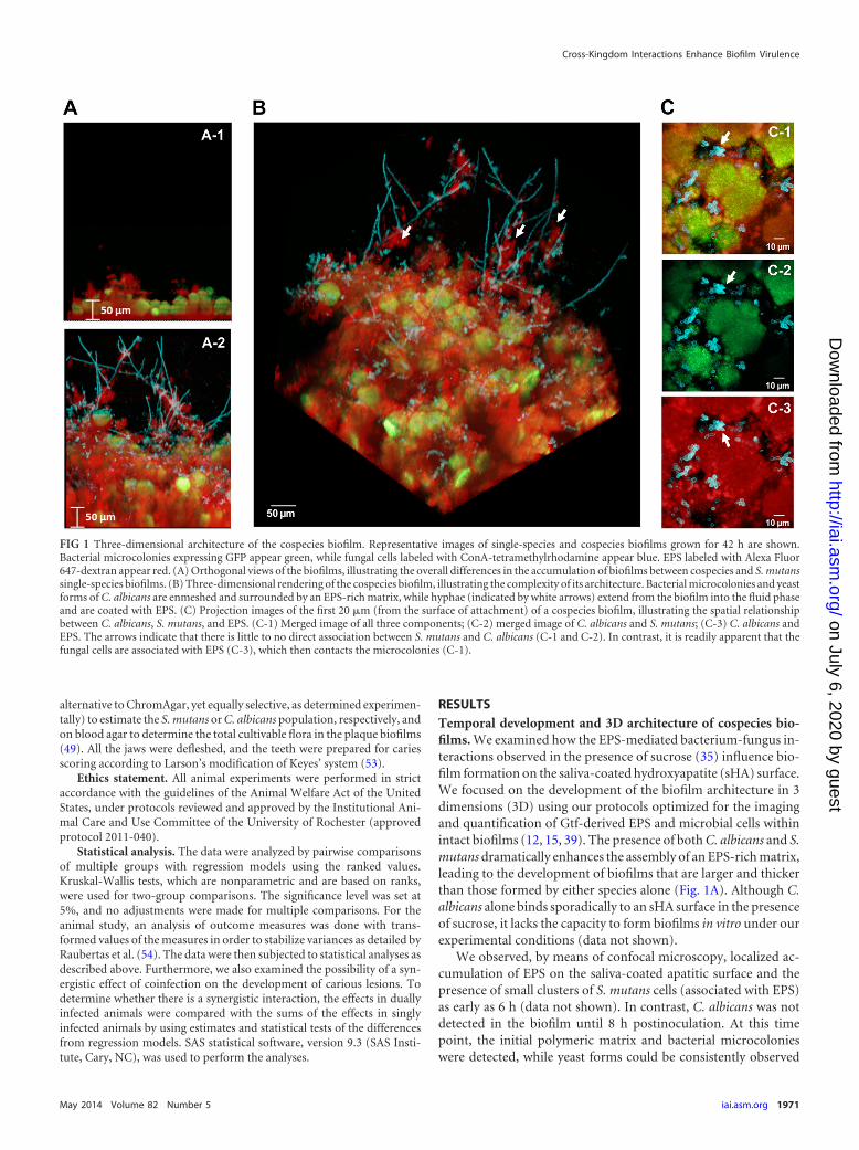
FIG 1 Three-dimensional architecture of the cospecies biofilm. Representative images of single-species and cospecies biofilms grown for 42 h are shown.Bacterial microcolonies expressing GFP appear green, while fungal cells labeled with ConA-tetramethylrhodamine appear blue. EPS labeled with Alexa Fluor647-dextran appear red. (A) Orthogonal views of the biofilms, illustrating the overall differences in the accumulation of biofilms between cospecies and S. mutanssingle-species biofilms. (B) Three-dimensional rendering of the cospecies biofilm, illustrating the complexity of its architecture. Bacterial microcolonies and yeastforms of C. albicans are enmeshed and surrounded by an EPS-rich matrix, while hyphae (indicated by white arrows) extend from the biofilm into the fluid phaseand are coated with EPS. (C) Projection images of the first 20 �m (from the surface of attachment) of a cospecies biofilm, illustrating the spatial relationshipbetween C. albicans, S. mutans, and EPS. (C-1) Merged image of all three components; (C-2) merged image of C. albicans and S. mutans; (C-3) C. albicans andEPS. The arrows indicate that there is little to no direct association between S. mutans and C. albicans (C-1 and C-2). In contrast, it is readily apparent that thefungal cells are associated with EPS (C-3), which then contacts the microcolonies (C-1).
Cross-Kingdom Interactions Enhance Biofilm Virulence
May 2014 Volume 82 Number 5 iai.asm.org 1971
on July 6, 2020 by guesthttp://iai.asm
.org/D
ownloaded from
throughout the biofilm (see Fig. S1A in the supplemental mate-rial). At 8 h, the appearance of pseudohyphal or hyphal forms wasinfrequent (see Fig. S1A). At 18 h, the EPS matrix and microbialbiomass had developed further, while microcolonies of S. mutansand hyphal forms of C. albicans appeared with greater prevalence(see Fig. S1B in the supplemental material). After 42 h, the size ofthe biofilm had increased, revealing large microcolonies (whichform as the initial microcolonies merge), abundant fungal cells(both yeast and hyphal), and the presence of an EPS-rich matrix(Fig. 1 and Table 1).
The resulting 3D architecture of mature cospecies biofilms ishighly intricate (Fig. 1B). Both yeast and hyphal cells were de-tected, along with sizable microcolonies, which were enmeshed inand surrounded by EPS. The hyphae extended out from the bio-film into the surrounding medium and were coated with EPS (Fig.1B, white arrows). In contrast, yeast cells tended to cluster near thesurface of the biofilm attachment and were closely associated withthe EPS surrounding bacterial microcolonies. We were mostlyunable to detect yeast and bacterial cells associated with one an-other without glucan as the intermediary (Fig. 1C, arrows), whichis in line with a lack of cell-cell binding in the absence of sucrose,as observed previously (30, 35). Furthermore, neither yeast cellsnor hyphal cells are found within the microcolony structuresformed by S. mutans; rather, they are associated with the periph-ery. These observations could be a product of sequential assemblyof the biofilm, where the colonization of yeast cells (and laterdifferentiation into hyphae) occurs after the initial EPS is formedon sHA and the basic microcolony structure has been initiated.However, it is also possible that competitive interactions may oc-cur locally between these organisms (55, 56), which could poten-tially explain their spatial relationship and physical proximity inthe biofilm.
The prevalence of EPS-coated hyphae prompted us to investi-gate the ability of purified GtfB to bind to and produce glucan insitu on hyphal cells. We had demonstrated previously that GtfBbinds to yeast cells in an active form, but we had not determined itsability to interact with hyphal cells (35). Despite the differences insize and membrane composition between yeast and hyphal cells(57), we found that the enzyme was equally efficient in producingglucans when adsorbed to either cell type (see Fig. S2 in the sup-plemental material); these results demonstrate that GtfB attachedto C. albicans can enhance EPS-rich matrix production in the ab-sence of bacteria. Our data also show that GtfB binds in an activeform to mannan and �-1,3-glucan (see Fig. S3 and Protocol S1 in
the supplemental material), which are present in the cell walls ofboth yeast and hyphal forms (57). Mannan is located at the out-ermost layer of the Candida cell wall (57). Although �-1,3-glucanis present close to the inner cell wall, it can be secreted extracellu-larly (58) (see below). It is possible that GtfB may bind to one ormore of the carbohydrate components, such as glucose (12).However, the exact location and/or structure of the Gtf bindingsites remains to be identified.
Clearly, C. albicans provides an abundance of binding sites forGtfs derived from S. mutans; the fungus is converted to a de factoglucan producer when exposed to sucrose. Thus, the formation ofcopious amounts of EPS on the large surface areas of C. albicanscells facilitates the assembly of a dense and abundant EPS-richmatrix in cospecies biofilms.
Enhanced microbial carriage in cospecies biofilms. The in-teractions of bacterial and fungal cells led to the development ofcospecies biofilms on the sHA surface, which contained more EPSand microbial biomass than S. mutans single-species biofilms (Ta-ble 1). Furthermore, cospecies biofilms displayed more (at theearly stage of 18 h) and larger (at 42 h) microcolonies than S.mutans-only biofilms (P, �0.05). Cross-sectional imaging analy-sis of cospecies biofilms revealed that microcolonies were com-posed of densely packed S. mutans cells alone, while C. albicanscells were located around the microcolonies. The enhanced mi-crocolony development corresponded to a nearly 2.5-fold in-crease in EPS accumulation in cospecies biofilms relative to bio-films with S. mutans alone (Table 1). This observation agrees withearlier reports that the amount of EPS present in the biofilm di-rectly affects the formation and size of the microcolony, sinceGtf-derived glucans help to cluster the bacterial cells (15, 39).
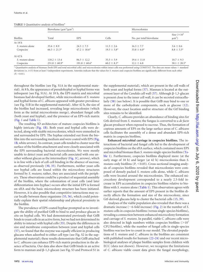
Analyses of the viable population also revealed that there was adramatic increase (�6-fold increase) (Fig. 2) in the number of S.mutans cells in cospecies biofilms (versus single-species biofilms),revealing a connection between enhanced microcolony formationand carriage of S. mutans. In parallel, viable C. albicans cells werealso detected in high numbers within cospecies biofilms (107
CFU/biofilm), while the number of fungal cells in single-speciesbiofilms was too low to count in our model. The elevated popula-tions of S. mutans and C. albicans in the biofilms are consistentwith published results for humans (22–24) and our recent micro-biological analyses of plaque biofilm samples from children withECC (data not shown). However, we recognize the limitationsof C. albicans viable count data given the fungal morphology,
TABLE 1 Quantitative analysis of biofilmsa
Biofilm
Biovolume (�m3/�m2) Microcolonies
Total EPS Cells No. per total biovolumeSize (�103
�m3)
At 18 hS. mutans alone 35.6 � 8.9 24.3 � 7.5 11.5 � 2.6 16.3 � 7.7 3.6 � 0.6Cospecies 66.5 � 21.5* 47.2 � 18.6* 19.3 � 5.8* 33.8 � 6.0* 8.8 � 5.5*
At 42 hS. mutans alone 118.2 � 13.4 84.3 � 12.2 35.5 � 5.9 19.4 � 11.0 10.7 � 9.3Cospecies 251.8 � 60.9* 191.8 � 60.6* 60.2 � 8.3* 12.1 � 6.8 30.1 � 20.5*
a Quantitative analysis of biomass (both EPS and total microbial cells) and microcolonies within intact cells was performed using COMSTAT. The data are mean values � standarddeviations (n, �15) from at least 3 independent experiments. Asterisks indicate that the values for S. mutans and cospecies biofilms are significantly different from each other(P, �0.01).
Falsetta et al.
1972 iai.asm.org Infection and Immunity
on July 6, 2020 by guesthttp://iai.asm
.org/D
ownloaded from
since hyphal development would increase biomass but not neces-sarily CFU.
The pH values of the medium surrounding cospecies biofilmswere highly acidic (final pH ranging from 4.5 to 4.7), as monitoredduring biofilm development (data not shown). However, the pHvalues were similar to those for single-species S. mutans biofilms atall stages of development, despite the differences in microcolonysize and bacterial density, although we did not measure the pHwithin the biofilm.
S. mutans-C. albicans interactions enhance the virulence ofplaque biofilms in vivo. The data from our in vitro biofilm studiesindicate that the infectivity and virulence of cospecies biofilmsmay be enhanced in vivo. Thus, we sought to determine whetherthe association of S. mutans and C. albicans influences the onset ofdental caries by using a rodent model. We used hyposalivatoryrats, which were provided a high-sucrose diet and sugared waterad libitum. The protracted feeding of sugars, coupled with therestricted access of saliva to teeth, used in our model mimics thesevere conditions experienced clinically by children afflicted withECC (3, 5, 6, 8, 20). The animals were readily infected with S.mutans, C. albicans, or both using our model, and then the impacton the development of carious lesions was assessed for each exper-imental condition.
Coinfection with S. mutans and C. albicans in vivo produceddramatic effects on both the level of microbial colonization andthe development of carious lesions. We detected significant in-creases in the viable populations of both S. mutans (3-fold in-crease) and C. albicans (20-fold increase) in plaque biofilmsfrom coinfected animals over those from animals infected witheither species alone (Table 2). This observation is consistent withthe data from our in vitro investigation. The uninfected animalsremained free of infection by S. mutans and/or C. albicans.
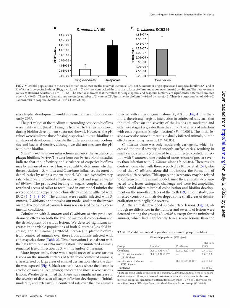
More importantly, there was a rapid onset of severe cariouslesions on the smooth surfaces of teeth from coinfected animals,characterized by large areas of enamel destruction where the den-tin was exposed (Fig. 3, black arrows). Areas where the dentin iseroded or missing (red arrows) indicate the most severe cariouslesions. We also determined that there was a significant increase inthe severity of disease at all stages of lesion development (initial,moderate, and extensive) in coinfected rats over that for animals
infected with either organism alone (P, �0.05) (Fig. 4). Further-more, there is a synergistic interaction in coinfected rats, such thatthe total effect on the severity of the lesions (at moderate andextensive stages) is greater than the sum of the effects of infectionwith each organism (single infection) (P, �0.001). The initial le-sions were also more numerous in dually infected animals, but theeffects were not synergistic (P, 0.05).
C. albicans alone was only moderately cariogenic, which in-creased the initial severity of smooth-surface caries, resulting insmall carious lesions (compared to an uninfected control). Infec-tion with S. mutans alone produced more lesions of greater sever-ity than infection with C. albicans alone (P, �0.05). These resultscontrast somewhat with those reported by Klinke et al. (59), whonoted that C. albicans alone did not induce the formation ofsmooth-surface caries. This apparent discrepancy may be relatedto differences in the animal model, since their animals were sub-jected to a lesser cariogenic challenge and were fed ampicillin,which could affect microbial colonization and biofilm develop-ment on the smooth surfaces of the teeth (59). In our study, un-infected (control) animals developed some small areas of demin-eralization with negligible severity.
All the animals developed sulcal-surface lesions (Fig. 5), al-though no differences in the number and severity of lesions weredetected among the groups (P, 0.05), except for the uninfectedanimals, which had significantly fewer severe lesions than the
FIG 2 Microbial populations in the cospecies biofilm. Shown are the total viable counts (CFU) of S. mutans in single-species and cospecies biofilms (A) and ofC. albicans in cospecies biofilms (B), grown for 42 h. C. albicans alone lacked the capacity to form biofilms under our experimental conditions. The data are meanvalues � standard deviations (n 16). (A) The asterisk indicates that the values for single-species and cospecies biofilms are significantly different from eachother (P, �0.05). There is a dramatic increase in the number of S. mutans CFU in cospecies biofilms (�6-fold increase). (B) There is a large number of viable C.albicans cells in cospecies biofilms (�107 CFU/biofilm).
TABLE 2 Viable microbial populations in animals’ plaque biofilms
Group
Microbial population (CFU/jaw)
S. mutans C. albicansTotal flora(107)
Coinfected (6.1 � 1.3) � 106 (2.9 � 1.2) � 105 2.4 � 0.2Infected with S. mutans
UA159 alone(1.9 � 0.3) � 166* — 1.6 � 0.1
Infected with C. albicansSC5314 alone
— (1.0 � 0.3) � 104* 2.7 � 0.4
Uninfected — — 1.3 � 0.2
a Data are mean viable populations of S. mutans, C. albicans, and total flora � standarddeviations (n 11). —, not detected. Asterisks indicate that the values for the twoinfection groups are significantly different from each other (P, �0.05). The values fortotal flora do not differ significantly for the different infection groups (P, 0.05).
Cross-Kingdom Interactions Enhance Biofilm Virulence
May 2014 Volume 82 Number 5 iai.asm.org 1973
on July 6, 2020 by guesthttp://iai.asm
.org/D
ownloaded from
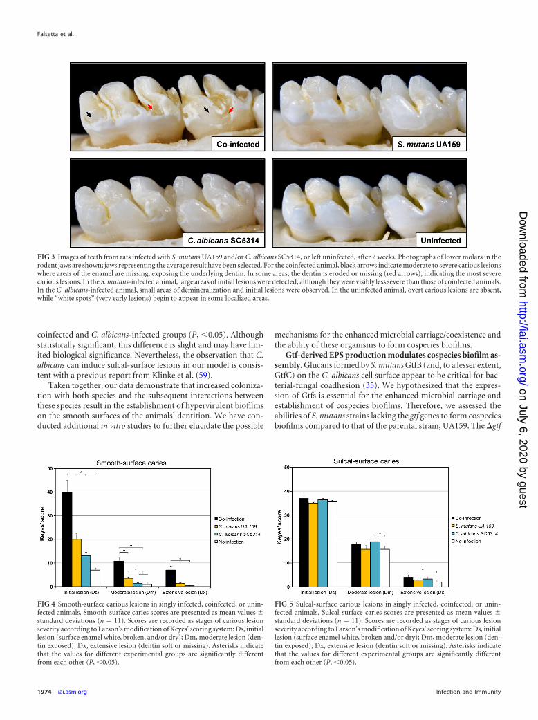
coinfected and C. albicans-infected groups (P, �0.05). Althoughstatistically significant, this difference is slight and may have lim-ited biological significance. Nevertheless, the observation that C.albicans can induce sulcal-surface lesions in our model is consis-tent with a previous report from Klinke et al. (59).
Taken together, our data demonstrate that increased coloniza-tion with both species and the subsequent interactions betweenthese species result in the establishment of hypervirulent biofilmson the smooth surfaces of the animals’ dentition. We have con-ducted additional in vitro studies to further elucidate the possible
mechanisms for the enhanced microbial carriage/coexistence andthe ability of these organisms to form cospecies biofilms.
Gtf-derived EPS production modulates cospecies biofilm as-sembly. Glucans formed by S. mutans GtfB (and, to a lesser extent,GtfC) on the C. albicans cell surface appear to be critical for bac-terial-fungal coadhesion (35). We hypothesized that the expres-sion of Gtfs is essential for the enhanced microbial carriage andestablishment of cospecies biofilms. Therefore, we assessed theabilities of S. mutans strains lacking the gtf genes to form cospeciesbiofilms compared to that of the parental strain, UA159. The �gtf
FIG 3 Images of teeth from rats infected with S. mutans UA159 and/or C. albicans SC5314, or left uninfected, after 2 weeks. Photographs of lower molars in therodent jaws are shown; jaws representing the average result have been selected. For the coinfected animal, black arrows indicate moderate to severe carious lesionswhere areas of the enamel are missing, exposing the underlying dentin. In some areas, the dentin is eroded or missing (red arrows), indicating the most severecarious lesions. In the S. mutans-infected animal, large areas of initial lesions were detected, although they were visibly less severe than those of coinfected animals.In the C. albicans-infected animal, small areas of demineralization and initial lesions were observed. In the uninfected animal, overt carious lesions are absent,while “white spots” (very early lesions) begin to appear in some localized areas.
FIG 4 Smooth-surface carious lesions in singly infected, coinfected, or unin-fected animals. Smooth-surface caries scores are presented as mean values �standard deviations (n 11). Scores are recorded as stages of carious lesionseverity according to Larson’s modification of Keyes’ scoring system: Ds, initiallesion (surface enamel white, broken, and/or dry); Dm, moderate lesion (den-tin exposed); Dx, extensive lesion (dentin soft or missing). Asterisks indicatethat the values for different experimental groups are significantly differentfrom each other (P, �0.05).
FIG 5 Sulcal-surface carious lesions in singly infected, coinfected, or unin-fected animals. Sulcal-surface caries scores are presented as mean values �standard deviations (n 11). Scores are recorded as stages of carious lesionseverity according to Larson’s modification of Keyes’ scoring system: Ds, initiallesion (surface enamel white, broken and/or dry); Dm, moderate lesion (den-tin exposed); Dx, extensive lesion (dentin soft or missing). Asterisks indicatethat the values for different experimental groups are significantly differentfrom each other (P, �0.05).
Falsetta et al.
1974 iai.asm.org Infection and Immunity
on July 6, 2020 by guesthttp://iai.asm
.org/D
ownloaded from
mutants are well described and well characterized (with no polarmutations or growth defects) in the published literature (15, 37).
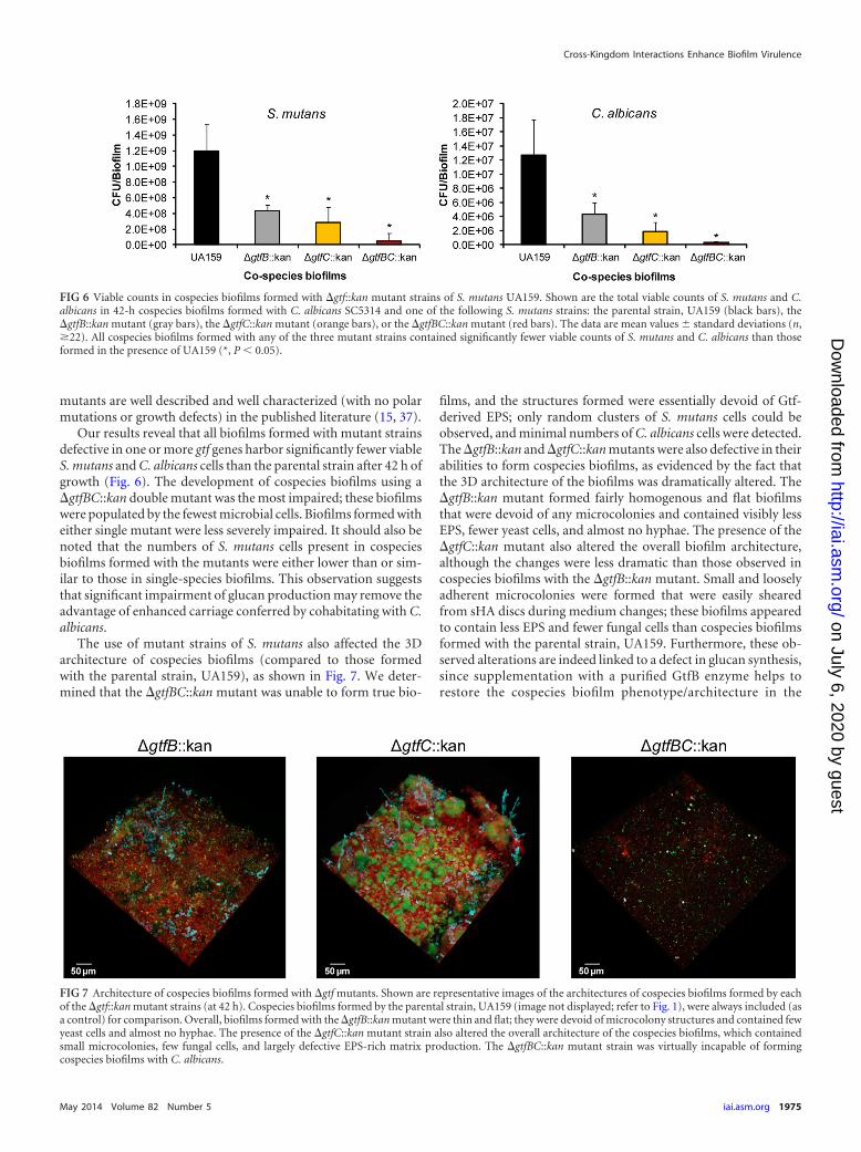
Our results reveal that all biofilms formed with mutant strainsdefective in one or more gtf genes harbor significantly fewer viableS. mutans and C. albicans cells than the parental strain after 42 h ofgrowth (Fig. 6). The development of cospecies biofilms using a�gtfBC::kan double mutant was the most impaired; these biofilmswere populated by the fewest microbial cells. Biofilms formed witheither single mutant were less severely impaired. It should also benoted that the numbers of S. mutans cells present in cospeciesbiofilms formed with the mutants were either lower than or sim-ilar to those in single-species biofilms. This observation suggeststhat significant impairment of glucan production may remove theadvantage of enhanced carriage conferred by cohabitating with C.albicans.
The use of mutant strains of S. mutans also affected the 3Darchitecture of cospecies biofilms (compared to those formedwith the parental strain, UA159), as shown in Fig. 7. We deter-mined that the �gtfBC::kan mutant was unable to form true bio-
films, and the structures formed were essentially devoid of Gtf-derived EPS; only random clusters of S. mutans cells could beobserved, and minimal numbers of C. albicans cells were detected.The �gtfB::kan and �gtfC::kan mutants were also defective in theirabilities to form cospecies biofilms, as evidenced by the fact thatthe 3D architecture of the biofilms was dramatically altered. The�gtfB::kan mutant formed fairly homogenous and flat biofilmsthat were devoid of any microcolonies and contained visibly lessEPS, fewer yeast cells, and almost no hyphae. The presence of the�gtfC::kan mutant also altered the overall biofilm architecture,although the changes were less dramatic than those observed incospecies biofilms with the �gtfB::kan mutant. Small and looselyadherent microcolonies were formed that were easily shearedfrom sHA discs during medium changes; these biofilms appearedto contain less EPS and fewer fungal cells than cospecies biofilmsformed with the parental strain, UA159. Furthermore, these ob-served alterations are indeed linked to a defect in glucan synthesis,since supplementation with a purified GtfB enzyme helps torestore the cospecies biofilm phenotype/architecture in the
FIG 6 Viable counts in cospecies biofilms formed with �gtf::kan mutant strains of S. mutans UA159. Shown are the total viable counts of S. mutans and C.albicans in 42-h cospecies biofilms formed with C. albicans SC5314 and one of the following S. mutans strains: the parental strain, UA159 (black bars), the�gtfB::kan mutant (gray bars), the �gtfC::kan mutant (orange bars), or the �gtfBC::kan mutant (red bars). The data are mean values � standard deviations (n,�22). All cospecies biofilms formed with any of the three mutant strains contained significantly fewer viable counts of S. mutans and C. albicans than thoseformed in the presence of UA159 (*, P � 0.05).
FIG 7 Architecture of cospecies biofilms formed with �gtf mutants. Shown are representative images of the architectures of cospecies biofilms formed by eachof the �gtf::kan mutant strains (at 42 h). Cospecies biofilms formed by the parental strain, UA159 (image not displayed; refer to Fig. 1), were always included (asa control) for comparison. Overall, biofilms formed with the �gtfB::kan mutant were thin and flat; they were devoid of microcolony structures and contained fewyeast cells and almost no hyphae. The presence of the �gtfC::kan mutant strain also altered the overall architecture of the cospecies biofilms, which containedsmall microcolonies, few fungal cells, and largely defective EPS-rich matrix production. The �gtfBC::kan mutant strain was virtually incapable of formingcospecies biofilms with C. albicans.
Cross-Kingdom Interactions Enhance Biofilm Virulence
May 2014 Volume 82 Number 5 iai.asm.org 1975
on July 6, 2020 by guesthttp://iai.asm
.org/D
ownloaded from
presence of the �gtfB::kan mutant (see Fig. S4 in the supple-mental material).
Although the diminished and unstable structure of the bio-films formed with each of the mutant strains prevented accuratequantification of biofilms, it is readily apparent that altering theamount and type of Gtf-derived EPS significantly impacts the ar-chitecture of cospecies biofilms.
C. albicans also contributes to the biofilm matrix. AlthoughS. mutans-derived EPS appears to be an articulation point betweenthe two species, it is conceivable that C. albicans may contribute itsown extracellular substances that help mediate this interaction. C.albicans alone produces matrix materials (�-glucans, chitin, �-N-acetylglucosamine) during biofilm formation on other surfaces,and these appear to confer protection from antifungal agents (58,60–62). Results from previous biochemical studies reveal that�-glucans are likely among the major constituents of the matricesof C. albicans biofilms, although these substances largely have notbeen visualized within the intact matrix (63–65). The presence of�-glucans was sought within the matrices of our cospecies bio-films by means of a �-glucan-specific antibody labeled with a flu-orescent secondary antibody, which was visualized via confocalimaging (66).
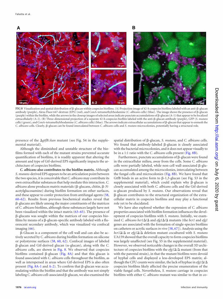
�-Glucan is a component of the cell wall and can also be ac-tively secreted by C. albicans during biofilm formation on siliconeor polystyrene surfaces (58, 60, 62). Confocal images of labeled�-glucan and Gtf-derived glucan (�-glucan), along with the C.albicans cells, are shown in Fig. 8. We observed that cospeciesbiofilms contained �-glucan (Fig. 8A) and that this glucan isfound associated with C. albicans cells throughout the biofilm, aswell as interspersed in areas where Gtf-derived EPS is also oftenpresent (Fig. 8A-1 and A-2). To confirm that �-glucan was accu-mulating within the biofilm and that the antibody was not simplylabeling C. albicans cell-associated �-glucan, we also examined the
spatial distribution of �-glucan, S. mutans, and C. albicans cells.We found that antibody-labeled �-glucan is closely associatedwith the bacterial microcolonies, and it does not appear visually tobe in a 1:1 ratio with the C. albicans cells present (Fig. 8B).
Furthermore, punctate accumulations of �-glucan were foundin the extracellular milieu, away from the cells. Some C. albicanscells were partially labeled, while non-cell wall-associated �-glu-can accumulated among the microcolonies, intercalating betweenthe fungal cells and microcolonies (Fig. 8B). We have found thatGtfB binds in an active form to �-1,3-glucan (see Fig. S3 in thesupplemental material), which could explain why �-glucan isclosely associated with both C. albicans cells and the Gtf-derived�-glucan produced by S. mutans. Our observations reveal that�-glucan contributes to the structural organization of the extra-cellular matrix in cospecies biofilms and may play a functionalrole yet to be elucidated.
We have also explored whether the expression of C. albicansproperties associated with biofilm formation influences the devel-opment of cospecies biofilms with S. mutans. Initially, we exam-ined C. albicans bcr1�/� and efg1�/� mutants (the bcr1 and efg1genes are associated with the formation of single-species biofilmson catheters or acrylic surfaces in vivo [58, 67]). Analysis using thebcr1�/� or efg1�/� deletion mutant cocultured with S. mutansUA159 showed that the overall capacity to form cospecies biofilmswas largely unaffected (see Fig. S5 in the supplemental material).However, we observed noticeable changes in the overall 3D archi-tecture of cospecies biofilms with the efg1�/� mutant (from thatwith the parental strain), in that the mutant biofilms were devoidof hyphal cells and displayed a less-developed EPS matrix; al-though the CFU counts were similar, the lack of hyphae in efg1�/�cospecies biofilms likely affected the precise enumeration of theviable fungal cells. Nevertheless, S. mutans carriage in cospeciesbiofilms with either C. albicans mutant was similar to that in co-
FIG 8 Visualization and spatial distribution of �-glucan within cospecies biofilms. (A) Projection image of 42-h cospecies biofilms labeled with an anti-�-glucanantibody (purple), Alexa Fluor 647-dextran (EPS) (red), and ConA-tetramethylrhodamine (C. albicans cells) (blue). The image shows the presence of �-glucan(purple) within the biofilm, while the arrows in the closeup images of selected areas indicate punctate accumulations of �-glucan (A-1) that appear to be localizedextracellularly (A-2). (B) Three-dimensional projection of a separate 42-h cospecies biofilm labeled with the anti-�-glucan antibody (purple), GFP (S. mutanscells) (green), and ConA-tetramethylrhodamine (C. albicans cells) (blue). The arrows indicate extracellular accumulations of �-glucan that appear to enmesh theC. albicans cells. Clearly, �-glucan can be found intercalated between C. albicans cells and S. mutans microcolonies, potentially having a structural role.
Falsetta et al.
1976 iai.asm.org Infection and Immunity
on July 6, 2020 by guesthttp://iai.asm
.org/D
ownloaded from
species biofilms formed with the parental C. albicans strain. Theseobservations suggest that the ability of C. albicans to form biofilmson other surfaces may not be crucial for the observed cooperativitywith S. mutans in our biofilm model at least. The data emphasizethe importance of S. mutans-derived Gtfs in mediating cospeciesbiofilm development on sHA, although the impact of other C.albicans biofilm-related properties needs additional exploration.
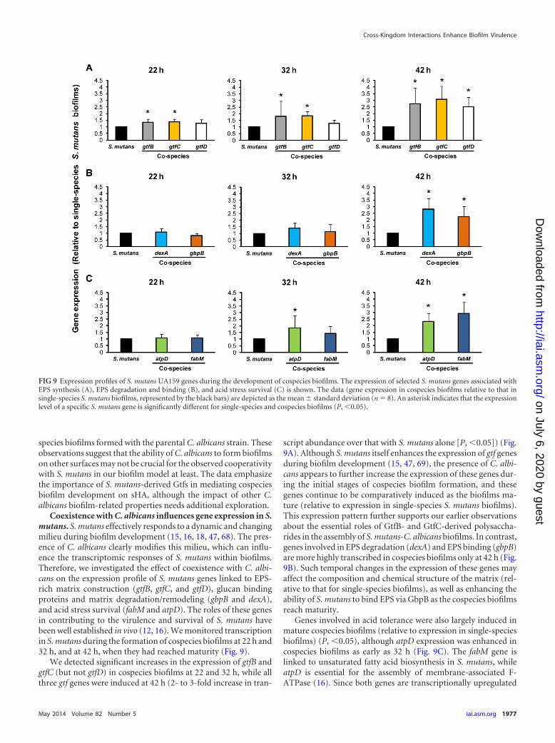
Coexistence with C. albicans influences gene expression in S.mutans. S. mutans effectively responds to a dynamic and changingmilieu during biofilm development (15, 16, 18, 47, 68). The pres-ence of C. albicans clearly modifies this milieu, which can influ-ence the transcriptomic responses of S. mutans within biofilms.Therefore, we investigated the effect of coexistence with C. albi-cans on the expression profile of S. mutans genes linked to EPS-rich matrix construction (gtfB, gtfC, and gtfD), glucan bindingproteins and matrix degradation/remodeling (gbpB and dexA),and acid stress survival (fabM and atpD). The roles of these genesin contributing to the virulence and survival of S. mutans havebeen well established in vivo (12, 16). We monitored transcriptionin S. mutans during the formation of cospecies biofilms at 22 h and32 h, and at 42 h, when they had reached maturity (Fig. 9).
We detected significant increases in the expression of gtfB andgtfC (but not gtfD) in cospecies biofilms at 22 and 32 h, while allthree gtf genes were induced at 42 h (2- to 3-fold increase in tran-
script abundance over that with S. mutans alone [P, �0.05]) (Fig.9A). Although S. mutans itself enhances the expression of gtf genesduring biofilm development (15, 47, 69), the presence of C. albi-cans appears to further increase the expression of these genes dur-ing the initial stages of cospecies biofilm formation, and thesegenes continue to be comparatively induced as the biofilms ma-ture (relative to expression in single-species S. mutans biofilms).This expression pattern further supports our earlier observationsabout the essential roles of GtfB- and GtfC-derived polysaccha-rides in the assembly of S. mutans-C. albicans biofilms. In contrast,genes involved in EPS degradation (dexA) and EPS binding (gbpB)are more highly transcribed in cospecies biofilms only at 42 h (Fig.9B). Such temporal changes in the expression of these genes mayaffect the composition and chemical structure of the matrix (rel-ative to that for single-species biofilms), as well as enhancing theability of S. mutans to bind EPS via GbpB as the cospecies biofilmsreach maturity.
Genes involved in acid tolerance were also largely induced inmature cospecies biofilms (relative to expression in single-speciesbiofilms) (P, �0.05), although atpD expression was enhanced incospecies biofilms as early as 32 h (Fig. 9C). The fabM gene islinked to unsaturated fatty acid biosynthesis in S. mutans, whileatpD is essential for the assembly of membrane-associated F-ATPase (16). Since both genes are transcriptionally upregulated
FIG 9 Expression profiles of S. mutans UA159 genes during the development of cospecies biofilms. The expression of selected S. mutans genes associated withEPS synthesis (A), EPS degradation and binding (B), and acid stress survival (C) is shown. The data (gene expression in cospecies biofilms relative to that insingle-species S. mutans biofilms, represented by the black bars) are depicted as the mean � standard deviation (n 8). An asterisk indicates that the expressionlevel of a specific S. mutans gene is significantly different for single-species and cospecies biofilms (P, �0.05).
Cross-Kingdom Interactions Enhance Biofilm Virulence
May 2014 Volume 82 Number 5 iai.asm.org 1977
on July 6, 2020 by guesthttp://iai.asm
.org/D
ownloaded from
when C. albicans is present, the ability of S. mutans to cope andthrive in an acidified environment may be enhanced in cospeciesbiofilms. Clearly, coexistence with C. albicans induces the expres-sion of key virulence genes in S. mutans that are critical for theability of the bacterium to persist within biofilms and to causedisease.
DISCUSSION
The results of our study provide striking evidence that S. mutansand C. albicans develop a symbiotic relationship that enhances thevirulence of cospecies plaque biofilms formed on tooth surfaces,ultimately amplifying the severity of disease. Our data support andfurther advance the initial concept that C. albicans may be associ-ated with the pathogenesis of early-childhood caries (ECC) (22–24, 59). More importantly, the rapid onset of disease and the en-hanced number, extent, and severity of carious lesions on the freesmooth surfaces of the teeth show very clearly that the presence ofC. albicans and its association with S. mutans have a synergisticeffect on the virulence of the disease. The presence of C. albicanswith S. mutans more than doubles the number and severity ofsmooth-surface lesions relative to the effect of infection with ei-ther organism alone. Furthermore, our results reveal an over-whelming infection by S. mutans and C. albicans when they aregrowing together in the presence of sucrose. This observation cer-tainly offers at least a partial explanation for the extent and rapid-ity of tooth destruction and the detection of elevated levels of bothorganisms seen clinically (22–24).
It may appear surprising that coinfection with C. albicans andS. mutans had a comparatively smaller effect on the number orseverity of sulcal-surface carious lesions in our model. It is wellrecognized that the development of smooth-surface caries ishighly dependent on the formation of Gtf-derived EPS (70, 71).EPS facilitates the adherence of S. mutans (and other organisms)and modulates the formation of cariogenic plaque biofilms in vivo(10), which is in line with the findings of our study. In contrast, thesulcal surfaces provide natural entrapment sites, and the retentionof microorganisms is not dependent on EPS. Nevertheless, a ma-jor clinical feature of ECC is the presence of extensive lesions onthe smooth surfaces, a condition that is clearly mimicked in our invivo model.
The data from the in vitro studies may offer a further explana-tion for the enhanced infectivity/carriage and cooperative coexis-tence in the presence of sucrose and may also explain why thepresence of C. albicans together with S. mutans causes the ob-served synergistic enhancement in virulence. The ability of C. al-bicans to colonize and develop cospecies biofilms with S. mutans islargely dependent on the actions of GtfB and GtfC. We proposethat the Gtfs play key roles in the development of highly virulentcospecies biofilms in at least three ways: (i) they convert C. albi-cans cells into glucan producers, which promote the assembly ofthe EPS-rich matrix scaffold; (ii) they enable the fungus to colo-nize EPS-coated surfaces readily; and (iii) they enhance fungal-bacterial coadherence.
The surface area of C. albicans is significantly larger than that ofS. mutans and provides plentiful Gtf binding sites (35). It shouldbe noted that GtfB binds to both yeast and hyphal cell forms andremains enzymatically active. This surface-bound enzyme pro-duces higher quantities of insoluble EPS, with more �1,6-linkages,than GtfB in solution or bound to the surfaces of S. mutans cells(35). The �1,6-linked glucosyl residues in the glucan structure in
turn provide a site to which S. mutans cells adhere avidly (12, 35,72). Thus, the enhanced surface area, together with an increasednumber of binding sites, offers a plausible explanation for en-hanced S. mutans carriage in cospecies biofilms. These phenom-ena were absent when C. albicans cells were grown with S. mutansstrains defective in gtfB and/or gtfC.
Gtf-derived glucans formed on the C. albicans surface enhancethe ability of the fungal cells to colonize and form cospecies bio-films. Results from previous studies have shown that S. mutans-derived Gtfs (particularly GtfC) present on sHA surface rapidlyform an amorphous glucan layer (13, 14, 72), which masks host-derived microbial binding sites in the salivary pellicle (72). Theseobservations are relevant because C. albicans itself adheres poorlyto the preformed EPS layer on sHA surfaces, or binds poorly to S.mutans, unless the fungal cells are first coated with Gtf-derivedglucans (28, 30, 35). Our study reveals that fungal cells are de-tected only after the initial polymeric matrix and S. mutans micro-colonies are formed on the sHA. Furthermore, the lack of gtfBand/or gtfC expression by S. mutans severely disrupts the ability ofC. albicans to colonize, accumulate, and form cospecies biofilms.These findings are supported by the observation that C. albicans isdetected at low numbers or not at all in the plaque of ECC-freechildren (22–24) and at lower number in rats infected with C.albicans alone than in coinfected rats under our experimental con-ditions.
Our data provide a feasible explanation for the previous re-ports showing that the ability of S. mutans and C. albicans to formbiofilms together was promoted in the presence of sucrose (32–34), while other sugars (e.g., glucose), which are not substrates forEPS synthesis, had no effect (33). A similar mechanism may alsoenhance C. albicans and S. gordonii biofilm formation in vitro (73).Altogether, we demonstrate the importance of Gtfs in mediatingthe cooperativity between C. albicans and S. mutans. This type ofinteraction represents a truly unique physical interaction where abacterially produced product adheres to, and functions on, thesurface of an organism from another kingdom, transforming arelatively innocuous organism (in terms of dental caries) into afierce stimulator of cariogenic biofilm formation.
The potential of C. albicans to contribute to the pathogenesis ofcaries disease has often been associated with its ability to produceand tolerate acids (22–24, 59, 75). We found that the pH values ofthe culture medium surrounding cospecies biofilms were highlyacidic, though not significantly different from those of single-spe-cies S. mutans biofilms. There may have been differences in the pHvalues within the biofilm, but this measurement was beyond thescope of the present study. Although an acidic pH is undeniablythe immediate cause of tooth enamel dissolution, the environ-ment within which the acid is produced plays a crucial role incariogenesis (12). The results of our previous studies have shownthat the synthesis of Gtf-derived glucans leads to the formation ofan insoluble EPS-rich matrix scaffold that acts as a diffusion-lim-iting barrier (15). In parallel, the metabolic activity of S. mutansclustered within the microcolony can produce copious amountsof acids that accumulate locally (15, 74). It is conceivable that thealterations in the extracellular matrix containing a dense pop-ulation of bacterial cells help to prevent acid within the biofilmfrom diffusing outward, thus prolonging and intensifying theacid attack.
The presence of C. albicans dramatically modifies the physicalenvironment and the 3D architecture of the biofilm. It alters the
Falsetta et al.
1978 iai.asm.org Infection and Immunity
on July 6, 2020 by guesthttp://iai.asm
.org/D
ownloaded from
volume and the structure of the extracellular matrix by (i) increas-ing the amount of insoluble Gtf-derived EPS, which has beenshown to have diffusion-limiting properties (15), and (ii) inde-pendently contributing to the matrix through the production ofextracellular �-glucans. It is also possible that the presence of in-soluble �-1,3-glucan embedded in the extracellular matrices ofcospecies biofilms may help limit diffusion while contributing tostability of the 3D matrix scaffold. Furthermore, S. mutans micro-colonies form more rapidly, and their size more than doubles,when the biofilms are grown in the presence of C. albicans. Wehave shown previously that the pH inside the microcolony be-comes more acidic as the structure increases in size, due to a highdensity of acidogenic organisms and limited diffusion into andout of the structure (15). Thus, the elevated and localized concen-tration of S. mutans cells sheltered by an abundant extracellularmatrix would maximize the ability of acids to demineralize teethby retaining the acids in close proximity to the tooth surface. Inthis scenario, EPS may be both the point of articulation for thecoexistence of S. mutans and C. albicans and a diffusion barrierthat helps to maintain an acidic environment, which could explainwhy the transcription of S. mutans acid tolerance genes (fabM andatpD) is induced in cospecies biofilms relative to single-speciesbiofilms. We are currently mapping the spatial distributions ofpH, EPS, and microbial cells by using a fluorescent pH indicatorthat is incorporated into the matrix scaffold to determine the exactlocations of acidic niches within undisturbed biofilms. At thesame time, such changes in expression also suggest that S. mutansmay be able to sense C. albicans within the surrounding biofilmmilieu, in turn increasing the production of proteins involved invirulence and/or stress defense. Overall, the data indicate that thepresence of C. albicans might accentuate the fitness of S. mutans,which may help to account for the enhanced virulence observed inour rodent model.
We recognize the complexity of this bacterium-fungus associ-ation. The interactions between these two organisms are multifac-eted and could presumably induce additional responses in oneanother and/or alter the immediate environment to influencepathogenesis. Although we focus on the influence of the presenceof C. albicans on S. mutans accumulation, biofilm formation, andvirulence expression in this publication, it is possible that S. mu-tans may also provide benefits to C. albicans, such as enhancedcolonization of the tooth surface. Although we have begun to in-vestigate the consequences of this cross-kingdom interaction,much is yet unknown. Certainly, further studies are needed toinvestigate the changes in C. albicans virulence and matrix pro-duction.
In summary, we propose that there is a novel mutualistic rela-tionship between a fungus and an oral bacterial pathogen thatresults in synergistic enhancement of the virulence of an infectiousdisease. The association between C. albicans and S. mutans ap-pears to be largely mediated by a physical interaction that relies onthe production of glucans, which are produced by bacterial exoen-zymes (Gtfs), on yeast and hyphal cell surfaces. These interactionsare essential for the assembly of an EPS-rich matrix, the formationof enlarged microcolonies containing densely packed S. mutanscells, and the development of cospecies biofilms. These findingsillustrate how Gtfs can convert a moderately cariogenic organisminto a major contributor to the formation of virulent plaque bio-films, ultimately modulating the pathogenesis of dental caries in asusceptible and vulnerable population. Since we have shown un-
equivocally that S. mutans-C. albicans association can drasticallyincrease virulence in vivo and that our in vitro studies point to aGtf-dependent mechanism, testing of individual �gtf mutantswith C. albicans (in the context of infection and cariogenesis) iscertainly warranted. In addition, it is clear that C. albicans alsocontributes independently to EPS production in cospecies bio-films. However, additional factors may be at play, including sig-naling interactors, since the presence of C. albicans augments theexpression of virulence genes in S. mutans.
Clinical implications. We offer plausible data to support theclinical importance of the association between C. albicans and S.mutans in the pathogenesis of ECC, one of the most virulent,painful, and costly infectious diseases afflicting children. A keyfinding of this study is that the interactions between C. albicansand S. mutans via EPS production increase both fungal and bac-terial carriage, and these organisms together synergistically en-hance the virulence of plaque biofilms. Our data help to explainthe high level of recovery of these organisms from the plaque ofchildren afflicted with ECC and the overt demineralization andrampant carious lesions that typically occur on the free smoothsurfaces of their primary teeth (22–24). Clearly, our data providenew perspectives for devising efficacious therapies to control ECC.For example, blocking Gtf binding to the Candida cell wall orincluding antifungals as part of the treatment to reduce or preventfungal infection may be effective therapeutic approaches. Further-more, increased knowledge concerning the identities of the mo-lecular interactors that facilitate C. albicans-S. mutans associationsand the assembly of hypervirulent biofilms may provide addi-tional avenues for the prevention of this disease.
In addition, enhanced colonization by C. albicans and in-creased fungal carriage in cospecies plaque biofilms may also pro-vide a fungal reservoir that could promote Candida infections ofsoft tissue and oral mucosal surfaces, as reported recently (31). Inour preliminary data, we observed elevated fungal colonization oftongues from coinfected animals relative to that for animals in-fected with C. albicans alone, as shown in Fig. S6 in the supple-mental material (albeit further quantitative analysis is warranted).Thus, our findings likely have relevance beyond teeth and themouth, since localized bacterium-fungus interactions are associ-ated with other polymicrobial infections and systemic complica-tions at various sites in humans (25–27).
ACKNOWLEDGMENTS
This work was supported in part by research grants from the NationalScience Foundation (EFRI-1137186) and from the National Institute forDental and Craniofacial Research (T90DE021985).
REFERENCES1. Hall-Stoodley L, Stoodley P. 2009. Evolving concepts in biofilm infec-
tions. Cell. Microbiol. 11:1034 –1043. http://dx.doi.org/10.1111/j.1462-5822.2009.01323.x.
2. Dye BA, Tan S, Smith V, Lewis BG, Barker LK, Thornton-Evans G, EkePI, Beltran-Aguilar ED, Horowitz AM, Li CH. 2007. Trends in oralhealth status: United States, 1988 –1994 and 1999 –2004. Vital Health Stat.11:1–92.
3. Berkowitz RJ, Turner J, Hughes C. 1984. Microbial characteristics of thehuman dental caries associated with prolonged bottle-feeding. Arch. OralBiol. 29:949 –951. http://dx.doi.org/10.1016/0003-9969(84)90097-9.
4. Milnes AR, Bowden GH. 1985. The microflora associated with develop-ing lesions of nursing caries. Caries Res. 19:289 –297. http://dx.doi.org/10.1159/000260858.
5. Hallett KB, O’Rourke PK. 2002. Early childhood caries and infant feedingpractice. Community Dent. Health 19:237–242.
Cross-Kingdom Interactions Enhance Biofilm Virulence
May 2014 Volume 82 Number 5 iai.asm.org 1979
on July 6, 2020 by guesthttp://iai.asm
.org/D
ownloaded from

6. Chestnutt IG, Murdoch C, Robson KF. 2003. Parents and carers’ choiceof drinks for infants and toddlers, in areas of social and economic disad-vantage. Community Dent. Health 20:139 –145.
7. Bowen WH, Lawrence RA. 2005. Comparison of the cariogenicity of cola,honey, cow milk, human milk, and sucrose. Pediatrics 116:921–926. http://dx.doi.org/10.1542/peds.2004-2462.
8. Karp J, Berkowitz RJ. 2008. Clinical outcomes for severe early child-hood caries. Clin. Rev. Pediatr. 4:169 –173. http://dx.doi.org/10.2174/157339608785855965.
9. Palmer CA, Kent R, Jr, Loo CY, Hughes CV, Stutius E, Pradhan N,Dahlan M, Kanasi E, Arevalo Vasquez SS, Tanner AC. 2010. Diet andcaries-associated bacteria in severe early childhood caries. J. Dent. Res.89:1224 –1229. http://dx.doi.org/10.1177/0022034510376543.
10. Kanasi E, Dewhirst FE, Chalmers NI, Kent R, Jr, Moore A, Hughes CV,Pradhan N, Loo CY, Tanner AC. 2010. Clonal analysis of the microbiotaof severe early childhood caries. Caries Res. 44:485– 497. http://dx.doi.org/10.1159/000320158.
11. Gross EL, Leys EJ, Gasparovich SR, Firestone ND, Schwartzbaum JA,Janies DA, Asnani K, Griffen AL. 2010. Bacterial 16S sequence analysis ofsevere caries in young permanent teeth. J. Clin. Microbiol. 48:4121– 4128.http://dx.doi.org/10.1128/JCM.01232-10.
12. Bowen WH, Koo H. 2011. Biology of Streptococcus mutans-derived glu-cosyltransferases: role in extracellular matrix formation of cariogenic bio-films. Caries Res. 45:69 – 86. http://dx.doi.org/10.1159/000324598.
13. Vacca-Smith AM, Bowen WH. 1998. Binding properties of streptococcalglucosyltransferases for hydroxyapatite, saliva-coated hydroxyapatite, andbacterial surfaces. Arch. Oral Biol. 43:103–110. http://dx.doi.org/10.1016/S0003-9969(97)00111-8.
14. Vacca-Smith AM, Venkitaraman AR, Schilling KM, Bowen WH. 1996.Characterization of glucosyltransferase of human saliva adsorbed ontohydroxyapatite surfaces. Caries Res. 30:354 –360. http://dx.doi.org/10.1159/000262342.
15. Xiao J, Klein MI, Falsetta ML, Lu B, Delahunty CM, Yates JR, III,Heydorn A, Koo H. 2012. The exopolysaccharide matrix modulates theinteraction between 3D architecture and virulence of a mixed-species oralbiofilm. PLoS Pathog. 8:e1002623. http://dx.doi.org/10.1371/journal.ppat.1002623.
16. Lemos JA, Quivey RG, Jr, Koo H, Abranches J. 2013. Streptococcusmutans: a new Gram-positive paradigm? Microbiology 159:436 – 445.http://dx.doi.org/10.1099/mic.0.066134-0.
17. Nyvad B, Crielaard W, Mira A, Takahashi N, Beighton D. 2013. Dentalcaries from a molecular microbiological perspective. Caries Res. 47:89 –102. http://dx.doi.org/10.1159/000345367.
18. Burne RA. 1998. Oral streptococci. products of their environment. J. Dent.Res. 77:445–452. http://dx.doi.org/10.1177/00220345980770030301.
19. Marsh PD. 2003. Are dental diseases examples of ecological catastrophes?Microbiology 149:279 –294. http://dx.doi.org/10.1099/mic.0.26082-0.
20. Acharya S, Tandon S. 2011. The effect of early childhood caries on thequality of life of children and their parents. Contemp. Clin. Dent. 2:98 –101. http://dx.doi.org/10.4103/0976-237X.83069.
21. Skeie MS, Raadal M, Strand GV, Espelid I. 2006. The relationshipbetween caries in the primary dentition at 5 years of age and permanentdentition at 10 years of age—a longitudinal study. Int. J. Paediatr. Dent.16:152–160. http://dx.doi.org/10.1111/j.1365-263X.2006.00720.x.
22. de Carvalho FG, Silva DS, Hebling J, Spolidorio LC, Spolidorio DM.2006. Presence of mutans streptococci and Candida spp. in dental plaque/dentine of carious teeth and early childhood caries. Arch. Oral Biol. 51:1024 –1028. http://dx.doi.org/10.1016/j.archoralbio.2006.06.001.
23. Raja M, Hannan A, Ali K. 2010. Association of oral candidal carriage withdental caries in children. Caries Res. 44:272–276. http://dx.doi.org/10.1159/000314675.
24. Yang XQ, Zhang Q, Lu LY, Yang R, Liu Y, Zou J. 2012. Genotypicdistribution of Candida albicans in dental biofilm of Chinese childrenassociated with severe early childhood caries. Arch. Oral Biol. 57:1048 –1053. http://dx.doi.org/10.1016/j.archoralbio.2012.05.012.
25. Peleg AY, Hogan DA, Mylonakis E. 2010. Medically important bacterial-fungal interactions. Nat. Rev. Microbiol. 8:340 –349. http://dx.doi.org/10.1038/nrmicro2313.
26. Shirtliff ME, Peters BM, Jabra-Rizk MA. 2009. Cross-kingdom interac-tions: Candida albicans and bacteria. FEMS Microbiol. Lett. 299:1– 8. http://dx.doi.org/10.1111/j.1574-6968.2009.01668.x.
27. Harriott MM, Noverr MC. 2011. Importance of Candida-bacterial poly-
microbial biofilms in disease. Trends Microbiol. 19:557–563. http://dx.doi.org/10.1016/j.tim.2011.07.004.
28. Jenkinson HF, Douglas LJ. 2002. Candida interactions with bacterialbiofilms, p 357–373. In Brogden KA, Guthmiller JM (ed), Polymicrobialdiseases. ASM Press, Washington, DC.
29. Diaz PI, Xie Z, Sobue T, Thompson A, Biyikoglu B, Ricker A, Ikono-mou L, Dongari-Bagtzoglou A. 2012. Synergistic interaction betweenCandida albicans and commensal oral streptococci in a novel in vitro mu-cosal model. Infect. Immun. 80:620 – 632. http://dx.doi.org/10.1128/IAI.05896-11.
30. Jenkinson HF, Lala HC, Shepherd MG. 1990. Coaggregation of Strepto-coccus sanguis and other streptococci with Candida albicans. Infect. Im-mun. 58:1429 –1436.
31. Xu H, Sobue T, Thompson A, Xie Z, Poon K, Ricker A, Cervantes J,Diaz PI, Dongari-Bagtzoglou A. 17 September 2013. Streptococcal co-infection augments Candida pathogenicity by amplifying the mucosal in-flammatory response. Cell. Microbiol. http://dx.doi.org/10.1111/cmi.12216.
32. Branting C, Sund ML, Linder LE. 1989. The influence of Streptococcusmutans on adhesion of Candida albicans to acrylic surfaces in vitro. Arch.Oral Biol. 34:347–353. http://dx.doi.org/10.1016/0003-9969(89)90108-8.
33. Pereira-Cenci T, Deng DM, Kraneveld EA, Manders EM, Del Bel CuryAA, Ten Cate JM, Crielaard W. 2008. The effect of Streptococcus mutansand Candida glabrata on Candida albicans biofilms formed on differentsurfaces. Arch. Oral Biol. 53:755–764. http://dx.doi.org/10.1016/j.archoralbio.2008.02.015.
34. Metwalli KH, Khan SA, Krom BP, Jabra-Rizk MA. 2013. Streptococcusmutans, Candida albicans, and the human mouth: a sticky situation. PLoSPathog. 9:e1003616. http://dx.doi.org/10.1371/journal.ppat.1003616.
35. Gregoire S, Xiao J, Silva BB, Gonzalez I, Agidi PS, Klein MI, Ambati-pudi KS, Rosalen PL, Bauserman R, Waugh RE, Koo H. 2011. Role ofglucosyltransferase B in interactions of Candida albicans with Streptococ-cus mutans and with an experimental pellicle formed on hydroxyapatitesurfaces. Appl. Environ. Microbiol. 77:6357– 6367. http://dx.doi.org/10.1128/AEM.05203-11.
36. Falsetta ML, Gregoire S, Colonne M, Koo H. 2013. Streptococcus mutansand Candida albicans interactions during cariogenic biofilm formation. J.Dent. Res. 92(Spec Iss A):abstr 385. https://iadr.confex.com/iadr/13iags/webprogram/Paper173866.html.
37. Koo H, Xiao J, Klein MI, Jeon JG. 2010. Exopolysaccharides produced byStreptococcus mutans glucosyltransferases modulate the establishment ofmicrocolonies within multispecies biofilms. J. Bacteriol. 192:3024 –3032.http://dx.doi.org/10.1128/JB.01649-09.
38. Klein MI, Duarte S, Xiao J, Mitra S, Foster TH, Koo H. 2009. Structuraland molecular basis of the role of starch and sucrose in Streptococcus mu-tans biofilm development. Appl. Environ. Microbiol. 75:837– 841. http://dx.doi.org/10.1128/AEM.01299-08.
39. Xiao J, Koo H. 2010. Structural organization and dynamics of exopoly-saccharide matrix and microcolonies formation by Streptococcus mutansin biofilms. J. Appl. Microbiol. 108:2103–2113. http://dx.doi.org/10.1111/j.1365-2672.2009.04616.x.
40. Koo H, Hayacibara MF, Schobel BD, Cury JA, Rosalen PL, Park YK,Vacca-Smith AM, Bowen WH. 2003. Inhibition of Streptococcus mutansbiofilm accumulation and polysaccharide production by apigenin and tt-farnesol. J. Antimicrob. Chemother. 52:782–789. http://dx.doi.org/10.1093/jac/dkg449.
41. Dongari-Bagtzoglou A, Kashleva H, Dwivedi P, Diaz P, Vasilakos J.2009. Characterization of mucosal Candida albicans biofilms. PLoS One4:e7967. http://dx.doi.org/10.1371/journal.pone.0007967.
42. Jin Y, Zhang T, Samaranayake YH, Fang HH, Yip HK, SamaranayakeLP. 2005. The use of new probes and stains for improved assessment of cellviability and extracellular polymeric substances in Candida albicans bio-films. Mycopathologia 159:353–360. http://dx.doi.org/10.1007/s11046-004-6987-7.
43. Hamada S, Gill K, Slade HD. 1977. Binding of lectins to Streptococcusmutans cells and type-specific polysaccharides, and effect on adherence.Infect. Immun. 18:708 –716.
44. London R, Schwedock J, Sage A, Valley H, Meadows J, Waddington M,Straus D. 2010. An automated system for rapid non-destructive enumer-ation of growing microbes. PLoS One 5:e8609. http://dx.doi.org/10.1371/journal.pone.0008609.
45. Chalmers NI, Palmer RJ, Jr, Cisar JO, Kolenbrander PE. 2008. Char-acterization of a Streptococcus sp.-Veillonella sp. community microma-
Falsetta et al.
1980 iai.asm.org Infection and Immunity
on July 6, 2020 by guesthttp://iai.asm
.org/D
ownloaded from
nipulated from dental plaque. J. Bacteriol. 190:8145– 8154. http://dx.doi.org/10.1128/JB.00983-08.
46. Cury JA, Koo H. 2007. Extraction and purification of total RNA fromStreptococcus mutans biofilms. Anal. Biochem. 365:208 –214. http://dx.doi.org/10.1016/j.ab.2007.03.021.
47. Klein MI, Xiao J, Lu B, Delahunty CM, Yates JR, III, Koo H. 2012.Streptococcus mutans protein synthesis during mixed-species biofilm de-velopment by high-throughput quantitative proteomics. PLoS One7:e45795. http://dx.doi.org/10.1371/journal.pone.0045795.
48. Koo H, Seils J, Abranches J, Burne RA, Bowen WH, Quivey RG, Jr.2006. Influence of apigenin on gtf gene expression in Streptococcus mutansUA159. Antimicrob. Agents Chemother. 50:542–546. http://dx.doi.org/10.1128/AAC.50.2.542-546.2006.
49. Bowen WH, Madison KM, Pearson SK. 1988. Influence of desalivation inrats on incidence of caries in intact cagemates. J. Dent. Res. 67:1316 –1318.http://dx.doi.org/10.1177/00220345880670101401.
50. Koo H, Rosalen PL, Cury JA, Park YK, Ikegaki M, Sattler A. 1999. Effectof Apis mellifera propolis from two Brazilian regions on caries develop-ment in desalivated rats. Caries Res. 33:393– 400. http://dx.doi.org/10.1159/000016539.
51. Keyes PH, White CL. 1959. Dental caries in the molar teeth of rats. III.Bio-assay of sodium fluoride and sodium lauroyl sarcosinate as caries-inhibitory agents. J. Am. Dent. Assoc. 58:43–55.
52. Klein MI, Scott-Anne KM, Gregoire S, Rosalen PL, Koo H. 2012.Molecular approaches for viable bacterial population and transcriptionalanalyses in a rodent model of dental caries. Mol. Oral Microbiol. 27:350 –361. http://dx.doi.org/10.1111/j.2041-1014.2012.00647.x.
53. Larson R. 1981. Merits and modifications of scoring rat dental caries byKeyes’ method, p 195–203. In Tanzer J. (ed), Animal models in cariology.IRL Press, Washington, DC.
54. Raubertas RF, Davis BA, Bowen WH, Pearson SK, Watson GE. 1999.Litter effects on caries in rats and implications for experimental design.Caries Res. 33:164 –169. http://dx.doi.org/10.1159/000016511.
55. De Sordi L, Muhlschlegel FA. 2009. Quorum sensing and fungal-bacterial interactions in Candida albicans: a communicative network reg-ulating microbial coexistence and virulence. FEMS Yeast Res. 9:990 –999.http://dx.doi.org/10.1111/j.1567-1364.2009.00573.x.
56. Merritt J, Qi F. 2012. The mutacins of Streptococcus mutans: regulationand ecology. Mol. Oral Microbiol. 27:57– 69. http://dx.doi.org/10.1111/j.2041-1014.2011.00634.x.
57. Gow NA, van de Veerdonk FL, Brown AJ, Netea MG. 2012. Candidaalbicans morphogenesis and host defence: discriminating invasion fromcolonization. Nat. Rev. Microbiol. 10:112–122. http://dx.doi.org/10.1038/nrmicro2711.
58. Nobile CJ, Nett JE, Hernday AD, Homann OR, Deneault JS, Nantel A,Andes DR, Johnson AD, Mitchell AP. 2009. Biofilm matrix regulation byCandida albicans Zap1. PLoS Biol. 7:e1000133. http://dx.doi.org/10.1371/journal.pbio.1000133.
59. Klinke T, Guggenheim B, Klimm W, Thurnheer T. 2011. Dental cariesin rats associated with Candida albicans. Caries Res. 45:100 –106. http://dx.doi.org/10.1159/000324809.
60. Nett JE, Sanchez H, Cain MT, Andes DR. 2010. Genetic basis of Candidabiofilm resistance due to drug-sequestering matrix glucan. J. Infect. Dis.202:171–175. http://dx.doi.org/10.1086/651200.
61. Nett JE, Crawford K, Marchillo K, Andes DR. 2010. Role of Fks1p andmatrix glucan in Candida albicans biofilm resistance to an echinocandin,pyrimidine, and polyene. Antimicrob. Agents Chemother. 54:3505–3508.http://dx.doi.org/10.1128/AAC.00227-10.
62. Taff HT, Nett JE, Zarnowski R, Ross KM, Sanchez H, Cain MT,Hamaker J, Mitchell AP, Andes DR. 2012. A Candida biofilm-inducedpathway for matrix glucan delivery: implications for drug resistance. PLoSPathog. 8:e1002848. http://dx.doi.org/10.1371/journal.ppat.1002848.
63. Al-Fattani MA, Douglas LJ. 2006. Biofilm matrix of Candida albicans andCandida tropicalis: chemical composition and role in drug resistance. J.Med. Microbiol. 55:999 –1008. http://dx.doi.org/10.1099/jmm.0.46569-0.
64. Baillie GS, Douglas LJ. 2000. Matrix polymers of Candida biofilms andtheir possible role in biofilm resistance to antifungal agents. J. Antimicrob.Chemother. 46:397– 403. http://dx.doi.org/10.1093/jac/46.3.397.
65. Nett J, Lincoln L, Marchillo K, Massey R, Holoyda K, Hoff B, Van-Handel M, Andes D. 2007. Putative role of �-1,3 glucans in Candidaalbicans biofilm resistance. Antimicrob. Agents Chemother. 51:510 –520.http://dx.doi.org/10.1128/AAC.01056-06.
66. Humbel BM, Konomi M, Takagi T, Kamasawa N, Ishijima SA, OsumiM. 2001. In situ localization of �-glucans in the cell wall of Schizosaccha-romyces pombe. Yeast 18:433– 444. http://dx.doi.org/10.1002/yea.694.
67. Nobile CJ, Fox EP, Nett JE, Sorrells TR, Mitrovich QM, Hernday AD,Tuch BB, Andes DR, Johnson AD. 2012. A recently evolved transcrip-tional network controls biofilm development in Candida albicans. Cell148:126 –138. http://dx.doi.org/10.1016/j.cell.2011.10.048.
68. Lemos JA, Burne RA. 2008. A model of efficiency: stress tolerance byStreptococcus mutans. Microbiology 154:3247–3255. http://dx.doi.org/10.1099/mic.0.2008/023770-0.
69. Li Y, Burne RA. 2001. Regulation of the gtfBC and ftf genes of Streptococ-cus mutans in biofilms in response to pH and carbohydrate. Microbiology147:2841–2848.
70. Tanzer JM, Freedman ML, Fitzgerald RJ. 1985. Virulence of mutantsdefective in glucosyltransferase, dextran-mediated aggregation, or dextra-nase activity, p 204 –211. In Mergenhagen SE, Rosan B (ed), Molecularbasis of oral microbial adhesion: proceedings of a workshop held in Phil-adelphia, Pennsylvania, 5– 8 June 1984. American Society for Microbiol-ogy, Washington, DC.
71. Yamashita Y, Bowen WH, Burne RA, Kuramitsu HK. 1993. Role of theStreptococcus mutans gtf genes in caries induction in the specific-pathogen-free rat model. Infect. Immun. 61:3811–3817.
72. Schilling KM, Bowen WH. 1992. Glucans synthesized in situ in experi-mental salivary pellicle function as specific binding sites for Streptococcusmutans. Infect. Immun. 60:284 –295.
73. Ricker A, Vickerman M, Dongari-Bagtzoglou A. 29 January 2014. Strep-tococcus gordonii glucosyltransferase promotes biofilm interactions withCandida albicans. J. Oral Microbiol. 6:23419. http://dx.doi.org/10.3402/jom.v6.23419.
74. Guo L, Hu W, He X, Lux R, McLean J, Shi W. 2013. Investigating acidproduction by Streptococcus mutans with a surface-displayed pH-sensitivegreen fluorescent protein. PLoS One 8:e57182. http://dx.doi.org/10.1371/journal.pone.0057182.
75. Klinke T, Kneist S, de Soet JJ, Kuhlisch E, Mauersberger S, Forster A,Klimm W. 2009. Acid production by oral strains of Candida albicans andlactobacilli. Caries Res. 43:83–91. http://dx.doi.org/10.1159/000204911.
Cross-Kingdom Interactions Enhance Biofilm Virulence
May 2014 Volume 82 Number 5 iai.asm.org 1981
on July 6, 2020 by guesthttp://iai.asm
.org/D
ownloaded from