Embed Size (px)
Citation preview
Sgw
DDKHa
Ab
Fc
d
e
f
g
G
a
ARRA
KFGRSTT
0
Veterinary Parasitology 205 (2014) 653–665
Contents lists available at ScienceDirect
Veterinary Parasitology
jo u r nal homep age: www.elsev ier .com/ locate /vetpar
urveillance of feral swine for Trichinella spp. and Toxoplasmaondii in the USA and host-related factors associatedith infection
.E. Hill a,∗, J.P. Dubeya, J.A. Barochb, S.R. Swaffordc, V.F. Fourneta,
. Hawkins-Coopera, D.G. Pyburnd, B.S. Schmitb, H.R. Gamblee,. Pedersenb, L.R. Ferreiraa, S.K. Vermaa, Y. Yinga, O.C.H. Kwoka,. Feidas f, G. Theodoropoulosg
United States Department of Agriculture, Agricultural Research Service, Beltsville Agricultural Research Center,nimal Parasitic Diseases Laboratory, Beltsville, MD, 20705-2350, USAUnited States Department of Agriculture, Animal and Plant Health Inspection Service, Wildlife Services, 4101 LaPorte Ave,
ort Collins, CO 80521, USAUnited States Fish and Wildlife Service, 595 Yazoo Refuge Road, MS 38748-9729, USANational Pork Board, 1776 NW 114th St., Clive, IA 50325, USANational Academy of Sciences, 500 5th Street N.W., Washington, D.C., 20001, USADepartment of Meteorology and Climatology, School of Geology, Aristotle University of Thessaloniki, Thessaloniki 54124, GreeceDepartment of Anatomy and Physiology of Farm Animals, Agricultural University of Athens, 75 Iera Odos, Votanikos, Athens 11855,reece
r t i c l e i n f o
rticle history:eceived 1 May 2014eceived in revised form 23 July 2014ccepted 25 July 2014
eywords:eral swineeographic distributionisk factorspatial scan statisticoxoplasma gondiirichinella
a b s t r a c t
Trichinella spp. and Toxoplasma gondii are important zoonotic parasites that infect warmblooded animals and humans worldwide. Among domesticated food animals, pigs are themain host for Trichinella spiralis. Pigs, chickens, sheep, and goats are known to be infectedwith T. gondii at varying rates, depending on husbandry. Infections in wildlife with theseparasites are generally higher than in domesticated species. Feral swine act as reservoirsof infection in the sylvatic ecosystem for Trichinella spp. and T. gondii, acting as sourcesof infection for peridomestic carnivores whose home ranges overlap with domestic pigs.Feral swine can have direct contact with non-biosecure domestic pigs, presenting oppor-tunity for direct disease transmission through cannibalistic behavior. Determination of theprevalence of Trichinella spp. and T. gondii infection in feral swine is needed to understandthe risk of transmission of these parasites to domestic pigs. A cross-sectional serologi-cal survey was conducted between 2006 and 2010 to estimate the antibody prevalenceof Trichinella spp. and T. gondii and risk factors associated with infection in feral swinein the USA. Serum samples were tested from 3247 feral pigs from 32 states; results arereported from 26 states. Maximum entropy ecological niche modeling and spatial scan
statistic were utilized to predict the geographic range and to examine clusters of infectionof Trichinella spp. and T. gondii in feral pigs. The seroprevalence of antibodies to Trichinellaas 3.0% and 17.7%, respectively. Species distribution modeling indicated
spp. and T. gondii w that the most probable distribution areas for both parasites was similar, concentrated pri-marily in the South and the Midwest regions of the USA. A follow up survey conductedduring 2012–2013 revealed that 2.9% of 984 sampled feral swine were seropositive for∗ Corresponding author. Tel.: +1 301 504 8770; fax: +1 301 504 5558.E-mail address: [email protected] (D.E. Hill).
http://dx.doi.org/10.1016/j.vetpar.2014.07.026304-4017/Published by Elsevier B.V.
654 D.E. Hill et al. / Veterinary Parasitology 205 (2014) 653–665
Trichinella spp., and 28.4% were seropositive for T. gondii. Three hundred and thirty (330)tongues were collected from the 984 sampled animals during 2012–2013; 1.81% were tissuepositive for T. spiralis muscle larvae; no other genotypes were found. The potential existsfor introduction of these pathogens into domestic herds of non-biosecure domestic pigs asa result of increasing overlap of the range of feral pigs with non-biosecure domestic pigsproduction facilities in the USA.
1. Introduction
The USA feral swine population is estimated at five mil-lion and is growing rapidly. Feral swine are now found inat least 39 states due to natural range expansion, acci-dental or intentional release, and illegal movement ofanimals for hunting opportunities (USDA, 2011, APHISBulletin # 2086). Feral swine approach domestic pig facil-ities that overlap their home range due to the presenceof breeding sows, access to food resources, and commin-gling. As a result, localized populations of feral swine posean increasing risk to non-biosecure domestic pig facili-ties by serving as reservoirs for pathogens which mightbe transmitted to domestic pigs. Trichinella spp. and Toxo-plasma gondii are important parasites worldwide whichhave been largely eliminated from domestic pigs in theUSA (Pyburn et al., 2005; Hill et al., 2010). Human trichinel-losis results from ingestion of larvae in raw or undercookedmeat (Gottstein et al., 2009); T. gondii infection results fromcongenital infection, accidental ingestion of oocysts in theenvironment, or from ingestion of tissue cysts in raw orundercooked meat (Dubey et al., 2005). Among domes-ticated food animals, pigs are most commonly infectedwith Trichinella spp., while chickens, sheep, goats, and pigsare known to be infected with T. gondii at varying rates,depending on husbandry (Gamble et al., 1999; Dubey et al.,2002; Hill et al., 2010). Infection rates in wildlife with theseparasites are generally higher than in domestic species(Zarnke et al., 2000; Dubey et al., 2009; Aubert et al., 2010).
Despite the zoonotic potential of trichinellosis andtoxoplasmosis, very little is known of the prevalence ofTrichinella spp. and T. gondii in feral swine in the USA.Also of concern is the potential for direct contact of feralswine with non-biosecure domestic pigs, which has beendocumented, and which presents opportunity for dis-ease transmission (Wychoff et al., 2009). Transmission ofTrichinella and T. gondii has been observed to occur dueto cannibalism in free-ranging pigs (Dubey et al., 1986a;Hanbury et al., 1986; Hill et al., 2010), and commingling offeral swine and non-biosecure domestic swine provides anideal circumstance for this behavior to occur. Hunters com-monly field dress feral swine, resulting in partial carcassesbeing left in the field and available for scavenging by free-ranging hogs (Giurgiutiu et al., 2009). In addition, somestates allow aerial hunting of feral swine, leaving entirecarcasses in the field for scavenging (Moarthland, 2011).
A cross-sectional, serological survey was conductedfrom 2006 to 2010 to determine the seroprevalence of
Trichinella spp. and T. gondii in feral swine in the USA. Inaddition, a predictive map based on environmental condi-tions using the maximum entropy (Maxent) approach toPublished by Elsevier B.V.
species distribution modeling (Phillips et al., 2006) wascreated for both parasites to highlight the geographicalareas with high probability for occurrence. The spatialscan statistic (SaTScan) was utilized to investigate clus-ters of infections. A follow up study was conducted in 2012through 2013 to assess continuing prevalence of T. gondiiand Trichinella spiralis, and to attempt isolation and geno-typing of muscle larvae from tongues collected from feralhogs to determine the species of Trichinella spp. circulatingin these animals.
2. Material and methods
2.1. Animals and sampled areas
Samples were collected from feral swine trappedor hunted in 32 states according to guidelines devel-oped by USDA’s Animal and Plant Health InspectionService, Wildlife Services (Wildlife Services, 2008, USDA-APHIS-WS, NWDP, Comprehensive Feral Swine DiseaseSurveillance Procedure Manual) during 2006–2010. Themain disease that drives feral pig surveillance is classi-cal swine fever (CSF), and feral pig sampling targeted highrisk areas based on potential entry pathways for CSF, suchas international borders, and in areas near domestic pigproduction facilities, landfills, and high risk pig produc-ers (small producers of non-contract animals for personaluse or limited distribution). The 32 states sampled for feralpigs were divided into West, Midwest, South, or Northeastregions based on designations applied by the USA Cen-sus Bureau (www.census.gov). In the program, the numberof pigs targeted for collection was determined from thenumber of samples needed per year for point prevalenceestimates for CSF based on the estimated population of feralpigs within each state. The location of collected animalswas determined using GPS units standardized to WorldGeodetic System (WGS-84) datum, collected in decimaldegrees (Fig. 1). The longitude and latitude coordinates ofthe collection location, gender, and age class of the ani-mals based on lower jaw tooth eruption criteria (incisor#2 absent = juveniles, less than two months old; incisor#2 erupted, deciduous canine = sub-adults, between twomonths and one year old; permanent canine = adults, overone year old; Matschke, 1967) were recorded for each col-lected sample.
Whole blood was collected directly from the heart, clav-icle well, or orbital sinus into serum separator tubes. Tubeswere labeled with a unique subject ID to link the sam-
ples and corresponding results back to the individual pig.Blood was allowed to clot for 5–10 min at ambient temper-ature before being placed in a cooler. Blood was centrifuged
D.E. Hill et al. / Veterinary Parasitology 205 (2014) 653–665 655
eral swi
waafse−tsa
2
etfT9wptvdcewTn
2
fttsd(
2
Fig. 1. Sampled areas for f
ithin 12 h of collection, and the serum sample was splitnd transferred to cryovials. Serum was refrigerated at 4 ◦Cnd shipped within three days post-collection (p.c.), orrozen at −20 ◦C for shipping within two weeks p.c. Serumamples were accessioned into the National Wildlife Dis-ase Program Feral Swine Tissue Archive, and stored at80 ◦C. Samples were periodically batched shipped frozen
o the Agricultural Research Survey (ARS) Animal Para-itic Diseases Laboratory, where they were stored frozent −20 ◦C until tested.
.2. Testing
Serum samples were tested in duplicate for the pres-nce of antibodies to Trichinella spp. and T. gondii usingwo commercial ELISA kits as recommended by the manu-acturer (SafePath Laboratories, Carlsbad, CA, USA). For therichinella ELISA, sera were diluted 1:200 (specificity (Sp),9.6%; sensitivity (Sn), 98.4%); for the T. gondii ELISA, seraere tested at a 1:50 dilution (Sp = 98%; Sn = 88.6%). Specificarasite positive and negative control pig sera supplied byhe manufacturer were included on each ELISA plate. ELISAalues were reported as the mean optical density (OD) ofuplicate wells after subtraction of the OD for the negativeontrol well. Optical densities for the Trichinella test whichxceeded 0.30 after subtraction of the negative control ODere considered positive, while optical densities for the
. gondii test which exceeded 0.20 after subtraction of theegative control OD were considered positive.
.3. Species’ distribution modeling
The maximum entropy method, Maxent 3.2.1 programor model building (Phillips et al., 2006), was used to modelhe Trichinella spp. and T. gondii geographic distribution inhe USA. Geographic range prediction maps for both para-ites were produced using the same types of environmental
ata and ecological niche modeling as previously describedMasuoka et al., 2009; Koum et al., 2010).WorldClim version 1.4 climate data (Hijmans et al.,005) were obtained from the WorldClim website
ne in 32 states in the USA.
(http://www.worldclim.org). WorldClim provides globalgridded climate data with a high spatial resolution ofabout 1 km2 in a form appropriate for mapping and spa-tial ecological modeling in a GIS or with other computerprograms. Input data were gathered from a variety ofsources primarily between 1950 and 2000 period to obtainthe best possible spatial representation since this signifi-cantly increased the number of records for some areas. TheWorldClim database comprises means for each month forprecipitation, and minimum and maximum temperatures.The data are further processed into a series of bioclimaticvariables which have an approximate spatial resolution of1 km and were used for this project.
Elevation data were also downloaded from the World-Clim website. WorldClim processed this data set from NASAShuttle Radar Topography Mission (SRTM) data to have thesame projection and resolution as the other WorldClim lay-ers.
Land cover data were downloaded from the UnitedStates Geological Survey’s (USGS) Global Land Cover Char-acteristics Database, version 2 Global (http://edc2.usgs.gov/glcc/glcc.php). The land cover data were developedfrom 1-km Advanced Very High Resolution Radiometer(AVHRR) satellite data from April 1992 to March 1993. Thishigh resolution dataset is still widely used in studies ofglobal change, in climate, weather, and ecological model-ing, biodiversity assessment, fire hazard and other issues,and is used in many current ecological modeling studies asa reference land cover dataset. Land cover data are availablein multiple classification schemes; the Global Ecosystemsland cover classification, which contains 100 classes, wasused for the modeling work.
Presence locations for both parasites were obtainedby matching the positive ELISA tests from the serologicalsurvey with the coordinates of the location of the corre-sponding animal.
2.4. Spatial cluster analysis
The presence of clusters was investigated using thespatial scan statistic as described by Kulldorff (1997) and
Parasito
656 D.E. Hill et al. / Veterinaryimplemented in SaTScan software, version 8.0 (www.satscan.org). The analysis was performed under theBernoulli probability model, using test-positive animalsas cases and test-negative animals as controls. The max-imum cluster size was set as 5% and 20% of the populationat risk for Trichinella and T. gondii infection respectively,based on the prevalence observed in the present study.The significance of cluster was assessed using Monte Carlohypothesis testing (999 simulations). The most likely clus-ter was considered significant at 5% and the secondarycluster at 10%, as the secondary clusters have conserva-tive p-values (Kulldorff and Nagarwalla, 1995; Odoi et al.,2004).
2.5. Statistical analysis
The Chi-square test was used to investigate any signifi-cant relationship between various characteristics (gender,age, location) and parasitic infections and to assess thedifference in prevalence within groups, using Epi Info soft-ware (version 3.5.1). Whenever an expected value was lessthan 5, the exact test probability was computed using theSISA tables program. A p-value of <0.05 was consideredsignificant.
2.6. Follow up survey
From 2012 through 2013, a follow up cross-sectionalsurvey was conducted by testing serum and tissue sam-ples collected from an additional 984 feral swine trappedor hunted in 26 states during the Feral Swine DiseaseSurveillance Program (APHIS-WS) described above to con-tinue monitoring of seroprevalence to the two parasitesand to determine the species of Trichinella circulating inferal swine. In the follow up survey, sera were testedfor antibodies to T. gondii by the modified agglutinationtest (MAT; (Sp = 90.29%; Sn = 82.9%; Dubey et al., 1995))as described by Dubey and Desmonts (1987). Initially serawere screened at 25, 50, 100, and 200 dilution; seroposi-tive samples were titrated further to 3200. Sera were alsotested for antibodies to T. spiralis using the commerciallyavailable kit described above. In addition, tongues werecollected from 330 of the feral swine to attempt isola-tion of Trichinella spp. from muscle larvae for genotypingto determine the species of Trichinella circulating in feralswine. Tongue collections were concentrated in counties ofstates where feral swine were identified in 2006–2010 ashaving high seroprevalence to Trichinella to maximize thechances of successful larval isolation. For isolation of mus-cle larvae, the mucosal surface of the entire tongue wasremoved using a sharp knife, and the remaining musclewas coarsely chopped before being ground using a table topgrinder (Magic Bullet, Homeland Housewares, Los Angeles,CA, USA). The ground meat was placed in a 500-1 L beaker(depending on the size of the tongue). Digest solutionwas prepared by adding granular pepsin (1:10,000 U/mg;American Laboratories Inc., Omaha, NE, USA) to 45 ◦C acid-
ified water (1% HCl)) to a final pepsin concentration of 1%.Digest solution was added to the beaker containing theground tongue sample at 10 times the volume of the groundmeat along with a teflon-coated stir bar, and the beaker waslogy 205 (2014) 653–665
covered with Parafilm to prevent evaporation. The beakerwas then placed on a magnetic stirrer with stirring to createa vortex, and digested for 45 min at 45 ◦C in a floor modelincubator (model RI40, Sheldon Mfg., Cornelius, OR, USA).
Following digestion, the samples were each pouredthrough a #45 copper sieve (355 �m mesh) into a 300 mlPilsner-style (conical bottom) glass. Samples were indi-vidually washed and settled in the Pilsner glasses twotimes in warm (37 ◦C) tap water to remove debris and clar-ify the suspension. Each washing and settling step wascarried out for 30 min. After the last settling step, the Pil-sner glasses were gently poured off, leaving ∼15–20 ml ofsediment in the bottom of the glass. This remaining sed-iment was poured into a square, gridded Petri dish, andallowed to settle for 1 min, then examined using a dis-secting microscope for the presence of muscle larvae. Allmuscle larvae observed in each sample were collected intoa 1 ml microfuge tube using a 20 �l pipette (Eppendorf) andfrozen at −80 ◦C until processed for genotyping. All mus-cle larvae from each sample were genotyped by PCR fordetermination of the species composition by the methodof Zarlenga et al. (1999). Amplification of the internal tran-scribed spacer (ITS) region of the ribosomal DNA using aset of multiplex primers produces a characteristic ampli-con (173 bp) in T. spiralis that differentiates it from allsylvatic species extant in North America, and producesamplicons from T. murrelli of 127 and 316 bp, Trichinellapseudospiralis of 310 and 330 bp, Trichinella nativa and T-6of 127 bp, differentiating them from each other. Ampliconswere visualized using an eGene HDA-GT 12 Genetic Ana-lyzer; molecular weight was calculated using the installedBiocalculator software (eGene, Inc., Irvine, CA, USA).
2.7. Ethics
All experiments were performed in accordance to pro-tocols approved by the United States Department ofAgriculture.
3. Results
3.1. General consideration
From 2006 to 2010, sera from feral swine in 12 statesfrom the South (Alabama (AL), Arkansas (AR), Florida (FL),Georgia (GA), Kentucky (KY), Mississippi (MS), North Car-olina (NC), Oklahoma (OK), Tennessee (TN), Texas (TX),Virginia (VA), West Virginia (WV)); six states from theMidwest (Iowa (IA), Kansas (KS), Michigan (MI), Missouri(MO), Nebraska (NE), Ohio (OH)); five states from the West(Arizona (AZ), California (CA), Colorado (CO), Hawaii (HI),New Mexico (NM)); and three from the Northeast (NewHampshire (NH), New Jersey (NJ), Pennsylvania (PA)) weresampled and tested by ELISA (totalling 26 states). Serafrom feral swine in six additional states were also sam-pled (Louisiana (LA), North Dakota (ND), New York (NY),Nevada (NV), Oregon (OR), and Wisconsin (WI)); however
feral swine populations in these states are localized or notwell established, and too few samples were received; theywere not included in the analysis. The overall seropreva-lence in 3247 feral swine to Trichinella spp. and T. gondii,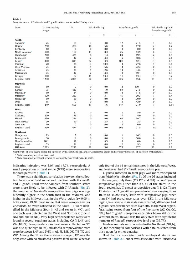
D.E. Hill et al. / Veterinary Parasitology 205 (2014) 653–665 657
Table 1Seroprevalence of Trichinella and T. gondii in feral swine in the USA by state.
State State samplingtarget
N Trichinella spp. Toxoplasma gondii Trichinella spp. andToxoplasma gondii
n % n % n %
SouthAlabamaa 25 79 3 3.8 17 21.5 1 1.3Floridaa 250 288 16 5.6 49 17.0 2 0.7Kentucky 10 8 0 0.0 0 0.0 0 0.0North Carolinaa 100 180 15 8.3 25 13.9 6 3.3Oklahomaa 200 425 6 1.4 83 19.5 2 0.5Tennessee 50 47 2 4.3 5 10.6 1 2.1Texasa 300 814 27 3.3 101 12.4 4 0.5Virginiaa 20 29 3 10.3 8 27.6 1 3.4West Virginia 25 18 1 5.6 4 22.2 0 0.0Arkansas 75 38 1 2.6 13 34.2 1 2.6Mississippi 75 47 2 4.3 9 19.1 0 0.0Georgia 100 82 11 13.4 11 13.4 3 3.7Regional total 1230 2055 87 4.2 325 15.8 21 1.0
MidwestIowa 10 2 0 0.0 2 100 0 0.0Kansasa 75 413 4 1.0 89 21.5 0 0.0Michigana 10 34 0 0.0 4 11.8 0 0.0Missouria 120 223 7 3.1 45 20.2 1 0.4Nebraskaa 10 20 0 0.0 4 20.0 0 0.0Ohio 15 7 0 0.0 3 42.9 0 0.0Regional total 240 699 11 1.6 147 21.0 1 0.14
WestArizona 40 17 0 0.0 1 5.9 0 0.0California 200 176 0 0.0 7 4.0 0 0.0Hawaii 250 234 0 0.0 93 39.7 0 0.0New Mexico 50 40 1 2.5 1 2.5 0 0.0Colorado 10 7 0 0.0 0 0.0 0 0.0Regional total 550 474 1 0.0 102 21.5 0.0 0.0
NortheastNew Jersey 25 7 0 0.0 1 14.3 0 0.0Pennsylvania 30 2 0 0.0 1 50.0 0 0.0New Hampshire NAb 12 1 8.3 0 0.0 0 0.0Regional total 55 21 1 4.8 2 9.5 0 0.0USA total 2075 3247 98 3.0 576 17.7 22 0.7
N Toxopla
te.
isf
tawtnhbTtoNfVwwWo
umber (n) of feral swine tested for infection with Trichinella spp. and/ora State sampling target was reached.b State sampling target not set due to low numbers of feral swine in sta
ndicating infection, was 3.0% and 17.7%, respectively. Amall proportion of feral swine (0.7%) were seropositiveor both parasites (Table 1).
There was a significant correlation between the collec-ion location of feral swine and infection with Trichinelland T. gondii. Feral swine sampled from southern statesere more likely to be infected with Trichinella (Fig. 2);
he number of Trichinella seropositive feral pigs was sig-ificantly higher in the South than in the Midwest, andigher in the Midwest than in the West region (p < 0.05 inoth cases). Of 98 feral swine that were seropositive forrichinella, 85 were collected in the South, 11 were iden-ified in the Midwest (four in KS and seven in MO), andne each was detected in the West and Northeast (one inM and one in NH). Very high seroprevalence rates were
ound in several southern states, including GA (13.4%) andA (10.3%). Seroprevalence in feral swine collected in NC,
as also quite high (8.3%). Trichinella seroprevalence ratesere between 1.4% and 5.6% in AL, FL, MS, OK, TN, TX, andV. Among the 12 southern states surveyed, KY was thenly state with no Trichinella positive feral swine, whereas
sma gondii and seroprevalence (%) of infection within states.
only four of the 14 remaining states in the Midwest, West,and Northeast had Trichinella seropositive pigs.
T. gondii infection in feral pigs was more widespreadthan Trichinella infection (Fig. 3). Of the 26 states includedin the analysis, only three (CO, KY, and NH) had no T. gondiiseropositive pigs. Other than KY, all of the states in theSouth region had T. gondii seropositive pigs (11/12). These11 states had T. gondii seroprevalence rates ranging from10.6% to 34.2%; every state with seropositive pigs otherthan TN had prevalence rates over 12%. In the Midwestregion, feral swine in six states were tested; all but one hadT. gondii seroprevalence rates over 20%. In the West region,feral swine tested from four of the five states (AZ, CA, CO,NM,) had T. gondii seroprevalence rates below 6%. Of theWestern states, Hawaii was the only state with significantnumbers of T. gondii seropositive feral pigs (39.7%).
Too few animals were collected in the Northeast (NH, NJ,
PA) for meaningful comparisons with data collected fromthis region for either parasite.Host factors associated with serological status areshown in Table 2. Gender was associated with Trichinella
658 D.E. Hill et al. / Veterinary Parasitology 205 (2014) 653–665
Fig. 2. Predicted probability of occurrence for Trichinella spp. infection in feral swine in the USA.
Fig. 3. Predicted probability of occurrence for T. gondii infection in feral swine in the USA.
Table 2Risk factors and geographic variables associated with seroprevalence of Trichinella and T. gondii in feral swine in the USA.
Factor N Trichinella spp. Toxoplasma gondii Trichinella spp. andToxoplasma gondii
n % n % n %
GenderMale 1427 36 2.5b 252 17.7 7 0.5Female 1516 58 3.8b 263 17.3 14 0.9Unknown 304
AgeAdult 1947 54 2.8b 388 19.9b,c 17 0.9Sub-adult 583 30 5.1b,c 80 13.7b 2 0.3Juvenile 432 10 2.3c 52 12.0c 2 0.5Unknown 285
Regiona
Northeast 21 1 4.8 2 9.5 0 0.0Midwest 699 11 1.6c 147 21.0c 1 0.1South 2053 85 4.1b,c 325 15.8b,c 21 1.0b
West 474 1 0.2b 102 21.5b 0 0.0b
Total 3247 98 3.0 576 17.7 22 0.7
Number (n) of feral swine tested positive for infection with Trichinella spp. and/or Toxoplasma gondii and seroprevalence (%) of infection within gender,age, and region.Values significantly different (p < 0.05) between groups are labeled with the same letter (b or c).
a Source: 〈http://www.eia.doe.gov/emeu/reps/maps/us census.html〉.
D.E. Hill et al. / Veterinary Parasitology 205 (2014) 653–665 659
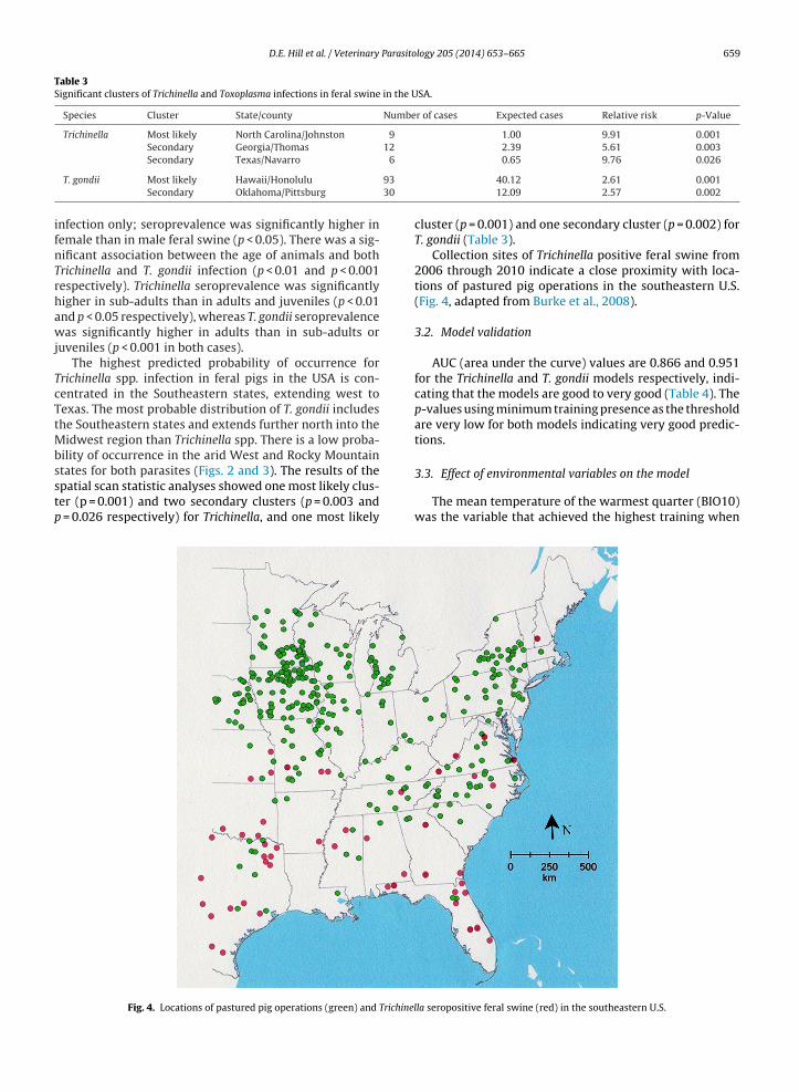
Table 3Significant clusters of Trichinella and Toxoplasma infections in feral swine in the USA.
Species Cluster State/county Number of cases Expected cases Relative risk p-Value
Trichinella Most likely North Carolina/Johnston 9 1.00 9.91 0.001Secondary Georgia/Thomas 12 2.39 5.61 0.003
6
93
30
ifnTrhawj
TcTtMbsstp
Secondary Texas/Navarro
T. gondii Most likely Hawaii/Honolulu
Secondary Oklahoma/Pittsburg
nfection only; seroprevalence was significantly higher inemale than in male feral swine (p < 0.05). There was a sig-ificant association between the age of animals and bothrichinella and T. gondii infection (p < 0.01 and p < 0.001espectively). Trichinella seroprevalence was significantlyigher in sub-adults than in adults and juveniles (p < 0.01nd p < 0.05 respectively), whereas T. gondii seroprevalenceas significantly higher in adults than in sub-adults or
uveniles (p < 0.001 in both cases).The highest predicted probability of occurrence for
richinella spp. infection in feral pigs in the USA is con-entrated in the Southeastern states, extending west toexas. The most probable distribution of T. gondii includeshe Southeastern states and extends further north into the
idwest region than Trichinella spp. There is a low proba-ility of occurrence in the arid West and Rocky Mountaintates for both parasites (Figs. 2 and 3). The results of the
patial scan statistic analyses showed one most likely clus-er (p = 0.001) and two secondary clusters (p = 0.003 and= 0.026 respectively) for Trichinella, and one most likely
Fig. 4. Locations of pastured pig operations (green) and Trichinel
0.65 9.76 0.026
40.12 2.61 0.00112.09 2.57 0.002
cluster (p = 0.001) and one secondary cluster (p = 0.002) forT. gondii (Table 3).
Collection sites of Trichinella positive feral swine from2006 through 2010 indicate a close proximity with loca-tions of pastured pig operations in the southeastern U.S.(Fig. 4, adapted from Burke et al., 2008).
3.2. Model validation
AUC (area under the curve) values are 0.866 and 0.951for the Trichinella and T. gondii models respectively, indi-cating that the models are good to very good (Table 4). Thep-values using minimum training presence as the thresholdare very low for both models indicating very good predic-tions.
3.3. Effect of environmental variables on the model
The mean temperature of the warmest quarter (BIO10)was the variable that achieved the highest training when
la seropositive feral swine (red) in the southeastern U.S.

660 D.E. Hill et al. / Veterinary Parasitology 205 (2014) 653–665
Table 4Statistical evaluation of the Maxent model.
Species Number of presence records(training/testing)
AUC for training data AUC for test data p-Value using minimum trainingpresence as threshold
Trichinella 46/15 0.949
T. gondii 205/68 0.951
used to build a model for Trichinella with no other variables(Fig. 5; Table 5; Hijmans et al., 2005). Land cover was thevariable that achieved the highest training when used tobuild a model for T. gondii with no other variables. Othervariables that show high training gain when modeling onlya single variable included elevation, annual mean tempera-ture (BIO1), and land cover for Trichinella and annual meantemperature (BIO1), mean temperature of coldest quarter(BIO11), and min temperature of coldest month (BIO6) for T.gondii. Land cover types that occurred at the species samp-ling areas are shown in Table 6. The primary land covers forTrichinella are forest and crops while for T. gondii they arewoody savanna, crops, grasses, and forest.
During the follow up survey conducted in 2012–2013,antibodies to T. gondii were found in 280 (28.4%) of 984feral swine with titers of 25–3200 (Table 7).
Most samples were from the South region where 209(28.3%) of 736 pigs were seropositive. Host factors associ-ated with serological status are shown in Table 8. Age was
Fig. 5. Training gain achieved by models using single variables. Length of bar repgain.
0.866 0.00010.951 0.0001
the main risk assessment factor; seroprevalence in olderpigs was about twice that in juvenile pigs. With respect togeography, prevalences had a similar trend although thenumber of animals sampled from the West and the North-east was relatively small.
Antibodies to Trichinella were detected in 29 (2.9%) of984 tested animals in the follow up survey. All seroposi-tive animals were collected in the South, and West to MOand TX. Of 330 tongues collected from a subset of thesesame animals, six (1.81%) were tissue positive for Trichinellamuscle larvae. All six tongue samples from which larvaewere isolated were seropositive; three additional tonguesamples was serologically positive but parasitologicallynegative. Tissue positive samples were collected from MO(Reynolds County; n = 2), NC (Swain County; n = 2), and TX(Baylor and Hemphill Counties; n = 2). All larvae collected
and genotyped from tongue tissue were identified as T. spi-ralis (data not shown). No other genotypes and no mixedinfections were detected.resents the training gain value. A longer bar represents a higher training
D.E. Hill et al. / Veterinary Parasitology 205 (2014) 653–665 661
Table 5List of WorldClim bioclimatic variables used in the model (Hijmans et al.,2005).
Bioclimaticvariable
Description
BIO1 Annual mean temperatureBIO2 Mean diurnal range (mean of monthly (max
temp–min temp))BIO3 Isothermality (P2/P7) (×100)BIO4 Temperature seasonality (standard
deviation × 100)BIO5 Max temperature of warmest monthBIO6 Min temperature of coldest monthBIO7 Temperature annual range (P5–P6)BIO8 Mean temperature of wettest quarterBIO9 Mean temperature of driest quarterBIO10 Mean temperature of warmest quarterBIO11 Mean temperature of coldest quarterBIO12 Annual precipitationBIO13 Precipitation of wettest monthBIO14 Precipitation of driest monthBIO15 Precipitation seasonality (coefficient of variation)BIO16 Precipitation of wettest quarterBIO17 Precipitation of driest quarter
4
d(peo
Table 6Number of sample localities within different land cover categories of theUSGS Global Ecosystems land cover classification.
Land cover (class number) Trichinella T. gondii
Tall grasses and shrubs (7) 3 7Wooded wet swamp (13) – 1Inland water (14) – 2Sea water (15) – 4Cool rain forest (20) – 2Cool conifer forest (22) – 3Cool mixed forest (23) – 13Mixed forest (24) 3 7Cool broadleaf forest (25) 3 43Deciduous broadleaf forest (26) 14 37Conifer forest (27) 7 35Cool crops and towns (30) 5 18Crops and Town (31) 13 33Corn and beans cropland (35) 3 5Hot irrigated cropland (37) – 4Cool irrigated cropland (38) – 2Cool grasses and shrubs (40) 1 3Hot and mild grasses and shrubs (41) 8 47Savanna (woods) (43) – 2Dry woody scrub (47) – 2Semi desert shrubs (51) – 1Cool fields and woods (55) – 8Forest and field (56) 3 18Cool forest and field (57) 10 29
siderable periods of time (>1 year), and potentially for the
TS
BIO18 Precipitation of warmest quarterBIO19 Precipitation of coldest quarter
. Discussion
Seroprevalence of T. gondii in confinement raisedomestic pigs in the USA was recently reported to be 2.7%Hill et al., 2010); seroprevalence in pastured/free-range
igs has been reported to be as high as 50–100% (Gamblet al., 1999). Results of this study indicate high prevalencef T. gondii in feral swine in the USA. T. gondii persists inable 7eroprevalence of T. gondii in feral pigs in the USA (2012–2013).
State No. of pigs No. of pigs positive with titers of:
25 50 100 200 400
Alabama 64 6 7 6 3 4
Arizona 136 3 6 3 13 6
Arkansas 17 0 0 0 1 0
California 20 0 1 0 0 0
Florida 93 9 3 2 2 1
Georgia 49 3 2 3 0 1
Hawaii 34 5 3 11 3 3
Illinois 13 0 0 0 1 0
Indiana 23 0 1 3 0 0
Kansas 43 3 6 4 10 1
Kentucky 5 3 0 1 0 0
Louisiana 98 14 5 3 5 2
Michigan 6 0 0 2 0 0
Missouri 22 1 1 5 0 0
Mississippi 90 8 7 8 7 3
North Carolina 39 1 1 3 3 1
New Jersey 3 0 0 0 0 0
New Mexico 22 1 0 0 0 0
Nevada 5 0 0 0 0 0
Ohio 5 1 1 0 1 0
Oklahoma 64 3 3 1 0 1
South Carolina 20 3 2 0 1 2
Tennessee 17 0 2 0 1 2
Texas 90 5 1 2 1 4
Virginia 5 0 0 0 0 0
West Virginia 1 0 1 0 0 0
Total 984 69 53 57 52 31
Woody savanna (91) 5 53Crops, grass, shrubs (94) 14 48
the tissues of infected pigs in an infective state for con-
life of the host. To our knowledge there were three previ-ous reports of T. gondii infection in feral swine in the USA(Diderrich et al., 1996; Dubey et al., 1997; Sandfoss et al.,
No. positives % positives
800 1600 ≥3200
2 0 0 28 43.71 0 0 32 23.50 0 0 1 5.80 0 0 1 50.01 0 1 19 20.40 0 0 9 18.30 0 0 25 73.50 0 0 1 7.60 0 0 4 17.31 0 0 25 58.30 0 0 4 80.01 1 1 32 32.00 0 0 2 33.00 0 0 7 31.82 0 0 35 38.81 1 0 11 28.21 0 0 1 33.00 0 0 1 4.50 0 0 0 00 0 0 3 60.01 0 0 9 14.01 0 0 9 45.01 0 0 6 35.21 0 0 14 15.50 0 0 0 00 0 0 1 10014 2 2 280 28.4

662 D.E. Hill et al. / Veterinary Parasito
Table 8Risk factors and geographic variables associated with seroprevalence of agondii in feral swine according with 2 surveys in the USA.
Factor (2006–2010) (2012–2013)
Year ELISA MATMAT
Test n % n %
GenderMale 252 17.7 460 28.4Female 263 17.3 372 28.4Unknown – – 4 25.0
AgeAdult 388 19.9b,c 728 33.3Sub-adult 80 13.7b 200 14.0Juvenile 52 12.0c 54 14.8Unknown – – 2 50.0
Regiona
Northeast 2 9.5 3 33.3Midwest 147 21.0 112 37.5South 325 15.8 736 28.3
West 102 21.5 98 28.5Total 576 17.7 984 28.4
2011). Seroprevalence in these studies varied from 0.9-to-50.0%, depending on the geography and the serologic testused. Diderrich et al. (1996) detected T. gondii antibodiesin 34.2% of 257 feral swine from the Great Smoky Moun-tains National Park, SC. Sandfoss et al. (2011) detected T.gondii antibodies in 27.7% of 83 feral swine from the HowellWoods Environmental Learning Center in eastern NC. Inboth Carolina surveys, the MAT test was considered posi-tive at a titer of 32 or higher; the MAT was the same testused here during the 2012–2013 survey, and thus resultsare comparable. Dubey et al. (1997) reported that a cat-free environment controlled seroprevalence in feral swinefrom GA. Anti-T. gondii antibodies were detected in 18.2%of 170 feral swine from the mainland and 0.9% of 1264 feralswine from a remote island (Ossabaw Island) by MAT witha titer of 25 considered positive. The low seroprevalenceon Ossabaw Island was attributed to the absence of cats onthis island; seroprevalence in a few of the feral swine (11of 1264) was attributed to cat fecal contamination of thegrain that was imported from the mainland and fed to feralswine.
Worldwide seroprevalences of T. gondii in feral swine upto 2009 were recently summarized (Dubey, 2010). Sincethen, seroprevalences of 17.6–55% were reported in feralswine from Europe, Brazil, and Malayasia (Richomme et al.,2009; Berger-Schoch et al., 2011; Opsteegh et al., 2011;Antolová et al., 2007, Bártová et al., 2006; Deksne andKirjusina, 2013). The successful isolation of viable T. gondiifrom pigs from France (Richomme et al., 2009) and fromMalaysia (Puvanesuaran et al., 2013) from tissues of pigsseropositive by MAT support the validity and usefulness ofMAT for the diagnosis of toxoplasmosis in pigs. Richommeet al. (2009) isolated viable T. gondii from the tissues of 21pigs with MAT titers of 1:6 out of a total of 60 pigs andPuvanesuaran et al. (2013) isolated viable T. gondii from
tissues of six of 30 wild boars with MAT titer of 1:24 orhigher.The Trichinella species and genotypes endemic in car-nivorous wildlife in the USA are the encapsulated species
logy 205 (2014) 653–665
T. murrelli, T. spiralis, T. nativa; the genotype Trichinella T6(Masuoka et al., 2009); and the non-encapsulated speciesT. pseudospiralis, which is the only Trichinella species pre-viously documented in feral swine in the USA (Gambleet al., 2005). The sylvatic genotypes are minimally infec-tive to domestic pigs as opposed to the domestic genotype(T. spiralis), and show poor persistence in pig tissues (Kapeland Gamble, 2000). On the other hand, even though T. spi-ralis reproduces well in most carnivore hosts, it is found inthe USA almost exclusively in pigs and peridomestic car-nivores. Most feral swine in the USA are descendants ofescaped or deliberately released domestic pigs, with inter-breeding with imported and released European wild boar.T. spiralis is rarely detected in wildlife species that do notassociate with pigs; T. murrelli is the predominant genotypeidentified in sylvatic carnivores in North America (Zarlengaet al., 1991; Pozio and La Rosa, 2000; Hill et al., 2008). Theseroprevalence of Trichinella spp. in feral pigs varies world-wide, 0.11% in Slovakia (Hurníková and Dubinsky, 2009),0.2% in Switzerland (Frey et al., 2009a), 0.77% in Spain(García Sánchez et al., 2009), and 19.9% in Vietnam (Vu Thiet al., 2010). The Trichinella spp. seroprevalence of 3.0% inferal swine in the USA seen in the current 2006–2010 sur-vey was four times higher than in feral swine of Europe. Thereason for the higher Trichinella spp. seroprevalence in theUSA compared to Europe may be related to the fact that incontrast to Europe, there has never been a Trichinella spp.control program in domestic pigs in the USA.
Available serological tests cannot distinguish Trichinellainfections at the species level; therefore, the seropreva-lence documented in the present study reflects infectionsdue to all possible Trichinella species in feral swine. Iso-lation of larvae from the tongue of infected animals andgenotyping by multiplex PCR is the typical method used toidentify larvae to species. Naturally infected animals typi-cally have very low worm burdens (<10 larvae per gram oftissue); the detection limit of the digestion assay used hereis 1–3 larvae per gram. As a result, larvae are frequentlymissed even in seropositive animals, which explains thedifference in the number of seropositive animals seen in2012–2013 (9/330) as compared to the number of tonguesfrom which larvae were successfully recovered (6/330). Ourresults here confirm that T. spiralis is circulating in feralswine, while other sylvatic genotypes were not detected.
The effect of gender and age on the seroprevalence ofTrichinella across host species is inconsistent. Trichinellaseroprevalence in the present study was higher in femalethan male feral swine. In humans, a higher seroprevalencehas been observed in females in Mexico (de-la-Rosa et al.,1998) but higher in males in Papua New Guinea (Owenet al., 2005). No gender based differences were found in redfoxes and Eurasian lynxes (Frey et al., 2009b) and wolves(Bagrade et al., 2009).
Trichinella seroprevalence in the present study washigher in sub-adult feral swine (5.1%) than adults (2.8%) andjuveniles (2.3%). Previous studies in our lab (Hill et al., 2010)demonstrated that naturally infected, juvenile feral swine
are capable of seroconversion and of harboring infectiousmuscle larvae of T. spiralis, so the finding of 10 seropositivejuveniles in this study was not suprising. Seroprevalencefor Trichinella infection was not statistically correlated with
Parasito
a2a
2tmtssco
attMp(WTfTt(stmStsrTiaoeMcfwpb
Tt(utab
g3Iitetvd
D.E. Hill et al. / Veterinary
ge in foxes (Frey et al., 2009b) and wolves (Bagrade et al.,009), while seroprevalence was significantly higher indult lynx compared to juveniles (Frey et al., 2009b).
In contrast, T. gondii seroprevalence in both the006–2010 and 2012–13 surveys was higher in adultshan juveniles, suggesting postnatal exposure and trans-
ission of T. gondii (Dubey, 2009). Transplacental orranscolostral immunity does not play a role in the lowereroprevalence for T. gondii observed in younger animalsince antibodies are not transmitted via the placenta, andolostrum-derived antibodies disappear by three monthsf age (Dubey and Urban, 1990).
In the prediction maps (Figs. 2 and 3), the most prob-ble distribution areas for Trichinella spp. are located inhe South, and for T. gondii the South and Midwest, whilehe parasites are virtually absent in the sampled Rocky
ountain and arid Western states. The low T. gondii sero-revalence rates (<6%) in feral swine in the West regionsCA, AZ, NM, CO) may reflect the hot, dry climate in these
estern states which could adversely impact survival of. gondii oocysts in the soil, a likely source of infection foreral swine. The high seroprevalence of Trichinella spp. and. gondii in feral pigs observed in the South in comparison tohe other regions is probably related to a number of factors:1) feral swine populations are concentrated in southerntates, and consequently more samples were collected inhese states, increasing the likelihood that seropositive ani-
als would be sampled; (2) warm temperatures in theouth promote prolonged survival of Trichinella larvae inissues of dead animals and T. gondii oocysts in soil, whicherve as sources of infection for feral swine; (3) the freezeesistance limits of both T. gondii and Trichinella (other thanrichinella nativa and Trichinella-T6, which are minimallynfective to pigs) and the prevailing low temperatures andrid climates of the other regions preclude the survivalf the parasites in the tissues of dead animals and short-ns oocyst survival in soil (Hershey and McGregor, 1987;asuoka et al., 2009). Interestingly, 1 of 12 serum samples
ollected in Sullivan County, New Hampshire was positiveor Trichinella; the site where the samples were collectedas part of a Trichinella eradication effort on a private gamereserve where hunted imported wild boar were found toe infected with Trichinella (Worley et al., 1994).
The significant clusters of infection for Trichinella and. gondii detected in North Carolina and Hawaii respec-ively during 2006–2010 fall in areas with high probabilityp > 0.7) of presence predicted by the Maxent program. Thenderlying causes of increased risk of infection in the iden-ified areas are not known but they may be linked to bothbiotic (climate, vegetation, and landscape attributes) andiotic factors (host density).
During the first survey from 2006 through 2010 of T.ondii in feral swine, antibodies were found in 17.7% of247 pigs; sera were tested using a commercial ELISA kit.n the follow up study of T. gondii in feral swine testedn 2012–2013, seroprevalence was higher using the MATest (28%). The difference seen may be related to the differ-
nce in serological tests used during the two time periods,he duration period of the surveys (5 years—2006–2010,ersus 1 year—2012–2013, so more animals were surveyeduring part one of the study). Further, the predominantlogy 205 (2014) 653–665 663
locations from which the feral swine were collected dif-fered between the two surveys; in the follow up study, 75%of the feral pigs were collected from the South. Feral pigsfrom this region had a higher rate of seropositivity for bothT. gondii and Trichinella than other regions of the countryin the 2006–2010 surveys. Whatever the reason for thesedifferent seroprevalence the results indicate stable rate ofT. gondii exposure of feral swine in the USA over 7 years.
Sport and game hunters that target feral swine are at riskwhen handling and consuming meat from these animals.Food-borne transmission of the parasites is an importantroute of infection particularly for people eating under-cooked or improperly processed meats. Care should betaken while butchering and handling raw meat to avoidinfection with T. gondii because of the presence of viable,infectious organisms in the tissues of infected animals.Pregnant women should avoid contact with raw meat dueto the risk associated with the presence of T. gondii in var-ious infective stages. To prevent infection of humans byT. gondii, thorough washing of hands with soap and waterafter handling raw meat is essential for killing the para-site. All cutting boards, sink tops, knives, and other utensilscoming in contact with uncooked meat should be washedthoroughly with soap and water. Trichinella spp. larvaeand T. gondii organisms in meat can be killed by heatingthroughout to 67 ◦C for at least 4 min before consumption,or cooling to −13 ◦C for three days (Kotula et al., 1983, 1990;Gamble et al., 2000; Hill et al., 2009). Tasting meat priorto cooking should be avoided. Adherence to good hygienicmeasures and safe handling and processing of meats is themost practical and effective method available to minimizetransmission of Trichinella spp. (Gamble et al., 2014) and T.gondii to humans from feral swine meat.
Feral swine also pose a significant risk for introductionof Trichinella and T. gondii into non-biosecure domestic pigsas a result of increasing overlap of the range of feral swinewith domestic swine production facilities in the South andMidwestern regions of the USA. Rearing of pigs outdoorshas been identified as a major risk factor for domestic piginfection with both Trichinella and T. gondii due to increasedexposure to potentially infected reservoir hosts, as well asexposure to oocyst contaminated soil in the case of T. gondii(Gamble et al., 2000, 2001; Pyburn et al., 2005; Hill et al.,2010). Transmission of T. spiralis and T. gondii has beenobserved to occur due to cannibalism in free-ranging pigs(Hanbury et al., 1986; Dubey et al., 1986a; Hill et al., 2010).Tail biting is common in pigs and T. gondii is known toencyst in musculature of pig tails (Dubey et al., 1986b). Con-sumer demand for ‘organically raised’, ‘humanely raised’and ‘free range’ pork products has resulted in increas-ing numbers of pigs being raised in non-confinementsystems (Honeyman et al., 2006). Pig producers havebeen recruited to produce animals for the organic mar-ket to fulfill a consumer demand that has increased20% per year in sales since 1990 (Dimitri and Greene,2002;http://www.ams.usda.gov/nop/). Though ‘humanelyraised’ and ‘free range’ products have standards that are
less stringently defined, outdoor access is also considereda requirement for labeling. These practices substantiallyincrease the risk of exposure of pigs to Trichinella andT. gondii. Burke et al. (2008) identified pastured pig
Parasito
664 D.E. Hill et al. / Veterinaryoperations in the eastern USA; there is close proximitybetween some of these operations and the locations of col-lection spots for Trichinella and T. gondii positive feral pigsidentified in this study (Fig. 4). The practice of field dressinghunted feral pigs and aerial hunting which leaves carcassesin the field should be discouraged, as feral pig carcasses andoffals can serve as sources of infection for grazing domesticpigs and for sylvatic carnivores that serve as reservoirs ofinfection for both parasites. Hunters should be encouragedto wear gloves when dressing carcasses and educated inthe potential hazards of contact with these parasites.
5. Conclusions
Contact between feral swine and domestic pigs shouldbe prevented. Increased surveillance efforts coupled withefforts to reduce or eliminate feral swine populationsshould be focused in regions with significant numbersof pasture raised pigs to prevent introduction of theseparasites into domestic animals destined for human con-sumption.
Conflict of interest
None.
Acknowledgments
The authors would like to thank Jorrell Fredericks andDominique Carter, Tuskegee University, Marc K. Kouam,Agricultural University of Athens, Greece Kelsey Zarlenga,Towson University, and Wildlife Services, state directorsand wildlife disease biologists in the 32 participating states,and Sarah Goff at the National Wildlife Disease ProgramFeral Swine Tissue Archive.
This work was funded in part by grant # 08-216 fromthe National Pork Board, Des Moines, Iowa.
References
Antolová, D., Reiterová, K., Dubinsky, P., 2007. Seroprevalence of Toxo-plasma gondii in wild boars (Sus scrofa) in the Slovak Republic. Ann.Agric. Environ. Med. 14, 71–73.
Aubert, D., Ajzenberg, D., Richomme, C., Gilot-Fromont, E., Terrier, M.E.,de Gevigney, C., Game, Y., Maillard, D., Gibert, P., Dardé, M.L., Villena,I., 2010. Molecular and biological characteristics of Toxoplasma gondiiisolates from wildlife in France. Vet. Parasitol. 171, 346–349.
Bagrade, G., Kirjusina, M., Vismanis, K., Ozolins, J., 2009. Helminth parasitesof the wolf Canis lupus from Latvia. J. Helminthol. 83, 63–68.
Bártová, E., Sedlák, K., Literák, I., 2006. Prevalence of Toxoplasma gondiiand Neospora caninum antibodies in wild boars in the Czech Republic.Vet. Parasitol. 142, 150–153.
Berger-Schoch, A.E., Bernet, D., Doherr, M.G., Gottstein, B., Frey, C.F., 2011.Toxoplasma gondii in Switzerland: a serosurvey based on meat juiceanalysis of slaughtered pigs, wild boar, sheep and cattle. ZoonosesPublic Health 58, 472–478.
Burke, R., Masuoka, P., Murrell, K.D., 2008. Swine Trichinella infectionand geographic information system tools. Emerg. Infect. Dis. 14,1109–1111.
de-la-Rosa, J.L., Aranda, J.G., Padilla, E., Correa, D., 1998. Prevalence and riskfactors associated with serum antibodies against Trichinella spiralis.Int. J. Parasitol. 28, 317–321.
Deksne, G., Kirjusina, M., 2013. Seroprevalence of Toxoplasma gondii indomestic pigs (Sus scrofa domestica) and wild boars (Sus scrofa) inLatvia. J. Parasitol. 99, 44–47.
Diderrich, V., New, J.C., Noblet, G.P., Patton, S., 1996. Serologic survey ofToxoplasma gondii antibodies in free-ranging wild hogs (Sus scrofa)
logy 205 (2014) 653–665
from the Great Smoky Mountains National Park and from sites in SouthCarolina. J. Eukaryot. Microbiol. 43, 122S.
Dimitri, C., Greene, C., 2002. Recent growth patterns in the U.S.organic foods market. Agriculture Information Bulletin No. (AIB777),September, 1–42. 〈http://www.ers.usda.gov/publications/aib777/〉(accessed 23.09.10).
Dubey, J.P., Murrell, K.D., Hanbury, R.D., Anderson, W.R., Doby, P.B., Miller,H.O., 1986a. Epidemiologic findings on a swine farm with enzootictoxoplasmosis. J. Am. Vet. Med. Assoc. 189, 55–56.
Dubey, J.P., Murrell, K.D., Fayer, R., Schad, G.A., 1986b. Distribution of Toxo-plasma gondii tissue cysts in commercial cuts of pork. J. Am. Vet. Med.Assoc. 188, 1035–1037.
Dubey, J.P., Desmonts, G., 1987. Serological responses of equids fed Toxo-plasma gondii oocysts. Equine Vet J. 19 (4), 337–339.
Dubey, J.P., Urban, J.F., 1990. Diagnosis of transplacentally induced toxo-plasmosis in pigs. Am. J. Vet. Res. 51, 1295–1299.
Dubey, J.P., Thulliez, P., Weigel, R.M., Andrews, C.D., Lind, P., Powell, E.C.,1995. Sensitivity and specificity of various serologic tests for detectionof Toxoplasma gondii infection in naturally infected sows. Am. J. Vet.Res. 56 (8), 1030–1036.
Dubey, J.P., Rollor, E.A., Smith, K., Kwok, O.C.H., Thulliez, P., 1997. Lowseroprevalence of Toxoplasma gondii in feral pigs from a remote islandlacking cats. J. Parasitol. 83, 839–841.
Dubey, J.P., Gamble, H.R., Hill, D., Sreekumar, C., Romand, S., Thulliez,P., 2002. High prevalence of viable Toxoplasma gondii infection inmarket weight pigs from a farm in Massachusetts. J. Parasitol. 88,1234–1238.
Dubey, J.P., Hill, D.E., Jones, J.L., Hightower, A.W., Kirkland, E., Roberts,J.M., Marcet, P.L., Lehmann, T., Vianna, M.C.B., Miska, K., Sreekumar,C., Kwok, O.C.H., Shen, S.K., Gamble, H.R., 2005. Prevalence of viableToxoplasma gondii in beef, chicken, and pork from retail meat storesin the United States: risk assessment to consumers. J. Parasitol. 91,1082–1093.
Dubey, J.P., 2009. Toxoplasmosis in pigs—the last 20 years. Vet. Parasitol.164, 89–103.
Dubey, J.P., Jenkins, M.C., Kwok, O.C.H., Zink, R.L., Michalski, M.L., Ulrich,V., Gill, J., Carstensen, M., Thulliez, P., 2009. Seroprevalence ofNeospora caninum and Toxoplasma gondii antibodies in white-taileddeer (Odocoileus virginianus) from Iowa and Minnesota using fourserologic tests. Vet. Parasitol. 161, 330–334.
Dubey, J.P., 2010. Toxoplasmosis of Animals and Humans, second. CRC,Boca Raton, FL, pp. 1–313.
Frey, C.F., Schuppers, M.E., Muller, N., Ryser-Degiorgis, M.P., Gottstein, B.,2009a. Assessment of the prevalence of Trichinella spp. in red foxesand Eurasian lynxes from Switzerland. Vet. Parasitol. 159, 295–299.
Frey, C.F., Schuppers, M.E., Eidam, V., Boujon, P., Waldvogel, A., Gottstein,B., 2009b. Occurrence of Trichinella spp. in wild boar in Switzerland.Schweiz. Arch. Tierheilkd. 151, 485–489.
Gamble, H.R., Brady, R.C., Dubey, J.P., 1999. Prevalence of Toxoplasma gondiiinfection in domestic pigs in the New England states. Vet. Parasitol.82, 129–136.
Gamble, H.R., Pyburn, D., Anderson, L.A., Miller, L.E., 2001. Verification ofgood production practices that reduce the risk of exposure of pigs toTrichinella. Parasite 8, S233–S235.
Gamble, H.R., Pozio, E., Lichtenfels, J.R., Zarlenga, D.S., 2005. Trichinellapseudospiralis from a wild pig in Texas. Vet. Parasitol. 132, 147–150.
Gamble, H.R., Bessonov, A.S., Cuperlovic, K., Gajadhar, A.A., van Knapen,F., Nöckler, K., Schenone, H., Zhu, X., 2014. International commis-sion on trichinellosis: recommendations on methods for the controlof Trichinella in domestic and wild animals intended for human con-sumption. Vet. Parasitol. 93, 393–408.
García Sánchez, R.N., Nogal-Ruiz, J.J., Manzano-Lorenzo, R., Díaz, J.M.,López, G.P., Ruano, F.J., Casas, A.R., Bascón, C.C., Bolás-Fernández,F., Martínez-Fernández, A.R., 2009. Trichinellosis survey in the wildboar from the Toledo mountains in south-western Spain (2007-2008): molecular characterization of Trichinella isolates by ISSR-PCR.J. Helminthol. 83, 117–120.
Giurgiutiu, D., Banis, C., Hunt, E., Mincer, J., Nicolardi, C., Weltman, A.,De, B., Stauffer, K., 2009. Brucella suis infection associated with feralswine hunting—three states, 2007–2008. Morb. Mortal. Wkly. Rep. 58,618–621.
Gottstein, B., Pozio, E., Nöckler, K., 2009. Epidemiology, diagnosis, treat-ment, and control of trichinellosis. Clin. Microbiol. Rev. 22, 127–145.
Hanbury, R.D., Doby, P.B., Miller, H.O., Murrell, K.D., 1986. Trichinellosis in
a herd of swine: cannibalism as a major mode of transmission. J. Am.Vet. Med. Assoc. 188, 1155–1159.Hershey, D.W., McGregor, J.A., 1987. Low prevalence of Toxoplasma infec-tion in a Rocky Mountain prenatal population. Obstet. Gynecol. 70,900–902.
Parasito
H
H
H
H
H
H
K
K
K
K
K
K
M
M
M
O
O
D.E. Hill et al. / Veterinary
ijmans, R.J., Cameron, S.E., Parra, J.L., Jones, P.G., Jarvis, A., 2005. Veryhigh resolution interpolated climate surfaces for global land areas.Int. J. Climatol. 25, 1965–1978.
ill, D.E., Samuel, M.D., Nolden, C.A., Sundar, N., Zarlenga, D.S., Dubey, J.P.,2008. Trichinella murrelli in scavenging mammal species from Wis-consin, USA. J. Wildl. Dis. 44, 629–635.
ill, D.E., Forbes, L., Zarlenga, D.S., Urban, J.F., Gajadhar, A.A., Gamble, H.R.,2009. Survival of North American genotypes of Trichinella in frozenpork. J. Food Prot. 72, 2565–2570.
ill, D.E., Pierce, V., Murrell, K.D., Ratliffe, N., Rupp, B., Fournet, V.M.,Zarlenga, D.S., Rosenthal, B.M., Gamble, H.R., Kelly, K., Dulin, M., 2010.Cessation of Trichinella spiralis transmission among scavenging mam-mals after the removal of infected pigs from a poorly managed farm:implications for trichinae transmission in the US. Zoonoses PublicHealth 57, e116–e123.
oneyman, M.S., Pirog, R.S., Huber, G.H., Lammers, P.J., Hermann, J.R.,2006. The United States pork niche market phenomenon. J. Anim. Sci.84, 276–280.
urníková, Z., Dubinsky, P., 2009. Long-term survey on Trichinella preva-lence in wildlife of Slovakia. Vet. Parasitol. 159, 276–280.
apel, C.M., Gamble, H.R., 2000. Infectivity, persistence, and antibodyresponse to domestic and sylvatic Trichinella spp. in experimentallyinfected pigs. Int. J. Parasitol. 30, 215–221.
otula, A.W., Murrell, K.D., Acosta-Stein, L., Lamb, L., Douglass, L., 1983.Trichinella spiralis: effect of high temperature on infectivity in pork.Exp. Parasitol. 156, 15–19.
otula, A.W., Sharar, A., Paroczay, E., Gamble, H.R., Murrell, K.D., Douglass,L., 1990. Infectivity of Trichinella from frozen pork. J. Food Prot. 53,571–573.
oum, M.K., Masuoka, P.M., Kantzoura, V., Theodoropoulos, G., 2010. Geo-graphic distribution modeling and spatial cluster anaysis for equinepiroplasms in Greece. Infect. Genet. Evol. 10, 1013–1018.
ulldorff, M., Nagarwalla, N., 1995. Spatial disease clusters: detection andinference. Stat. Med. 15, 707–715.
ulldorff, M., 1997. A spatial scan statistic. Commun. Stat.—Theory Method26, 1481–1496.
asuoka, P.M., Burke, R., Colaccico, M., Razuri, H., Hill, D.E., Murrell,K.D., 2009. Predicted geographic ranges for North American sylvaticTrichinella species. J. Parasitol. 95, 829–837.
atschke, G.H., 1967. Aging European wild hogs by dentition. J. Wildl.Management. 31, 109–113.
oarthland, J., 2011. A plague of pigs in Texas. Smithsonian Mag-azine. 〈http://www.smithsonianmag.com/science-nature/A-Plague-of-Pigs-in-Texas.html〉.
doi, A., Martin, S.W., Michel, P., Middleton, D., Holt, J., Wilson, J., 2004.Investigation of clusters of giardiasis using GIS and a spatial scan
statistic. Int. J. Health Geogr. 3, 11-.psteegh, M., Swart, A., Fonville, M., Dekkers, L., van der Giessen, J.,2011. Age-related Toxoplasma gondii seroprevalence in Dutch wildboar inconsistent with lifelong persistence of antibodies. PLoS One6, e16240.
logy 205 (2014) 653–665 665
Owen, I.L., Morales, M.A.G., Pezzottic, P., Pozio, E., 2005. Trichinella infec-tion in a hunting population of Papua New Guinea suggests an ancientrelationship between Trichinella and human beings. Trans. R. Soc. Trop.Med. Hyg. 99, 618–624.
Phillips, S.J., Anderson, R.P., Schapire, R.E., 2006. Maximum entropy mod-eling of species geographic distributions. Ecol. Model. 190, 231–259.
Pozio, E., La Rosa, G., 2000. Trichinella murrelli n. sp: etiological agent ofsylvatic trichinellosis in temperate areas of North America. J. Parasitol.86, 134–139.
Puvanesuaran, V.R., Noordin, R., Balakrishnan, V., 2013. Genotyping ofToxoplasma gondii isolates from wild boars in peninsular Malaysia.PLoS One 8, e61730.
Pyburn, D.G., Gamble, H.R., Wagstrom, E.A., Anderson, L.A., Miller, L.E.,2005. Trichinae certification in the United States pork industry. Vet.Parasitol. 132, 179–183.
Richomme, C., Aubert, D., Gilot-Fromont, E., Ajzenberg, D., Mercier, A.,Ducrot, C., Ferté, H., Delorme, D., Villena, I., 2009. Genetic characteri-zation of Toxoplasma gondii from wild boar (Sus scrofa) in France. Vet.Parasitol. 164, 296–300.
Sandfoss, M., DePerno, C., Patton, S., Flowers, J., Kennedy-Stoskopf, S.,2011. Prevalence of antibody to Toxoplasma gondii and Trichinellaspp. in feral pigs (Sus scrofa) of eastern North Carolina. J Wildl Dis. 47(2), 338–343.
United States Department of Agriculture, 2011. Animal and Plant HealthInspection Service. Feral Swine: damage and disease threats. Agri-culture Information Bulletin No. 2086. Available from PublicationsDistribution, USDA, Animal and Plant Health Inspection Service, Unit1, 4700 River Road, Riverdale, MD 20737-1229.
Vu Thi, N., Dorny, P., La Rosa, G., To Long, T., Nguyen Van, C., Pozio, E., 2010.High prevalence of anti-Trichinella IgG in domestic pigs of the Son Laparovince. Vietnam Vet. Parasitol. 168, 136–140.
Wildlife Services, 2008. National Wildlife Disease, Surveillance andEmergency Response Program Comprehensive Feral Swine Disease,Surveillance Procedure Manual, Version 1.1, October 1, 2008.
Worley, D.E., Seesee, F.M., Zarlenga, D.S., Murrell, K.D., 1994. Attempts toeradicate trichinellosis from a wild boar population in a private gamepark (USA). Trichinellosis 611–616 (Istituto Superiore di Sanita Press,Rome).
Wychoff, A.C., Henke, S.E., Campbell, T.A., Hewitt, D.G., Vercauteren, K.C.,2009. Feral swine contact with domestic swine: a serologic surveyand assessment of potential for disease transmission. J. Wildl. Dis. 45,422–429.
Zarlenga, D.S., Al-Yaman, F., Minchella, D.J., La Rosa, G., 1991. A repet-itive DNA probe specific for a North American sylvatic genotype ofTrichinella. Mol. Biochem. Parasitol. 48, 131–137.
Zarlenga, D.S., Chute, M.B., Martin, A., Kapel, C.M., 1999. A multiplex
PCR for unequivocal differentiation of all encapsulated and non-encapsulated genotypes of Trichinella. Int. J. Parasitol. 29, 1859–1867.Zarnke, R.L., Dubey, J.P., Kwok, O.C.H., Ver Hoef, J.M., 2000. Serologic surveyfor Toxoplasma gondii in selected wildlife species from Alaska. J. Wildl.Dis. 36, 219–224.