Embed Size (px)
Citation preview
�Bulletin of the NYU Hospital for Joint Diseases 2007;6�(1):�-18
Abstract
Modern advances in total hip arthroplasty have included im-proved biomaterials, alternative bearings, so-called fourth-generation cement techniques, and enhanced in-growth prostheses. Recently, much attention has been focused on improving the surgical technique for total hip arthroplasty. This endeavor has included the development of mini-incision arthroplasty and has now progressed to minimally invasive procedures. Minimally invasive techniques are modified versions of the classic approaches to the hip, with the goal of decreasing soft tissue dissection and trauma. While these new surgical exposures may result in improved short-term outcomes, their long-term results have yet to be proven. In addition, they are technically demanding, may require specialized training, and can be associated with a difficult learning curve. Only the future will determine if mini-inci-sion and minimally invasive arthroplasty are as successful as the well-proven standard surgical approaches used for the last three to four decades.
Total hip arthroplasty (THA) has been one of the most successful orthopaedic procedures over the past 30 years. During this time, several classic surgical ap-
proaches to the hip have been described, including anterior, anterolateral, direct lateral (with multiple modifications),
transtrochanteric, and posterior.1-3 There are advantages and disadvantages to each exposure, yet overall success rates are similar at long-term follow-up. The Charnley transtro-chanteric approach has the longest available follow-up data. Callaghan and colleagues have reported on 34 THAs from the 27 patients alive at a minimum 30-year follow-up; they found 88% of prostheses were intact at the time of latest fol-low-up or death.4 Despite relatively large incisions and the use of trochanteric osteotomies in all cases, these patients did extremely well. Similarly, excellent long-term results have been shown in several other studies.�,6
As the comfort level of surgeons and the techniques for THA have evolved, incision length has gradually decreased. Simultaneously, patient, surgeon, and industry pressures have driven the push for shorter hospital stays and an early return to function. Together, these factors led to the evolution of mini-incision approaches and minimally invasive surgery (MIS) for THA. Mini-incision approaches are based on the classic expo-sures previously mentioned, but with an abbreviated skin incision of less than 10 cm. However, MIS approaches, have been developed to minimize soft tissue damage and avoid sectioning any tendons or muscles during the dissection. Both mini-incision and MIS THA may require advanced training and special instrumentation (i.e., surgical tables, reamers, retractors, and insertion devices) to be performed safely and accurately. Early results with these techniques have been promising, yet controversial, with conflicting reports on whether there are true benefits other than the cosmesis of a smaller incision(s).7-14
In critically evaluating mini-incision and MIS THA approaches, one must compare their features to the ideals of a “perfect approach.” Such a technique should be easy to understand, teach, and perform, while allowing precise, reproducible implantation of various prosthetic options (cemented, cementless, proximal-fit, or distal-fit). Addition-
Surgical Approaches in Total Hip ArthroplastyA Review of the Mini-Incision and MIS Literature
Brett R. Levine, M.D., M.S., Gregg R. Klein, M.D., and Paul E. Di Cesare, M.D.
Brett R. Levine, M.D., M.S., was a Chief Resident in the NYU Hospital for Joint Diseases Department of Orthopaedic Surgery, New York, New York. Gregg R. Klein, M.D., was Assistant Profes-sor of Orthopaedic Surgery in the NYU School of Medicine and Attending in the Adult Reconstructive Service, NYU Hospital for Joint Diseases Department of Orthopaedic Surgery, New York, New York. Paul E. Di Cesare, M.D., was Professor of Orthopaedic Sur-gery and Cell Biology, NYU School of Medicine and Director of the Musculoskeletal Research Center, NYU Hospital for Joint Diseases Department of Orthopaedic Surgery, New York, New York. Correspondence: Brett Levine, M.D., 6000 N. Allen Road, Peoria, Illinois 61614.
Bulletin of the NYU Hospital for Joint Diseases 2007;6�(1):�-186
ally, long-term results ultimately need to be equal or better than the current gold standard. In order to be universally accepted, the approach must require a minimal number of assistants and afford only a nominal risk to the surrounding neurovascular structures. The overall goals should be to de-crease pain, length of hospital stay, and time to ambulatory independence, while yielding negligible risk for concomitant morbidities. Currently, the “perfect approach” does not exist, and it is important to keep these ideals in mind, as the media and internet advertising have increased patient awareness and demands for mini-incision and MIS THA.1�
Patient SelectionOne of the most crucial aspects in performing a limited-inci-sion approach for THA is patient selection. These exposures should not be used for all patients and component positioning should never be compromised secondary to limited anatomi-cal visualization. Table 1 represents general guidelines for relative and absolute contraindications with mini-incision and MIS THA.16-18
The ideal patient would have minimal proximal femur and acetabular deformity, a primary THA setting, and a body mass index (BMI) of less than 30. Even with the perfect patient characteristics and radiographs, all patients should be counseled on the possibility of incision extension based
upon the intraoperative course of the procedure. Goldstein and Branson found that component size may dictate incision length, as they determined a four-inch incision is required
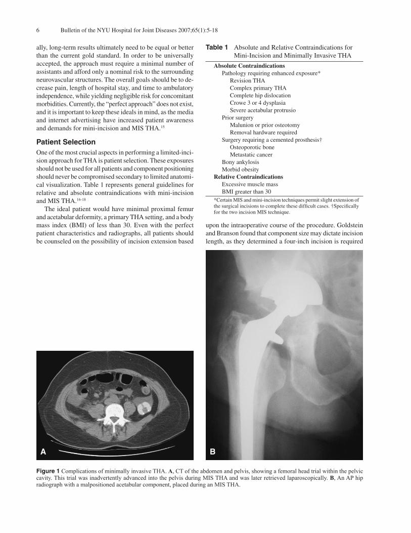
Figure 1 Complications of minimally invasive THA. A, CT of the abdomen and pelvis, showing a femoral head trial within the pelvic cavity. This trial was inadvertently advanced into the pelvis during MIS THA and was later retrieved laparoscopically. B, An AP hip radiograph with a malpositioned acetabular component, placed during an MIS THA.
BA
Table 1 Absolute and Relative Contraindications for Mini-Incision and Minimally Invasive THA
AbsoluteContraindications Pathology requiring enhanced exposure* Revision THA Complex primary THA Complete hip dislocation Crowe 3 or 4 dysplasia Severe acetabular protrusio Prior surgery Malunion or prior osteotomy Removal hardware required Surgery requiring a cemented prosthesis† Osteoporotic bone Metastatic cancer Bony ankylosis Morbid obesity RelativeContraindications Excessive muscle mass BMI greater than 30
*Certain MIS and mini-incision techniques permit slight extension of the surgical incisions to complete these difficult cases. †Specifically for the two incision MIS technique.
7Bulletin of the NYU Hospital for Joint Diseases 2007;6�(1):�-18
to insert a size �6 acetabular component without contact between the prosthesis and the skin or subcutaneous fat.1�
Learning CurveWhen embarking upon mini-incision and MIS THA proce-dures, there can be a significant initial learning curve. During this time, significantly higher complication rates have been reported to occur.16,19,20 Figure 1 depicts two examples of complications related to the learning curve of MIS THA. Many of these techniques require specialized training and cadaver dissection prior to attempting them in the op-erating room. Each implant company has designed specific instrumentation and retractors for these limited-incision ap-proaches, which necessitate a certain level of familiarity. For mini-incision approaches, current recommendations include starting with a standard incision approach using the associ-ated modified instruments and gradually decreasing the inci-sion length as the surgeon becomes more comfortable with the approach and instrumentation.21,22 MIS THA is thought to be still more challenging, as it represents approaches and techniques that are unfamiliar to most orthopaedic surgeons. It is recommended that surgeons first be comfortable with mini-incision THA before attempting MIS techniques.
Mini-Incision ApproachesCurrently, multiple mini-incision approaches have been de-scribed to include anterior, anterolateral, direct lateral, pos-terolateral, and a direct two-incision approach.11,1�,22-27 The basic premise of these approaches is utilization of a smaller skin incision (defined as less than 10 cm) to create a mobile window that allows intermittent complete visualization of the surgical anatomy. The same respective surgical approach and bone resection are performed beneath the skin incision. Overall, there is conflicting data as to the efficacy of these approaches regarding blood transfusions, pain control, length of hospital stay, and duration of recovery.14,27-32 However, most studies have reported improved cosmesis and patient satisfaction with these smaller incision approaches.27-30 Howell and associates lent significant importance to the psy-chological impact of improved cosmesis on patient attitude, satisfaction, and motivation for recovery, and cautioned that this appeal should not be underestimated.21 However, some recent studies involving evaluation of surgical wounds by plastic surgeons have shown worse cosmetic results with mini-incision approaches.33
Anterior ApproachSiguer and coworkers26 have described a modified anterior mini-incision approach, based on the standard surgical approach described by Smith-Petersen.34 The patient is positioned supine on a Judet orthopaedic table (Tasserit, Sens, France). This table accommodates the use of trac-tion, external and internal rotation of the lower extremity, and hip extension during the procedure. THA can be safely performed with one or two assistants via this approach.
The incision is limited to � cm to 10 cm and is made obliquely 2 cm posterior to an imaginary line drawn from the anterosuperior iliac spine to the fibula head. Using the greater trochanter as a reference, two-thirds of the incision extends superior and one-third inferior to this landmark. Sharp dissection is followed to the tensor fascia lata (TFL) aponeurosis, which is incised in-line with the skin inci-sion. Extension of this split is carried proximal and distal underneath the skin edges, using the superficial layers as a mobile window. The TFL is retracted posteriorly and the sartorius anteriorly to reveal the innominate aponeurosis. In this layer, the lateral circumflex femoral vessels and their branches are encountered and must be protected or ligated. Once the innominate aponeurosis is opened the entire length of the incision, a deep fatty layer is found over the anterior hip capsule. With appropriate dissection and placement of retractors, the anterior hip capsule can be exposed. Capsulotomy or capsulectomy may be performed at this point, and the remainder of the THA is performed through this exposure. Visualization of the acetabulum is straight-forward; however, the Judet table is vital to the exposure of the femur with this approach. The leg must be lowered (hip extension) and externally rotated with continual adjustments of the applied traction. Retractors are then used to elevate the proximal femur from the wound in preparation for prosthetic implantation. Reduction is performed with the trial compo-nents in place and the limb lengths can be checked easily on the operating room table, due to the supine positioning of the patient. Siguer and colleagues also retrospectively reviewed 1037 THAs using this approach, with the majority of pa-tients (92%) having a primary diagnosis of osteoarthritis.26 Cemented monoblock stainless Charnley LFA MKII stems (Sanortho, Smith and Nephew, Orthez, France) and poly-ethylene hemispherical cups were used in all cases. Patients were allowed to be weightbearing as tolerated starting on the second postoperative day. Forty-five patients (4.3%) were lost to follow-up, and a total of 10 dislocations (0.96%) were reported. Seven of the 10 dislocations were treated with closed reduction without further episodes of instability. Eight cases (0.77%) underwent revision surgery as follows: three cases of septic loosening, three cases of aseptic loos-ening, and two cases of recurrent dislocations. No patients developed significant heterotopic ossification, clinical limp, or Trendelenburg gait. There was difficulty with exposure in 1� cases of obese patients, and in eight muscular males the piriformis tendon was sectioned for enhanced visualization. Overall, the authors concluded this was a safe technique that afforded a low dislocation rate and reproducible implantation of THA components via an approach that avoids sectioning of any muscles or tendons. Matta reported on �06 unselected, consecutive THAs us-ing a fluoroscopic-assisted anterior mini-incision approach: 386 primary THAs, 92 with prior hip surgery, and 22 revision THAs.3�
Bulletin of the NYU Hospital for Joint Diseases 2007;6�(1):�-188
The surgery was performed on a PROfx™ fracture table (OSI, Union City, CA), which can be used intraoperatively to manipulate the operative lower extremity. The average patient was 6� years of age. The mean fluoroscopy time was 30 sec. There were �0 hybrid THA and 336 cementless THA, with an overall average length of surgery of 90 min (range, 36 min to 240 min). The average postoperative abduction and anteversion angles were 41° and 23°, respectively. The median length of stay was four days, with an average of 10 days to walking without external support. There were five complications (one infection, two anterior dislocations, one posterior dislocation, and one temporary femoral nerve palsy), and the average leg length discrepancy was 3 mm. Matta concluded that the mini-incision anterior approach with an orthopaedic table allows for no postoperative hip precautions with good outcomes, minimal complications, and maintenance of accurate leg lengths.3�
Most recently, Rachbauer and associates prospectively reported on a similar anterior mini-incision approach in 100 consecutive THAs without exclusion criteria.36 Excellent component positioning was noted with mean cup inclination and stem varus/valgus angles of 44.1° and 0°, respectively. Minimal postoperative pain was described and the average WOMAC (Western Ontario and McMaster osteoarthritis questionaire) score at six weeks was 90.4. Blood loss, postoperative pain, and length of hospital stay were noted to be reduced, and overall rehabilitation found to be quicker in these patients compared to standard incision THA. There were a total of three complications (one proximal femur fracture, one acetabular perforation, and one deep infection). The investigators concluded that this minimally invasive anterior approach was safe and advantageous.
Anterolateral ApproachThe classic anterolateral approach was initially described by Watson-Jones in 1936.1 This approach followed the intermuscular plane between the gluteus medius and the TFL. Perceived advantages of such an approach were de-creased abductor weakness (compared to the direct lateral approach) and lower rates of dislocation (compared to the posterior approach). These goals were obtained by insert-ing a prosthesis through the intermuscular interval, leaving the abductor musculature and posterior hip capsule/short external rotators intact. Bertin and Rottinger recently described a mini-incision modification of the Watson-Jones approach.22 The patient is placed in the lateral position, using a hip positioner, on a Jupiter table (TRUMPF Inc., Charleston, SC) in which the posterior half of the table distal to the pelvis is detachable. A 6 to 7 cm skin incision is effected along a line from the anterior tubercle of the greater trochanter towards the an-terosuperior iliac spine. One-fourth of the incision is over the trochanter, with the remainder extending proximally. The fascia is divided inline with the skin incision and blunt dissection follows, dividing the plane between the gluteus
medius/minimus and the TFL. Modified Hohmann retractors (with an attached light) are then placed along the superior and inferior borders of the femoral neck to expose the hip capsule. A U-shaped capsulotomy is made along the femoral neck and the Hohmann retractors are moved to an intracapsular location. The femoral neck is cut in situ using two osteotomies, one in the subcapital region and the other at the predetermined level of the final neck cut. The interca-lary piece is then removed using a threaded Steinman pin, followed by extraction of the femoral head using a second pin. The acetabulum is then exposed with traction and external rotation of the femur, improving acetabular visualization. The acetabular component (cemented or cementless) is im-planted in the standard fashion. The femur is prepared with the leg placed in a sterile bag or pocket and extended off the posterior half of the detachable table. With the hip in 30° of extension, adduction, and external rotation of the proximal femur can be addressed. A retractor is placed beneath the proximal femur to elevate it out of the wound, and a second lighted-retractor is positioned to protect the gluteus medius muscle. The proximal femur is prepared in standard fashion for any implant type. The wound is closed in layers over a suction drain, with 0.2�% marciane infiltration of the tissues at each layer. Patients can be discharged the same day or after a 24-hour stay with this approach.22
Bertin and Rottinger have performed over 300 THAs using this approach; long-term follow-up and clinical data is still pending for these cases.22 For obese and muscular patients, they recommended 1 to 2 cm extension of the inci-sion. Division of the inferior and posterior capsule (without these releases, increasing pressure is placed on the femur and can result in fracture of the shaft) may also be neces-sary to facilitate difficult exposures. Overall, these authors are cautiously optimistic that this versatile approach will provide universally successful long-term results. Further experience (as exposure of femur is challenging), improved instrumentation, and possibly computer navigation (as con-sistent acetabular component positioning is difficult) may enhance outcomes with this approach in the future.
Lateral ApproachThe direct lateral approach (also referred to as an anterolat-eral, lateral, or transgluteal approach) was initially described by Kocher and has been subsequently been modified by Hardinge (1982) and Mullikan and coworkers (1998).2,37,38 This exposure can be performed with the patient supine, semilateral, or in the lateral decubitus position. There is no true internervous plane, and the dissection involves splitting the gluteus medius and vastus lateralis muscles (multiple modifications describe variations of this split). Division of the gluteus medius is limited to � cm proximal to the greater trochanter or 4 cm proximal to the superior acetabulum, as further extension places the superior gluteal neurovascular bundle at risk for injury. Controversy exists
9Bulletin of the NYU Hospital for Joint Diseases 2007;6�(1):�-18
as to the clinical significance and morbidity of dividing the abductor musculature. Several studies show an increase limp/lurch (10% to 26% of cases) and significant denervation of the abductors (23% decrease in abductor strength) after a direct lateral approach.39-42 However, there are also reports of no significant increase in the incidence of a postopera-tive limp.43-46 Regardless, very low rates of hip dislocation (typically less than 1%) have been documented when using this approach.47-49
Ilizaliturri and colleagues described a mini-incision direct lateral approach with the patient in the lateral decubitus posi-tion using standard instrumentation.2� A “hockey stick” split of the gluteus medius and vastus lateralis is done to expose the anterior hip capsule. The hip is dislocated anteriorly after either incision or excision of the anterior capsule. The femoral neck is osteotomized, and the femur is prepared first in this approach. The investigators reported on 40 patients (five cemented, nine hybrid, and 26 cementless THAs) with an average age of 60 years. Complications included one pulmonary embolism and 19 small skin abrasions, all of which healed without complication. There were no cases of dislocation, and three cases (7.�%) necessitated conver-sion to standard length incisions (greater than 10 cm, due to initial exposure difficulty). These cases were subsequently excluded from the series. Overall, this approach afforded good results (using standard instrumentation) in this small patient population; however, meticulous care of the skin edges must be emphasized. Higuchi and associates retrospectively reported on 212 patients undergoing THA via a direct lateral approach in the lateral decubitus position.�0 Patients were divided into three groups: mini-incision (less than 10 cm), short incision (10 cm to 1� cm), and conventional incision (greater than 1� cm). Postoperative protocols were similar; however, patients with shorter incisions were allowed full weightbearing at two weeks versus those with conventional incision THA, who were kept partial weightbearing for a total of six weeks. As the length of the incision was made smaller, decreases in operative times (94.9 min to 69.7 min), intraoperative blood loss (388.2 g to 184.3 g), and total blood loss (891.� g to 668.0 g) were observed. There were no differences in postoperative bleeding or complications among the three groups. The overall dislocation rate was 2.4% (three mini-, one short-, and one conventional incision) and acetabular loosening, 1.9% (one mini-, one short- and two conventional incision). Earlier rehabilitation and full weightbearing did not adversely influence outcomes in the patients with mini- and short-incision THA. These investigators concluded that a shorter incision with standard instrumentation is safe, with comparable results, shorter operative times, and lower blood loss, via a direct lateral approach. de Beer and coworkers compared matched pair cohorts (from a prospective THA database) of 30 patients undergo-ing primary THA using mini-incision versus a conventional direct lateral approach.11 The average incision length was 7.7
cm for mini-incision THAs and 13.9 cm for conventional THAs. There were no significant differences in operative time, opioid consumption, postoperative blood loss, com-plications, length of hospital stay, or Harris hip and Oxford hip scores at six weeks postoperatively. They concluded there was no advantage conferred with the mini-incision direct lateral approach. The authors also cautioned that the increased technical difficulty with these small incision ap-proaches could possibly lead to some untoward complica-tions, due to poor visualization and the demanding nature of the procedure. Recently, Howell and colleagues described a similar direct lateral mini-incision approach.21 This incision differs from those of the previous study in that it is centered on a point 2 cm distal to the tip of the greater trochanter and extends 3 cm proximal and distal to this point (proximal half is angled 30° posterior to the long axis of the femur and the distal half is 30° anterior). The gluteus medius muscle is divided at the junction of the anterior one-third and pos-terior two-thirds and elevated with a flake of bone from the greater trochanter. The exposure of the acetabulum follows as above, and an anterior capsulectomy is performed. The femur is prepared using retractors to elevate the proximal portion from the wound, with the hip flexed, adducted, and in maximal external rotation. Howell and associates prospectively compared �0 mini-incision THAs with �7 conventional THAs via a direct lateral approach.21 The MIS group had a higher proportion of males (2:1 versus 0.9:1) and a lower overall BMI (26.2 versus 28.8) compared to the control group. Mini-incision THA increased the average length of surgery from 84 minutes to 97 minutes compared to conventional THA. There were no differences in rate of transfusion for either group; however, the length of hospital stay was significantly reduced from a mean of �.7 days to 4.4 days with mini-incision THA. Surgical complications included two intraoperative proximal femur fractures treated with cerclage wiring and two hematomas in the mini-incision group compared with one hematoma in the conventional group. Overall, the mini-incision direct lateral approach was ob-served to be safe and may decrease the length of hospital stay. This study was performed during this group of surgeons’ initial learning curve and they anticipate an improvement in the comparative length of surgery over time.
Posterior ApproachThe posterior approach to the hip was initially popularized by Moore,�1with subsequent modifications by Gibson3 and Marcy and Fletcher.�2 It is the most commonly used approach in the United States, considered technically the easiest exposure to perform, and requires only one assistant. The biggest criti-cism of this approach remains the higher associated rate of postoperative dislocation (1% to 9%), secondary to the de-tachment of the short external rotator muscles and posterior
Bulletin of the NYU Hospital for Joint Diseases 2007;6�(1):�-1810
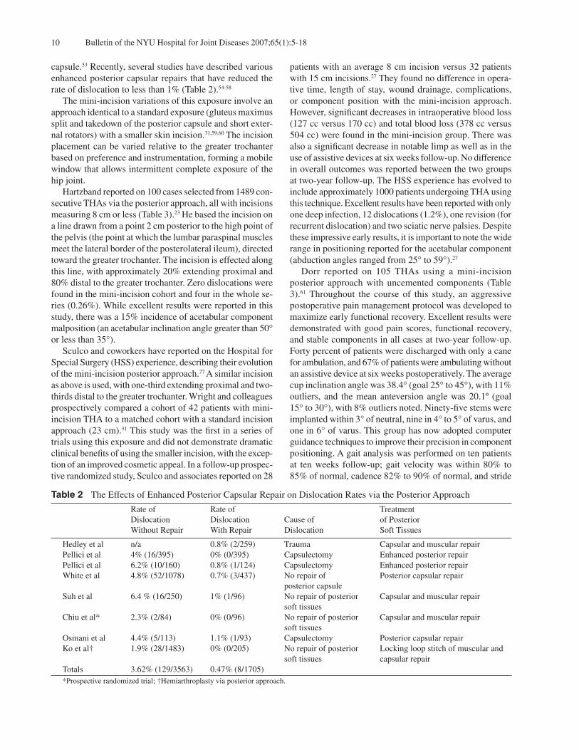
capsule.�3 Recently, several studies have described various enhanced posterior capsular repairs that have reduced the rate of dislocation to less than 1% (Table 2).�4-�8
The mini-incision variations of this exposure involve an approach identical to a standard exposure (gluteus maximus split and takedown of the posterior capsule and short exter-nal rotators) with a smaller skin incision.31,�9,60 The incision placement can be varied relative to the greater trochanter based on preference and instrumentation, forming a mobile window that allows intermittent complete exposure of the hip joint. Hartzband reported on 100 cases selected from 1489 con-secutive THAs via the posterior approach, all with incisions measuring 8 cm or less (Table 3).23 He based the incision on a line drawn from a point 2 cm posterior to the high point of the pelvis (the point at which the lumbar paraspinal muscles meet the lateral border of the posterolateral ileum), directed toward the greater trochanter. The incision is effected along this line, with approximately 20% extending proximal and 80% distal to the greater trochanter. Zero dislocations were found in the mini-incision cohort and four in the whole se-ries (0.26%). While excellent results were reported in this study, there was a 1�% incidence of acetabular component malposition (an acetabular inclination angle greater than �0° or less than 3�°). Sculco and coworkers have reported on the Hospital for Special Surgery (HSS) experience, describing their evolution of the mini-incision posterior approach.27 A similar incision as above is used, with one-third extending proximal and two-thirds distal to the greater trochanter. Wright and colleagues prospectively compared a cohort of 42 patients with mini-incision THA to a matched cohort with a standard incision approach (23 cm).31 This study was the first in a series of trials using this exposure and did not demonstrate dramatic clinical benefits of using the smaller incision, with the excep-tion of an improved cosmetic appeal. In a follow-up prospec-tive randomized study, Sculco and associates reported on 28
patients with an average 8 cm incision versus 32 patients with 1� cm incisions.27 They found no difference in opera-tive time, length of stay, wound drainage, complications, or component position with the mini-incision approach. However, significant decreases in intraoperative blood loss (127 cc versus 170 cc) and total blood loss (378 cc versus �04 cc) were found in the mini-incision group. There was also a significant decrease in notable limp as well as in the use of assistive devices at six weeks follow-up. No difference in overall outcomes was reported between the two groups at two-year follow-up. The HSS experience has evolved to include approximately 1000 patients undergoing THA using this technique. Excellent results have been reported with only one deep infection, 12 dislocations (1.2%), one revision (for recurrent dislocation) and two sciatic nerve palsies. Despite these impressive early results, it is important to note the wide range in positioning reported for the acetabular component (abduction angles ranged from 2�° to �9°).27
Dorr reported on 10� THAs using a mini-incision posterior approach with uncemented components (Table 3).61 Throughout the course of this study, an aggressive postoperative pain management protocol was developed to maximize early functional recovery. Excellent results were demonstrated with good pain scores, functional recovery, and stable components in all cases at two-year follow-up. Forty percent of patients were discharged with only a cane for ambulation, and 67% of patients were ambulating without an assistive device at six weeks postoperatively. The average cup inclination angle was 38.4° (goal 2�° to 4�°), with 11% outliers, and the mean anteversion angle was 20.1º (goal 1�° to 30°), with 8% outliers noted. Ninety-five stems were implanted within 3° of neutral, nine in 4° to �° of varus, and one in 6° of varus. This group has now adopted computer guidance techniques to improve their precision in component positioning. A gait analysis was performed on ten patients at ten weeks follow-up; gait velocity was within 80% to 8�% of normal, cadence 82% to 90% of normal, and stride
Table 2 The Effects of Enhanced Posterior Capsular Repair on Dislocation Rates via the Posterior Approach
Rate of Rate of Treatment Dislocation Dislocation Cause of of Posterior Without Repair With Repair Dislocation Soft Tissues
Hedley et al n/a 0.8% (2/2�9) Trauma Capsular and muscular repairPellici et al 4% (16/39�) 0% (0/39�) Capsulectomy Enhanced posterior repairPellici et al 6.2% (10/160) 0.8% (1/124) Capsulectomy Enhanced posterior repairWhite et al 4.8% (�2/1078) 0.7% (3/437) No repair of Posterior capsular repair posterior capsule Suh et al 6.4 % (16/2�0) 1% (1/96) No repair of posterior Capsular and muscular repair soft tissuesChiu et al* 2.3% (2/84) 0% (0/96) No repair of posterior Capsular and muscular repair soft tissuesOsmani et al 4.4% (�/113) 1.1% (1/93) Capsulectomy Posterior capsular repairKo et al† 1.9% (28/1483) 0% (0/20�) No repair of posterior Locking loop stitch of muscular and soft tissues capsular repairTotals 3.62% (129/3�63) 0.47% (8/170�)*Prospective randomized trial; †Hemiarthroplasty via posterior approach.
11Bulletin of the NYU Hospital for Joint Diseases 2007;6�(1):�-18
length 62% to 74% of normal. There was one deep infection and one transient sciatic nerve palsy in this study.61 Dorr cautioned that, while early results are encouraging, it is dif-ficult to distinguish between the benefits of aggressive pain management and the mini-incision surgical techniques. Nakamura and coworkers retrospectively compared �0 posterior mini-incision and 42 standard incision THAs in two demographically similar patient cohorts.18 Decreased opera-
tive times and intraoperative blood loss were observed using the mini-incision approach (Table 3). There were no differ-ences in component positioning, postoperative blood loss, or mean Merle d’Aubigne and Postel scores at six-month follow-up. No cases of dislocation or pulmonary embolus were described, and it was concluded that the mini-incision approach afforded less surgical invasion with similar short-term follow-up to a standard posterior-incision THA.
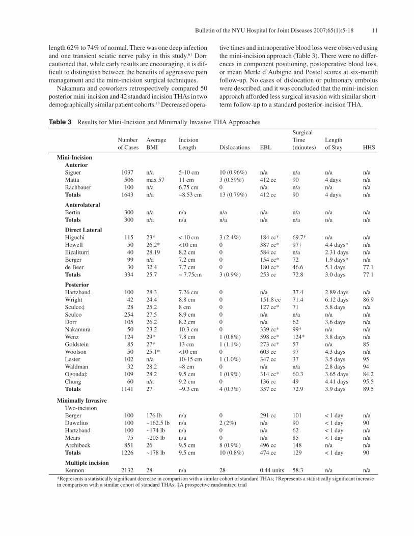
Table 3 Results for Mini-Incision and Minimally Invasive THA Approaches
Surgical Number Average Incision Time Length of Cases BMI Length Dislocations EBL (minutes) of Stay HHS
Mini-Incision Anterior Siguer 1037 n/a �-10 cm 10 (0.96%) n/a n/a n/a n/a Matta �06 max �7 11 cm 3 (0.�9%) 412 cc 90 4 days n/a Rachbauer 100 n/a 6.7� cm 0 n/a n/a n/a n/a Totals 1643 n/a ~8.�3 cm 13 (0.79%) 412 cc 90 4 days n/a
Anterolateral Bertin 300 n/a n/a n/a n/a n/a n/a n/a Totals 300 n/a n/a n/a n/a n/a n/a n/a
DirectLateral Higuchi 11� 23* < 10 cm 3 (2.4%) 184 cc* 69.7* n/a n/a Howell �0 26.2* <10 cm 0 387 cc* 97† 4.4 days* n/a Ilizaliturri 40 28.19 8.2 cm 0 �84 cc n/a 2.31 days n/a Berger 99 n/a 7.2 cm 0 1�4 cc* 72 1.9 days* n/a de Beer 30 32.4 7.7 cm 0 180 cc* 46.6 �.1 days 77.1 Totals 334 2�.7 ~ 7.7�cm 3 (0.9%) 2�3 cc 72.8 3.0 days 77.1
Posterior Hartzband 100 28.3 7.26 cm 0 n/a 37.4 2.89 days n/a Wright 42 24.4 8.8 cm 0 1�1.8 cc 71.4 6.12 days 86.9 Sculco‡ 28 2�.2 8 cm 0 127 cc* 71 �.8 days n/a Sculco 2�4 27.� 8.9 cm 0 n/a n/a n/a n/a Dorr 10� 26.2 8.2 cm 0 n/a 62 3.6 days n/a Nakamura �0 23.2 10.3 cm 0 339 cc* 99* n/a n/a Wenz 124 29* 7.8 cm 1 (0.8%) �98 cc* 124* 3.8 days n/a Goldstein 8� 27* 13 cm 1 (1.1%) 273 cc* �7 n/a 8� Woolson �0 2�.1* <10 cm 0 603 cc 97 4.3 days n/a Lester 102 n/a 10-1� cm 1 (1.0%) 347 cc 37 3.� days 9� Waldman 32 28.2 ~8 cm 0 n/a n/a 2.8 days 94 Ogonda‡ 109 28.2 9.� cm 1 (0.9%) 314 cc* 60.3 3.6� days 84.2 Chung 60 n/a 9.2 cm 0 136 cc 49 4.41 days 9�.� Totals 1141 27 ~9.3 cm 4 (0.3%) 3�7 cc 72.9 3.9 days 89.�
MinimallyInvasive Two-incision Berger 100 176 lb n/a 0 291 cc 101 < 1 day n/a Duwelius 100 ~162.� lb n/a 2 (2%) n/a 90 < 1 day 90 Hartzband 100 ~174 lb n/a 0 n/a 62 < 1 day n/a Mears 7� ~20� lb n/a 0 n/a 8� < 1 day n/a Archibeck 8�1 26 9.� cm 8 (0.9%) 496 cc 148 n/a n/a Totals 1226 ~178 lb 9.� cm 10 (0.8%) 474 cc 129 < 1 day 90
Multipleincision Kennon 2132 28 n/a 28 0.44 units �8.3 n/a n/a*Represents a statistically significant decrease in comparison with a similar cohort of standard THAs; †Represents a statistically significant increase in comparison with a similar cohort of standard THAs; ‡A prospective randomized trial

Bulletin of the NYU Hospital for Joint Diseases 2007;6�(1):�-1812
Wenz and colleagues compared 6� standard incision di-rect lateral THAs with 124 posterior mini-incision THAs.14 Significant decreases in operative time (164 minutes versus 124 minutes), estimated blood loss (727 cc versus �98 cc), and intraoperative transfusions (0.6 units versus 0.3 units) were demonstrated in the mini-incision cohort. While a similar length of hospital stay was noted for both approaches, three times more patients with a mini-incision THA ambulated on the first postoperative day and required significantly less assistance for supine-to-sit, sit-to-stand, and bed-to-chair transfers. Fewer patients in the mini-inci-sion group required a transfer to a skilled nursing facility than with the direct lateral approach (7% versus 14%). There were no differences in wound infections and four and three intraoperative femur fractures were noted with the lateral and mini-incision approaches, respectively. Again, while excellent results were reported, there was a wide range of acetabular inclination angles noted, 3�° to 64°, with an average of 47°. Goldstein and associates, using a similar posterior ap-proach (incision centered 2 cm posterior and proximal to the apex of a standard posterior incision), retrospectively reviewed 8� THAs using a mini-incision and 8� THAs us-ing a standard incision (mean, 36 cm).8,1� The average BMI for the mini-incision group was significantly lower than for the standard incision group (27 versus 31), which may have attributed to some of the findings (Table 3). Operative time, acetabular inclination angles, Harris hip scores, and transfu-sion rates were similar between the two cohorts. There was one posterior dislocation reported for each group. Patients were extremely satisfied with their outcome in 96% of the mini-incision and 90% of the standard THAs. There were seven superficial wound complications, five with standard THA and two with mini-incision THAs; all resolved with antibiotics and retention of the components. These investi-gators have been reluctant to promote early discharge for minimal incision THAs, as the surgery beneath the skin is the same and unrelated to the length of the surgical incision.8 Despite similar results, they cautioned against suggesting any advantages to the posterior mini-incision approach other than cosmesis. Chung and coworkers, in a prospective nonrandomized study, reported on their posterior mini-incision approach in 60 patients with osteoarthritis, using only cementless com-ponents.62 This group was compared to a matched cohort undergoing standard length incision THA via a posterior approach. The average length of follow-up was 14 months (range 9 to 26 months). Significant differences in length of hospital stay, intraoperative blood loss, and use of walking aids (24.8 to 21.4 days) were noted between the two groups (Table 3). Although not statistically significant, operative time was decreased by 6 min with the mini-incision ap-proach. No significant differences in mean Harris hip scores, narcotic requirements, or complications for either approach were reported. All femoral stems were implanted within 3°
of neutral, and all acetabular components were found to have abduction angles between 40° and �0°. Conclusions of the study were that cementless THA may be performed safely via a posterior mini-incision approach. While many authors have reported on the accolades of the mini-incision approaches, Woolson and colleagues remain skeptical regarding the overall advantages of such limited-incision THA. In 2004, they retrospectively compared the results of 8� standard and �0 mini-incision THAs.63 No dif-ferences in transfusion rates, estimated blood loss, operative time, or length of hospital stay were demonstrated between the two groups. However, they did note an increased number of malpositioned components in the mini-incision cohort. The acetabular inclination angle was found to be less than 30° or more than �0° in 30% of the mini-incision cases (1�% for standard THAs), and the femoral stem observed in varus in 12% of mini-incision cases (4% for standard THAs). They also reported a significant increase in wound complications in the mini-incision cohort compared to the standard incision THA group when hemodialysis patients were excluded. In a follow-up study by Woolson and Mow, two plastic surgeons independently graded wounds from 2� mini-in-cision and 2� standard incision THAs performed via the posterior approach.33 Thirty-three percent of the mini-inci-sion wounds were graded as poor compared to 14% with a standard incision. Overall, 12% of those with a mini-incision approach complained of wound complications compared to zero in the standard group. When surveyed, 98% of the patients felt that scar length was not as impor-tant as pain relief or implant longevity. In a recent survey, Goldstein and associates echoed this concern, reporting on the cosmetic outcomes in �72 primary THAs (41% mini-incision and �9% standard incision).64 The mini-incision cohort reported a significantly higher incidence of wound edges curling into the scar. Both groups agreed in ranking the highest priority for THA as ability to regain normal activity, avoidance of infection, and decreasing hip pain. The lowest levels of patient concern were for cosmetic appearance, length of in-patient stay, and time away from work. The efficacy of the posterior mini-incision approach con-tinues to be questioned by others as well. In 200�, Ogonda and coworkers reported on 219 prospectively randomized THAs performed by a single, experienced surgeon.6� All patients underwent hybrid THA: 109 through a posterior mini-incision and 110 via a standard posterior incision (16 cm). All patients and nursing staff were blinded to the inci-sion length during the hospital stay. No significant differ-ences were found in postoperative hematocrit, rate of blood transfusion, pain scores, analgesic use, length of hospital stay, functional outcomes (WOMAC, Oxford, and Harris hip scores), component placement (inclination angle 4�.6° versus 46.7°), or grade of cement mantle between the two groups. A random cohort of 100 patients was selected from this group for stride analysis. Subsequently, no significant
13Bulletin of the NYU Hospital for Joint Diseases 2007;6�(1):�-18
differences were demonstrated in average stride length, cadence, or walking speed based on incision length. The main determinant for discharge to home was adequate family support, while age and preoperative hematocrit correlated with a shorter hospital stay. Conclusions were that THA performed via a mini-incision posterior approach confers no early advantage to a standard incision THA. It is noted that these results are in the hands of a high-volume surgeon and the investigator cautioned against widespread use of the mini-incision approach, as results may not be as good with lower volume or less experienced surgeons.6�
Implant malposition with a mini-incision approach is quite common and is a complication that may compromise long-term outcomes. To improve component positioning, some groups have advocated the use of computer guidance with limited-incision approaches. Murphy reported on the computer-assisted modified posterior approach in 72 THAs, which utilizes a superior capsulotomy to gain exposure.66,67 Specially designed instrumentation is required, as well as either a CT- or fluoroscopic-based navigation system. Accurate cup placement with excellent precision (range 3�° to 49° versus 26° to ��° without computer guidance) using computer navigation systems has been demonstrated. Combined with a mini-incision approach, he reported a decreased length of stay (from 4.2� to 3.� days), accel-erated return to normal, unaided walking, and excellent overall results at 14.� months follow-up.67 DiGioia and colleagues have reported similar results combining com-puter navigation with a posterior mini-incision approach.32 They compared 33 mini-incision THAs and 33 standard THAs and found a �0% decrease in soft tissue dissection, improvements in postoperative limp and stair climbing at six months, and a decreased rate of transfusion (0.7 units versus 1.1 units), all favoring the shorter incision group. While the operative time was increased approximately 20 min with the use of computer guidance, all components were implanted within �° of the preoperative plan (4�° abduction and 20° flexion).
Direct Two-Incision ApproachIrving described a direct two-incision approach for THA without fluoroscopic guidance.24 This combines a mini-pos-terior approach using a modification of the Moore approach (ideal for femoral canal preparation) and a mini-anterior Smith-Petersen approach (affords the best visualization for placement of the acetabular component) via two separate in-cisions. The exposure involves both an anterior capsulectomy and takedown of the short external rotators and posterior capsule. He reported on 192 THAs using this approach with a maximum of two-year follow-up. The mean BMI for pa-tients was 27.4 (range, 17.4 to 46) and the combined length of the two incisions averaged 13.4� cm. Five intraoperative proximal femur fractures (2.6%, treated with cerclage wir-ing) and three anterior dislocations (1.6%) occurred. Conclu-sions were that this approach is effective and reproducible
for primary THA at short-term follow-up.
Minimally Invasive (MIS)Minimally invasive THA involves a modified surgical dis-section that utilizes internervous planes while minimizing any tendon or muscle trauma during the exposure.17,68 Cur-rently, there are three described techniques for MIS THA: an anterior approach (discussed under anterior mini-inci-sion section), an anterior approach with accessory portals, and the two-incision fluoroscopy-assisted approach. These techniques are quite challenging and require specially designed instruments and training to be performed safely. They maintain long learning curves and patient selection is crucial, particularly during one’s early experiences with the procedure.
Anterior Approach with Accessory PortalsKennon and associates have described an anterior MIS THA approach, using accessory portals as needed, that has been utilized for more than 30 years.69 The surgery is performed in the supine position (allowing for bilateral THA without redraping or changing positions) with a sand bag placed under the pelvis on the operative side. The anterior skin inci-sion is made in-line with the femoral neck and the superficial interval is between the TFL and sartorius muscle bellies. The lateral circumflex vessels are identified and preserved, if possible. The rectus femoris and gluteus medius muscles are then retracted to expose the anterior hip capsule. After an anterior capsulectomy and in situ femoral neck osteotomy, the acetabulum and femur can be prepared. Often a distal lateral or a posterior superior accessory portal is necessary to introduce a reamer or broach, as determined by the surgeon’s discretion. Preparation of the femur can be difficult with this approach; however, this can be improved by using a rigid retractor under the greater trochanter to elevate the femur, lowering the foot of the table to place the hip in hyperexten-sion, releasing the TFL from the anterior iliac crest, releasing the medial joint capsule or posterior capsule, and performing a short external rotator release. Limb length can easily be checked with this approach, as the patient is supine, allowing direct palpation of anatomi-cal landmarks for more precise measurements. Cemented or noncemented THAs have been performed safely through this exposure although cementless modular components have been recommended. Kennon and coworkers have performed over 6000 THAs via this multiple incision approach.69 Recently, they described their results in 2132 consecutive THAs, which included 1281 cemented and 8�1 noncemented THAs. Only 30 patients (1.4%) were lost to follow-up. There was a mini-mum of six months follow-up for all patients. Complications included 28 (1.3%) dislocations, 12 of which were early and 16 late (one required an open reduction). Thirty-one (1.�%) hematomas, five (0.23%) infections, 87 (4.1%) fractures (61 with noncemented THA and 26 with cemented THA) and

Bulletin of the NYU Hospital for Joint Diseases 2007;6�(1):�-1814
nine nerve injuries (five lateral femoral cutaneous nerve and four patients with a foot drop) were reported in this series. The rate of thromboembolic events was 0.8%. Patients were typically discharged on the third or fourth postoperative day. Although patients were physically capable of an earlier discharge, the investigators did not recommend discharge prior to postoperative day three for concerns over patient safety. They concluded that their anterior approach (with or without accessory portals) is versatile, providing excellent long-term outcomes with minimal complications via a small incision and muscle sparing exposure.
Two-Incision Technique (Fluoroscopy-Assisted)In 1997, a novel two-incision THA was developed by Mears and Duwelius and colleagues and was subsequently patented in 2000.70,71 The goal was to develop an approach to THA that would avoid the sectioning of any muscles or tendons. To do so safely, image intensification is necessary and modified re-tractors and reamers have been designed.12 This technique involves a Smith-Petersen approach of 4 to 6 cm centered over the femoral neck, confirmed with intraoperative fluoros-copy. The femoral neck is cut in situ, with two osteotomies, one in the subcapital region and one along the preoperatively planned level of the femoral neck. The intercalary neck segment is removed with a Steinman pin, followed by the femoral head. The completed cut is checked under image intensification to assure an accurate osteotomy has been achieved. The posterior incision is typically 3 to 4 cm in length, in-line with the femur and is confirmed under image intensi-fication. Blunt dissection through the gluteus maximus fibers is performed superficially. Subsequently, a deep internervous plane between the gluteus medius and piriformis muscles is developed blindly to gain access to the medullary canal. The components of the THA are implanted in standard fashion using cementless components and intermittent fluoroscopy to check positioning. Typically, diaphyseal fitting stems are used with this approach; however, proximal filling stems can be used. This approach is not suitable for cemented fixation at this time. In the initial multicenter study, 37� primary two-incision THAs were performed, while 10�1 concomitant standard primary THAs were performed by the four participating surgeons (Table 3).71 Excellent results were reported dur-ing this trial with minimal complications and included: six proximal femur fractures, two partial femoral nerve palsies, 16 lateral femoral cutaneous nerve (LFCN) palsies (nine resolved), one late infection, two cases of nonrecurrent dislocation, and one deep vein thrombosis. Approximately, 7�% of these patients were discharged home within the first 24 hours after surgery. Berger10 and Berger and Duwelius17 reported on 100 two-incision MIS THAs, all performed as the first surgery of the day (Table 3). During this study period, �34 standard
THAs were performed simultaneously. There have been no reoperations or readmissions within this cohort. One intraoperative calcar fracture occurred in case number ten, which was treated with a cerclage wire and without further complications. There have been no dislocations or other complications reported at this time. Of the first 12 patients undergoing this procedure, the average length of stay was 1.� days, and for the second 88 patients, all were discharged within 23 hours. In a radiographic analysis of 30 patients with an average of 17-month follow-up, in the same stud-ies, the investigators reported excellent alignment of the components. All of the femoral stems were between neutral and 3° of valgus, with 91% having a neutral alignment. The average acetabular inclination angle was 4�° (range, 36° to �4°), and no components were noted to migrate.10,17
Berger and associates, in another study, coupled the MIS THA with a rapid rehabilitation program in order maximize the advantages of this approach.29 One hundred patients undergoing MIS THA were evaluated after completing this rapid rehabilitation program. They found that with MIS THA, 97% of patients were able to meet their physical therapy goals (transfer in and out of bed from standing, rising from a chair to standing, and sit from standing, walk 100 feet, and ascend and descend a full flight of stairs) on the day of surgery. Outpatient physical therapy was started immediately in 9%, at one week in 62%, and at two weeks in all patients. The mean time for return to work was 8 days, weaning from all assistive devices was accomplished by 9 days, and ambulation of 1.2 miles was achieved by 16 days. Of note, two patients did not drive, 13 never walked half of a mile, and 22 did not work. Preoperative Harris hip scores improved from �6 to 91, 94, and 96, at 3, 6, and 12 weeks, respectively. No readmissions or complications were noted from this aggressive approach to rehabilitation, and it is felt to be a safe protocol for postoperative MIS THA patients. While there has been a strong push for MIS THA, par-ticularly the two-incision fluoroscopy-assisted technique, several recent reports have questioned the efficacy/safety of this procedure. In a consecutive clinical study from the Mayo Clinic, Pagnano and coworkers compared the complication rates of MIS and standard THA in 80 and 160 patients, respectively.20 Overall, there was a 14% complication rate (four intraoperative femur fractures, three postoperative femur fractures, one anterior dislocation, and one com-ponent subsidence) in the MIS group compared to 3.7�% (four intraoperative fractures and two dislocations) in the standard group. Bal and Haltom recently presented similar results using the dual-incision MIS THA in 87 patients (89 hips).72 Data available at six-month follow-up revealed a 10% reoperation in this group: two cases of postoperative femur fractures, one acute dislocation requiring treatment, two superficial wound debridements, and four revisions for early subsidence and femoral loosening. There were 22 hips with LFCN palsies, all of which improved at latest follow-up. In comparison, 96 mini-incision direct lateral THAs were
1�Bulletin of the NYU Hospital for Joint Diseases 2007;6�(1):�-18
performed with a similar follow-up and an overall complica-tion rate of 6% and a reoperation rate of 3%. These groups concluded that the two-incision technique is demanding, even in the hands of experienced total joint specialists. In a cadaver study from the Mayo Clinic, MIS and mini-incision THAs were performed to evaluate the damage be-neath the skin incision to the muscles and tendons.73 Twenty matched pairs of THAs were performed on cadavers using a posterior mini-incision approach on one hip and a two-inci-sion MIS approach on the contralateral hip. Once completed, the wounds were opened to evaluate the soft tissues about the hip. In the MIS approach, all cadavers experienced dam-age or sectioning of the gluteus medius, gluteus minimus, piriformis, or conjoint tendon during component insertion. Nivbrant and colleagues described a clinical and cadaveric study documenting anatomical variations of the LFCN and injury during dual-incision MIS THA.74 They found a 38% incidence of paraesthesia in the LFCN distribution postoperatively, with almost �0% resolution at six-month follow-up. Upon cadaveric dissection (97 specimens), they identified a significant lateral branch of the LFCN in 31% of the cases, which correlates to proximal and lateral thigh sensory loss. They now recommend modifying the anterior dissection (specific isolation of the two branches) of the dual-incision MIS THA to acknowledge this high incidence of a significant lateral branch of the LFCN and protect it, as well as the larger medial trunk. In another cadaver study, Noble and associates described the pathomechanics of a femoral shaft fracture with the two-incision MIS approach.7� Using a cadaveric hip, they noted that broaching and stem insertion may assert out-of-plane forces, generating large cortical strains during this technique. Such loads may increase the risk for intraoperative and postoperative femur fractures in these patients. Archibeck and White have reviewed the results of the first 10 cases performed by surgeons completing the MIS two-inci-sion training course.19 There were 1�9 surgeons, with a total of 8�1 MIS THAs performed, mostly in thinner and younger patients. A challenging learning curve was noted, as the aver-age incision lengths had great variation for both the anterior (range, 1.� cm to 19 cm) and posterior (1 to 21 cm) incisions. In evaluating all complications, it was found that there were no complications in 47% of cases, one complication in 37%, and greater than one complication in 22%. Fractures of the femur occurred in 6.�% of the cases (4.1% proximal femur, 1.3% femoral shaft, and 1.1% greater trochanter). Nerve injury occurred in 3.2% and consisted mostly of LFCN and femoral nerve injury (an isolated case of sciatic nerve injury was reported). There were seven early revisions (0.9%), seven infections, and eight dislocations (three recurrent). Operative time averaged 148 minutes (range, �0 min to 4�� min), with a trend towards decreasing to 130 min by the surgeon’s tenth case. The mean fluoroscopy time was two min (range, zero min to 1� min) and was also noted to decrease to 99 sec by the tenth case. Estimated blood loss averaged 496 cc (range,
30 cc to 2800 cc), with a notable decline from �47 cc to 427 cc by a surgeon’s tenth case. The results of this study showed the learning curve for the two-incision MIS THA is quite striking and this procedure may be performed best by those specially trained surgeons who perform a high volume of these cases. A complication rate of 26.�% was found for those surgeons performing 2� to �0 THAs per year, which was significantly higher than for those performing �0 to 100 (7.1%), 100 to 1�0 (8.6%), or greater than 1�0 (7.1%) THAs per year. The latter surgeons were 2.� times more likely to not have a complication using the MIS approach. It was also demonstrated that patients with a BMI greater than 30 had more complications than those with lower BMIs, 16.3% versus 8.3%. However, the most striking finding was that the number of complications did not significantly decrease from the first to the tenth case in a single surgeon’s experience. This suggests that this learning curve may extend beyond one’s first 10 cases.
DiscussionMini-incision and MIS THA remains a controversial topic, as there are few studies with Level I evidence that compare these techniques with traditional methods. Early enthusiasm has centered on the potential for earlier return to function, less pain, and improved cosmetic results.76 These relatively unsubstantiated claims have been driven by patient requests, the companies developing this technology, and surgeon mar-keting campaigns. In a recent study, Labovitch and Bozic evaluated the information provided over the internet concern-ing MIS THA.77 They found many sites offered inaccurate and misleading information, often making unsubstantiated claims (68% cited less pain and 91% proffered shorter recov-ery times) to serve marketing purposes and increase referrals. Based on this Internet search, 4�% of the sites they found provided the opportunity to schedule an appointment online; however, only 1�% described the eligibility criteria for the procedure, and only 13% actually documented the ascribed risks of MIS THA.77 While some have used the Internet to advertise these new techniques, a recent online search of the Hip Society’s members showed 17% made reference to MIS in their web sites.78 Only 8% of members and 3% of web sites discussed two-incision techniques. The investigators concluded that unproven techniques are not often promoted via the internet by many of the leaders in THA. While excellent early results have been reported by some, it is important to critically evaluate these studies and note the paucity of Level I evidence currently available. An additional factor that deserves mention is the development of postoperative pain management and physical therapy protocols. The evolution of mini-incision and MIS THA has spurned the creation of more aggressive treatment protocols that have been shown to be safe and effective.12,29 There is question as to whether the surgical technique or protocols are truly responsible for these perceived improvements. Further studies using more aggressive pain management and
Bulletin of the NYU Hospital for Joint Diseases 2007;6�(1):�-1816
postoperative physical therapy in conjunction with standard incision THA may resolve this issue in the future. It can be quite difficult to temper thoughts of progress-ing to less invasive techniques with hip arthroplasty, as historically has been done in other surgical fields, such as arthroscopy and laparoscopy.76 Careful scientific study and evaluation are warranted prior to widespread acceptance of mini-incision and MIS techniques. In the most recent study by Ogonda and coworkers, an experienced surgeon with more than 300 mini-incision THAs performed prior to their report, found no difference between mini-incision and standard THA results.6� In this well-controlled study, it is important to note that this surgeon was well past the “learn-ing curve” of the mini-incision technique, and these results may be difficult to reproduce by the general orthopaedist. Fehring and Mason recently reported on three catastrophic complications after mini-incision (two cases) and MIS (one case) THA.79 There was one segmental acetabular defect created during the primary THA, one malpositioned component that led to multiple dislocations and revisions with a subsequent transverse acetabular dissociation, and one greater trochanter fracture with an extended operative time of over 9 hours. In all cases, the primary surgery was performed by a general orthopaedist. While these examples are quite extreme, they do represent the reality of what can happen during cases in which THA is performed via limited visualization. Mini-incision and MIS THAs are early in their evolution and there will likely be a role for these techniques in the future as instrumentation and methods are further refined. Computer guidance is currently advocated by some and may allow for virtual visualization of the minimally exposed surgical anatomy.32,80 While early enthusiasm for these tech-niques is warranted, there are no long-term follow-up studies at this time. In the next 10 to 20 years, data will be available to compare the results with our current gold standard.
References1. Watson-Jones R. Fractures of the neck of the femur. Br J Surg.
1936;23:787-808.2. Hardinge K. The direct lateral approach to the hip. J Bone
Joint Surg Br. 1982;64:17-9.3. Gibson A. Posterior exposure of the hip joint. J Bone Joint
Surg Br. 19�0;32(2):183-6.4. Callaghan JJ, Templeton JE, Liu SS, et al. Results of Charnley
total hip arthroplasty at a minimum of thirty years. A con-cise follow-up of a previous report. J Bone Joint Surg Am. 2004;86(4):690-�.
�. Berry DJ, Harmsen WS, Cabanela ME, et al. Twenty-five-year survivorship of two thousand consecutive primary Charnley total hip replacements: Factors affecting survivorship of acetabular and femoral components. J Bone Joint Surg Am. 2002;84(2):171-7.
6. Wroblewski BM, Siney PD, Fleming PA. Charnley low-frictional torque arthroplasty in patients under the age of �1 years. Follow-up to 33 years. J Bone Joint Surg Br. 2002;84(4):�40-3.
7. Sculco TP. Minimally invasive total hip arthroplasty: In the affirmative. J Arthroplasty. 2004;19(4 Suppl 1):78-80.
8. Goldstein WM, Branson JJ, Berland KA, et al. Minimal-incision total hip arthroplasty. J Bone Joint Surg Am. 2003;8�(Suppl 4):33-8.
9. Dorr LD. The mini-incision hip: Building a ship in a bottle. Orthopedics. 2004;27(2):192, 194.
10. Berger RA. Total hip arthroplasty using the minimally invasive two-incision approach. Clin Orthop Relat Res. 2003;(417):232-41.
11. de Beer J, Petruccelli D, Zalzal P, et al. Single-incision, mini-mally invasive total hip arthroplasty: Length doesn’t matter. J Arthroplasty. 2004;19(8):94�-�0.
12. Berry DJ, Berger RA, Callaghan JJ, et al. J Bone Joint Surg Am. 2003;8�(11):223�-46.
13. Hungerford DS. Minimally invasive total hip arthroplasty: In opposition. J Arthroplasty. 2004;19(4 Suppl 1):81-82.
14. Wenz JF, Gurkan I, Jibodh SR. Mini-incision total hip arthro-plasty: A comparative assessment of peri-operative outcomes. Orthopedics. 2002;2�(10):1031-43.
1�. Goldstein WM, Branson JJ. Posterior-lateral approach to minimal incision total hip arthroplasty. Orthop Clin North Am. 2004;3�(2):131-6.
16. Jando VT, Duncan CP. Two-incision technique for mini-mally invasive total hip arthroplasty. Oper Tech Orthop. 2004;14(2):102-10.
17. Berger RA, Duwelius PJ. The two-incision minimally invasive total hip arthroplasty: Technique and results. Orthop Clin North Am. 2004;3�(2):163-72.
18. Nakamura S, Matsuda K, Arai N, et al. Mini-incision posterior approach for total hip arthroplasty. Int Orthop. 2004;28(4):214-7.
19. Archibeck MJ, White RE Jr. Learning curve for the two-incision total hip replacement. Clin Orthop Relat Res. 2004;(429):232-8.
20. Pagnano MW, Lewallen DG, Hanssen AD. Two-inicision THA in 80 consecutive unselected patients: Prevalence of compli-cations. Presented at the American Academy of Orthopaedic Surgeons 72nd Annual Meeting; Washington, DC, February 23-27, 2005.
21. Howell JR, Masri BA, Duncan CP. Minimally invasive versus standard incision anterolateral hip replacement: A comparative study. Orthop Clin North Am. 2004;3�(2):1�3-62.
22. Bertin KC, Rottinger H. Anterolateral mini-incision hip re-placement surgery: A modified Watson-Jones approach. Clin Orthop Relat Res. 2004;(429):248-��.
23. Hartzband MA. Posterolateral minimal incision for total hip replacement: Technique and early results. Orthop Clin North Am. 2004;3�(2):119-29.
24. Irving JF. Direct two-incision total hip replacement without fluoroscopy. Orthop Clin North Am. 2004;3�(2):173-181.
2�. Ilizaliturri VMJ, Chaidez PA, Valero FS, et al. Small incision total hip replacement by the lateral approach using standard instruments. Orthopedics. 2004;27(4):377-81.
26. Siguier T, Siguier M, Brumpt B. Mini-incision anterior ap-proach does not increase dislocation rate: A study of 1037 total hip replacements. Clin Orthop Rela Res. 2004;(426):164-73.
27. Sculco TP, Jordan LC, Walter WL. Minimally invasive total hip arthroplasty: The Hospital for Special Surgery experience.
17Bulletin of the NYU Hospital for Joint Diseases 2007;6�(1):�-18
Orthop Clin North Am. 2004;3�(2):137-42.28. Sherry E, Egan M, Henderson A, et al. Minimally invasive
techniques for total hip arthroplasty. J Bone Joint Surg Am. 2002;84(8):1481; Author reply, 1481-2.
29. Berger RA, Jacobs JJ, Meneghini RM, et al. Rapid rehabilita-tion and recovery with minimally invasive total hip arthro-plasty. Clin Orthop Relat Res. 2004;(429):239-47.
30. Sculco TP, Jordan LC. The mini-incision approach to total hip arthroplasty. Instr Course Lect. 2004;�3:141-7.
31. Wright JM, Crockett HC, Delgado S, et al. Mini-incision for total hip arthroplasty: A prospective, controlled inves-tigation with �-year follow-up evaluation. J Arthroplasty. 2004;19(�):�38-4�.
32. DiGioia AM 3rd, Plakseychuk AY, Levison TJ, et al. Mini-incision technique for total hip arthroplasty with navigation. J Arthroplasty. 2003;18(2):123-8.
33. Woolson ST, Mow CS. Cosmetic appearance of mini-versus standard-length total hip incisions. Presented at the Hip So-ciety Annual Meeting, Santa Fe, NM, October 6-10, 2004.
34. Smith-Petersen MN. Approach and exposure of the hip joint for mold arthroplasty. J Bone Joint Surg Am. 1949;31:40-6.
3�. Matta JM. Anterior approach for THA on the orthopedic table. Presented at the Hip Society Annual Meeting, Pittsburgh, PA, September 22-24, 200�.
36. Rachbauer F, Nogler M, Krismer M, et al. Minimal invasive total hip arthroplasty via direct anterior single incision ap-proach. Paper presented at: 72nd Annual Meeting of the American Academy of Orthopaedic Surgeons, Washington, DC, February 23-27, 2005.
37. McFarland B, Osborne G. Approach to the hip. A suggested improvement on Kocher’s method. J Bone Joint Surg Br 19�4;36B(3):364-7.
38. Mulliken BD, Rorabeck CH, Bourne RB, et al. A modified direct lateral approach in total hip arthroplasty. J Arthroplasty. 1998;13(7):737-46.
39. Moskal JT, Mann JW 3rd. A modified direct lateral approach for primary and revision total hip arthroplasty. A prospective analysis of 4�3 cases. J Arthroplasty. 1996;11(3):2��-66.
40. Frndak PA, Mallory TH, Lombardi AV Jr. Translateral surgical approach to the hip. The abductor muscle “split.” Clin Orthop Relat Res. 1993;(29�):13�-41.
41. Abitbol JJ, Gendron D, Laurin CA, et al. Gluteal nerve dam-age following total hip arthroplasty. A prospective analysis. J Arthroplasty. 1990;�(4):319-22.
42. Obrant KJ, Ringsberg K, Sanzen L. Decreased abduction strength after Charnley hip replacement without trochanteric osteotomy. Acta Orthop Scand. 1989;60(3):30�-7.
43. Downing ND, Clark DI, Hutchinson JW, et al. Hip abductor strength following total hip arthroplasty: A prospective com-parison of the posterior and lateral approach in 100 patients. Acta Orthop Scand. 2001;72(3):21�-20.
44. Jolles BM, Bogoch ER. Surgical approach for total hip arthroplasty: Direct lateral or posterior? J Rheumatol. 2004;31(9):1790-6.
4�. Jolles BM, Bogoch ER. Posterior versus lateral surgical ap-proach for total hip arthroplasty in adults with osteoarthritis. Cochrane Database Syst Rev. 2004;(1):CD003828.
46. Ritter MA, Harty LD, Keating ME, et al. A clinical comparison of the anterolateral and posterolateral approaches to the hip. Clin Orthop Relat Res. 2001;(38�):9�-9.
47. Demos HA, Rorabeck CH, Bourne RB, et al. Instability in primary total hip arthroplasty with the direct lateral approach. Clin Orthop Relat Res. 2001;(393):168-80.
48. Mallory TH, Lombardi AV Jr, Fada RA, et al. Dislocation after total hip arthroplasty using the anterolateral abductor split approach. Clin Orthop Relat Res. 1999;(3�8):166-72.
49. Masonis JL, Bourne RB. Surgical approach, abductor function, and total hip arthroplasty dislocation. Clin Orthop Relat Res. 2002;(40�):46-�3.
�0. Higuchi F, Gotoh M, Yamaguchi N, et al. Minimally inva-sive uncemented total hip arthroplasty through an antero-lateral approach with a shorter skin incision. J Orthop Sci. 2003;8(6):812-7.
�1. Moore AT. The self locking metal hip prosthesis. J Bone Joint Surg Am. 19�7;39:811-27.
�2. Marcy GH, Fletcher RS. Modification of the posterolateral approach to the hip for insertion of femoral head prosthesis. J Bone Joint Surg Am. 19�4;36:142-3.
�3. Woo RY, Morrey BF. Dislocations after total hip arthroplasty. J Bone Joint Surg Am 1982;64(9):129�-306.
�4. Suh KT, Park BG, Choi YJ. A posterior approach to primary total hip arthroplasty with soft tissue repair. Clin Orthop Relat Res. 2004;(418):162-7.
��. Pellicci PM, Bostrom M, Poss R. Posterior approach to total hip replacement using enhanced posterior soft tissue repair. Clin Orthop Relat Res. 1998;(3��):224-8.
�6. Ko CK, Law SW, Chiu KH. Enhanced soft tissue repair using locking loop stitch after posterior approach for hip hemiar-throplasty. J Arthroplasty. 2001;16(2):207-11.
�7. Hedley AK, Hendren DH, Mead LP. A posterior approach to the hip joint with complete posterior capsular and muscular repair. J Arthroplasty 1990;�(Suppl):S�7-66.
�8. Osmani ON, Walz B, Baker D, et al. Posterior capsular repair decreases incidence of dislocation following primary total hip arthroplasty. Presented at the 71st Annual Meeting of the American Academy of Orthopaedic Surgeons, San Francisco, CA, March 10-14, 2004.
�9. Waldman BJ. Minimally invasive total hip replacement and peri-operative management: Early experience. J South Orthop Assoc. 2002;11(4):213-7.
60. Lester DK, Helm M. Mini-incision posterior approach for hip arthroplasty. Orthop Traumatol. 2001;4:24�-�3.
61. Dorr LD. Single-incision minimally invasive total hip arthro-plasty. J Bone Joint Surg. Am. 2003;8�(11):2236-8.
62. Chung WK, Liu D, Foo LS. Mini-incision total hip replace-ment--surgical technique and early results. J Orthop Surg (Hong Kong). 2004;12(1):19-24.
63. Woolson ST, Mow CS, Syquia JF, et al. Comparison of primary total hip replacements performed with a standard incision or a mini-incision. J Bone Joint Surg Am. 2004;86(7):13�3-8.
64. Goldstein WM, Ali R, Murphy SI, et al. Patient priorities and importance of cosmesis after THA: Standard versus minimal incision. Presented at the American Academy of Orthopaedic Surgeons 72nd Annual Meeting, Washington, DC, February 23-27, 2005.
6�. Ogonda L, Wilson R, Archbold P, et al. A minimal-incision technique in total hip arthroplasty does not improve postop-erative outcomes: A prospective randomized controlled trial. J Bone Joint Surg Am. 200�;87:701-10.
66. Murphy SB. Technique of tissue-preserving, minimally-inva-

Bulletin of the NYU Hospital for Joint Diseases 2007;6�(1):�-1818
sive total hip arthroplasty using a superior capsulotomy. Oper Tech Orthop. 2004;14(2):94-101.
67. Murphy SB. Alumina ceramic-ceramic total hip arthroplasty using computer-assisted surgical navigation and a new mini-mally invasive technique. In: Lazennec JY, Dietrich M (eds): Bioceramics in Joint Arthroplasty. Darmstadt, Germany: Steinkopff Verlag, 2004, pp 61-64.
68. Berger RA. The technique of minimally invasive total hip arthroplasty using the two-incision approach. Instr Course Lect. 2004;�3:149-��.
69. Kennon RE, Keggi JM, Wetmore RS, et al. Total hip ar-throplasty through a minimally invasive anterior surgical approach. J Bone Joint Surg Am. 2003;8�(Suppl 4):39-48.
70. Mears DC. Development of a two-incision minimally invasive total hip replacement. J Bone Joint Surg Am. 2003;8�(11):2238-40.
71. Duwelius PJ, Berger RA, Hartzband MA, et al. Two-incision minimally invasive total hip arthroplasty: Operative technique and early results from four centers. J Bone Joint Surg Am. 2003;8�(11):2240-2.
72. Bal BS, Haltom JD. Complications associated with the two-incision technique in primary total hip arthroplasty. Paper presented at: 72nd Annual Meeting of the American Academy of Orthopaedic Surgeons, Washington, DC, February 23-27, 2005.
73. Mardones R, Pagnano MW, Trousdale RT, et al. Muscle dam-age after total hip arthroplasty done with the two-incision minimally invasive and mini-posterior techniques. Presented
at the American Academy of Orthopaedic Surgeons 72nd Annual Meeting, Washington, DC, February 23-27, 2005.
74. Nivbrant B, Khan R, Wood DJ, et al. The lateral femoral cutaneous nerve of the thigh in minimally invasive hip arthro-plasty. Presented at the American Academy of Orthopaedic Surgeons 72nd Annual Meeting, Washington, DC, February 23-27, 2005.
7�. Noble PC, Johnston JD, Alexander JW, et al. The pathome-chanics of femoral fractures occurring during MIS THA performed via a dual incision. Presented at the Hip Society Annual Meeting, Pittsburgh, PA, September 22-2�, 200�.
76. Berry DJ. “minimally invasive” total hip arthroplasty. J Bone Joint Surg Am 200�;87A:699-700.
77. Labovitch RS, Bozic K: The quality of information on the internet regarding minimally invasive hip replacement. Pre-sented at the American Academy of Orthopaedic Surgeons 72nd Annual Meeting, Washington, DC, February 23-27, 200�.
78. Klein GR, Parvizi J, Sharkey PF, et al. Minimally invasive total hip arthroplasty internet claims made by hip society members. Presented at the Hip Society Annual Meeting, Santa Fe, NM, October 6-10, 2004.
79. Fehring TK, Mason JB. Catastrophic complications of minimally invasive hip surgery. J Bone Joint Surg Am 200�;87:711-4.
80. DiGioia AM 3rd, Blendea S, Jaramaz B, et al. Less invasive total hip arthroplasty using navigational tools. Instr Course Lect. 2004;�3:1�7-64.


![Pseudotumors following Total Hip and Knee Arthroplasty' [2MB]](https://img.dokumen.tips/doc/110x75/6204e70f4c89d3190e0c558c/pseudotumors-following-total-hip-and-knee-arthroplasty-2mb.jpg)