Embed Size (px)
Citation preview
Cancer National Specialist Advisory Group Summary of evidence: management of early colorectal cancer October 2013
Cancer NSAG summary of evidence: management of early colorectal cancer.
2 | P a g e
Contents
1 Executive summary ............................................................................................. 3 2 Background to, purpose of and use of this summary ........................................... 5 3 Introduction .......................................................................................................... 6 4 Definition of early cancer ..................................................................................... 7 5 Pathology overview .............................................................................................. 8
5.1 Invasion .................................................................................................................. 8
5.2 High-grade dysplasia .............................................................................................. 8
5.3 Prediction of recurrence and/or nodal involvement.................................................. 8
5.3.1 Tumour morphology ......................................................................................... 9 5.3.2 Margin involvement .......................................................................................... 9 5.3.3 Depth of invasion ........................................................................................... 10 5.3.4 Lymphovascular invasion ............................................................................... 13 5.3.5 Tumour budding ............................................................................................. 14 5.3.6 Location ......................................................................................................... 14 5.3.7 Size ............................................................................................................... 14 5.3.8 Molecular markers ......................................................................................... 14
6 Assessment of the risk of recurrence in individuals ........................................... 15 7 Risk adjusted mortality: balancing the risk of recurrence against operative mortality .................................................................................................................... 15 8 Pre-treatment staging ........................................................................................ 17
8.1 Endoscopy ............................................................................................................ 17
8.2 Imaging ................................................................................................................. 18
9 Treatment options after local excision, including endoscopic resection ............. 19 9.1 Surgery ................................................................................................................. 19
9.1.1 Local excision ................................................................................................ 20 9.1.2 Transanal Endoscopic Microsurgery (TEMS) ................................................. 20
9.2 The place of pre-operative adjuvant radiotherapy for rectal cancers ..................... 21
9.3 Oncology .............................................................................................................. 21
10 The role of the MDT........................................................................................ 22 11 Follow-up ........................................................................................................ 22 12 Audit ............................................................................................................... 22 13 Research and development ............................................................................ 23 14 References ..................................................................................................... 24 15 Acknowledgements ........................................................................................ 28 Appendix 1: CHIRP reporting choices ...................................................................... 29 Appendix 2: Meta analysis of histopathological factors ............................................ 30 Appendix 3: Methods of risk assessment for individual patients .............................. 31 Appendix 4: An example of a referral pathway for early colorectal cancer ............... 33
Cancer NSAG summary of evidence: management of early colorectal cancer.
3 | P a g e
1 Executive summary Background
Early colorectal cancer, defined as pT1 (i.e. confined to the sub mucosa) has a good
prognosis. With the advent of Bowel Cancer Screening in Wales (BSW) the incidence has
risen. At present, there is a lack of high quality (level 1) evidence to guide management
decisions in early colorectal cancer; this may be responsible for current variation in
management.
Evidence
To inform this document the following have been included: an analysis of T1 lesions by the
Welsh Bowel Cancer Audit (WBCA); published National, European and Japanese guidance;
and a detailed literature search of prognostic factors including a meta-analysis. National
Institute of Health and Clinical and Excellence (NICE) guidance was published in November
20111 but was unable to produce recommendations due to the lack of high order evidence.
This document does not duplicate or supersede the advice published by NICE and is
intended to act as a practical resource for MDTs.
Summary of recommendations for early colorectal cancer Evidence grade is in brackets where this is appropriate (using methodology from Health
Services Research unit, University of Aberdeena).
a. NICE recommend that early colorectal cancer specialist MDTs should be established
and should discuss each case of early colorectal cancer. (C) see section
b. Each specialist and colorectal MDT should have agreed local protocols for the
management of early colorectal cancer. (C) see section
c. Each MDT should have a method of estimating the risks of local recurrence and
lymph node metastasis for each case of early colorectal cancer either developed
locally or based on a method from the literature to advise patients on their further
management. (B) see section
d. In each case, the perceived oncological risk should be balanced against locally
determined risk adjusted post-operative mortality, and other patient related factors.
(B) see section a Available at http://g-i-n.net
Cancer NSAG summary of evidence: management of early colorectal cancer.
4 | P a g e
e. Computer programs that determine individual patient’s risk for major surgery (for
example CR-POSSUM) used by each colorectal MDT. (B) see section
f. The role of the pathologist is critical to establish a diagnosis of malignancy and
provide sufficient assessment to allow risk stratification for recurrence. see section
g. Therefore:
i. The detailed pathology report should inform MDT discussion on further
treatment and any need for a second opinion (B)
ii. For early colorectal cancer (CRC) or locally excised CRC there should be a
pathology review of difficult cases by the regional expert panel and/or UK
national panel. (C )
iii. The consistency of pathological assessment should be quality assured and
satisfactory outcomes confirmed by audit.
iv. There should be accurate recording of this relevant data within Canisc.
v. There should be electronic reporting of these cases using CHIRP.
h. Rectal lesions with endoscopic features of potential malignancy should only be
biopsied (not excised) and should be fully staged by endoscopic ultrasound and
magnetic resonance imaging (MRI), prior to more formal treatment by local excision
(endomucosal resection [EMR] or transanal endoscopic micro surgery [TEMS]) or by
formal segmental resection. (C ) see section
i. Where appropriate, patients with early colonic and rectal cancer should be
considered for entry into randomised trials aimed at assessing the treatment of these
conditions (e.g. Transanal Endoscopic Microsurgery and Radiotherapy in Early
Rectal Cancer [TREC]). If not locally available, patients should be referred to centres
participating in these trials. see section
j. The colorectal subgroup of the Cancer NSAG will work with partners to investigate
developing a U.K or European collaborative database to collect data, assess current
practice, establish outcomes and ensure consistent pathological reporting. Further
research will ultimately define the optimum management of early colorectal cancer.
see section
Cancer NSAG summary of evidence: management of early colorectal cancer.
5 | P a g e
2 Background to, purpose of and use of this summary A recent study of the surgical management of T1 cancers within Wales2 concluded that there
is significant variance in approaches to management between MDTs. More detailed
pathological data than is currently routinely collected within the all Wales clinical patient
management system for cancer (Canisc) is needed to fully understand the differences. With
increasing numbers of early cancers detected by the BSW programme, a lack of high quality
evidence based guidance and differences in practice, a summary of evidence is urgently
needed on an all Wales basis. NICE guidance was published in November 20113 following a
review of evidence and stakeholder consultation, but concluded that there was little high
order evidence on stage 1 cancer and did not include specific guidance on early colorectal
cancer (pT1). The Association of Coloproctology of Great Britain and Ireland (ACPGBI) has
published a position statement on the management of polyp cancers4. This document is not
intended to duplicate or supersede the advice published by NICE/ACPGBI but to act as a
practical resource for MDTs, and will be reviewed in light of subsequent publications.
The current evidence base for the optimal management of early cancer is weak. BSW and
the Bowel Cancer Screening Programme in England (NHS BCSP) are prospectively
collecting information as detailed in European guidelines5 using a dataset proforma. This will
facilitate future analysis of T1 adenocarcinomas. In Wales, this collection of data will be
facilitated by CHIRP which is a web-based reporting system linked to the Pathology
Laboratory Information Management System (LIMS). This will include size, tumour grade
(differentiation), depth of invasion, lymphovascular invasion and completeness of excision.
The pathological analysis of a large national cohort of these early cancers will highlight
features important in deciding between a conservative, non-operative approach or advising
an oncological resection or other treatment.
This summary is intended to assist in decision making until more data on outcomes in early
colorectal cancer are available. The evidence and references cited are only indicative. It
should be used in conjunction with National and European guidance but informed by local
assessment of the relevant literature. It is hoped that it will form a useful framework for
discussion at MDTs. All involved in the management of early colorectal cancer should be
conversant with the relevant literature and its limitations.
Cancer NSAG summary of evidence: management of early colorectal cancer.
6 | P a g e
The following evidence has been used in compiling this statement for Wales:
a. National Guidelines from NICE, ACPGBI, Scottish Intercollegiate Guidelines Network
(SIGN), British Society of Gastroenterology (BSG), Royal College of Pathologists
(RCPath), NHS BCS), and the Japanese Society for Cancer of the Colon and
Rectum 2010.
b. European Guidelines.
c. A systematic literature review and meta-analysis of the risk of lymph node
metastasis.
d. Analysis of T1 tumours 2009-2011 in Wales on behalf of the Cancer National
Specialist Advisory Group (Cancer NSAG) and WBCA.
e. The ACPGBI and CR-POSSUM risk adjusted operative mortality models to predict
individual risk
Grading of evidence and recommendations are according to the Health Services Research
Unit, University of Aberdeenb. It must be recognised that there is little level 1 evidence
currently available.
3 Introduction
This document focuses on the evidence available for MDTs to inform the management of
early colorectal cancers or “polyp cancers”. If a lesion or polyp has been locally excised the
debate is whether the patient should undergo radical surgery due to the risk of local
recurrence and/or of lymph node metastasis (see below for more detailed discussion), or
whether the risks of surgery, including morbidity, outweigh the risk of recurrence based on
the histopathology report. The evidence should be presented to the patient and a decision
made on an individual basis, balancing these potential risks.
NICE has recently published “Guidelines for the management of colorectal cancer”3 which
includes the management of stage 1 (pT1/2N0) colorectal cancer (section 3.2). NICE did not
specifically address early colorectal cancer (pT1) due to the lack of high order evidence
necessary to formulate guidelines. The guidelines do highlight the importance of addressing
the issue of early disease as it is being detected more commonly and reiterate their 20046
advice that early colorectal cancer MDTs be established. b Available at http://g-i-n.net
Cancer NSAG summary of evidence: management of early colorectal cancer.
7 | P a g e
Both colon and rectal cancers have been considered, and the evidence regarding both have
been presented together because of the similarities in diagnosis, morphological appearance
and histology. There are some differences in treatment, and where appropriate these have
been presented separately.
4 Definition of early cancer
Historically, early colorectal cancer was regarded as Dukes’ A (T1/T2N0 or Stage 1) having
a 5-year cancer specific survival of 95% following R0 resection7.
In 2007 the RCPath defined early colorectal cancer as “invasion into the sub-mucosa”.
Therefore for the purposes of this summary early colorectal cancer is defined as pT1 as
stated in the RCPath “Data set for Colorectal Cancer 2nd edition 2007”8 and BSCP
“Reporting lesions in the NHS BCSP” 20079.
Carcinoma “in situ” (invasion of the lamina propria) is not recognised as malignant in the UK
(in contrast to Japan10) but is classified as high-grade dysplasia11. Therefore interpretation of
the Japanese literature requires caution.
Nodal status can be confirmed after segmental resection but cannot reliably be determined
beforehand by current imaging techniques. T1/T2 lesions can be identified following
pathological assessment after local excision but for accurate T staging the specimen needs
to be full thickness or at least contain muscle.
In the absence of reliable imaging for the determination of lymph node involvement3 a
number of observational studies have identified prognostic pathological factors for the risk of
loco regional recurrence. The evidence base on which to apply these in any individual T1
case is, however, weak. These factors include resection margins, depth of invasion, lympho-
vascular invasion and tumour budding. These have been refined by classifications of polyp
morphology largely based on the level of invasion12, 13, 14, 15, 16, 17, 18.
Cancer NSAG summary of evidence: management of early colorectal cancer.
8 | P a g e
5 Pathology overview Accurate pathological assessment is pivotal in the management of early colorectal cancer.
5.1 Invasion
A difficult area of assessment in the diagnosis of early colorectal cancer is whether actual
invasion is present or whether there is only epithelial misplacement (pseudo invasion). Many
prolapsed polyps, particularly in the sigmoid colon, show epithelial misplacement and in the
presence of ulceration and inflammation it can be difficult to differentiate this from invasive
adenocarcinoma19.
5.2 High-grade dysplasia
The features of high grade dysplasia together with the diagnostic pitfalls are described in
detail in “Reporting lesions in the NHS bowel cancer screening programme” 9, 19. In particular
it is worth noting that high-grade dysplasia can be over diagnosed from biopsies.
A report of high-grade dysplasia should be taken in the context of the endoscopic
appearances. The presence of high-grade dysplasia in a biopsy should raise the suspicion of
a co-existing focus of cancer and lead to “en bloc” resection of the polyp. To enable
subsequent identification of polyps that may need further excision the BSG and BSW
guidelines advise that all polyps greater than 1 cm should be marked by tattooing at the time
of endoscopic resection or biopsy.
To prevent over treatment of these benign adenomas BSW and the NHS BCSP are
proposing the double reporting of all early colorectal cancers and that all slides from pT1
cancers are collected and digitally scanned for assessment and reproducibility of diagnostic
risk factors.
5.3 Prediction of recurrence and/or nodal involvement
The prediction of recurrence, after local excision or polypectomy, is central to patient
management12, 20, 21.
Whilst the aetiology of local recurrence is debated, particularly in the rectum, the following
definitions are used here22.
a. Intraluminal: recurrence in the mucosa or bowel wall.
Cancer NSAG summary of evidence: management of early colorectal cancer.
9 | P a g e
b. Nodal: recurrence in the regional lymph nodes.
c. Loco regional: this embraces both intraluminal and loco regional as in the
case of the rectum it may be difficult to determine the origin if the recurrence
involves the bowel and regional nodes.
d. Distant: outside that segment of bowel and lymph node drainage e.g. liver,
lung, para aortic nodes.
The factors that may influence the risk of recurrence are discussed below.
Excepting margin involvement, which predicts intraluminal recurrence, most systems use
histopathological factors to try to predict the risk of nodal involvement and therefore
locoregional recurrence. A number of systems have been described which estimate this risk
but their reliability is compromised by incomplete/piecemeal excision and therefore is subject
to inconsistent reproducibility between pathologists. There is also debate about the
definitions in reporting these variables and most studies are retrospective. Direct
observations can be made from studies of radical resections but cohort numbers in
publications are usually low. Studies following local resection require longer follow up to be
assured of their reliability.
5.3.1 Tumour morphology
A polyp cancer with an area of either irregularly folded, distorted, small tubules or the lack of
any tubular formation should be classed as poorly differentiated. Marked cytological
pleomorphism is usually seen in these poorly differentiated adenocarcinomas. Ulcerated or
depressed polyps should be reported as non-polypoid. These lesions have an increased risk
of lymph node metastasis and of local recurrence23.
5.3.2 Margin involvement
Peripheral and deep margin involvement by adenocarcinoma should be recorded. Margins
are currently described as involved if the clearance is <1mm (current RCPath
recommendation8). Some authors prefer 2mm or even 5mm, which emphasises the
confusion surrounding this topic in the literature, but further collaborative studies will clarify.
Allowance can be made for cauterised tissue.
Further surgery, whether local excision or oncological resection, is recommended for margin
involvement because of the high risk of local recurrence3. Determining this in polypectomy
Cancer NSAG summary of evidence: management of early colorectal cancer.
10 | P a g e
specimens from endoscopic treatments can prove difficult. Whilst the pathology report may
suggest an involved margin the depth of tissue destruction beyond this created at the time of
polypectomy may, in reality, be more. The clinical margin of clearance may therefore be
greater than that reported by the pathologist. Endoscopy reports should be specific as to the
method of polypectomy performed and a statement should be made by the endoscopist
about their assessment of complete or incomplete excision of any resected polyp.
Comments should also be included in the report as to whether any further treatment was
given to the polyp base (such as argon plasma coagulation).
5.3.3 Depth of invasion
There are many methods for measuring the depth of invasion into the submucosa that can
sub-stage pT1 lesions24 and each has its advantages and disadvantages.
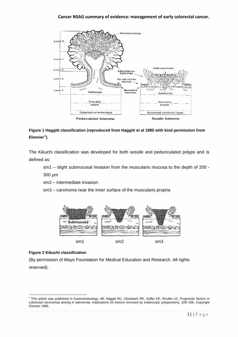
The Haggitt system defines the level of invasion according to the following criteria (see figure
1):
Level 1 - carcinoma invading through the muscularis mucosae into the submucosa
but limited to the head of the polyp.
Level 2 - carcinoma invading to the level of the neck of the adenoma.
Level 3 – carcinoma invading any part of the stalk.
Level 4 – carcinoma invading into the submucosa but above the muscularis propria.
This system applies only to pedunculated polyps as sessile polyps were considered as level
4 lesions due to involvement of the submucosa. Haggitt concluded that only in level 4 lesions
was there a significant risk of nodal involvement (27%). Although level 3 had no involved
nodes some developed recurrence. This emphasises the need to take all factors into
account. Matsuda et al25 reported a large series of pedunculated polyps (n=384) using
Haggitt lines and came to the same conclusion, putting the risk for level 3 & 4 at 6%.
Cancer NSAG summary of evidence: management of early colorectal cancer.
11 | P a g e
Figure 1 Haggitt classification (reproduced from Haggitt et al 1985 with kind permission from
Elsevierc).
The Kikuchi classification was developed for both sessile and pedunculated polyps and is
defined as:
sm1 – slight submucosal invasion from the muscularis mucosa to the depth of 200 -
300 µm
sm2 – intermediate invasion
sm3 – carcinoma near the inner surface of the muscularis propria
Figure 2 Kikuchi classification
(By permission of Mayo Foundation for Medical Education and Research. All rights
reserved).
c This article was published in Gastroenterology, 89, Haggitt RC, Glotzbach RE, Soffer EE, Wruble LD, Prognostic factors in colorectal carcinomas arising in adenomas: implications for lesions removed by endoscopic polypectomy, 328–336. Copyright Elsevier 1985.
sm1 sm2 sm3
Cancer NSAG summary of evidence: management of early colorectal cancer.
12 | P a g e
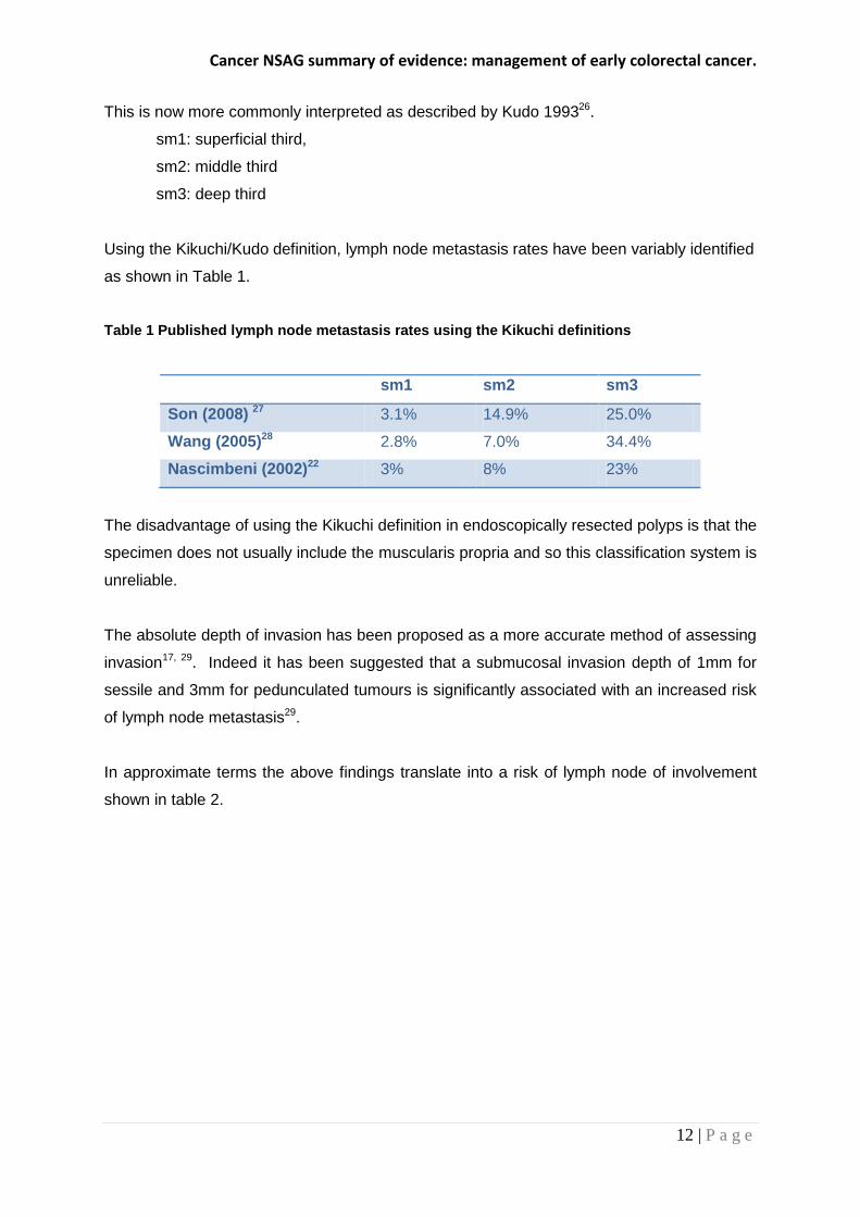
This is now more commonly interpreted as described by Kudo 199326.
sm1: superficial third,
sm2: middle third
sm3: deep third
Using the Kikuchi/Kudo definition, lymph node metastasis rates have been variably identified
as shown in Table 1.
Table 1 Published lymph node metastasis rates using the Kikuchi definitions
sm1 sm2 sm3
Son (2008) 27 3.1% 14.9% 25.0%
Wang (2005)28 2.8% 7.0% 34.4%
Nascimbeni (2002)22 3% 8% 23%
The disadvantage of using the Kikuchi definition in endoscopically resected polyps is that the
specimen does not usually include the muscularis propria and so this classification system is
unreliable.
The absolute depth of invasion has been proposed as a more accurate method of assessing
invasion17, 29. Indeed it has been suggested that a submucosal invasion depth of 1mm for
sessile and 3mm for pedunculated tumours is significantly associated with an increased risk
of lymph node metastasis29.
In approximate terms the above findings translate into a risk of lymph node of involvement
shown in table 2.
Cancer NSAG summary of evidence: management of early colorectal cancer.
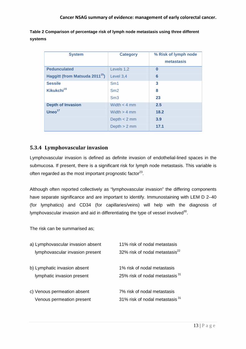
13 | P a g e
Table 2 Comparison of percentage risk of lymph node metastasis using three different
systems
System Category % Risk of lymph node
metastasis
Pedunculated Haggitt (from Matsuda 201125)
Levels 1,2 0
Level 3,4 6
Sessile
Kikukchi15 Sm1 3
Sm2 8
Sm3 23 Depth of Invasion
Uneo17 Width < 4 mm 2.5
Width > 4 mm 18.2
Depth < 2 mm 3.9
Depth > 2 mm 17.1
5.3.4 Lymphovascular invasion
Lymphovascular invasion is defined as definite invasion of endothelial-lined spaces in the
submucosa. If present, there is a significant risk for lymph node metastasis. This variable is
often regarded as the most important prognostic factor23.
Although often reported collectively as “lymphovascular invasion” the differing components
have separate significance and are important to identify. Immunostaining with LEM D 2–40
(for lymphatics) and CD34 (for capillaries/veins) will help with the diagnosis of
lymphovascular invasion and aid in differentiating the type of vessel involved30.
The risk can be summarised as;
a) Lymphovascular invasion absent 11% risk of nodal metastasis
lymphovascular invasion present 32% risk of nodal metastasis22
b) Lymphatic invasion absent 1% risk of nodal metastasis
lymphatic invasion present 25% risk of nodal metastasis 31
c) Venous permeation absent 7% risk of nodal metastasis
Venous permeation present 31% risk of nodal metastasis 31
Cancer NSAG summary of evidence: management of early colorectal cancer.
14 | P a g e
5.3.5 Tumour budding
Small islands or single cell infiltration of cancer cells at the front of tumour invasion constitute
tumour budding. This has been identified as an unfavourable risk factor (15.5% risk of lymph
node metastasis)23, 31, but there are problems with reproducibility and consistency of
definition17, 32. The most commonly accepted definition is provided by Morodomi32 whereby
budding is defined as either isolated undifferentiated cancer cells or clusters of five or six
cancer cells forming a microtubular structure, which appeared to bud from large cancerous
glands.
Adoption of this definition would allow evaluation of the value of budding as a prognostic
indicator.
5.3.6 Location
Screen detected early colorectal cancer tends to be more distal and a significant number are
low in the rectum14. This has implications for further treatment, particularly surgery. There is
debate as to whether location of tumour has any impact on the risk of lymph node
metastasis, however data is inconclusive33, 22.
5.3.7 Size
Ever since Morson’s original paper34 there has been debate as to importance of lesion size. Bach et al (2009)35 in a prospective study of TEMS resections concluded that lesions larger
than 3cm are at high risk of loco regional recurrence. An objective measure of depth and
width of the adenocarcinoma has been recommended by Ueno (2004)17.
5.3.8 Molecular markers
In the future molecular markers may improve selection criteria for subsequent management.
Where appropriate, informed consent from the patient should be sought and samples
submitted for research.
Cancer NSAG summary of evidence: management of early colorectal cancer.
15 | P a g e
6 Assessment of the risk of recurrence in individuals The purpose of pathological assessment is to determine which patients are at “high risk” of
locoregional recurrence and thus require further treatment, mainly surgery, based on the risk
factors considered above set against the risk of surgery. Such lesions would include those
with5:
a. cancer within 1mm of the resection margin
b. poor differentiation
c. lymphovascular invasion
d. Sm3 and Haggitt 4
Conversely other lesions (low risk), have a risk of recurrence that is probably below the risk
of intervention e.g. SM1, Haggitt 1, 2, or 3.
There are factors to be taken into account determining an individual’s risk other than Kikuchi
and Haggitt levels. These are outlined in appendix 2 and 3.
As most series have low power, larger data bases are required. Attempts have been made
to use computer models for accurate prediction and clarification33.
Attention is drawn to the papers by Mainprize and Tytherleigh that discuss this in relation to
the rectum7, 36.
7 Risk adjusted mortality: balancing the risk of recurrence against operative mortality
The risk of recurrence following local excision needs to be weighed against potential
operative morbidity and mortality.
A recent audit of Welsh patients treated by surgical resection with T1N0 disease, between
April 2009 and March 2011, found a 30 day mortality rate of 3%2. This data highlights that
oncological resection is not without considerable risk. This mortality data is for “all comers”
and it must be remembered that in any individual the risk of post-operative death is not
uniform37.
Cancer NSAG summary of evidence: management of early colorectal cancer.
16 | P a g e
Individual mortality risk is dependent on several variables relating to the patient and their co-
morbidities. There are two readily available risk adjustment models3 that attempt to predict
an individual patient’s mortality risk, having adjusted for other risk factors. These are the
ACPGBI Mortality Model3, 37, which is specific to colorectal cancer patients, and the CR-
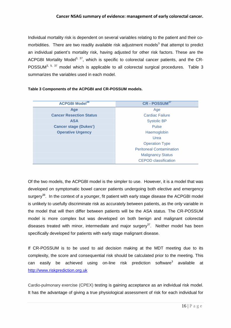
POSSUM3, 5, 37 model which is applicable to all colorectal surgical procedures. Table 3
summarizes the variables used in each model.
Table 3 Components of the ACPGBI and CR-POSSUM models.
ACPGBI Model38 CR - POSSUM37 Age Age
Cancer Resection Status Cardiac Failure ASA Systolic BP
Cancer stage (Dukes’) Pulse Operative Urgency Haemoglobin
Urea Operation Type Peritoneal Contamination Malignancy Status CEPOD classification
Of the two models, the ACPGBI model is the simpler to use. However, it is a model that was
developed on symptomatic bowel cancer patients undergoing both elective and emergency
surgery38. In the context of a younger, fit patient with early stage disease the ACPGBI model
is unlikely to usefully discriminate risk as accurately between patients, as the only variable in
the model that will then differ between patients will be the ASA status. The CR-POSSUM
model is more complex but was developed on both benign and malignant colorectal
diseases treated with minor, intermediate and major surgery37. Neither model has been
specifically developed for patients with early stage malignant disease.
If CR-POSSUM is to be used to aid decision making at the MDT meeting due to its
complexity, the score and consequential risk should be calculated prior to the meeting. This
can easily be achieved using on-line risk prediction software3 available at
http://www.riskprediction.org.uk
Cardio-pulmonary exercise (CPEX) testing is gaining acceptance as an individual risk model.
It has the advantage of giving a true physiological assessment of risk for each individual for
Cancer NSAG summary of evidence: management of early colorectal cancer.
17 | P a g e
major surgery. However, for various reasons, certain patients may not be able to undertake
or complete the test so it cannot be used in every patient.
8 Pre-treatment staging
8.1 Endoscopy
Polyp assessment by the endoscopist can identify lesions at high risk of containing early
cancer and sub mucosal invasion before any endoscopic treatment is undertaken.
Accurately identifying such lesions will allow the initial endoscopist to determine if an
immediate attempt at polypectomy should be performed or if it is best to take simple biopsies
followed by further staging investigations and a complex polypectomy at a later date with
adequate informed consent from the patient. If local expertise is not available, referral should
be made to a specialist in complex endoscopic polypectomy techniques.
Assessment of the polyp includes:
a. using the Paris classification18, of pit pattern recognition and granularity etc;
b. combinations of techniques such as high definition endoscopy, narrow band imaging
and dye spraying;
c. inspection of macroscopic appearances such as a flat or hard lesion with depression,
ulceration, (Paris 0-11a+c), non-granular, advanced pit pattern or failure to lift on
sub mucosal injection are suspicious features of invasive disease;
d. polyp size is more controversial. If sub mucosal invasion is present and there is a low
risk of recurrence (not sm3) then en-bloc resection (by EMR or TEMS for rectal
lesions) may be curative and gives the pathologist the best chance to assess the
resection margins and other risk factors more accurately than simple
polypectomy39.
For rectal lesions there are important differences in treatment options available and in the
consequences of radical treatment. Because of this, rectal lesions should be accurately
staged radiologically prior to any formal attempt at polypectomy as doing so may affect the
findings of these important staging investigations and lead to a less than optimal treatment
strategy for the patient.
Cancer NSAG summary of evidence: management of early colorectal cancer.
18 | P a g e
8.2 Imaging
In the context of early colorectal cancer the important considerations are: 3, 40, 41, 42,
a. exclusion of distant metastasis;
b. assessment of N stage;
c. determination of T stage (particularly in the rectum if N and M staging are
negative).
CT scanning of the chest, abdomen and pelvis should be performed to determine if there is
metastatic disease. These findings may well change the treatment strategy from that of a
radical approach to one aimed at achieving acceptable local symptom control whilst allowing
the patient to commence treatment for systemic disease more quickly.
For rectal lesions, digital examination cannot be relied on for staging3. For early rectal
lesions an MRI and/or endoscopic ultrasound (EUS) should be performed. This will assist in
clarifying T stage and local nodal status. It is not uncommon to find a discrepancy between
imaging, endoscopic and ultimate path staging. If the lesion is invasive, MRI allows
assessment of the likelihood of a R0 resection with clear circumferential margins. EUS is the
most accurate modality for the determination of rectal T stage and it should be performed if
local excision is contemplated3. Therefore in endoscopically suspicious early rectal lesions,
EUS should be performed prior to any formal polypectomy as the latter may distort the subtle
anatomy of the layers in the rectal wall making interpretation of EUS inaccurate. If an
attempt at complete removal by polypectomy has been previously performed, identification
of the polyp site may not be possible rendering EUS assessment unreliable.
Over staging a benign rectal polyp or an early rectal cancer as a more advanced lesion may
preclude acceptable local excision and lead to unnecessary radical rectal resection or
chemoradiotherapy with its significant morbidity. It may also increase the potential risk of
permanent stoma formation and deleterious effects on quality of life. If EUS is unavailable,
MRI/ endocoil can also be used.
The following should be noted:
a. there is no reliable imaging modality to determine N stage. Accuracy of the
available imaging modalities range from 56-75%3;
b. there are particular problems with imaging low in the rectum in assessing
local spread3;
Cancer NSAG summary of evidence: management of early colorectal cancer.
19 | P a g e
c. a PET scan may be positive in the presence of a benign polyp43;
d. in the future endoscopic ultrasound of the colon may be employed to
determine the local T stage for colonic cancer.
9 Treatment options after local excision, including endoscopic resection
Treatment options include:
a. intensive clinical follow-up;
b. further local excision;
c. further local excision and/or radiotherapy/chemo-radiotherapy (awaiting
further evaluation in trials such as TREC);
d. oncological resection.
T1 lesions can be stratified as low or high risk to inform this choice. Completely removed
low risk lesions do not require further treatment other than follow up. The timing should be
established locally but may include such measures as early endoscopic inspection of the
polypectomy site (e.g. at 3 months).
There is a very limited place for re-excision of incompletely excised lesions, even for low risk
lesions as there is an increased risk of local recurrence particularly low in the rectum20, 49.
9.1 Surgery
Surgeons managing early colorectal cancer should be fully cognisant of the relevant
literature22. They should be able to offer laparoscopic surgery to suitable patients, have
sufficient volume of work in this area, be able to present audited outcomes and follow
national guidelines for the management of colorectal cancer44. They should have developed
networks for accessing treatment of suitable cases by ESD and TEMS.
Patients should be offered laparoscopic or laparoscopically assisted resection if suitable with
early colorectal cancer where formal resection is advised or chosen by the patient 45, 46.
Cancer NSAG summary of evidence: management of early colorectal cancer.
20 | P a g e
Centres offering laparoscopic resection should have sufficient volume of work in this area,
be able to present audited outcomes and follow national guidelines for the management of
colorectal cancer.
The need for oncological resection of rectal lesions may be compromised by initial local
excision particularly for low rectal tumours 23, 27, 47, 48. This may necessitate non restorative
surgery with stoma formation.
9.1.1 Local excision
The selection criteria for cases that are suitable for local resection are not completely
defined. However, local excision has the advantages of low operative mortality and morbidity
together with preservation of organ function and a low stoma rate.
With local excision, the main issue is the risk of developing local recurrence. In many series
this has been reported to be higher than for radical surgery39. Recurrence rates after local
excision by any method can be significant but low rates can be achieved by following
guidelines such as those advocated by Bach et al 200935 or Tytherleigh et al 200849.
Salvage rates for local recurrence can be poor and worse results have been reported in
distal rectal tumours.
9.1.2 Transanal Endoscopic Microsurgery (TEMS)
TEMS is a minimally invasive form of local excision for rectal lesions performed through a
resectoscope50, 51, 52, 53, 54. Complex polypoid lesions may be better resected by TEMS than
endoscopically (EMR) as full (or partial) thickness resection of muscle can be undertaken by
TEMS allowing accurate determination of T stage and sm level. This makes TEMS a
treatment option for T1-Sm1/ Sm2. There may also be a place for re-excision of low risk pT1
lesions by TEMS following endoscopic polypectomy if clear margins cannot be guaranteed.
TEMS may be a first line treatment for early rectal cancers in preference to endoscopic
treatment if EUS suggests a depth of invasion of Sm2 or greater, there is severe dysplasia
or if endoscopic appearances are not in favour of allowing a complete endoscopic resection
(e.g. non-lifting during sub mucosal injection).
Cancer NSAG summary of evidence: management of early colorectal cancer.
21 | P a g e
Appropriate patients should be entered into trials of early colorectal cancer management
such as TREC. The long term results of TEMS should be judged against those of
oncological resection.
9.2 The place of pre-operative adjuvant radiotherapy for rectal
cancers
Preoperative adjuvant radiotherapy is addressed in recent NICE guidance3. However there
is an emerging view that radiotherapy and or chemotherapy can be combined with local
excision as an alternative to major surgery in early rectal cancer. Local excision of high risk
(T1) lesions together with External Beam Radiotherapy (EBRT) should only be undertaken in
the context of a formal clinical trial (e.g. TREC) or in a patient unfit for major radical resection
or when it is the patient’s informed and objective preference.
The Papillon technique (contact radiotherapy) has been used to treat small early tumours
particularly in the medically unfit55.
9.3 Oncology
At present, the place of chemo-radiotherapy in the treatment of early rectal cancer is
controversial, particularly as a primary treatment in patients otherwise suitable for surgical
treatment. Recent NICE guidance suggests it should not be used routinely in early disease
but advocates of its use exist56. A detailed outline of emerging trends of the use of
radiotherapy for rectal preservation is outside the scope of this paper. MDTs should however
be aware of:
a. the use of radiotherapy and local excision in combination;
b. EBRT with palliative intent in the unfit patient;
c. relevant trials that are in progress e.g. TREC (TEMS and Radiotherapy in
Early Rectal Cancer).
Down staging lesions by chemo radiotherapy to ypT1 is also outside the scope of this paper.
Cancer NSAG summary of evidence: management of early colorectal cancer.
22 | P a g e
10 The role of the MDT
Wales is formally signed up to follow NICE guidance and it is against this backdrop that the
following guidance is included in this document:
a. NICE Improving Outcomes Guidance for Colorectal Cancer6 recommended the
establishment of specialist early cancer MDTs on a Network basis. This advice has
been reinforced by recent NICE guidance (2011)3 for Stage 1 disease.
b. Specialist MDTs are being increasingly formed in parts of the UK, which in England
are subject to evaluation via the peer review programme.
MDTs should at least have access to specialist opinion as suggested by NICE1 and patient
information provided. Consideration should be given to the establishment of specialist early
colorectal cancer MDTs in Wales. The educational training of MDTs in the management of
early cancer will be supported by the Cancer NSAG. Literature is available to help
organisations develop these specialist MDTs57.
11 Follow-up
Follow up programmes, after local excision or resection, differ only in emphasis. The aim is
to detect recurrence early, at a stage where further curative treatment may be possible.
After oncological resection or local excision, colonoscopic surveillance should be as for high-
risk adenomas as described in BSG and European Guidelines. NICE guidance states that
stage 1 colorectal cancer should be followed up with surveillance CT3.
Detection of local recurrence in rectal lesions requires special consideration. This will require
endoscopy, EUS, digital exam, MRI or combinations of these and the frequency with which
these need to be performed is debatable.
12 Audit
The 2009-11 audit of T1N0 cancers in Wales showed an apparently wide inter-unit variability
in the management of stage 1 cancer2. Whilst the number of cases treated by each MDT
was small, surgical resection rates varied between 25 and 100% following local excision2.
Cancer NSAG summary of evidence: management of early colorectal cancer.
23 | P a g e
The reasons for this are not clear and need to be defined through audit on an all Wales
basis. This can be achieved by collaboration between NBOCAP and BSW’s Quality
Assurance Programme.
Central to formulating management guidelines for T1 lesions is the availability of reliable
pathological data. BSW relies on Canisc but completion of the pathological fields in this is
suboptimal. Although Canisc has been revised to include additional fields particularly
relevant to screening (including extended laparoscopic, pathology, follow-up and
complication fields) and CHIRP is being implemented, it is important that complete
pathological data is prospectively collected for comparative audit. Reliable data will inform
future guidelines.
Audit should also confirm the safety and appropriateness of different treatments, including
surgery, against both local and national guidelines (see also implications for BSW).
13 Research and development
Given the lack of high level evidence on which to base management decisions, in the long
term the gathering of further observational data is of paramount importance. Potential ways
forward are:
a. collaboration both locally and nationally in pathology studies to help establish quality
assured pathology reporting of risk factors;
b. entry in to trials e.g. TREC; a phase 2 trial;
c. establishment of a database allowing prospective data collection and international
collaboration.
Cancer NSAG summary of evidence: management of early colorectal cancer.
24 | P a g e
14 References 1 Colorectal cancer: the diagnosis and management of colorectal cancer. NICE (Oct 2011) available
from http://guidance.nice.org.uk/CG131/Guidance/pdf/English 2 Khalid U, Evans M.D., Williams G.L., Hanson J., Davies M. The variability in management of T1
colorectal cancer in Wales: time for a consensus? Annals Royal College of Surgeons (in press)
3 Colorectal cancer: the diagnosis and management of colorectal cancer. NICE (Oct 2011) available
from http://guidance.nice.org.uk/CG131/Guidance/pdf/English 4 Williams JG, Pullan RD, Hill J, Horgan PG, Salmo E, Buchanan GN, Rasheed S, McGee SG, Haboubi N.
Colorectal Dis. Management of the malignant colorectal polyp: ACPGBI position statement. 2013
Aug;15 Suppl 2:1-38. 5 von Karsa L, Segnan N, Patnick J (Eds.), (2010) European guidelines for quality assurance in
colorectal cancer screening and diagnosis Luxembourg: Publications Office of the European Union
6 Improving outcomes in colorectal cancers: manual update. Cancer service guidance (2004).
Available from http://guidance.nice.org.uk/CSGCC.
7 Yamauchi H, Togashi K, Kawamura YJ, Horie H, Sasaki J, Tsujinaka S, Yasuda Y, Konishi F.
Pathological predictors for lymph node metastasis in T1 colorectal cancer Surg Today. 2008;38:905-
10. 8 Williams G T et al. Standards & datasets for reporting Cancers .Royal College of Pathology 2nd ed.
2007. 9 Reporting lesions in the NHS, Bowel Cancer Screening Programme, 2007 available from
www.cancerscreening.nhs.uk 10 Japanese classification of Colorectal cancer 1st ed. Tokyo 1997.
11 Schlemper RJ, Kato Y, Stolte M. Review of histological classifications of gastrointestinal epithelial
neoplasia: differences in diagnosis of early carcinomas between Japanese and Western pathologists.
J Gastroenterol. 2001 Jul;36(7):445-56.
12 Nivatvongs S, Rojanasakul A, Reiman HM, et al. The risk of lymph node metastasis in colorectal
polyps with invasive adenocarcinoma. Dis Colon Rectum 1991; 34:323–328. 13 Okabe S, Shia J, Nash G, Wong WD, Guillem JG, Weiser MR, Temple L, Sugihara K, Paty PB. Lymph
node metastasis in T1 adenocarcinoma of the colon and rectum. J Gastro Surg 2004 1032-9. 14 Volk EE, Goldblum JR, Petras RE, Carey WD, Fazio VW. Management and outcome of patients with
invasive carcinoma arising in colorectal polyps. Gastroenterology 1995; 109: 1801–1807.
Cancer NSAG summary of evidence: management of early colorectal cancer.
25 | P a g e
15 Kikuchi R, Takano M, Takagi K, Fujimoto N, Nozaki R, Fujiyoshi T et al. Management of early
invasive colorectal cancer. Risk of recurrence and clinical guidelines. Dis Colon Rectum 1995; 38:
1286–1295. 16 Haggitt RC, Glotzbach RE, Soffer EE, Wruble LD. Prognostic factors in colorectal carcinomas arising
in adenomas: implications for lesions removed by endoscopic polypectomy. Gastroenterology 1985;
89: 328–336. 17 Ueno H, Mochizuki H, Hashigushi Y, Shimazaki H, Aida S, Hase K, Matsukuma S, Kanai T, Kurihara H,
Ozawaka K, Yoshimura K, Bekku S. Risk factors for an adverse outcome in early invasive colorectal
carcinoma. Gastroenterology 2004; 127: 385-394. 18 Participants in the Paris Workshop. The Paris endoscopic classification of superficial neoplastic
lesions: esophagus, stomach, and colon: November 30 to December 1, 2002. Gastrointest Endosc
2003; 58: 3-43. 19 Quality assurance in pathology in colorectal cancer screening ed. Quirke P; European Guidelines
2005 www.irac.fr. 20 Cooper HS, Deppisch LM, Gourley WK, et al. Endoscopically removed malignant colorectal polyps:
clinicopathologic correlations. Gastroenterology 1995;108:1657-1665 21 Hase K, Shatney CH, Mochizuki H, Johnson DL, Tamakuma S, Vierra M et al. Long-term results of
curative resection of ‘minimally invasive’ colorectal cancer. Dis Colon Rectum 1995; 38: 19–26. 22 Nascimbeni R, Burgart LJ, Nivatvongs S, et al. Risk of lymph node metastasis in T1 carcinoma of the
colon and rectum. Dis Colon Rectum 2002;45:200–6. 23 Kobayashi H, Mochizuki H, Kato T, Mori T, Kameoka S, Shirouzu K, Saito Y, Watanabe M, Morita T,
Hida J, Ueno M, Ono M, Yasuno M, Sugihara K. Is Total Mesorectal Excision always necessary for T1-
T2 lower rectal cancer? Ann. Surg. Oncol. 2010;17: 973-80. 24 Kudo S, Early Colorectal cancer, Igaku-Shon. Tokyo 1996. 25 Matsuda T, Fukazawa M et al; Risk of lymph node metastasis in patients with pedunculated type
early invasive colorectal cancer. Cancer Sci 2011 102 1693-1697. 26 KudoS. Endoscopic Mucosal Resection of Flat and Depressed Types of Early Colorectal Cancer.
Endoscopy 1993; 25(7): 455-461. 27 Son HJ. Song SY, Lee WY, Yang SS, Park SH, Yang MH, Yoon SH, Chun HK. Characteristics of early
colorectal carcinomas with lymph node metastatic disease. Hepatogastroenterology. 2008; 55: 1293-
1297. 28 Wang H-S, Liang W-Y, Lin T-C, et al. Curative resection of T1 colorectal carcinoma: risk of lymph
node metastasis and long-term prognosis. Dis Colon Rectum. 2005;48:1182–1192.
Cancer NSAG summary of evidence: management of early colorectal cancer.
26 | P a g e
29 Kitajima K, Fujimori T, Fujii S. et al Correlations between lymph node metastasis and depth of
submucosal invasion in submucosal invasive colorectal carcinoma: a Japanese collaborative study. J
Gastroenterol 2004. 39534–543. 30 Bayar S, Saxena R, Emir B, Salem RR. Venous invasion may predict lymph node metastasis in early
rectal cancer. Eur J Surg Oncol. 2002 Jun;28:413-7. 31 Egashira Y, Yoshida T, Hirata I, Hamamoto N, Akutagawa H, Takeshita A, Noda N, Kurisu Y,
Shibayama Y. Analysis of pathological risk factors for lymph node metastasis of submucosal invasive
colon cancer. Mod Pathol. 2004;17:503-11. 32 Morodomi T, Isomoto H, Shirouzu K, Kakegawa K, Irie K, Morimatsu M. An index for estimating the
probability of lymph node metastasis in rectal cancers. Lymph node metastasis and the
histopathology of actively invasive regions of cancer. Cancer 1989;63:539-43. 33 Rasheed S, Bowley DM, Aziz O, Tekkis PP, Sadat AE, Guenther T, Boello ML, McDonald PJ, Talbot
IC, Northover JMA. Can depth of tumour invasion predict lymph node positivity in patients
undergoing resection for early rectal cancer? A comparative study between T1 and T2 cancers.
Colorectal Disease 2007;10:231-237. 34 Morson BC, Whiteway JE, Jones EA, Macrae FA, Williams CB. Histopathology and prognosis of
malignant colorectal polyps treated by endoscopic polypectomy. Gut 1984; 25: 437–444. 35 Bach SP, Hill J, Monson JR, Simson JN, Lane L, Merrie A, Warren B, Mortensen NJ; Association of
Coloproctology of Great Britain and Ireland Transanal Endoscopic Microsurgery (TEM) Collaboration.
A predictive model for local recurrence after transanal endoscopic microsurgery for rectal cancer. Br
J Surg. 2009;96:280-90. 36 Breen E, Brady R. Preservation of the anus in the therapy of distal rectal cancer. Surg Clin NA,
1997; 71: 77-83.
37 Tekkis PP, Prytherch DR, Kocher HM, Senapati A, Poloniecki JD, Stamatakis JD, Windsor AC.
Development of a dedicated risk-adjustment scoring system for colorectal surgery (colorectal
POSSUM). Br J Surg. 2004;91:1174-82.
38 Tekkis PP, Poloniecki JD, Thompson MR, Stamatakis JD. Operative mortality in colorectal cancer:
prospective national study BMJ. 2003;327:1196-201. 39 Moss A, Bourke MJ, Williams SJ, Hourigan LF, Brown G, Tam W, Singh R, Zanati S, Chen RY, Byth K.
Endoscopic mucosal resection outcomes and prediction of submucosal cancer from advanced
colonic mucosal neoplasia. Gastroenterology. 2011;140:1909-18.
40 Pre-treatment Staging of Colorectal Cancer. American College of Radiologists 2004.
41 Colorectal Cancer Screening. American College of Radiologists (2005).
Cancer NSAG summary of evidence: management of early colorectal cancer.
27 | P a g e
42 Royal College of Radiologists. Recommendations for Cross-Sectional Imaging in Cancer
Management. London: RCR, 2006.
http://www.rcr.ac.uk/docs/oncology/pdf/Cross_Sectional_Imaging_12.pdf 43 Gollub MJ, Akhurst T, Markowitz AJ, Weiser MR, Guillem JG, Smith LM, Larson SM, Margulis AR.
Combined CT Colonography and F-FDS PET of colon Polyps; Am J Roentgenol. 2007 Jan;188(1):130-8. 44 Laparoscopic surgery for the treatment of colorectal cancer (review). NICE technology appraisal
guidance 105 (2006). Available from www.nice.org.uk/TA105. 45 Bentrem DJ, Okabe S, Wong WD, et al. T1 adenocarcinoma of the rectum: transanal excision or
radical surgery? Ann Surg. 2005 Oct; 242(4):472-7. 46 Hotta T and Yamaue H. Laparoscopic surgery for rectal cancer: review of published literature 2000-
2009. Surg Today 2011; 41(12):1583-91. 47 Hahnloser D, Wolff B, Larson D, Ping J, Nivatvongs S. Immediate radical resection after local
excision of rectal cancer: an oncologic compromise? Diseases of the Colon and Rectum 48(3): 429-
37, 2005. 48 Yamamoto S, Watanabe M, Hasegawa H, et al. The risk of lymph node metastasis in T1 colorectal
carcinoma. Hepatogastroenterology. 2004;51:998–1000. 49 Tytherleigh MG, Warren B, Mortensen NJ. Management of early rectal cancer. Br J Surg 2008
95:409-23. 50 Middleton PF, Sutherland LM, Maddern GJ. Transanal endoscopic microsurgery; a systemic review.
Dis Colon Rectum. 2005;48:270-84. 51 Floyd ND, Saclarides TJ. Transanal endoscopic microsurgical resection of pT1 rectal tumors. Dis
Colon Rectum. 2006;49:164-8. 52 Winde G, Nottberg H, Keller R, Schmid KW, Bünte H., Surgical cure for early rectal cancer (T1)
Transanal endoscopic microsurgery vs anterior resection. Dis Colon Rectum. 1996;39:969-76. 53 Doornebosch PG, Tollenaar RA, De Graaf EJ, Is the increasing role of Transanal Endoscopic
microsurgery in curation for T1 rectal cancer justified? A systematic review. Acta Oncol.
2009;48:343-53. 54 Mitchell PJ, Haboubi NY. The malignant adenoma: when to operate and when to watch. Surg
Endosc. 2008;22:1563–1569. 55 Gérard JP, Ayzac L, Coquard R et al., Endocavitary irradiation for early rectal carcinomas T1 (T2). A
series of 101 patients treated by the Papillon technique. Int J Radiat Onc Biol Phys. 1996 ; 34 : 775-
83.
Cancer NSAG summary of evidence: management of early colorectal cancer.
28 | P a g e
56 Habr-Gama A, Perez RO, Nadalin W, Sabbaga J, Ribeiro U, Jr, Silva e Sousa AH, Jr, et al. Operative
versus nonoperative treatment for stage 0 distal rectal cancer following chemoradiation therapy:
long-term results. Ann Surg. 2004;240:711–17.
57 Case study: Implementing early rectal cancer MDTs in secondary care. Nice, London (November
2011) available at http://www.nice.org.uk/nicemedia/live/13597/56961/56961.pdf 58 Beaton C, Twine CP, Williams GL, Radcliffe AG. Systematic review and meta-analysis of
histopathological factors influencing the risk of lymph node metastasis in early colorectal cancer.
Colorectal Dis. 2013 Jul;15(7):788-97.
15 Acknowledgements This document was prepared on behalf of the colorectal group of the Cancer NSAG by the following draftees:
Andrew Radcliffe, Convener. Colorectal cancer NSAG Consultant Surgeon
Martyn Evans, WBCA and Colorectal group Cancer NSAG Consultant Surgeon
Meleri Morgan, Consultant Histopathologist
Gethin Williams, Colorectal group Cancer NSAG and WBCA Consultant Surgeon
Namor Williams, QA Advisor Pathology BSW Consultant Pathologist
Andrew Maw, Consultant Surgeon Colorectal group Cancer NSAG BSW screening
colonoscopist
Ceri Beaton, Consultant Surgeon
Hayley Heard, Head of Programme BSW
Mark Davies, Consultant Surgeon, Chair, Colorectal group Cancer NSAG
Richard Adams, Consultant Oncologist
Gareth Tudor, QA Advisor Radiology BSW Consultant Radiologist Dean Harris, Consultant Surgeon
Thanks must also go to MDTs across Wales who reviewed the document as part of a
consultation prior to publication.
Queries regarding this document should in the first instance be directed to Louise
Carrington, Cancer NSAG Core Team at [email protected]
Cancer NSAG summary of evidence: management of early colorectal cancer.
29 | P a g e
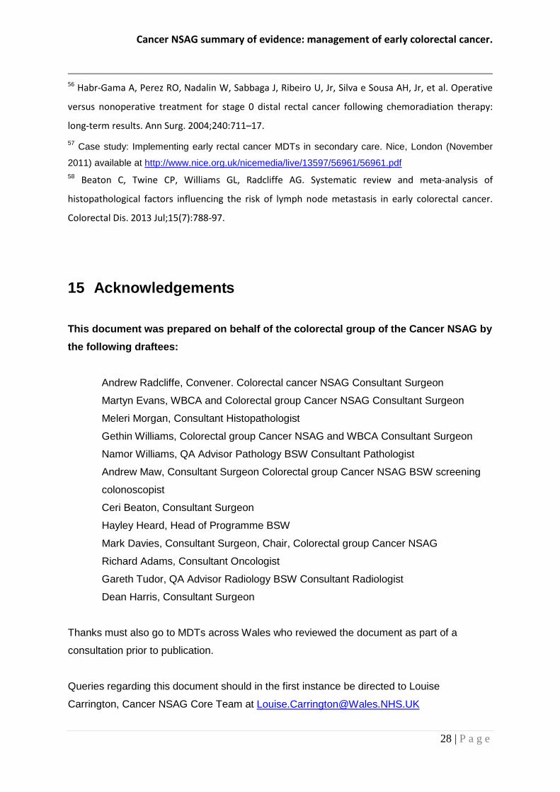
Appendix 1: CHIRP reporting choices
Polypoid cancer Polyp size (in mm) Tumour type:
Adenocarcinoma
Other (specify)
Differentiation:
Well
Moderate
Poor
Maximum thickness from muscularis mucosae (in mm) Haggitt level (for polypoid tumours): 1
2
3
4
Into muscularis propria
Not applicable
Kikuchi level (for sessile tumours): sm1
sm2
sm3
Into muscularis propria
Not applicable
Lymphatic or vascular invasion:
None
Possible
Definite
Background adenoma:
Yes
No
Margins:
Not involved
Involved by adenoma only
Peripheral margin involved by carcinoma
Distance from carcinoma to deep margin (in mm) Deep margin involved by carcinoma
Deep and peripheral margin involved by carcinoma
Not assessable
Invasive cancer (biopsy): Tumour type:
Adenocarcinoma
Other
Mucinous adenocarcinoma
Signet ring cell carcinoma
Adenosquamous carcinoma
Squamous cell carcinoma
Small cell carcinoma
Other pathology IBD - Ulcerative colitis
IBD - Crohns
IBD - Unclassified
Other inflammation (description)
Normal
Polyp Size (in mm) Type:
Tubular adenoma
Tubulovillous adenoma
Villous adenoma
Hyperplastic polyp
Serrated adenoma
Mixed hyperplastic polyp/adenoma
Inflammatory
Juvenile
Peutz-Jeghers
Other
Endocrine cell tumour (carcinoid)
Melanoma
Leiomyoma
Schwannoma
Neurofibroma
Ganglioneuroma
GIST
Lipoma
Other malignant neoplasm (specify)
Inadequate
Specimen site: Rectum
Rectosigmoid
Sigmoid colon
Descending colon
Splenic flexure
Transverse colon
Hepatic flexure
Ascending colon
Caecum
Suspicious of invasive malignancy
Cancer NSAG summary of evidence: management of early colorectal cancer.
30 | P a g e
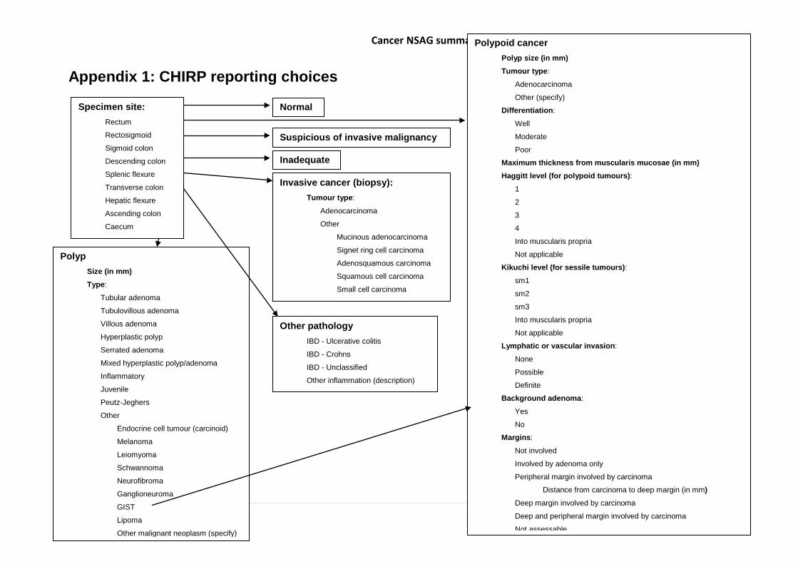
Appendix 2: Meta analysis of histopathological factors
The meta-analysis recently reported by Beaton et al analysed a cohort of 23 studies and
4510 patients58. There was a significantly higher risk of lymph node metastasis with a sub
mucosal invasion depth of greater than 1mm compared to lesser degrees of penetration (OR
0·3.87, 95% CI 1.50-10.00, p=0.005). Lymphovascular invasion was found to be significantly
associated with the risk of lymph node metastasis (OR 4·81, 3·14-7·37, p<0·00001). Poorly
differentiated tumours when compared to well or moderately differentiated tumours had a
higher risk of lymph node metastasis (OR 5.60, 2.90-10.82, p<0·00001). Tumour budding
was found to be significantly associated with the risk of lymph node metastasis (OR 7·74,
4·47-13·39, p<0·001).
The analysis concluded that in early colorectal cancer a sub mucosal invasion depth of
>1mm, lymphovascular invasion, poor differentiation and tumour budding are
histopathological factors that are significantly associated with a risk of lymph node
metastasis. Patients who have early colorectal cancers with any one of these
histopathological characteristics should be counselled regarding the risks and considered for
oncological resection.
Cancer NSAG summary of evidence: management of early colorectal cancer.
31 | P a g e
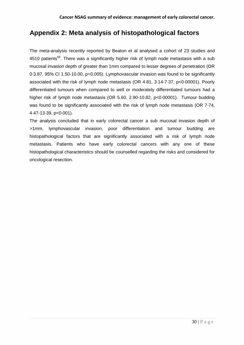
Appendix 3: Methods of risk assessment for individual patients
Each MDT needs to have a method of estimating these risks either developed locally or
based on a method in the literature. The three methods outlined below are illustrative.
i) Ueno et al (2004)17
If there is poor differentiation, lymph vascular permeation, tumour budding or extensive sub
mucosal invasion (Kikuchi sm3, Haggitt level 4 or width >4mm, depth >2mm), a formal
colectomy or rectal resection is advised.
ii) Egashiura et al (2004)31
Egashiura et al have devised a flow chart which also includes the histopathological presence
or absence and depth of a lymphatic invasion related to the adenocarcinoma.
Adapted from Eshigara et al 2004 with kind permission from Macmillan Publishers Ltd
(diagram first published in Modern Pathology: Egashira Y, Yoshida T, Hirata I, Hamamoto N,
Akutagawa H, Takeshita A, Noda N, Kurisu Y, Shibayama Y. Analysis of pathological risk factors for
lymph node metastasis of submucosal invasive colon cancer May;17(5):503-11, copyright 2004).
Cancer NSAG summary of evidence: management of early colorectal cancer.
32 | P a g e
iii) Japanese Society for Cancer of the Colon and Rectum 2010 guidelines for the treatment of
colorectal cancer
The Japanese guidelines provide a literature review and consensus expert opinion. They
recommend that the criteria for identifying curable T1 colorectal carcinoma after endoscopic
resection are well/moderately differentiated or papillary histological grade, no vascular
invasion, sub mucosal invasion depth less than 1mm and budding grade 1 (low grade).
In summary, the pathology report should inform MDT discussion on further treatment.
Cancer NSAG summary of evidence: management of early colorectal cancer.
33 | P a g e
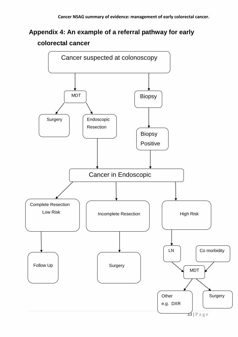
Appendix 4: An example of a referral pathway for early colorectal cancer
Cancer suspected at colonoscopy
Biopsy
Biopsy
Positive
Cancer in Endoscopic
MDT
Surgery Endoscopic
Resection
Complete Resection
Low Risk
Follow Up
Incomplete Resection
Surgery
High Risk
MDT
LN Co morbidity
Surgery Other
e.g. DXR