Embed Size (px)
Citation preview
209
1. Exposure Data
1.1 Identification of the agent
1.1.1 Nomenclature
Chem. Abstr. Serv. Reg. No.: 599-79-1 (O’Neil, 2001; SciFinder, 2013)Chem. Abstr. Serv. Name: Benzoic acid, 2-hy - droxy-5-[2-[4-[(2-pyridinylamino)sulf onyl]-phenyl]diazenyl] (O’Neil, 2001; SciFinder, 2013)IUPAC systematic name: 2-Hydroxy-5-[2-[4-[pyridine-2-ylsu lfamoyl)phenyl]diazenyl]benzoic acid (Lide, 2005; European Pharmacopoeia, 2008)United States nonproprietary name (USAN): SulfasalazineSynonyms: Benzoic acid, 2-hydroxy-5- [[4-[(2-pyridinylamino)sulfonyl]phenyl]azo]-; 5-[[p-(2-Pyridylsulamoyl)phenyl]azo]salicylic acid (US Pharmacopeia, 2007, 2013)
See WHO (2007) for names in other languages.
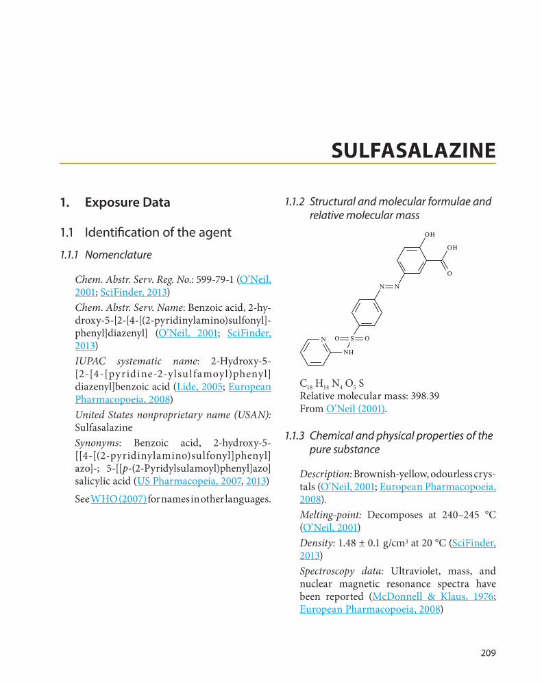
1.1.2 Structural and molecular formulae and relative molecular mass
OH
OH
N N
O
SO O
N
N
H
C18 H14 N4 O5 SRelative molecular mass: 398.39From O’Neil (2001).
1.1.3 Chemical and physical properties of the pure substance
Description: Brownish-yellow, odourless crys-tals (O’Neil, 2001; European Pharmacopoeia, 2008).Melting-point: Decomposes at 240–245 °C (O’Neil, 2001)Density: 1.48 ± 0.1 g/cm3 at 20 °C (SciFinder, 2013)Spectroscopy data: Ultraviolet, mass, and nuclear magnetic resonance spectra have been reported (McDonnell & Klaus, 1976; European Pharmacopoeia, 2008)
SULFASALAZINE
IARC MONOGRAPHS – 108
210
Solubility: Practically insoluble in water, ether, benzene, chloroform; very slightly soluble in ethanol; soluble in alkali hydrox-ides (McDonnell & Klaus, 1976; O’Neil, 2001)Octanol/water partition coefficient: Log P = 3.88 (Rosenbaum, 2011)Stability data: The compound did not degrade when dissolved in dimethylformamide and was subjected to thermal stress at 80 °C for 196 hours (McDonnell & Klaus, 1976; Jacoby, 2000)
1.1.4 Technical products and impurities
(a) Trade names
Azulfidine EN-tabs; Azulfidine; Azaline; Sulfazine; Sulfazine EC; Apo-Sulfasalazine; PMS-Sulfasalazine; Salazopyrin En-Tabs; Sala zo pyrin; Azulfidina; Azulfin; Bomecon; Colo-Pleon; Disalazin; Falazine; Gastropyrin; Lazafin; Pyralin EN; Rosulfant; Salazine; Salazodin; Salazopirina; Salazopyrin Entabs; Salazopyrin-EN; Salazopyrina; Salazopyrine; Salivon; Salopyr; Salopyrine; Saridine-E; Sulcolon; Sulfasalazin; Sulfitis; Ulcol; Zopyrin (Porter & Kaplan, 2013).
(b) Impurities
Impurities as given in European Pharmacopoeia (2008):
• 4,4′-[(4-hydroxy-1,3-phenylene)bis(diaze-ne d iy l)]bi s[N-(py r id i n-2-y l)ben z ene sulfo namide
• 2-hydroxy-3,5-bis[2-[4-(pyridin-2-ylsulfa-moyl)phenyl]diazenyl]benzoic acid
• 2-hydroxy-5-[2-[4-(2-iminopyridin-1(2H)- yl)phenyl]diazenyl]benzoic acid
• 4-[2-(2-hydroxyphenyl)diazenyl]-N-(pyri-din-2-yl)benzenesulfonamide
• 2-hydroxy-4′-(pyridin-2-ylsulfamoyl)-5-[2-[4-(pyridin-2-ylsulfamoyl) phenyl]diazenyl]biphenyl-3-carboxylic acid
• 2-hydroxy-3-[2-[4-(pyridin-2-ylsulfamoyl)phenyl]diazenyl]benzoic acid
• 5-[2-[4′,5-bis(pyridin-2-ylsulfamoyl)biphe-nyl-2-yl]diazenyl]-2-hydroxy benzoic acid
• salicylic acid• 2-hydroxy-5-[2-(4-sulfophenyl)diazenyl]
benzoic acid• 4-amino-N-(pyridin-2-yl)benzenesulfona-
mide (sulfapyridine).
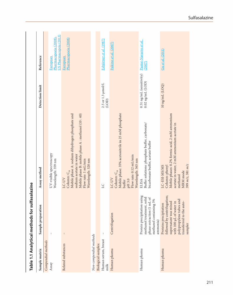
1.1.5 Analysis
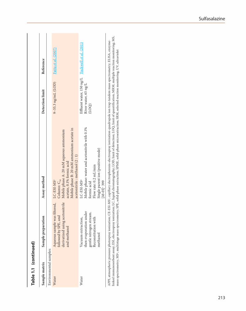
Selected compendial and non-compendial methods of analysis are presented in Table 1.1. Sulfasalazine in human plasma can be deter-mined by high-performance liquid chromatog-raphy using ultraviolet detection (Fukino et al., 2007). It can also be analysed through liquid chromatography-tandem mass spectrometry in human plasma using electron spray ioniza-tion techniques in multiple reaction monitoring mode, with a limit of quantification of 10 ng/mL (Gu et al., 2011).
In urban water, sulfasalazine can be quanti-fied by liquid chromatography-mass spectrom-etry using electron spray ionization. The limit of quantification is 65 ng/L (Tuckwell et al., 2011).
1.2 Production
1.2.1 Production process
Sulfasalazine does not occur in nature. Sulfasalazine is produced by reacting sulfanila-mide with salicyclic acid through a series of steps, with water as a solvent (Novacek et al., 1991).
1.2.2 Use
(a) Indications
Sulfasalazine is an aminosalicylate whose chief bioactive metabolite is 5-aminosalicyclic acid (5-ASA). Sulfasalazine, mesalazine (5-ASA),
Sulfasalazine
211
Tabl
e 1.
1 A
naly
tica
l met
hods
for s
ulfa
sala
zine
Sam
ple
mat
rix
Sam
ple
prep
arat
ion
Ass
ay m
etho
dD
etec
tion
lim
itR
efer
ence
Com
pend
ial m
etho
dsA
ssay
–U
V-vi
sible
spec
tros
copy
W
avel
engt
h: 3
59 n
mEu
rope
an
Phar
mac
opoe
ia (2
008)
, U
S Ph
arm
acop
eia
(201
3)Re
late
d su
bsta
nces
–LC
-UV
C
olum
n: C
18
Mob
ile p
hase
A: s
odiu
m d
ihyd
roge
n ph
osph
ate
and
sodi
um a
ceta
te in
wat
er
Mob
ile p
hase
B: m
obile
pha
se A
: m
etha
nol (
10 :
40)
Flow
rate
: 1 m
L/m
in
Wav
elen
gth:
320
nm
Euro
pean
Ph
arm
acop
oeia
(200
8)
Non
-com
pend
ial m
etho
dsBi
olog
ical
sam
ples
:H
uman
seru
m, b
reas
t m
ilk–
LC2.
5 or
1.3
µm
ol/L
(L
OD
)Es
björ
ner e
t al.
(198
7)
Hum
an p
lasm
aC
entr
ifuga
tion
LC-U
V
Col
umn:
C18
M
obile
pha
se: 3
5% a
ceto
nitr
ile in
25
mM
pho
spha
te
buffe
r pH
3.0
Fl
ow ra
te: 0
.12
mL/
min
W
avel
engt
h: 3
65 n
m
Fuki
no et
al.
(200
7)
Hum
an p
lasm
aPr
otei
n pr
ecip
itatio
n us
ing
met
hano
l tre
atm
ent,
solid
ph
ase
extr
actio
n (1
mL
of
met
hano
l con
tain
ing
5%
amm
onia
)
ELIS
A
Buffe
r sol
utio
ns: p
hosp
hate
buff
er, c
arbo
nate
/bi
carb
onat
e bu
ffer,
acet
ate
buffe
r
0.51
ng/
mL
(sen
sitiv
ity)
0.02
ng/
mL
(LO
D)
Past
or-N
avar
ro et
al.
(200
7)
Hum
an p
lasm
aPr
otei
ns p
reci
pita
tion
follo
wed
by
cent
rifu
gatio
n;
supe
rnat
ant w
as m
ixed
w
ith 1
00 µ
L of
wat
er in
po
lypr
opyl
ene
tube
s and
tr
ansf
erre
d to
the
auto
-sa
mpl
er
LC–E
SI-M
S/M
S C
olum
n: p
heny
l M
obile
pha
ses:
0.2%
form
ic a
cid,
2 m
M a
mm
oniu
m
acet
ate
in w
ater
, 2 m
M a
mm
oniu
m a
ceta
te in
m
etha
nol
MR
M m
ode
399
m/z
, 381
m/z
10 n
g/m
L (L
OQ
)G
u et
al.
(201
1)
IARC MONOGRAPHS – 108
212
Sam
ple
mat
rix
Sam
ple
prep
arat
ion
Ass
ay m
etho
dD
etec
tion
lim
itR
efer
ence
Mou
se p
lasm
a–
LC-U
V
Col
umn:
C18
M
obile
pha
se: m
etha
nol a
nd 2
5 m
M p
hosp
hate
bu
ffer (
64 :
36)
pH 2
.5
Flow
rate
: 1 m
L/m
in
0.32
nm
ol/m
L (L
OD
)Zh
eng
et a
l. (1
993)
Rat p
lasm
a–
LC-U
V
Col
umn:
C18
M
obile
pha
se: 2
5 m
M p
hosp
hate
buff
er :
met
hano
l (4
0 : 6
0)
pH 3
.0
Flow
rate
: 0.3
mL/
min
W
avel
engt
h: 3
60 n
m
40 n
g/m
L (L
OQ
)Le
e et
al.
(201
2)
Food
sam
ples
:Po
rk m
eat
Pres
suri
zed
liqui
d ex
trac
tion
with
hot
wat
er, c
lean
-up
usin
g oa
sis H
LB c
artr
idge
(p
oly(
divi
nylb
enze
ne-c
o-N
-py
rrol
idon
e)
CE-
ESI-
MS2
Shea
th li
quid
: met
hano
l, w
ater
and
form
ic a
cid
(49.
5 : 4
9.5
: 1)
Elec
trol
yte:
50
mM
am
mon
ium
ace
tate
pH
4.16
M
RM
mod
e [M
+H] + 3
98
317
m/z
, 156
m/z
, 108
m/z
6.25
µg/
kg (L
OD
) 21
.3 µ
g/kg
(LO
Q)
Font
et a
l. (2
007)
Hon
eyA
dded
10%
tric
hlor
oace
tic
acid
, hea
ted
at 6
5 °C
, fo
llow
ed b
y liq
uid–
liqui
d ex
trac
tion
(ace
toni
trile
, di
chlo
rom
etha
ne),
orga
nic
phas
e w
as e
vapo
rate
d,
reco
nstit
uted
usi
ng
met
hano
l : w
ater
(20
: 80)
LC-A
PPI-
MS/
MS
Col
umn:
C18
M
obile
pha
se: 0
.5%
form
ic a
cid
(v/v
) and
1 m
M
nony
lfluo
rope
ntan
oic
acid
(sol
vent
A) a
nd a
mix
ture
of
met
hano
l/ace
toni
trile
(50/
50, v
/v),
cont
aini
ng
0.5%
form
ic a
cid
(sol
vent
B)
Flow
rate
: 300
µL/
min
(S
RM
) pos
itive
ioni
zatio
n
0.4–
4.5
µg/k
g (L
OD
) 1.
2–15
.0 µ
g/kg
(LO
Q)
Moh
amed
et a
l. (2
007)
Vena
link
blis
ter p
acks
(m
onito
red
dosa
ge
syst
em)
Dis
solv
e dr
ug in
di
met
hylfo
rmam
ide,
di
lute
with
met
hano
l. In
tern
al st
anda
rd w
as 1
m
L of
0.1%
(w/v
) 4-N
,N-
dim
ethy
lam
inob
enza
ldeh
yde
LC-U
V
Col
umn:
C18
M
obile
pha
se: m
etha
nol,
wat
er, a
nd a
cetic
aci
d (7
0 :
29 :
1)
Flow
rate
: 1.5
mL/
min
W
avel
engt
h: 3
65 n
m
0.1
ng/m
L (L
OD
) 1
ng/m
L (L
OQ
)El
mas
ry et
al.
(201
1)
Tabl
e 1.
1 (
cont
inue
d)
Sulfasalazine
213
Sam
ple
mat
rix
Sam
ple
prep
arat
ion
Ass
ay m
etho
dD
etec
tion
lim
itR
efer
ence
Envi
ronm
enta
l sam
ples
:W
ater
Aqu
eous
sam
ple
was
filte
red,
fo
llow
ed b
y SP
E, a
nd
deri
vatiz
ed u
sing
ace
toni
trile
an
d m
etha
nol
LC-E
SI-M
S2 C
olum
n: C
18
Mob
ile p
hase
A: 2
0 m
M a
queo
us a
mm
oniu
m
acet
ate,
0.1%
form
ic a
cid
M
obile
pha
se B
: 20
mM
am
mon
ium
ace
tate
in
acet
onitr
ile :
met
hano
l (2
: 1)
9–55
.3 n
g/m
L (L
OD
)Fa
tta
et a
l. (2
007)
Wat
erVa
cuum
ext
ract
ion,
th
en e
vapo
ratio
n un
der
gent
le n
itrog
en st
ream
. Re
cons
titut
ion
with
m
etha
nol
LC-E
SI-M
Sn M
obile
pha
se: w
ater
and
ace
toni
trile
with
0.1%
fo
rmic
aci
d Fl
ow ra
te: 0
.2 m
L/m
in
Sing
le p
aren
t ion
(pos
itive
mod
e)
[M+H
]+ 399
Efflue
nt w
ater
, 150
ng/
L R
iver
wat
er, 6
5 ng
/L
(LO
Q)
Tuck
wel
l et a
l. (2
011)
APP
I, at
mos
pher
ic p
ress
ure
phot
ospr
ay io
niza
tion;
CE-
ESI-
MS2 ,
capi
llary
ele
ctro
phor
esis
-ele
ctro
spra
y io
niza
tion-
quad
rupo
le io
n tr
ap-t
ande
m m
ass s
pect
rom
etry
; ELI
SA, e
nzym
e-lin
ked
imm
unos
orbe
nt a
ssay
; ESI
, ele
ctro
spra
y io
niza
tion;
LC
, liq
uid
chro
mat
ogra
phy;
LO
D, l
imit
of d
etec
tion;
LO
Q, l
imit
of q
uant
ifica
tion;
MR
M, m
ultip
le re
actio
n m
onito
ring
; MS,
m
ass s
pect
rom
etry
; MSn ,
mul
tista
ge m
ass s
pect
rom
etry
; SPE
, sol
id-p
hase
ext
ract
ion;
SPM
E, so
lid-p
hase
mic
roex
trac
tion;
SR
M, s
elec
ted
reac
tion
mon
itori
ng; U
V, u
ltrav
iole
t
Tabl
e 1.
1 (
cont
inue
d)
IARC MONOGRAPHS – 108
214
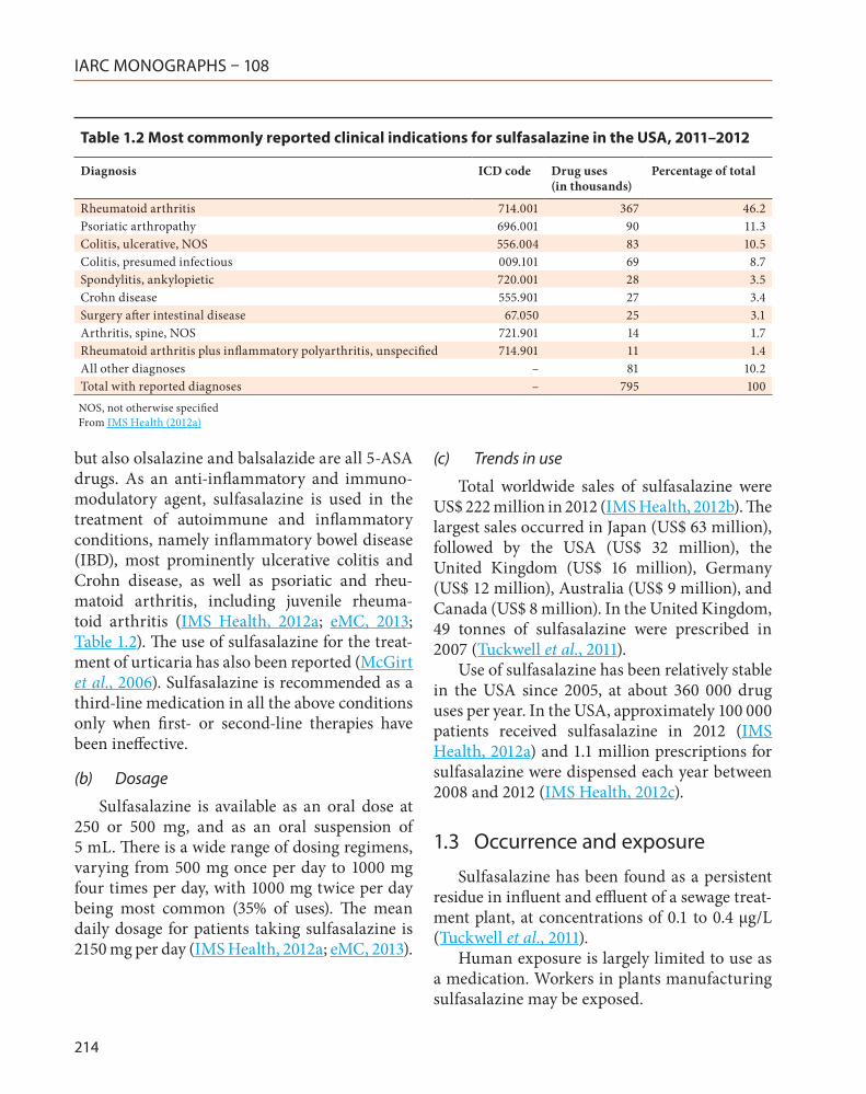
but also olsalazine and balsalazide are all 5-ASA drugs. As an anti-inflammatory and immuno-modulatory agent, sulfasalazine is used in the treatment of autoimmune and inflammatory conditions, namely inflammatory bowel disease (IBD), most prominently ulcerative colitis and Crohn disease, as well as psoriatic and rheu-matoid arthritis, including juvenile rheuma-toid arthritis (IMS Health, 2012a; eMC, 2013; Table 1.2). The use of sulfasalazine for the treat-ment of urticaria has also been reported (McGirt et al., 2006). Sulfasalazine is recommended as a third-line medication in all the above conditions only when first- or second-line therapies have been ineffective.
(b) Dosage
Sulfasalazine is available as an oral dose at 250 or 500 mg, and as an oral suspension of 5 mL. There is a wide range of dosing regimens, varying from 500 mg once per day to 1000 mg four times per day, with 1000 mg twice per day being most common (35% of uses). The mean daily dosage for patients taking sulfasalazine is 2150 mg per day (IMS Health, 2012a; eMC, 2013).
(c) Trends in use
Total worldwide sales of sulfasalazine were US$ 222 million in 2012 (IMS Health, 2012b). The largest sales occurred in Japan (US$ 63 million), followed by the USA (US$ 32 million), the United Kingdom (US$ 16 million), Germany (US$ 12 million), Australia (US$ 9 million), and Canada (US$ 8 million). In the United Kingdom, 49 tonnes of sulfasalazine were prescribed in 2007 (Tuckwell et al., 2011).
Use of sulfasalazine has been relatively stable in the USA since 2005, at about 360 000 drug uses per year. In the USA, approximately 100 000 patients received sulfasalazine in 2012 (IMS Health, 2012a) and 1.1 million prescriptions for sulfasalazine were dispensed each year between 2008 and 2012 (IMS Health, 2012c).
1.3 Occurrence and exposure
Sulfasalazine has been found as a persistent residue in influent and effluent of a sewage treat-ment plant, at concentrations of 0.1 to 0.4 μg/L (Tuckwell et al., 2011).
Human exposure is largely limited to use as a medication. Workers in plants manufacturing sulfasalazine may be exposed.
Table 1.2 Most commonly reported clinical indications for sulfasalazine in the USA, 2011–2012
Diagnosis ICD code Drug uses (in thousands)
Percentage of total
Rheumatoid arthritis 714.001 367 46.2Psoriatic arthropathy 696.001 90 11.3Colitis, ulcerative, NOS 556.004 83 10.5Colitis, presumed infectious 009.101 69 8.7Spondylitis, ankylopietic 720.001 28 3.5Crohn disease 555.901 27 3.4Surgery after intestinal disease 67.050 25 3.1Arthritis, spine, NOS 721.901 14 1.7Rheumatoid arthritis plus inflammatory polyarthritis, unspecified 714.901 11 1.4All other diagnoses – 81 10.2Total with reported diagnoses – 795 100NOS, not otherwise specifiedFrom IMS Health (2012a)
Sulfasalazine
215
1.4 Regulations and guidelines
Sulfasalazine has been widely approved by drug regulatory agencies around the world. In the USA, it was approved by the Food and Drug Administration in 1950 (FDA, 2013).
Sulfasalazine is listed as “known to cause cancer” by the State of California’s Office of Environmental Health Hazard Assessment, requiring public notice of potential environ-mental exposure (OEHHA, 2013).
2. Cancer in Humans
2.1 Background
Sulfasalazine, a member of the family of 5- ASA drugs (see Section 1.2.2), has been used since the 1950s to treat IBD (primarily ulcerative colitis) and, to a lesser extent, Crohn disease (Hanauer, 2004). IBD is associated with an increased risk of dysplasia and cancer of the colorectum. Risk factors for IBD-associated cancer of the colorectum include duration, severity and extent of colitis, the presence of coexistent primary sclerosing cholangitis, and a family history of cancer of the colorectum (Dyson & Rutter, 2012). Chronic inflammation has been proposed as a mechanism for colorectal cancer associated with IBD (or ulcerative colitis), and thus it has been suggested that 5-ASA drugs are chemopreventive agents, because of their anti-inflammatory, anti-oxidant, and pro-apoptotic properties (Rubin et al., 2006; Lakatos & Lakatos, 2008).
The available epidemiological studies included a surveillance study, two cohort studies, three nested case–control studies and three case–control studies of cancer of the colorectum among patients with IBD or ulcerative colitis. Some studies on IBD included patients with Crohn disease in addition to patients with ulcer-ative colitis, but none of the studies stratified by IBD subtype. A case–control study of cancer of
the colorectum and exposure to dihydrofolate reductase inhibitors (sulfasalazine, triamterene, and methotrexate) was identified (Coogan & Rosenberg, 2007), but was not considered to be informative because it did not provide a risk estimate specifically for sulfasalazine; for further information, see the Monograph on triamterene in the present volume).
2.2 Longitudinal, cohort, and nested case–control studies
See Table 2.1 and Table 2.2Moody et al. (1996) evaluated long-term
treatment with sulfasalazine and risk of cancer of the colorectum in a retrospective cohort of 175 patients with ulcerative colitis diagnosed between 1972 and 1989 in Leicestershire, England. Clinical information, including compliance with sulfasalazine treatment and history of cancer, was obtained from case records. A patient was considered to be “non-compliant” if there was clear evidence that the patient had ceased taking the medication or was instructed by the physi-cian to stop the medication without replacement by another 5-ASA drug. The crude proportion of cases of cancer of the colorectum in the sulfasala-zine non-compliant group (31%) was significantly higher than in the compliant group (3%), and a significant effect of compliance was observed in survival analyses using log-ranked and Wilcoxon methods. [This study was limited by lack of a true unexposed group (the use of sulfasalazine in the non-compliant group was not known), small numbers, and limited information on use of sulfasalazine (e.g. time period, dose, dura-tion), or other medications, and information on risk factors for cancer of the colorectum. It was not clear whether other 5-ASA drugs were used as a replacement for sulfasalazine in members of the compliant group who developed cancer, and whether the physician recommendations for stopping treatment with sulfasalazine would affect cancer outcome.]
IARC MONOGRAPHS – 108
216
Tabl
e 2.
1 Su
rvei
llanc
e an
d co
hort
stu
dies
of c
ance
r and
sul
fasa
lazi
ne
Ref
eren
ce
Loca
tion
, pe
riod
Tota
l No.
of
subj
ects
Expo
sure
as
sess
men
tO
rgan
site
(I
CD
cod
e)Ex
posu
re c
ateg
orie
sEx
pose
d ca
ses
Rel
ativ
e ri
sk
(95%
CI)
Cov
aria
tes
Com
men
ts
Moo
dy
et a
l. (1
996)
Le
ices
ter,
Engl
and
1981
–92
175
Cas
e re
cord
s –
use
and
com
plia
nce
Col
orec
tum
Cru
de p
ropo
rtio
n of
cas
es
(com
plia
nt v
s non
-com
plia
nt; χ
2 )P
< 0.
001
Retr
ospe
ctiv
e; c
ohor
t co
mpr
ised
pat
ient
s with
to
tal u
lcer
ativ
e co
litis
or
with
lim
ited
colit
is w
ho
wer
e de
ceas
ed; i
dent
ified
vi
a co
litis
dat
abas
e (c
ase
asce
rtai
nmen
t, 98
%).
Can
cer
case
s or d
yspl
asia
iden
tified
vi
a ca
se re
cord
s or r
egis
try;
bi
opsy
dia
gnos
is c
onfir
med
on
10%
sam
ple.
Sur
viva
l an
alys
is a
djus
ted
for a
ge a
nd
sex
[OR
for c
ompl
ianc
e][0
.07
(0.0
2–0.
30)]
Sulfa
sala
zine
non
-com
plia
nt g
roup
55/
16 (3
1%)
Sulfa
sala
zine
com
plia
nt g
roup
55/
152
(3%
)Su
rviv
al a
naly
sis (
canc
er-f
ree)
fo
r com
plia
nce
(log
rank
ed a
nd
Wilc
oxon
met
hods
)
P <
0.00
1
Lind
berg
et
al.
(200
1)
Swed
en,
Follo
w-u
p,
20 y
r
143
Hos
pita
l re
cord
s or
ques
tionn
aire
s
Col
orec
tum
(c
ance
r or
dysp
lasi
a)
Sulfa
sala
zine
4242
/124
(34%
)Su
rvei
llanc
e st
udy;
pat
ient
s w
ith u
lcer
ativ
e co
litis
pa
rtic
ipat
ing
in 2
0-yr
co
lono
scop
ic su
rvei
llanc
e pr
ogra
mm
e; 1
24 p
atie
nts i
n th
e tr
eatm
ent g
roup
No
sulfa
sala
zine
88/
18 (4
4%)
Sulfa
sala
zine
vs n
o su
lfasa
lazi
ne
(t-te
st)
P =
0.38
[OR
for s
ulfa
sala
zine
][0
.6 (0
.2–1
.7)]
Cum
ulat
ive
risk
of C
RC/d
yspl
asia
(s
ulfa
sala
zine
vs n
o tr
eatm
ent)
P =
0.40
Cum
ulat
ive
risk
ana
lysi
s ad
just
ed fo
r pri
mar
y sc
lero
sing
cho
lang
itis,
sex,
du
ratio
n of
ulc
erat
ive
colit
is,
cole
ctom
y Po
wer
, < 5
0%
Col
orec
tum
(c
ance
r on
ly)
Sulfa
sala
zine
vs n
o su
lfasa
lazi
ne
(t-te
st)
NR
Sulfa
sala
zine
55/
124
(4%
)N
o su
lfasa
lazi
ne2
2/18
(11%
)[O
R fo
r sul
fasa
lazi
ne ]
[0.3
(0.0
6–1.
87)]
Sulfasalazine
217
Ref
eren
ce
Loca
tion
, pe
riod
Tota
l No.
of
subj
ects
Expo
sure
as
sess
men
tO
rgan
site
(I
CD
cod
e)Ex
posu
re c
ateg
orie
sEx
pose
d ca
ses
Rel
ativ
e ri
sk
(95%
CI)
Cov
aria
tes
Com
men
ts
van
Staa
et
al.
(200
5)
Uni
ted
Kin
gdom
, 19
87–2
001
33 9
05 w
ith
5-A
SA d
rug
pres
crip
tions
GPR
DC
olor
ectu
m
(153
, 154
, 15
9)
Refe
renc
e co
hort
(num
ber n
ot
repo
rted
); no
his
tory
of I
BD o
r pr
escr
iptio
n fo
r 5-A
SA d
rug
116
1Pa
tient
s with
5-A
SA d
rug
pres
crip
tions
and
/or I
BD
iden
tified
from
GPR
D;
patie
nts w
ith h
isto
ry o
f CRC
ex
clud
ed. A
naly
sis a
djus
ted
for a
ge a
nd se
x
18 9
695-
ASA
dru
g/IB
D c
ohor
t (5-
ASA
dr
ug [e
xclu
ding
sulfa
sala
zine
] or
sulfa
sala
zine
and
IBD
); n
= 18
969
124
1.99
(1.5
4–2.
56)
Sulfa
sala
zine
rheu
mat
oid
arth
ritis
co
hort
(rem
aini
ng su
lfasa
lazi
ne
with
out I
BD);
num
ber,
NR
691.
26 (0
.94–
1.70
)
Nes
ted
case
–con
trol
an
alys
is (1
00
case
s and
600
co
ntro
ls)
Sulfa
sala
zine
use
12
mon
ths b
efor
e th
e in
dex
date
:C
ases
sele
cted
from
5-A
SA
drug
/IBD
coh
ort,
cont
rols
rand
omly
sele
cted
and
m
atch
ed fo
r age
, sex
, and
ca
lend
ar y
ear o
f ind
ex c
ase.
A
naly
sis a
djus
ted
for B
MI,
IBD
dur
atio
n, h
isto
ry o
f co
lore
ctal
pol
yps,
NSA
ID,
para
ceta
mol
, asp
irin
, im
mun
osup
pres
sive,
gl
ucoc
ortic
oids
, pri
or
hosp
italiz
atio
n fo
r ga
stro
inte
stin
al c
ondi
tion,
ph
ysic
ian
visit
s, co
lono
scop
y
Regu
lar u
se22
0.67
(0.3
6–1.
25)
6–12
pre
scri
ptio
ns3
0.95
(0.2
2–4.
11)
13–3
0 pr
escr
iptio
ns5
0.41
(0.14
–1.2
0)>
30 p
resc
ript
ions
140.
77 (0
.37–
1.60
)D
aily
dos
e, <
2 g
60.
84 (0
.29–
2.42
)D
aily
dos
e, ≥
2 g
150.
69 (0
.35–
1.37
)
5-A
SA, 5
-am
inos
alic
ylic
aci
d; B
MI,
body
mas
s ind
ex; C
RC, c
olor
ecta
l can
cer;
GPR
D, G
ener
al P
ract
ice
Rese
arch
Dat
abas
e; IB
D, i
nflam
mat
ory
bow
el d
isea
se; N
R, n
ot re
port
ed; N
SAID
, no
nste
roid
al a
nti-i
nflam
mat
ory
drug
s; O
R, o
dds r
atio
; vs,
vers
us
Tabl
e 2.
1 (
cont
inue
d)
IARC MONOGRAPHS – 108
218
Tabl
e 2.
2 Ca
se–c
ontr
ol s
tudi
es o
f can
cer o
f the
col
orec
tum
and
sul
fasa
lazi
ne
Ref
eren
ce
Loca
tion
, pe
riod
Tota
l ca
ses
Tota
l co
ntro
ls
Con
trol
so
urce
(h
ospi
tal,
popu
lati
on)
Expo
sure
as
sess
men
tO
rgan
site
(I
CD
cod
e)Ex
posu
re
cate
gori
esEx
pose
d ca
ses
Rel
ativ
e ri
sk
(95%
CI)
Cov
aria
tes
Com
men
ts
Pinc
zow
ski
et a
l. (1
994)
Sw
eden
, 19
65–8
3
102
196
Nes
ted
case
-con
trol
; ul
cera
tive
colit
is c
ohor
t
Med
ical
re
cord
sC
olor
ectu
m
Sulfa
sala
zine
us
e, o
ne o
r mor
e tr
eatm
ent c
ours
es
(> 3
mon
ths)
480.
38 (0
.20–
0.69
)A
ge, n
umbe
r of
exac
erba
tions
/yr
Con
trol
s mat
ched
by
sex,
ex
tent
of u
lcer
ativ
e co
litis
at
dia
gnos
is, a
nd y
r of
diag
nosi
sJe
ss e
t al.
(200
7)
Cop
enha
gen
Den
mar
k,
1962
–97;
M
inne
sota
, U
SA,
1940
–200
4
43
102
Nes
ted
case
–con
trol
: IB
D c
ohor
t; ul
cera
tive
colit
is o
r C
rohn
dis
ease
Med
ical
re
cord
sC
olor
ectu
m
(ade
noca
rcin
oma,
ad
enom
a,
or d
yspl
asia
[c
ombi
ned]
)
Sulfa
sala
zine
us
e, c
umul
ativ
e do
se: 2
.9/1
000
g (m
edia
n) fo
r cas
es
vs 2
.2 (m
edia
n) fo
r co
ntro
ls
NR
1.1
(1.0
–1.3
)A
ge a
nd c
alen
dar y
r of
diag
nosi
s C
ontr
ols f
rom
the
sam
e re
gion
al c
ohor
t mat
ched
on
sex,
IBD
(sub
type
, du
ratio
n, c
alen
dar y
r, an
d ag
e of
dia
gnos
is). U
SA c
ohor
t fo
llow
ed u
ntil
2004
, and
D
anis
h fo
llow
ed u
ntil
1997
. O
f the
43
case
s, 23
wer
e C
RC, 1
3 ad
enom
a, a
nd 7
dy
spla
sia
Sulfa
sala
zine
, re
gula
r use
(> 2
g/
day)
361.
1 (0
.3–3
.7)
Eade
n et
al.
(200
0)
Engl
and
and
Wal
es, [
date
, N
R]
102
102
IBD
pat
ient
sM
edic
al
reco
rds
Col
orec
tum
Su
lfasa
lazi
ne u
se:
Poss
ible
sele
ctio
n bi
as,
sour
ce o
f pop
ulat
ion
patie
nts
from
phy
sicia
n in
tere
sted
in
stud
y, co
ntro
ls fr
om IB
D
Leic
este
rshi
re d
atab
ase.
C
ontr
ols m
atch
ed fo
r sex
, ag
e (w
ithin
10
yr),
exte
nt a
nd
dura
tion
of d
isea
se, b
ut n
ot
hosp
ital o
r yr o
f dia
gnos
is.
“Adj
uste
d fo
r mos
t infl
uent
ial
vari
able
s”, o
ther
5-A
SA
drug
s, co
ntac
t with
hos
pita
l do
ctor
, col
onos
copi
es
diag
nosi
s, re
lativ
e w
ith C
RC
< 2
g/da
y7
0.93
(0.2
2–3.
91)
≥ 2
g/da
y32
0.85
(0.3
2–2.
26)
Sulfasalazine
219
Ref
eren
ce
Loca
tion
, pe
riod
Tota
l ca
ses
Tota
l co
ntro
ls
Con
trol
so
urce
(h
ospi
tal,
popu
lati
on)
Expo
sure
as
sess
men
tO
rgan
site
(I
CD
cod
e)Ex
posu
re
cate
gori
esEx
pose
d ca
ses
Rel
ativ
e ri
sk
(95%
CI)
Cov
aria
tes
Com
men
ts
Rutt
er e
t al.
(200
4)
Engl
and,
19
88–2
002
68
136
Hos
pita
l, co
lono
scop
y su
rvei
llanc
e
Med
ical
re
cord
s, in
terv
iew
s, an
d po
stal
qu
estio
nnai
res
Sulfa
sala
zine
use
:C
ontr
ols m
atch
ed fo
r sex
, ex
tent
and
dur
atio
n of
ul
cera
tive
colit
is, a
ge o
f ons
et
of u
lcer
ativ
e co
litis
, yr o
f in
dex
colo
nosc
opy;
als
o ha
d to
hav
e in
tact
col
on a
nd o
n su
rvei
llanc
e w
ithin
5 y
r of
case
dia
gnos
is
Col
orec
tum
(c
ance
r, ad
enom
a,
and
dysp
lasi
a)
≤ 10
yr
170.
97 (0
.41–
2.26
)>
10 y
r 37
1.58
(0.7
1–3.
51)
Col
orec
tum
(c
ance
r)≤
10 y
r5
4.89
(0.4
7–51
.00)
> 10
yr
86.
59 (0
.64–
67.9
2)
Terd
iman
et
al. (
2007
) U
SA, 2
001–
3
18 4
40
368
800
Popu
latio
n,
heal
th-c
are
data
base
Adm
inis
trat
ive
clai
ms i
n da
taba
se o
f la
rge
heal
th-
care
insu
ranc
e co
mpa
nies
Col
orec
tum
(IC
D-
9-C
M)
Sulfa
sala
zine
use
1
yr b
efor
e di
agno
sis
642.
33 (1
.80–
3.01
)C
ontr
ols f
ree
of c
ance
r and
bo
wel
surg
ery,
mat
ched
to
cas
es b
y ag
e, se
x, a
nd
cale
ndar
yea
r (20
: 1)
; CRC
bu
t not
IBD
dia
gnos
is
inte
rnal
ly v
alid
ated
. No
adju
stm
ent i
n an
alys
es
of a
ny u
se 1
yr b
efor
e di
agno
sis.
Dos
e–re
spon
se
anal
ysis
adj
uste
d fo
r age
, sex
, co
lono
scop
y, ph
ysic
ian
visit
s, ul
cera
tive
colit
is o
r Cro
hn
dise
ase,
hos
pita
lizat
ion,
N
SAID
, glu
coco
rtic
oste
roid
s, an
d im
mun
omod
ulat
ors
364
1172
IBD
pat
ient
sSu
lfasa
lazi
ne u
se 1
yr
bef
ore
diag
nosi
s, IB
D p
atie
nts
441.
19 (0
.83–
1.72
)
No.
of
pres
crip
tions
:0
320
1.0
(ref
.)1–
212
1.65
(0.8
0–3.
39)
3–4
111.
01 (0
.46–
2.21
)≥
521
1.10
(0.6
3–1.
92)
P fo
r tre
nd0.
27
5-A
SA, 5
-am
inos
alic
ylic
aci
d; C
RC, c
olor
ecta
l can
cer;
IBD
, infl
amm
ator
y bo
wel
dis
ease
; IC
D-9
CM
, Int
erna
tiona
l Cla
ssifi
catio
n of
Dis
ease
s Nin
th R
evis
ion,
Clin
ical
Mod
ifica
tion;
NR
, no
t rep
orte
d; N
SAID
, non
ster
oida
l ant
i-infl
amm
ator
y dr
ugs;
ref.,
refe
renc
e; v
s, ve
rsus
; yr,
year
Tabl
e 2.
2 (
cont
inue
d)
IARC MONOGRAPHS – 108
220
The association between sulfasalazine intake and colorectal cancer or dysplasia was evalu-ated in a study of 143 patients with ulcerative colitis who underwent regular colonoscopies and multiple biopsies in a 20-year surveillance programme in Sweden (Lindberg et al., 2001). Of the 143 patients, 124 were in the group that had received treatment with sulfasalazine (treated for at least 6 months between onset of ulcerative colitis and start of surveillance by colonoscopy). Dysplasia or cancer of the colon developed in 51 patients. No statistically significant differences in the adjusted cumulative risk analysis for devel-oping cancer or dysplasia of the colorectum, or the percentage of cancers in the two treat-ment groups (44% in the non-treatment group compared with 34% in the treatment group) were reported. [The study was limited by small numbers and limited exposure information.]
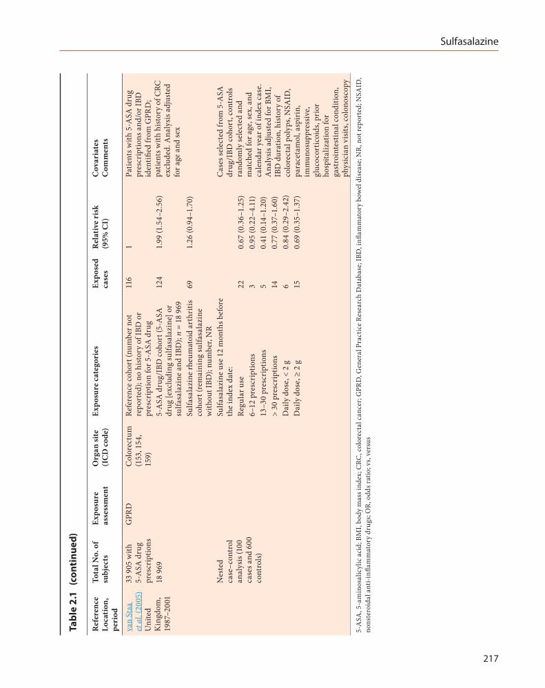
A cohort of users of 5-ASA drugs was identi-fied from the General Practice Research Database in the United Kingdom (van Staa et al., 2005). The cohort was divided into three subcohorts: (i) the 5-ASA drug/IBD cohort included 18 969 patients who either had a prescription for an 5-ASA drug (not including sulfasalazine) or who had taken sulfasalazine and had a diagnosis of IBD; (ii) the remaining patients who were taking sulfasalazine but did not have IBD; and (iii) a reference cohort consisting of patients without IBD or a prescription for a 5-ASA drug, matched by calendar year to participants receiving a 5-ASA drug. [The rationale for this approach was that sulfasalazine is used to treat IBD and other diseases in the United Kingdom, while the other 5-ASA drugs are only used to treat IBD.] Relative risks (RRs) for incidence of cancer of the colorectum were 1.99 (95% CI, 1.54–2.56) for the 5-ASA drug/IBD cohort, and 1.26 (95% CI, 0.94–1.70) for the sulfasalazine/non-IBD cohort.
A nested case–control analysis was conducted among the cohort of patients receiving 5-ASA drugs, which included 100 cases and 600 controls (matched to cases on age, sex, and
calendar year) who had had prescriptions in the 6 months preceding the case index date. The type of 5-ASA drug was classified according to the last prescription issued before the index date. Adjusted odds ratios (ORs) were < 1 for regular use or 6–12 prescriptions, 0.41 (0.14–1.20) for 13–30 prescriptions, and 0.77 (0.37–1.60) for > 30 prescriptions in the previous 12 months, and for daily doses of < 2 g and ≥ 2 g; however, they were not statistically significant and no clear exposure–response patterns were observed (see Table 2.1). Duration of IBD was a strong risk factor for cancer of the colorectum in the study and was controlled for in the analyses. [This study had several advantages, including the prospective design, population-base selection of subjects, analyses by different exposure catego-ries for specific 5-ASA drugs, and good clinical information on each patient. However, informa-tion on drug use was limited to the last prescrip-tion, information on lifetime drug use was not available, and there was limited information on follow-up procedures (e.g. no information was provided on tracking individuals who moved out of the region of the United Kingdom database). There was also a potential for misclassification of disease; classification appeared to be based on the General Practice Research Database with diagnosis confirmed via physician question-naire for a small subset of patients. The study had limited statistical power. Moreover, it was unclear whether all the variables in the statistical models were potential confounders, which may have further reduced the statistical power.]
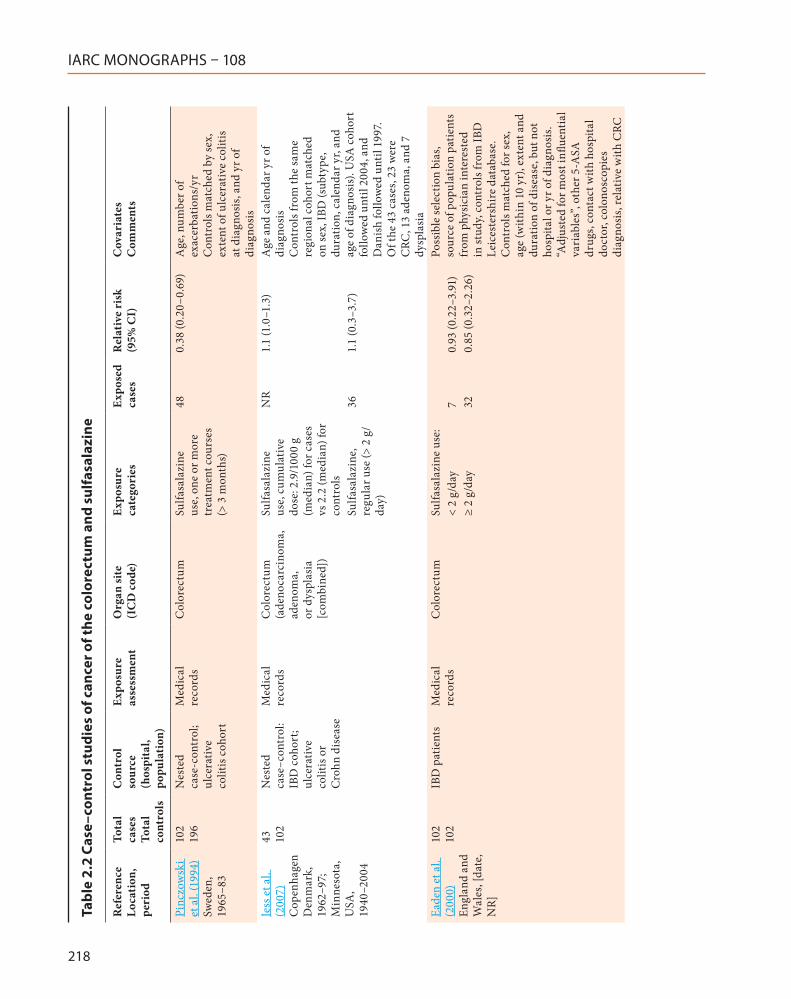
Two case–control studies were nested in population-based cohorts of patients with IBD (see Table 2.2). A Swedish study identified 102 cases of cancer of the colorectum via linkage to the national Swedish cancer registry, among a cohort of patients with ulcerative colitis (Pinczowski et al., 1994). Living controls (n = 196), with intact or partially intact colon, were matched to cases on sex, extent of disease, and time of diagnosis of disease. Information on pharmacological therapy
Sulfasalazine
221
(including sulfasalazine), clinical features of disease, smoking, and family history of IBD, cancer of the colorectum, and other diseases, was collected from the patient’s medical records. A decreased risk of cancer of the colorectum was found among individuals who had followed one or more treatment course of sulfasalazine (at least 3 months) (adjusted RR, 0.38; 95% CI, 0.20–0.69). [The strengths of the study were the prospective design, the use of a population-based cohort, and the use of controls matched for disease severity. The major limitations were the lack of detailed exposure information and small size.]
The second nested case–control study evalu-ated the risk of colorectal neoplasia (adenocarci-noma, adenoma, and dysplasia combined) in two cohorts of patients with IBD in Denmark and in Minnesota, USA (Jess et al., 2007). Both cohorts included patients with ulcerative colitis or Crohn disease. Cases were identified via linkage with cancer registries, and controls matched for sex, vital status, and age at diagnosis, and clinical factors related to IBD were identified from each regional cohort. Exposure and clinical informa-tion was obtained from medical records. The adjusted relative risk for colorectal neoplasia was close to unity for regular use (> 2 g/day) or cumu-lative dose of sulfasalazine. Primary sclerosing cholangitis was a strong risk factor for colorectal neoplasia. [The advantages and limitations of this study were similar to those of the Swedish study. An additional limitation of this study was that there was not a separate analysis for cancer of the colorectum alone; of the 43 cases of colorectal neoplasia, 23 were cancer.]
2.3 Case–control studies
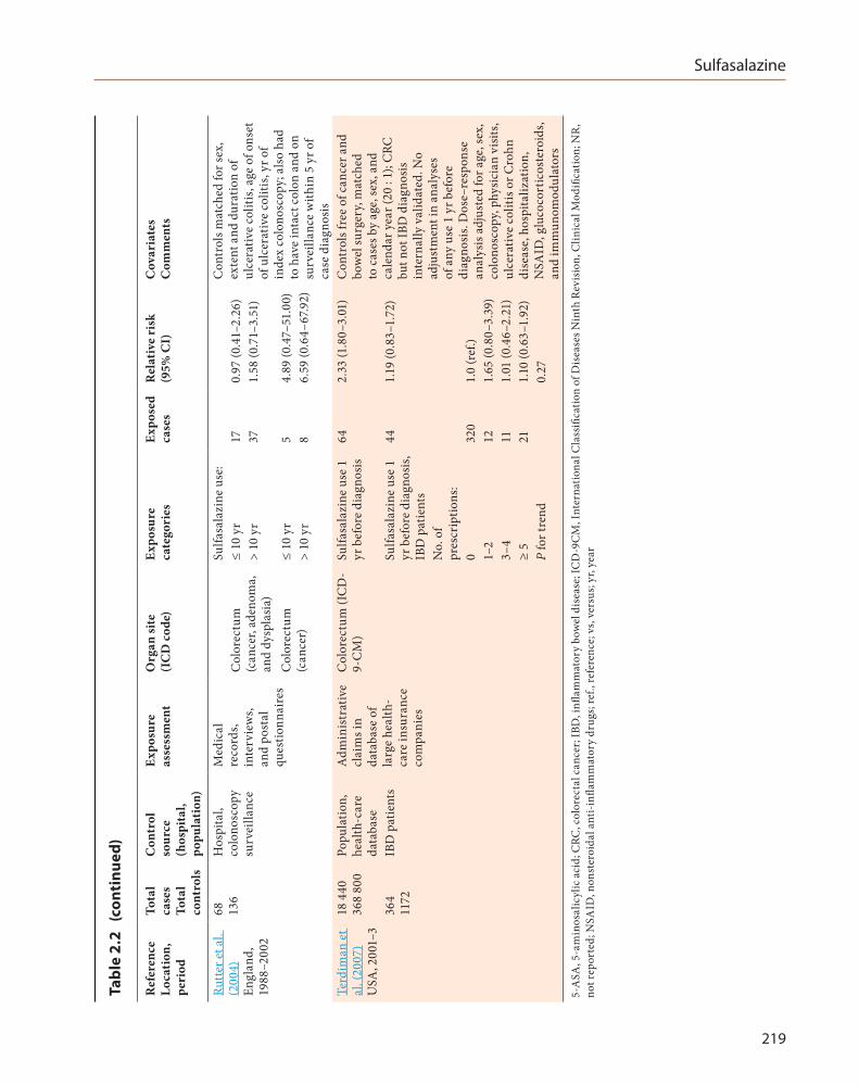
See Table 2.2Three studies selected cases and controls
from patients with ulcerative colitis. Eaden et al. (2000) evaluated the risk of colorectal cancer among patients with ulcerative colitis and controls in England and Wales. Cases (n = 102)
were identified from records of consenting gastroenterologists throughout England and Wales, and 102 controls matched by sex, age (categories of 10 years), extent and duration of IBD were identified from the Leicestershire data-base of IBD patients. Data were extracted from medical records. Sulfasalazine therapy at both < 2 g/day and ≥ 2 g/day was inversely associ-ated with increased risk of colorectal cancer in unadjusted analyses, while adjusted odds ratios were 0.93 (95% CI, 0.22–3.91) for the group at the lower dose and 0.85 (95% CI, 0.32–2.26) for the group at higher doses. [The limitations of the study were the potential for selection bias, inad-equate matching of the controls (using categories of 10 years of age, and not matching on hospital or year of diagnosis of IBD), limited exposure information, limited documentation of covari-ates controlled in the analyses. Odds ratios were adjusted for the use of other 5-ASA drugs, but this may not be appropriate since these drugs could work via the same mechanisms as sulfasalazine. The study population may have overlapped with the cohort reported by Moody et al. (1996); both studies identified patients from the same data-base of patients, but the years of study recruit-ment were not reported in the study by Eaden et al. (2000).]
One study evaluated risk factors for colorectal neoplasia (cancer, adenoma, and dysplasia) among patients with chronic ulcerative colitis who were part of a colonoscopy surveillance programme (Rutter et al., 2004). Sixty-eight cases with neoplasia were identified and 136 controls from the surveillance population were matched on sex and clinical characteristics of IBD. Long-term use (> 10 years) of sulfasalazine was associated with a non-statistically significant elevated odds ratio for colorectal neoplasia (cancer, adenoma, or dysplasia) of 1.58 (95% CI, 0.71–3.51; 37 exposed cases); use of sulfasalazine for > 3 months to 10 years was associated with an odds ratio of 0.97 (95% CI, 0.41–2.26; 17 exposed cases). Analysis of the cancer cases only found that both exposure
IARC MONOGRAPHS – 108
222
categories were associated with elevated, but very imprecise odds ratios based on small numbers (see Table 2.1). [A significant association between severity of inflammation and colorectal neoplasia was noted, and this hindered interpretation of the association of colorectal cancer and treatment with sulfasalazine. Other concerns included the small numbers of exposed cancer cases, lack of adjustment for risk factors, and limitations in the generalizability of the findings due to the selec-tion of subjects from a surveillance programme. In addition, drug use may have been related to duration of ulcerative colitis, and thus matching by duration of ulcerative colitis may bias the odds ratio towards the null.]
Terdiman et al. (2007) evaluated use of sulfasalazine in a population based case–control study consisting of 18 440 cases of cancer of the colorectum and 368 800 controls (matched by age, sex, and calendar year) identified from two administrative databases covering all regions in the USA, including 364 cases and 1172 controls with a diagnosis of IBD. Information on claims for sulfasalazine prescriptions and other clinical variables was obtained from the claim data-base. A statistically significant increased risk of cancer of the colorectum was found among all patients using sulfasalazine 1 year before diag-nosis (crude OR, 2.33; 95% CI, 1.80–3.01), while among patients with IBD only the adjusted odds ratio for sulfasalazine treatment was 1.19 (95% CI, 0.83–1.72); no exposure–response relation-ship was observed with number of prescriptions, regardless of adjustment for multiple covariates (P for trend, 0.27). [The strengths of this study were the large size and population-based design, and information on many (but not all) potential confounders, and some exposure–response anal-yses. The major limitation was the short exposure duration (1 year before diagnosis); in addition, information was not available on several key potential confounders, such as family history of colon cancer or IBD and other factors not related to medications.]
2.4 Meta-analysis
A meta-analysis of four cohort studies assessed the association between long-term treat-ment with sulfasalazine and risk of colorectal cancer (Diculescu et al., 2003). [The Working Group could not interpret this study due to the lack of information on analytical methods and the inclusion of studies that were not specific for treatment with sulfasalazine. Furthermore, summary measures of association were not calculated.]
3. Cancer in Experimental Animals
3.1 Mouse
See Table 3.1
Oral administration
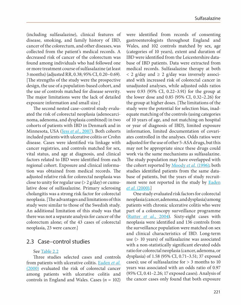
In one study, groups of 50 male and 50 female B6C3F1 mice (age, 6 weeks) were given sulfasala-zine (USP grade) at a dose of 0, 675, 1350, or 2700 mg/kg bw in corn oil by gavage once per day, 5 days per week, for 104 weeks. There was a 6–18% decrease in mean body weight in male and female mice at the highest dose compared with controls. The incidence of hepatocellular adenoma in males and females, and the inci-dence of hepatocellular adenoma or carcinoma (combined) in males and females, were signifi-cantly greater than those in controls, and the incidences increased with a positive trend. The incidence of hepatocellular carcinoma was signif-icantly increased in female mice (Iatropoulos et al., 1997; NTP, 1997a).
In a first experiment in a group of related studies, groups of 50–60 male B6C3F1 mice (age, 6 weeks) were given sulfasalazine (USP grade) at a dose of 0 or 2700 mg/kg bw by gavage in corn oil once per day, 5 days per week, for 103 weeks (~2 years); in a second experiment, an unexposed
Sulfasalazine
223
Tabl
e 3.
1 St
udie
s of
car
cino
geni
city
in m
ice
give
n su
lfas
alaz
ine
by g
avag
e
Stra
in (s
ex)
Dur
atio
n R
efer
ence
Dos
ing
regi
men
, A
nim
als/
grou
p at
star
tIn
cide
nce,
(%) a
nd/o
r mul
tipl
icit
y of
tum
ours
Sign
ifica
nce
Com
men
ts
B6C
3F1
(M, F
) 10
4 w
k N
TP (1
997a
), Ia
trop
oulo
s et
al.
(199
7)
Sulfa
sala
zine
(in
corn
oil)
at a
dos
e of
0, 6
75, 1
350,
or
2700
mg/
kg b
w p
er d
ay, 5
day
s per
wk,
for 1
04 w
k 50
M a
nd 5
0 F/
grou
p
Mal
es
Hep
atoc
ellu
lar a
deno
ma:
13
/501 ,
32/5
0*, 2
8/50
**, 4
2/50
* H
epat
ocel
lula
r car
cino
ma:
13
/50,
15/
50, 2
3/50
, 8/5
0
Hep
atoc
ellu
lar a
deno
ma
or c
arci
nom
a (c
ombi
ned)
: 24
/501 ,
38/5
0***
, 38/
50**
**, 4
4/50
* Fe
mal
es
Hep
atoc
ellu
lar a
deno
ma:
12
/501 ,
28/5
0*, 2
5/50
**, 2
8/49
* H
epat
ocel
lula
r car
cino
ma:
2/
50, 1
0/50
****
*, 10
/50*
****
, 9/4
9***
**
Hep
atoc
ellu
lar a
deno
ma
or c
arci
nom
a (c
ombi
ned)
: 14
/502 ,
32/5
0*, 2
8/50
*, 29
/49*
*P <
0.0
01 (P
oly-
3 te
st)
**P
= 0.
002
(Pol
y-3
test
) **
*P =
0.0
04 (P
oly-
3 te
st)
****
P =
0.00
5 (P
oly-
3 te
st)
****
*P ≤
0.0
5 (P
oly-
3 te
st)
1 P <
0.0
01 (P
oly-
3 tr
end
test
) 2 P
= 0
.005
(Pol
y-3
tren
d te
st)
Puri
ty, U
SP
grad
e
B6C
3F1 (
M)
Up
to 1
56 w
k N
TP (1
997b
), A
bdo
& K
ari
(199
6)
Exp.
1: s
ulfa
sala
zine
(in
corn
oil)
at a
dos
e of
0 o
r 27
00 m
g/kg
bw
per
day
, 5 d
ays p
er w
k, fo
r 103
wk
(~2
yr),
fed
ad li
bitu
m
Exp.
2: u
nexp
osed
gro
up fe
d su
ch th
at m
ean
body
w
eigh
t mat
ched
that
of t
he tr
eate
d gr
oup
fed
ad
libitu
m
Exp.
3 (d
ieta
ry re
stri
ctio
n): t
wo
grou
ps o
f 110
mic
e (o
ne c
ontr
ol a
nd o
ne d
osed
) wer
e gi
ven
iden
tical
qu
antit
ies o
f fee
d su
ch th
at th
e co
ntro
l gro
up w
ould
at
tain
bod
y w
eigh
ts o
f app
roxi
mat
ely
80%
that
of
the
ad li
bitu
m-fe
d co
ntro
ls; 6
0 m
ice
per g
roup
wer
e ev
alua
ted
at 1
03 w
k (~
2 yr
) and
the
rem
aini
ng 5
0 m
ice
per g
roup
at 1
56 w
k (3
yr)
, or w
hen
surv
ival
re
ache
d 20
%
50–6
0 M
/gro
up
Hep
atoc
ellu
lar a
deno
ma:
Ex
p. 1
: 13/
50, 4
2/50
* Ex
p. 2
: 8/5
0, 4
2/50
* Ex
p. 3
: 13/
52, 9
/50
(~2
yr)
Exp.
3: 1
0/48
, 14/
50 (u
p to
3 y
r)
Hep
atoc
ellu
lar c
arci
nom
a:
Exp.
1: 1
3/50
, 8/5
0 Ex
p. 2
: 6/5
0, 8
/50
Exp.
3: 7
/52,
1/5
0 (~
2 yr
) Ex
p. 3
: 16/
48, 6
/50
(up
to 3
yr)
H
epat
ocel
lula
r ade
nom
a or
car
cino
ma
(com
bine
d):
Exp.
1: 2
4/50
, 44/
50*
Exp.
2: 1
4/50
, 44/
50*
Exp.
3: 1
8/52
, 9/5
0 (~
2 yr
) Ex
p. 3
: 21/
48, 1
8/50
(up
to 3
yr)
*P <
0.0
01 (i
ncre
ase,
lo
gist
ic re
gres
sion
test
)Pu
rity
, USP
gr
ade
bw; b
ody
wei
ght;
Exp.
, exp
erim
ent;
F, fe
mal
e; M
, mal
e; w
k, w
eek;
USP
, Uni
ted
Stat
es P
harm
acop
oeia
; yr,
year
IARC MONOGRAPHS – 108
224
group was fed such that the mean body weight of the group matched that of the treated group fed ad libitum. In a third experiment (dietary restric-tion), two groups of 110 animals, one control group and one group given sulfasalazine at a dose of 2700 mg/kg bw in corn oil were offered identical quantities of feed such that the control group would attain body weights of approxi-mately 80% those of the control group fed ad libitum. Sixty mice from each group were eval-uated at 103 weeks and the remaining 50 mice from each group were evaluated at 156 weeks (3 years), or at the time when survival reached 20%.
The mean body weight at 1 year and survival at 103 weeks (~2 years) for the mice treated with sulfasalazine were decreased by 15% and 19%, respectively, relative to controls. The body weight and survival of the weight-matched vehicle-con-trol group were similar to those of the treated group fed ad libitum. Under the dietary restric-tion protocol, the control and treated groups weighed 42 g and 34 g at 1 year and had respective survival rates of 84% and 88% after 103 weeks.
Exposure to sulfasalazine under ad-libitum feeding conditions for 103 weeks (~2 years) caused significantly increased incidences of hepatocellular adenoma, and hepatocellular adenoma or carcinoma (combined) in exposed mice compared with the controls fed ad libitum and the weight-matched controls. In contrast to the findings of the first two experiments, the inci-dence of hepatocellular tumours in dietary-re-stricted mice was significantly decreased in the exposed group after 103 weeks, and was similar to that in non-treated mice after 3 years (Abdo & Kari, 1996; NTP, 1997b).
3.2 Rat
See Table 3.2
Oral administration
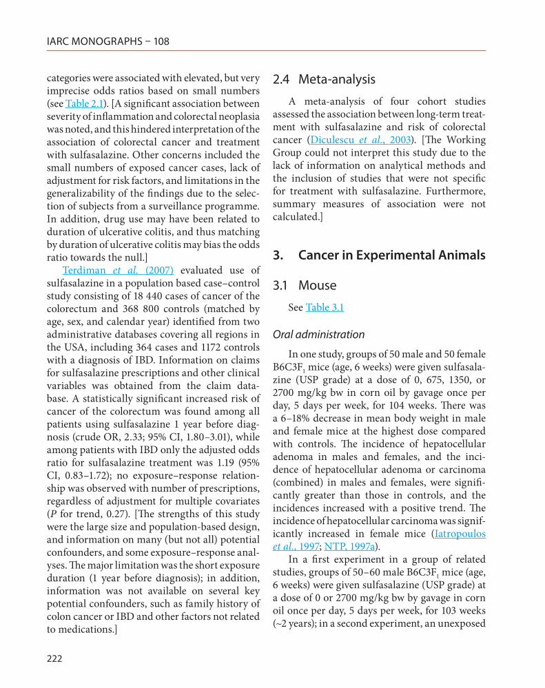
In one study, groups of 50 male and 50 female F344/N rats (age, 6 weeks) were given sulfasalazine (USP grade) at a dose of 0, 84, 168, or 337.5 mg/kg bw by gavage in corn oil once per day, 5 days per week, for 105 weeks (core study; continuous exposure). An additional group of male rats (stop-exposure group) was treated with sulfasalazine in corn oil at 337.5 mg/kg bw for 26 weeks, and then with corn oil only for the remainder of the study (79 weeks). Survival of male rats at the highest dose in the core study was significantly lower than that of controls, with most deaths occurring during the last 8 weeks of the study. Survival of all other treated groups was similar to that of controls.
The incidence of transitional cell papilloma of the urinary bladder in the core study was increased with a positive trend in the groups of treated male rats; the incidence in the group at the highest dose was significantly increased. The transitional cell neoplasms of the urinary tract observed in the core study were not observed in the stop-exposure group. In exposed females, there were also low incidences of [rare] transi-tional cell papilloma of the kidney and of the urinary bladder. All rats with transitional cell papillomas of the urinary tract also had grossly visible concretions (calculi) in the kidney and/or urinary bladder (Iatropoulos et al., 1997; NTP, 1997a).
In a first experiment in a group of related studies, groups of 50–60 male F344/N rats (age, 6 weeks) were given sulfasalazine (USP grade) at a dose of 0 or 337.5 mg/kg bw in corn oil by gavage once per day, 5 days per week, for up to 104 weeks; in a second experiment, an unexposed group was fed such that the mean body weight of the group matched that of the treated group fed ad libitum. In a third experiment (dietary restric-tion), two groups of 110 rats, one control group and one group given sulfasalazine at a dose of 337.5 mg/kg bw in corn oil were offered identical

Sulfasalazine
225
Tabl
e 3.
2 St
udie
s of
car
cino
geni
city
in ra
ts g
iven
sul
fasa
lazi
ne b
y ga
vage
Stra
in (s
ex)
Dur
atio
n R
efer
ence
Dos
ing
regi
men
, A
nim
als/
grou
p at
star
tFo
r eac
h ta
rget
org
an: I
ncid
ence
, (%
) and
/or
mul
tipl
icit
y of
tum
ours
Sign
ifica
nce
Com
men
ts
F344
/N
(M, F
) 10
5 w
k N
TP
(199
7a),
Iatr
opou
los
et a
l. (1
997)
Sulfa
sala
zine
(in
corn
oil)
at a
dos
e of
0, 8
4, 1
68, o
r 337
.5 m
g/kg
bw
, onc
e pe
r day
, 5 d
ays p
er w
k, fo
r 105
wk
(cor
e st
udy)
; an
addi
tiona
l gro
up o
f mal
e ra
ts (s
top-
expo
sure
gro
up) w
as tr
eate
d w
ith su
lfasa
lazi
ne (i
n co
rn o
il) a
t 337
.5 m
g/kg
bw
for 2
6 w
k an
d th
en w
ith c
orn
oil o
nly
for t
he re
mai
nder
of t
he st
udy
(79
wk)
50
M a
nd 5
0 F/
grou
p
Mal
es
Tran
sitio
nal c
ell p
apill
oma
of th
e ur
inar
y bl
adde
r: 0/
501 ,
0/49
, 2/5
0 (4
%),
6/50
(12%
)*;
stop
exp
osur
e, 0
/47
Fem
ales
Tr
ansit
iona
l cel
l pap
illom
a of
the
urin
ary
blad
dera :
0/49
, 0/5
0, 2
/50
(4%
), 0/
50
Tran
sitio
nal c
ell p
apill
oma
of th
e ki
dney
b : 0/
50, 0
/50,
0/5
0, 2
/50
(4%
)
1 P <
0.0
01
(Pol
y-3
tren
d te
st)
*P =
0.0
11
(Pol
y-3
test
)
Puri
ty, U
SP
grad
e
F344
/N (M
) U
p to
13
0 w
k N
TP
(199
7b),
Abd
o &
K
ari (
1996
)
Exp.
1: s
ulfa
sala
zine
in c
orn
oil a
t a d
ose
of 0
, or 3
37.5
mg/
kg b
w,
once
per
day
, 5 d
ays p
er w
k fo
r up
to 1
04 w
k, fe
d ad
libi
tum
Ex
p. 2
: une
xpos
ed g
roup
fed
such
that
mea
n bo
dy w
eigh
t m
atch
ed th
at o
f the
trea
ted
grou
p fe
d ad
libi
tum
. Ex
p. 3
– d
ieta
ry re
stri
ctio
n: tw
o gr
oups
of 1
10 ra
ts (o
ne c
ontr
ol
and
one
dose
d) w
ere
give
n id
entic
al q
uant
ities
of f
eed
such
that
th
e co
ntro
l gro
up w
ould
att
ain
body
wei
ghts
of a
ppro
xim
atel
y 80
% th
ose
of th
e ad
libi
tum
-fed
cont
rols
; 60
rats
/gro
up w
ere
eval
uate
d at
104
wk
(~2
yr) a
nd th
e re
mai
ning
50
rats
/gro
up a
t 13
0 w
k, o
r whe
n su
rviv
al re
ache
d 20
%
50–6
0 m
ales
/gro
up
Tran
sitio
nal c
ell p
apill
oma
of th
e ur
inar
y bl
adde
r: Ex
p. 1
: 0/5
0, 6
/50*
Ex
p. 2
: 0/5
0, 6
/50*
Ex
p. 3
: 0/5
1, 0
/50
(~2
yr)
Exp.
3: 0
/49,
1/4
9 (u
p to
130
wk)
*P =
0.0
11
(logi
stic
re
gres
sion)
Puri
ty, U
SP
grad
e
a H
isto
rica
l inc
iden
ce fo
r 2-y
ear s
tudi
es b
y th
e N
TP in
rats
fed
corn
oil
by g
avag
e (v
ehic
le c
ontr
ol g
roup
s): 3
/903
(0.3
% ±
0.8
%);
rang
e, 0
–2%
b H
isto
rica
l inc
iden
ce fo
r 2-y
ear s
tudi
es b
y th
e N
TP in
rats
fed
corn
oil
by g
avag
e (v
ehic
le c
ontr
ol g
roup
s): 0
/920
bw, b
ody
wei
ght;
Exp.
, exp
erim
ent;
F, fe
mal
e; M
, mal
e; N
TP, N
atio
nal T
oxic
olog
y Pr
ogra
m; w
k, w
eek;
USP
, Uni
ted
Stat
es P
harm
acop
oeia
; yr,
year
IARC MONOGRAPHS – 108
226
quantities of feed such that the control group would attain body weights of approximately 80% that of the controls fed ad libitum. Sixty rats from each group were evaluated at 104 weeks and the remaining 50 rats from each group were evalu-ated at 130 weeks, or at the time when survival reached 20%.
After 1 year, mean body weights for the control and treated rats in the first experiment were similar. Since there was negligible body weight loss throughout the study, no adjustments were made to the weight-matched control group, thereby yielding a redundant control group. Survival at 2 years in the first experiment was 70% and 46% for the control and treated rats, respectively.
The incidence of transitional cell papilloma of the urinary bladder was significantly greater in exposed rats than in the controls fed ad libitum in the first experiment, or weight-matched controls in the second experiment. All rats with transitional cell papilloma of the urinary bladder also had grossly visible concretions in the kidney and/or urinary bladder. In the third experiment, no significant increase in the incidence of transi-tional cell papilloma of the urinary bladder was observed (Abdo & Kari, 1996; NTP, 1997b).
3.3 Studies of co-carcinogenicity
A group of 12 male Wistar rats was given 1,2-dimethylhydrazine at a dose of 40 mg/kg bw as a single subcutaneous injection each week, concurrently with sulfasalazine at a dose of 60 mg/kg bw per day by gavage, for 20 weeks. One control group of 11 male Wistar rats was given 1,2-dimethylhydrazine only. Development of “colon tumours” (mainly adenocarcinomas) was assessed histologically at week 21. All rats developed tumours of the intestine. In the control group receiving 1,2-dimethylhydrazine only, there were 70 tumours of the intestine with a tumour multiplicity of 6.4 ± 0.69, while in the group given 1,2-dimethylhydrazine plus
sulfasalazine there were 141 tumours of the intestine (P ≤ 0.05, ANOVA test) with a tumour multiplicity of 11.8 ± 2.16 (P < 0.05, t-test) (Davis et al., 1992).
4. Mechanistic and Other Relevant Data
4.1 Absorption, distribution, metabolism, and excretion
4.1.1 Humans
(a) Absorption, distribution, metabolism, and excretion
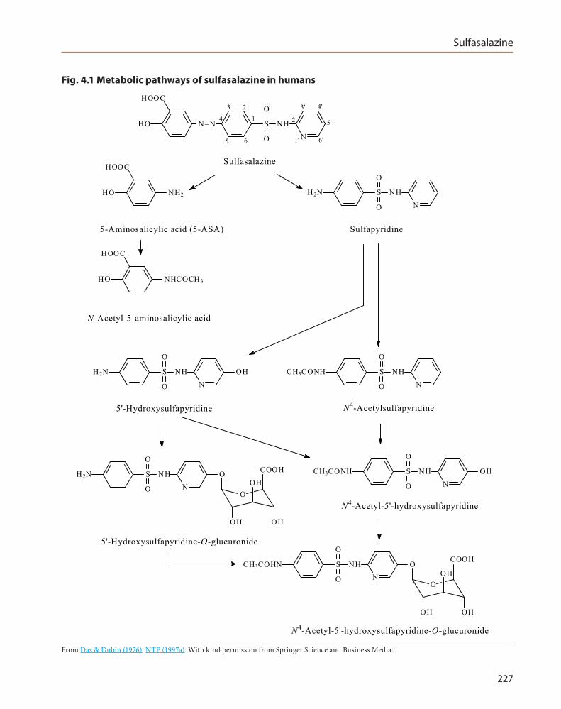
The metabolic scheme for sulfasalazine in humans is shown in Fig. 4.1 (Das & Dubin, 1976; NTP, 1997a).
Sulfasalazine is not absorbed to any signif-icant extent from the stomach (Das & Dubin, 1976). Slow absorption of small amounts (~10–30%) via the small intestine has been reported before enterohepatic recycling, and with the majority of unchanged drug reaching the colon (Das & Dubin, 1976; Azad Khan et al., 1982).
The sulfasalazine molecule comprises 5-ASA and sulfapyridine moieties, linked by an azo bond, which is cleaved by bacterial azoreductases in the colon, releasing 5-ASA and sulfapyridine (Azad Khan et al., 1982). This cleavage is the rate-limiting step for clearance of sulfasala-zine (Das & Dubin, 1976). Most of the 5-ASA is excreted; approximately 50% directly in the faeces, and at least 25% via the kidneys (after absorption and acetylation in the liver) (Das & Dubin, 1976; Azad Khan et al., 1982). In contrast, sulfapyridine is almost completely absorbed. In the liver, sulfapyridine undergoes hydroxylation and/or N-acetylation to 5′-hydroxysulfapyri-dine, N4-acetylsulfapyridine, and N4-acetyl-5′-hydroxysulfapyridine subsequently forming glucuronic acid conjugates, before excretion mainly in the urine (Das & Dubin, 1976; Azad Khan et al., 1982).
Sulfasalazine
227
Fig. 4.1 Metabolic pathways of sulfasalazine in humans
N=N S NH
NO
OHOOC
HO1
23
4
5 6 1'
2'
3' 4'
5'
6'
HOOC
HO NH2 S NH
N
H2N
O
O
HOOC
HO NHCOCH 3
S NHN
H2N
O
O
OH S NHN
CH3CONH
O
O
O
OHOH
S NH
N
H2N
O
O
O COOH S NH
N
CH3CONH
O
O
OH
Sulfasalazine
5-Aminosalicylic acid (5-ASA) Sulfapyridine
N-Acetyl-5-aminosalicylic acid
5'-Hydroxysulfapyridine N4-Acetylsulfapyridine
N4-Acetyl-5'-hydroxysulfapyridine
5'-Hydroxysulfapyridine-O-glucuronide
O
OHOH
S NHN
CH3COHN
O
O
O COOH
N4-Acetyl-5'-hydroxysulfapyridine-O-glucuronide
OH
OH
From Das & Dubin (1976), NTP (1997a). With kind permission from Springer Science and Business Media.
IARC MONOGRAPHS – 108
228
In studies of serum from 10 healthy male volunteers given single oral doses of 4 g of sulfasalazine, parent drug was detectable at 1.5 hours after dosing, and at maximum concen-trations at 3–5 hours in nine subjects, and after 7 hours in one subject (Schröder & Campbell, 1972). Metabolites (sulfapyridine, and acetylated and glucuronidated derivatives) were detected in the serum at 3–5 hours after dosing (Schröder & Campbell, 1972). The pharmacokinetics of rectally administered sulfasalazine have been studied in three healthy male Japanese volunteers (Tokui et al., 2002). Sulfasalazine (6.5 mmol), given as a single suppository, reached maximum plasma concentration (2.5 ± 0.4 µM) in 5 hours (Tmax), and an area under the curve (AUC) of 27.4 ± 4.8 µM.h. Parent drug was almost completely hydrolysed in the colon, and the urinary recovery was only approximately 0.2%. Maximum plasma concentration (Cmax) of the metabolite N-acetyl-5-aminosalicylic acid was 0.5 ± 0.2 µM, reached in 12 hours, while that of sulfapyridine was 1.2 ± 0.4 µM, reached in 5 hours. 5-ASA was not detected in the serum. Administration of an enema containing 6.5 mmol of 5-ASA, resulted in Cmax 5.8 ± 2.0 µM in 1 hour and an AUC of 29.4 ± 11.1 µM.h. In the urine, approximately 0.3% was recovered unchanged. The Cmax for the acetylated metabolite, N-acetyl-5-aminosalicylic acid, was 13.3 ± 3.6 µM in 7 hours. More than 10% of 5-ASA was excreted in the urine as acetyl-5-aminosalicylic acid, suggesting that absorption of 5-ASA is favoured when administered rectally (Tokui et al., 2002).
A study to compare the absorption and metabolism of oral preparations of sulfasalazine, mesalazine (5-ASA) and olsalazine (a dimer of 5-ASA) used regularly by patients (n = 12, 13, and 8, respectively) for treatment of ulcerative colitis, showed considerably greater absorption of 5-ASA and less acetylation, in patients receiving mesalazine than in those receiving olsalazine or sulfasalazine (Stretch et al., 1996).
(a) Variation in absorption, distribution, metabolism, and excretion
(i) IBD and rheumatoid arthritisThe characteristics of absorption, metabo-
lism, and excretion of the parent drug in four patients with IBD (ulcerative colitis or Crohn disease) were similar to those in four healthy subjects, each given a single oral dose of sulfasala-zine (3 or 4 g). However, absorption and urinary excretion of the metabolite, sulfapyridine, was decreased in patients with IBD. The metabolism of sulfasalazine was markedly reduced in patients taking antibiotics and after removal of the large bowel (Azad Khan et al., 1982).
Pharmacokinetic studies of sulfasalazine and its principal metabolites in 13 patients with rheumatoid arthritis and 8 patients with IBD given sulfasalazine as a single oral dose of 2 g (Astbury et al., 1990), showed that patients with rheumatoid arthritis had a significantly higher (and more sustained) plasma concentration of sulfapyridine than did patients with IBD (medians of 14.0 µg/mL and 7.4 µg/mL, respec-tively). Two factors may have contributed to high peak plasma concentrations of sulfapyridine in patients with rheumatoid arthritis: firstly, the metabolism of sulfapyridine may be impaired, and secondly, a larger quantity of sulfasalazine may reach the lower bowel leading to higher concentrations of the subsequent cleavage compounds. The pharmacokinetics of sulfasala-zine were variable among patients; maximum plasma concentrations of sulfapyridine ranged from 8 to 22 µg/mL in patients with rheumatoid arthritis, and from 5 to 18 µg/mL in patients with IBD. The elimination half-life of sulfasala-zine ranged from 3 to 8 hours in patients with rheumatoid arthritis, and from 4 to 9 hours in patients with IBD (Astbury et al., 1990).
Sulfasalazine
229
(ii) PregnancySulfasalazine and its primary metabolites are
able to cross the placenta (Azad Khan & Truelove, 1979; Järnerot et al., 1981). In five patients with ulcerative colitis treated with sulfasalazine (0.5 g, four times per day) throughout and after preg-nancy, sulfasalazine was detected in the umbilical cord blood (mean concentration, 50% of that in maternal serum) and at very low concentrations in the amniotic fluid (Azad Khan & Truelove, 1979). Analyses of metabolites showed that total concentrations of sulfapyridine were equal in maternal and cord sera, but concentrations of free sulfapyridine were significantly lower (P < 0.02) in cord sera. Total concentrations of acetylated sulfapyridine were significantly higher (P < 0.025) in cord sera than maternal sera. Total concentrations of 5-ASA were very low in all fluids analysed (Azad Khan & Truelove, 1979). In the study by Järnerot et al. (1981) of 11 preg-nant patients with IBD treated with sulfasalazine (1 g daily), concentrations of sulfasalazine were almost identical in cord and maternal serum. Seven of the women were analysed at a later date (4–24 months) after pregnancy; plasma concen-trations of sulfasalazine remained the same, but concentrations of sulfapyridine had increased, probably reflecting different extents of protein binding of these compounds, and the different distribution volume in pregnancy (Järnerot et al., 1981).
Small quantities of sulfasalazine and sulfapyridine have also been detected in breast milk (Azad Khan & Truelove, 1979; Järnerot & Into-Malmberg, 1979). Mean concentrations of sulfasalazine and total sulfapyridine in milk, compared with concentrations in maternal serum, were approximately 30% and 50%, respec-tively, as reported by Azad Khan & Truelove (1979), and negligible and 40%, respectively as reported by Järnerot & Into-Malmberg (1979). The various metabolites of sulfapyridine were present in approximately the same proportions
as in maternal serum (Azad Khan & Truelove, 1979). It was estimated that an infant would receive sulfapyridine at dose of 3–4 mg/kg bw, after a maternal dose of 2 g of sulfasalazine per day (Järnerot & Into-Malmberg, 1979). Sulfapyridine and its acetylated and glucuronidated metabo-lites have been shown to be excreted by babies, 1–2.5 months after maternal dosing (Järnerot & Into-Malmberg, 1979).
(c) Genetic polymorphisms
(i) N-AcetyltransferasesThe sulfasalazine molecule may be consid-
ered as a slow-release carrier for sulfapyridine, but there is large inter-individual variation in the rate of metabolism of sulfapyridine, which can affect steady-state serum concentrations (Das & Dubin, 1976). The rate of N-acetylation of 5-ASA to N-acetyl-5-aminosalicylic acid is under genetic control (Das & Dubin, 1976). The cytosolic N-acetyltransferase (NAT) family comprises NAT1 and NAT2, which catalyse the transfer of acetyl groups from activated acetyl-coenzyme A to the nitrogen of primary amines, hydrazines, or hydrazides (Kuhn et al., 2010). Single nucleo-tide polymorphisms have been detected in NAT2. The wildtype has been designated NAT2*4, but NAT2*5, NAT2*6, and NAT2*7 alleles encode N-acetyltransferase enzymes with amino-acid changes that cause reduced activity (slow acetyl-ating function). Patients with a “slow” acetylator phenotype generally show significantly higher, and more sustained plasma concentrations of sulfapyridine and its non-acetylated metabo-lites. The elimination half-life of sulfasalazine in patients with a slow-acetylator phenotype may be approximately 50–100% longer than in those with a fast-acetylator phenotype (Taggart et al., 1992).
A 1–3 year study of 185 patients with ulcer-ative colitis undergoing daily treatment with sulfasalazine (2 g per day) demonstrated that serum concentrations of sulfapyridine (both free
IARC MONOGRAPHS – 108
230
and total) were higher in patients with a slow-acetylator phenotype (Azad Khan et al., 1983). Concentrations of sulfasalazine and of 5-ASA were not significantly different in fast and slow acetylators. These findings were confirmed by later studies (Taggart et al., 1992).
The frequency of polymorphism in NAT2 varies in different racial or ethnic populations (Ma et al., 2009). Studies have shown that about 60% of patients with IBD, studied in Edinburgh, Scotland, are slow acetylators (Das et al., 1973), and a similar proportion was found in healthy volunteers in a study in Liverpool, England (Schröder & Evans, 1972). In Germany, a study of NAT2 genotype and acetylation, using sulfasala-zine as probe drug, showed that 24 out of 44 healthy volunteers (54.5%) were slow acetylators, in accordance with the “slight prevalence of slow acetylators in central European (Caucasian) populations” which has been reported in several studies (Kuhn et al., 2010).
The NAT2 genotype has also been investi-gated in Asian populations. Studies in 21 Japanese subjects (8 healthy subjects and 13 patients with IBD) given a single oral dose of sulfasalazine at 40 mg/kg bw demonstrated generally good correlation between three NAT2 genotypes (rapid, intermediate, slow acetylators) and the plasma or urinary concentrations of sulfapyri-dine and N4-acetyl sulfapyridine (Tanigawara et al., 2002). Similar analyses in seven healthy Japanese subjects after 8 days of continued (“multiple dosing”) oral doses of 1 g of sulfasala-zine once per day also demonstrated correlation with genotype (Kita et al., 2001). In 18 healthy Chinese men given 1 g of sulfasalazine as a single oral dose, the NAT2 gene was shown to be an important determinant of metabolite profiles; the frequency of slow acetylators in the Chinese population was lower than in Caucasians, but higher than in the Japanese population (Ma et al., 2009).
The effects of age and acetylator status on the pharmacokinetics of sulfasalazine were
compared in patients with rheumatoid arthritis (8 young and 12 elderly, with equal numbers of slow and fast acetylators in both age groups), each receiving sulfasalazine at 2 g per day for 21 days (Taggart et al., 1992). In the elderly, the elimi-nation half-life of sulfasalazine was increased [possibly partly due to slow cleavage of the azo bond (Tett, 1993)], and steady-state serum concentrations of N-acetyl-5-aminosalicylic acid were higher. The pharmacokinetics of sulfapyri-dine were unchanged with age, but were influ-enced by acetylation status, in particular, with increased steady-state serum concentrations in slow acetylators. Although age is a determinant of the steady-state concentration of salicylate moieties, the acetylator phenotype seems to play the greater role in determining serum concentra-tions of sulfapyridine (Taggart et al., 1992).
(ii) Role of transporter proteinsAn efflux ATP-binding cassette (ABC) trans-
porter, the breast cancer resistance BCRP protein (encoded by the ABCG2 gene), has a role in the pharmacokinetics of various drugs, including sulfasalazine. The BCRP protein is expressed at the luminal membrane of cells with key functions in drug transport, namely, placental trophoblast cells, hepatocyte bile canaliculi, kidney cells, and enterocytes.
The poor bioavailability of sulfasalazine has long been attributed to its low solubility and poor permeability (Das & Dubin, 1976). However, treatment of human T-cells with sulfasalazine was shown to cause cellular drug resistance that was mediated by induction of BCRP (ABCG2), suggesting that sulfasalazine may be a substrate for human BCRP (van der Heijden et al., 2004; Urquhart et al., 2008).
A subsequent study in vitro, using data on expression in human tissue, indicated an asso-ciation between reduced cell surface expression of the 421C > A variant and reduced BCRP-mediated transport of sulfasalazine in patients

Sulfasalazine
231
carrying an A allele at position 421 of the BCRP [ABCG2] gene (Urquhart et al., 2008).
Variation in the ABCG2 gene may impair the transport of drugs that are substrates for the ABC transporter, leading to increased intes-tinal absorption, and/or decreased biliary excre-tion, and result in high plasma concentrations, as demonstrated in 37 healthy Japanese people selected according to ABCG2 and NAT2 genotype and given a single oral dose of 2 g of sulfasala-zine (Yamasaki et al., 2008). They showed that in ABCG2-A/A subjects, mean plasma AUC0–48h and Cmax values for sulfasalazine were signifi-cantly higher, and the AUC for sulfapyridine was lower (except in those who also had the slow-acetylator NAT2 genotype) than in individ-uals without the ABCG2 variant. The increased AUC for sulfasalazine in ABCG2-A/A subjects may result from increased oral availability and/or decreased hepatic clearance, since BCRP is expressed on enterocytes and hepatocytes. The ratios of AUCacetylsulfapyridine/AUCsulfapyridine were significantly higher in subjects with the rapid-acetylator NAT2 phenotype than in those with intermediate or slow genotypes, demonstrating that inter-individual variability in the pharma-cokinetics of sulfasalazine can be attributed to genetic polymorphism in drug transport and metabolism (Yamasaki et al., 2008).
4.1.2 Experimental systems
(a) Absorption, distribution, metabolism, and excretion
Experimental studies in Sprague-Dawley rats given diets containing sulfasalazine have shown that most of the sulfasalazine is reductively cleaved by intestinal bacteria to two compounds, 5-ASA (which is poorly absorbed) and sulfapyri-dine (which is well absorbed) (Peppercorn & Goldman, 1972). After metabolism by mamma-lian enzymes, 5-ASA and sulfapyridine are excreted mainly in the faeces, and in the urine, respectively (Peppercorn & Goldman, 1972).
In male and female B6C3F1 mice given sulfasalazine as an intravenous dose at 5 mg/kg bw, plasma concentrations of the parent drug rapidly declined with a mean elimination half-life of 0.5 hour in males and 1.2 hour in females (Zheng et al., 1993). The sex-specific differences in clearance rate were reflected in AUCsulfasalazine values (9.21 µM.h-1 and 21.39 µM.h-1, in males and females, respectively).
In male and female B6C3F1 mice given sulfasalazine by gavage at various doses (67.5, 675, 1350, or 2700 mg/kg bw), the bioavailability of the parent drug was approximately 17% (range, 16–18%) at the lowest dose (67.5 mg/kg bw), and was lower (range, 3–9%) at the higher doses. Both sulfapyridine and N4-acetylsulfapyridine were identified in the plasma. Sulfapyridine was elim-inated more slowly than the parent compound, and thus accumulated in all mice given multiple doses of sulfasalazine. The differential between plasma concentrations of sulfapyridine and sulfasalazine varied, however, between males and females: the AUCs for sulfapyridine, at all four doses of sulfasalazine, were higher than those of parent drug by 21–32 times in males and by 5–25 times in females, while maximum plasma concentrations were higher than those of the parent drug by 6–8 times in male mice, and up to 4 times in females. Plasma concentrations of N4-acetylsulfapyridine were very low compared with those of sulfapyridine. [This indicated slow acetylation of sulfapyridine by B6C3F1 mice, comparable to that found in humans with the slow-acetylator phenotype.] The pharmacoki-netic pattern in B6C3F1 mice given multiple oral doses of sulfasalazine (i.e. daily doses for three consecutive days) was similar to that in mice given a single oral dose, but accumulation of sulfapyridine was evident, producing greater AUCsulfapyridine values in the multiple-dose study than in the single-dose study (Zheng et al., 1993).
In a study by the NTP (1997a), male F344/N rats were given sulfasalazine as an intravenous dose at 5 mg/kg bw, and pharmacokinetic
IARC MONOGRAPHS – 108
232
parameters were compared with those in the study in male B6C3F1 mice by Zheng et al. (1993). The rats retained sulfasalazine longer than mice; the AUC for sulfasalazine in rats was double that in mice, while values for systemic clearance and apparent volume of distribution in mice were double those in rats. The rate of elimination of sulfasalazine was similar in rats and mice; plasma elimination rate constants were 1.47 per hour and 1.28 per hour, respectively, and elimination half-lives, 0.53 hour and 0.54 hour, respectively. In F344/N rats given a low oral dose of 67.5 mg/kg bw, sulfasalazine and its metabolites were unde-tectable; however, after a higher dose (675 mg/kg bw), plasma concentrations of parent compound were detectable within 12 hours (NTP, 1997a).
(b) Role of transporter proteins
BCRP is a member of the ATP-dependent efflux transporters, which includes P-glycoprotein or multi-drug resistance protein 1 (MDR1/ABCB1) and multidrug resistance-associated protein 2 (MRP2/ABCC2). These transporter proteins have significant roles in the processes of drug absorption, distribution, and clearance, and are expressed at the apical membrane of cells in the liver, kidney, brain, placenta, colon, and intes-tine. From the latter location, on the villus tip of the apical brush-border membrane of intestinal enterocytes, they actively cause efflux of drugs from gut epithelial cells back into the intestinal lumen.
In experiments in Bcrp−/− [Abcg2−/−] knockout mice given sulfasalazine, the AUC for sulfasala-zine was greater than in wildtype mice by 13-fold after an intravenous dose (5 mg/kg bw) and 111-fold after an oral dose (20 mg/kg bw) (Zaher et al., 2006). This treatment in the mdr1a [Abcb1a] knockout mouse did not significantly change the AUC for sulfasalazine. Furthermore, studies in wildtype mice treated with an inhibitor of Bcrp (gefitinib) before an oral dose of sulfasalazine resulted in a 13-fold increase in the AUC of plasma sulfasalazine compared with nontreated
controls. This work thus demonstrated that Bcrp has a key role in controlling [i.e. maintaining a low (Dahan & Amidon, 2010)] oral bioavaila-bility of sulfasalazine (Zaher et al., 2006).
In Caco-2 cells, sulfasalazine normally exhibits a basolateral-to-apical permeability that is 19 times higher than apical-to-basolateral permeability, indicative of net mucosal secre-tion (Dahan & Amidon, 2009). In this study of three ATP-dependent efflux transporters (in Caco-2 cells and rat jejunum), specific inhibitors of BCRP and of MRP2 were shown to disrupt the normal direction of sulfasalazine perme-ability. The presence of both MRP2 and BCRP inhibitors produced an efflux ratio of 1, indi-cating no efflux of sulfasalazine. Inhibitors of P-glycoprotein had no effect on the movement of sulfasalazine. The results thus suggested that efflux transport of sulfasalazine is mediated by BCRP and MRP2 (Dahan & Amidon, 2009). A more recent study of sulfasalazine absorption has shown that curcumin is a potent inhibitor of human BCRP. Curcumin not only increased the plasma AUC0–8h eightfold in wildtype mice (but not in Bcrp–/– mice), but also increased the plasma AUC0–24h by twofold at microdoses of sulfasalazine and by 3.2-fold at therapeutic doses in humans (Kusuhara et al., 2012).
Further studies of the absorption charac-teristics of sulfasalazine in the isolated mouse intestine, have indicated that both influx and efflux transporters are involved in the intestinal absorption of sulfasalazine (Tomaru et al., 2013). OATP2B1 is a multispecific organic anion influx transporter. Like BCRP, it is localized at the brush-border membrane of intestinal epithelial cells, and mediates uptake of many endogenous and xenobiotic substrates from the lumen. Like BCRP, sulfasalazine is a substrate for OATP2B1 (Kusuhara et al., 2012; Tomaru et al., 2013). The study by Kusuhara et al. (2012) was inspired by the finding that pharmacokinetic data (plasma AUC for parent drug) from human subjects given sulfasalazine, at either a microdose (100 µg
Sulfasalazine
233
suspension) or a therapeutic dose (2 g as tablets), demonstrated nonlinearity between doses in the AUC of plasma sulfasalazine (Kusuhara et al., 2012). Investigations of sulfasalazine transport were performed in three systems in vitro, namely: (i) ATP-dependent uptake of sulfasalazine by membrane vesicles expressing human BCRP; (ii) oral bioavailability of sulfasalazine in vivo, in wildtype and Bcrp–/– mice; and (iii) uptake of sulfasalazine in HEK293 cells transfected with the influx transporter OATP2B1 (SLCO2B1). The results indicated that sulfasalazine is a substrate for OATP2B1, and that saturation of the influx transporter OATP2B1 at the therapeutic dose is a possible mechanism underlying nonlinearity in the dose–exposure relationship for sulfasalazine (Kusuhara et al., 2012).
The nonsteroidal anti-inflammatory drug indomethacin (an inhibitor of the MRP family that includes MRP2) has been shown to change, in a concentration-dependent manner, the direc-tion of membrane permeability to sulfasala-zine in Caco-2 cells; high concentrations of indomethacin substantially reduced efflux of sulfasalazine. Efflux was not however abolished, due to the contribution of BCRP to the control of absorption. Additionally, an indomethacin-in-duced increase in sulfasalazine permeability through the gut wall was also shown in the rat jejunal perfusion model. The concomitant intake of indomethacin and sulfasalazine may lead to increased absorption of sulfasalazine in the small intestine, reducing its concentration in the colon, and potentially altering its therapeutic effect (Dahan & Amidon, 2010).
4.2 Genetic and related effects
Several studies, particularly those conducted in vivo, have demonstrated genotoxicity associ-ated with sulfasalazine and some of its metab-olites. The mutagenicity of sulfasalazine and its two major metabolites, sulfapyridine and 5-ASA, was reviewed by Iatropoulos et al. (1997).
4.2.1 Humans
See Table 4.1Mitelman et al. (1982) reported that there was
no clear evidence of chromosomal damage in lymphocytes of patients receiving sulfasalazine for 1 month or 4 months at 3 g per day, although an effect could not be ruled out.
Increased frequencies of micronucleus forma-tion and sister chromatid exchange in patients with IBD receiving sulfasalazine have been reported, but confounding factors were apparent in the study (Erskine et al., 1984).
4.2.2 Experimental systems
See Table 4.1
(a) Mutagenicity
Sulfasalazine was not mutagenic in assays for gene mutation in bacteria, including a variety of strains of Salmonella typhimurium, Escherichia coli, and Klebsiella pneumoniae, in a variety of protocols, with or without metabolic acti-vation (Voogd et al., 1980; Zeiger et al., 1988; Iatropoulos et al., 1997). In addition, treatment with sulfasalazine did not result in an increase in mutations conferring 6-thioguanine resist-ance in mouse lymphoma L5178Y cells, with or without metabolic activation (Iatropoulos et al., 1997).
(b) Chromosomal damage
Mackay et al. (1989) reported positive results in a test for induction of sister chro-matid exchange in cultured human lymphocytes treated with sulfasalazine at a concentration of 20–160 μg/mL in the absence of metabolic acti-vation. In contrast, Bishop et al. (1990) observed no increase in the frequency of sister chromatid exchange in Chinese hamster ovary cells treated with sulfasalazine at up to 1000 μg/mL, with or without metabolic activation. An increase in the formation of micronuclei in cultured human
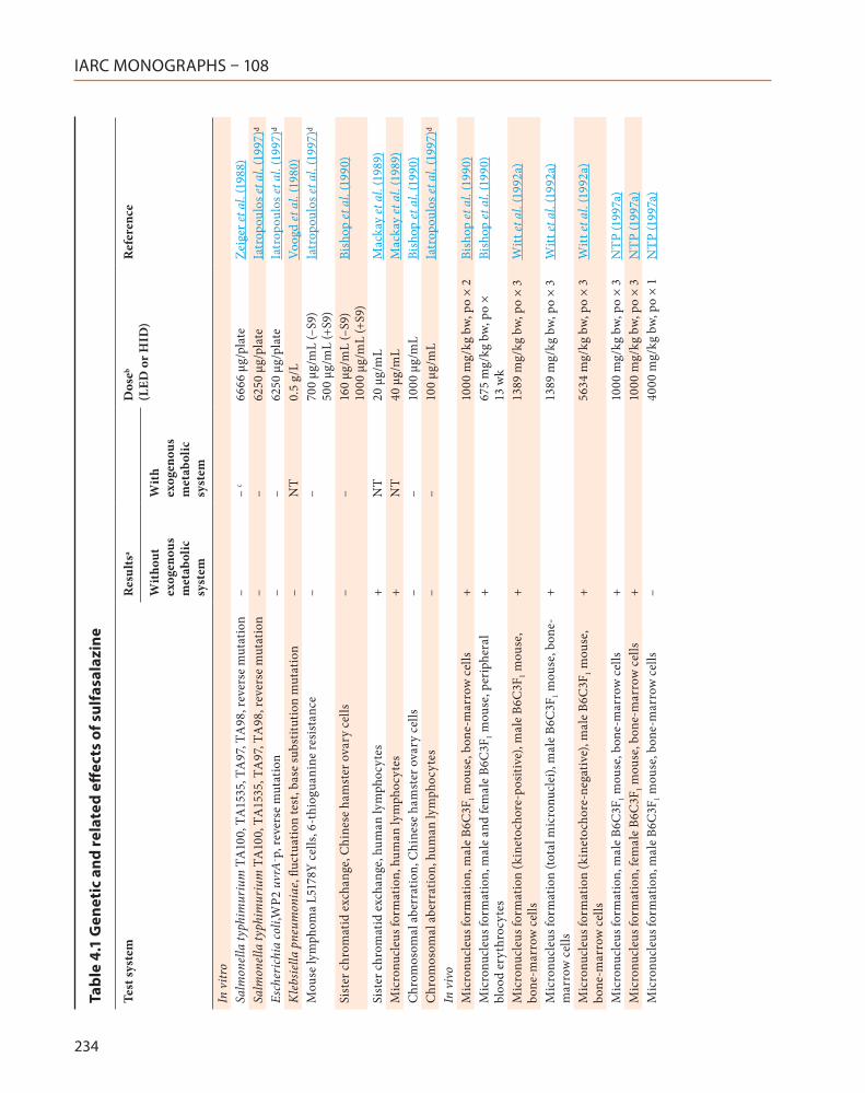
IARC MONOGRAPHS – 108
234
Tabl
e 4.
1 G
enet
ic a
nd re
late
d eff
ects
of s
ulfa
sala
zine
Test
syst
emR
esul
tsa
Dos
eb (L
ED o
r HID
)R
efer
ence
Wit
hout
ex
ogen
ous
met
abol
ic
syst
em
Wit
h ex
ogen
ous
met
abol
ic
syst
em
In v
itro
Salm
onel
la ty
phim
uriu
m T
A10
0, T
A15
35, T
A97
, TA
98, r
ever
se m
utat
ion
––
c66
66 μ
g/pl
ate
Zeig
er et
al.
(198
8)Sa
lmon
ella
typh
imur
ium
TA
100,
TA
1535
, TA
97, T
A98
, rev
erse
mut
atio
n–
–62
50 μ
g/pl
ate
Iatr
opou
los e
t al.
(199
7)d
Esch
eric
hia
coli,
WP2
uvr
A– p
, rev
erse
mut
atio
n–
–62
50 μ
g/pl
ate
Iatr
opou
los e
t al.
(199
7)d
Kle
bsie
lla p
neum
onia
e, flu
ctua
tion
test
, bas
e su
bstit
utio
n m
utat
ion
–N
T0.
5 g/
LVo
ogd
et a
l. (1
980)
Mou
se ly
mph
oma
L517
8Y c
ells
, 6-t
hiog
uani
ne re
sist
ance
––
700
μg/m
L (–
S9)
500
μg/m
L (+
S9)
Iatr
opou
los e
t al.
(199
7)d
Sist
er c
hrom
atid
exc
hang
e, C
hine
se h
amst
er o
vary
cel
ls–
–16
0 μg
/mL
(–S9
) 10
00 μ
g/m
L (+
S9)
Bish
op et
al.
(199
0)
Sist
er c
hrom
atid
exc
hang
e, h
uman
lym
phoc
ytes
+N
T20
μg/
mL
Mac
kay
et a
l. (1
989)
Mic
ronu
cleu
s for
mat
ion,
hum
an ly
mph
ocyt
es+
NT
40 μ
g/m
LM
acka
y et
al.
(198
9)C
hrom
osom
al a
berr
atio
n, C
hine
se h
amst
er o
vary
cel
ls–
–10
00 μ
g/m
LBi
shop
et a
l. (1
990)
Chr
omos
omal
abe
rrat
ion,
hum
an ly
mph
ocyt
es–
–10
0 μg
/mL
Iatr
opou
los e
t al.
(199
7)d
In v
ivo
Mic
ronu
cleu
s for
mat
ion,
mal
e B6
C3F
1 mou
se, b
one-
mar
row
cel
ls+
1000
mg/
kg b
w, p
o ×
2Bi
shop
et a
l. (1
990)
Mic
ronu
cleu
s for
mat
ion,
mal
e an
d fe
mal
e B6
C3F
1 mou
se, p
erip
hera
l bl
ood
eryt
hroc
ytes
+67
5 m
g/kg
bw,
po
× 13
wk
Bish
op et
al.
(199
0)
Mic
ronu
cleu
s for
mat
ion
(kin
etoc
hore
-pos
itive
), m
ale
B6C
3F1 m
ouse
, bo
ne-m
arro
w c
ells
+13
89 m
g/kg
bw,
po
× 3
Witt
et a
l. (1
992a
)
Mic
ronu
cleu
s for
mat
ion
(tota
l mic
ronu
clei
), m
ale
B6C
3F1 m
ouse
, bon
e-m
arro
w c
ells
+13
89 m
g/kg
bw,
po
× 3
Witt
et a
l. (1
992a
)
Mic
ronu
cleu
s for
mat
ion
(kin
etoc
hore
-neg
ativ
e), m
ale
B6C
3F1 m
ouse
, bo
ne-m
arro
w c
ells
+56
34 m
g/kg
bw,
po
× 3
Witt
et a
l. (1
992a
)
Mic
ronu
cleu
s for
mat
ion,
mal
e B6
C3F
1 mou
se, b
one-
mar
row
cel
ls+
1000
mg/
kg b
w, p
o ×
3N
TP (1
997a
)M
icro
nucl
eus f
orm
atio
n, fe
mal
e B6
C3F
1 mou
se, b
one-
mar
row
cel
ls+
1000
mg/
kg b
w, p
o ×
3N
TP (1
997a
)M
icro
nucl
eus f
orm
atio
n, m
ale
B6C
3F1 m
ouse
, bon
e-m
arro
w c
ells
–40
00 m
g/kg
bw,
po
× 1
NTP
(199
7a)
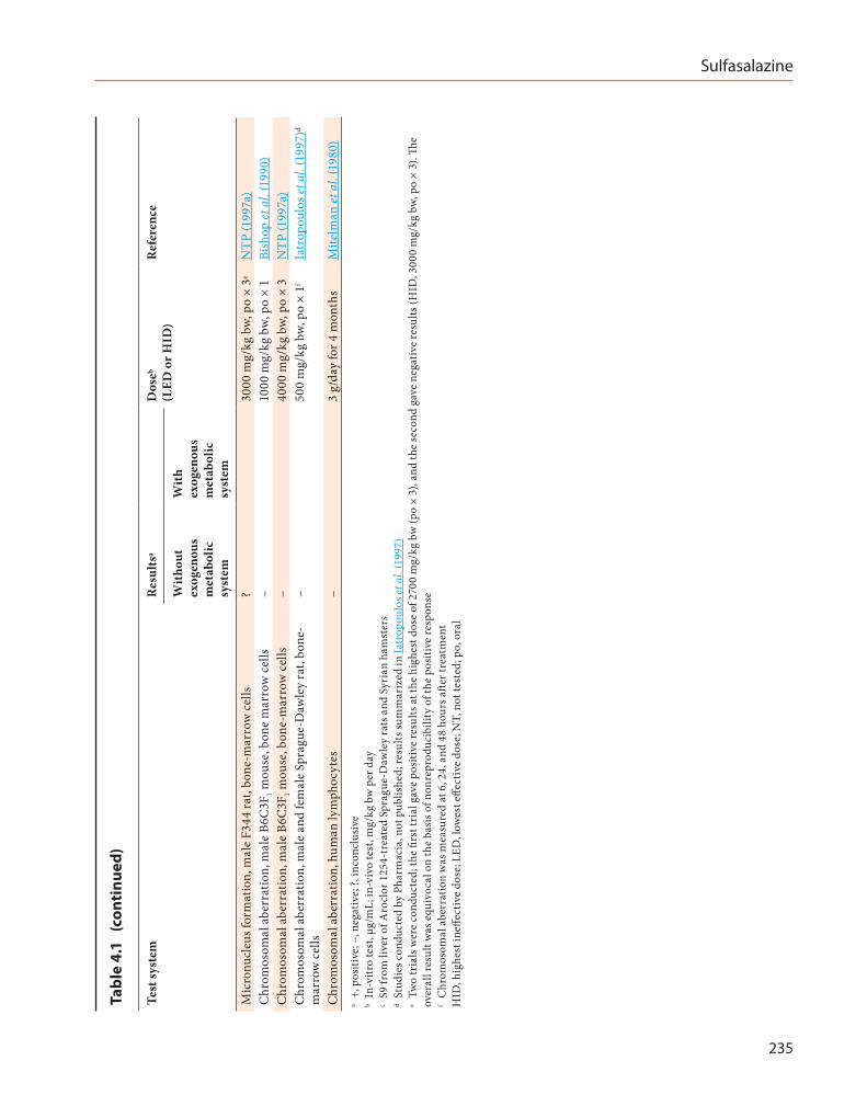
Sulfasalazine
235
Test
syst
emR
esul
tsa
Dos
eb (L
ED o
r HID
)R
efer
ence
Wit
hout
ex
ogen
ous
met
abol
ic
syst
em
Wit
h ex
ogen
ous
met
abol
ic
syst
em
Mic
ronu
cleu
s for
mat
ion,
mal
e F3
44 ra
t, bo
ne-m
arro
w c
ells
?30
00 m
g/kg
bw,
po
× 3e
NTP
(199
7a)
Chr
omos
omal
abe
rrat
ion,
mal
e B6
C3F
1 mou
se, b
one
mar
row
cel
ls–
1000
mg/
kg b
w, p
o ×
1Bi
shop
et a
l. (1
990)
Chr
omos
omal
abe
rrat
ion,
mal
e B6
C3F
1 mou
se, b
one-
mar
row
cel
ls–
4000
mg/
kg b
w, p
o ×
3N
TP (1
997a
)C
hrom
osom
al a
berr
atio
n, m
ale
and
fem
ale
Spra
gue-
Daw
ley
rat,
bone
-m
arro
w c
ells
–50
0 m
g/kg
bw,
po
× 1f
Iatr
opou
los e
t al.
(199
7)d
Chr
omos
omal
abe
rrat
ion,
hum
an ly
mph
ocyt
es–
3 g/
day
for 4
mon
ths
Mite
lman
et a
l. (1
980)
a +,
pos
itive
; –, n
egat
ive;
?, in
conc
lusi
ve
b In
-vitr
o te
st, μ
g/m
L; in
-viv
o te
st, m
g/kg
bw
per
day
c
S9 fr
om li
ver o
f Aro
clor
125
4-tr
eate
d Sp
ragu
e-D
awle
y ra
ts a
nd S
yria
n ha
mst
ers
d St
udie
s con
duct
ed b
y Ph
arm
acia
, not
pub
lishe
d; re
sults
sum
mar
ized
in Ia
trop
oulo
s et a
l. (1
997)
e Tw
o tr
ials
wer
e co
nduc
ted;
the
first
tria
l gav
e po
sitiv
e re
sults
at t
he h
ighe
st d
ose
of 2
700
mg/
kg b
w (p
o ×
3), a
nd th
e se
cond
gav
e ne
gativ
e re
sults
(HID
, 300
0 m
g/kg
bw
, po
× 3)
. The
over
all r
esul
t was
equ
ivoc
al o
n th
e ba
sis o
f non
repr
oduc
ibili
ty o
f the
pos
itive
resp
onse
f C
hrom
osom
al a
berr
atio
n w
as m
easu
red
at 6
, 24,
and
48
hour
s afte
r tre
atm
ent
HID
, hig
hest
ineff
ectiv
e do
se; L
ED, l
owes
t effe
ctiv
e do
se; N
T, n
ot te
sted
; po,
ora
l
Tabl
e 4.
1 (
cont
inue
d)
IARC MONOGRAPHS – 108
236
lymphocytes after treatment with sulfasalazine (effective concentration range, 40–160 μg/mL) in the absence of metabolic activation was reported by Mackay et al. (1989). No significant increases in the frequency of chromosomal aberration were observed in cultured Chinese hamster ovary cells (Bishop et al., 1990), or cultured human lymphocytes (Iatropoulos et al., 1997), treated with sulfasalazine (concentration, up to 1000 or 100 μg/mL, respectively). Thus the results of tests for chromosomal damage in vitro after treat-ment with sulfasalazine were generally negative, although sporadic positive results were reported.
In vivo, consistent with results reported in assays in vitro, no increases in the frequency of chromosomal aberration were observed in male mice or male and female rats treated with sulfasalazine by gavage at doses of up to 4000 mg/kg bw per day (Bishop et al., 1990; Iatropoulos et al., 1997; NTP, 1997a).
The results of assays for micronucleus forma-tion in male and female mice treated with sulfasalazine were uniformly positive when multiple treatments (at least two) were employed (Bishop et al., 1990; Witt et al., 1992a, NTP, 1997a). Further investigation of the nature of the induced micronuclei revealed that the majority were kinetochore-positive, suggesting that the micronuclei contained whole chromosomes rather than fragments, and were primarily due to aneuploidy events rather than chromosome breakage (Witt et al., 1992a). This observation was consistent with the negative results in assays for chromosomal aberration with sulfasalazine in vitro and in vivo (Mitelman et al., 1980; Bishop et al., 1990; Iatropoulos et al., 1997; NTP, 1997a). The negative results of one test in male mice given a single dose of sulfasalazine at 4000 mg/kg bw underscored the need for multiple treatments to induce an observable increase in micronucleus formation (NTP, 1997a).
In addition to the necessity for multiple treatments, sulfasalazine may also demonstrate selective activity in mice; the results of a study on
micronucleus formation in bone marrow of male rats given three doses of sulfasalazine (highest dose, 3000 mg/kg bw) were judged to be equiv-ocal (NTP, 1997a); in this assay, an initial trial gave a positive response at the highest dose of 2700 mg/kg bw, but a second trial, with a highest dose of 3000 mg/kg bw, gave negative results. This apparent preferential activity in mice was consistent with the observation that mice have a greater systemic exposure than rats to sulfapyri-dine, the active moiety, after administration of similar doses (Zheng et al., 1993).
In one study, no evidence for genotoxicity was obtained for sulfasalazine when tested for the induction of micronuclei in mouse bone marrow, with or without pretreatment with folate. Likewise, no evidence for formation of DNA adducts was detected by 32P-postlabelling in rat and mouse liver and urinary bladder (Iatropoulos et al., 1997). [The nuclease P1 enrichment proce-dure was used in the 32P-postlabelling method. Assuming that N-hydroxylation of sulfapyri-dine occurred in vivo, adducts derived from this metabolite would probably be lost.]
4.2.3 Genotoxicity of sulfasalazine metabolites
See Table 4.2Sulfasalazine has two major metabolites,
sulfapyridine (a carrier molecule that allows transport of sulfasalazine to the intestine, where it is activated) and 5-ASA, the therapeutically active moiety.
(a) 5-ASA
5-ASA has not shown activity in any assay for genotoxicity in vitro or in vivo. It does not induce mutations in any of a variety of Salmonella typh-imurium strains, with or without metabolic acti-vation or in Klebsiella pneumoniae in the absence of metabolic activation (Voogd et al., 1980). No induction of sister chromatid exchange, micro-nucleus formation, or chromosomal aberration
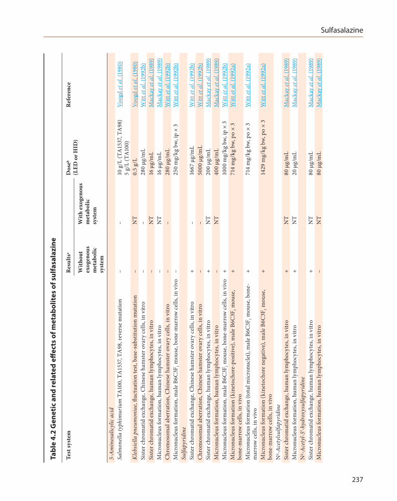
Sulfasalazine
237
Tabl
e 4.
2 G
enet
ic a
nd re
late
d eff
ects
of m
etab
olit
es o
f sul
fasa
lazi
ne
Test
syst
emR
esul
tsa
Dos
eb (L
ED o
r HID
)R
efer
ence
Wit
hout
ex
ogen
ous
met
abol
ic
syst
em
Wit
h ex
ogen
ous
met
abol
ic
syst
em
5-A
min
osal
icyl
ic a
cid
Salm
onel
la ty
phim
uriu
m T
A10
0, T
A15
37, T
A98
, rev
erse
mut
atio
n–
–10
g/L
(TA
1537
, TA
98)
5 g/
L (T
A10
0)Vo
ogd
et a
l. (1
980)
Kle
bsie
lla p
neum
onia
e, flu
ctua
tion
test
, bas
e-su
bstit
utio
n m
utat
ion
–N
T0.
5 g/
LVo
ogd
et a
l. (1
980)
Sist
er c
hrom
atid
exc
hang
e, C
hine
se h
amst
er o
vary
cel
ls, i
n vi
tro
––
280
μg/m
LW
itt et
al.
(199
2b)
Sist
er c
hrom
atid
exc
hang
e, h
uman
lym
phoc
ytes
, in
vitr
o–
NT
16 μ
g/m
LM
acka
y et
al.
(198
9)M
icro
nucl
eus f
orm
atio
n, h
uman
lym
phoc
ytes
, in
vitr
o–
NT
16 μ
g/m
LM
acka
y et
al.
(198
9)C
hrom
osom
al a
berr
atio
n, C
hine
se h
amst
er o
vary
cel
ls, i
n vi
tro
––
280
μg/m
LW
itt et
al.
(199
2b)
Mic
ronu
cleu
s for
mat
ion,
mal
e B6
C3F
1 mou
se, b
one-
mar
row
cel
ls, i
n vi
vo–
250
mg/
kg b
w, ip
× 3
Witt
et a
l. (1
992b
)Su
lfapy
ridi
neSi
ster
chr
omat
id e
xcha
nge,
Chi
nese
ham
ster
ova
ry c
ells
, in
vitr
o+
–16
67 μ
g/m
LW
itt et
al.
(199
2b)
Chr
omos
omal
abe
rrat
ion,
Chi
nese
ham
ster
ova
ry c
ells
, in
vitr
o–
–50
00 μ
g/m
LW
itt et
al.
(199
2b)
Sist
er c
hrom
atid
exc
hang
e, h
uman
lym
phoc
ytes
, in
vitr
o+
NT
200
μg/m
LM
acka
y et
al.
(198
9)M
icro
nucl
eus f
orm
atio
n, h
uman
lym
phoc
ytes
, in
vitr
o–
NT
400
μg/m
LM
acka
y et
al.
(198
9)M
icro
nucl
eus f
orm
atio
n, m
ale
B6C
3F1 m
ouse
, bon
e-m
arro
w c
ells
, in
vivo
+10
00 m
g/kg
bw,
ip ×
3W
itt et
al.
(199
2b)
Mic
ronu
cleu
s for
mat
ion
(kin
etoc
hore
-pos
itive
), m
ale
B6C
3F1 m
ouse
, bo
ne-m
arro
w c
ells
, in
vivo
+71
4 m
g/kg
bw,
po
× 3
Witt
et a
l. (1
992a
)
Mic
ronu
cleu
s for
mat
ion
(tota
l mic
ronu
clei
), m
ale
B6C
3F1 m
ouse
, bon
e-m
arro
w c
ells
, in
vivo
+71
4 m
g/kg
bw,
po
× 3
Witt
et a
l. (1
992a
)
Mic
ronu
cleu
s for
mat
ion
(kin
etoc
hore
neg
ativ
e), m
ale
B6C
3F1 m
ouse
, bo
ne-m
arro
w c
ells
, in
vivo
+14
29 m
g/kg
bw,
po
× 3
Witt
et a
l. (1
992a
)
N4 -A
cety
lsulfa
pyri
dine
Sist
er c
hrom
atid
exc
hang
e, h
uman
lym
phoc
ytes
, in
vitr
o+
NT
80 μ
g/m
LM
acka
y et
al.
(198
9)M
icro
nucl
eus f
orm
atio
n, h
uman
lym
phoc
ytes
, in
vitr
o+
NT
20 μ
g/m
LM
acka
y et
al.
(198
9)N
4 -Ace
tyl-5′-h
ydro
xysu
lfapy
ridi
neSi
ster
chr
omat
id e
xcha
nge,
hum
an ly
mph
ocyt
es, i
n vi
tro
+N
T80
μg/
mL
Mac
kay
et a
l. (1
989)
Mic
ronu
cleu
s for
mat
ion,
hum
an ly
mph
ocyt
es, i
n vi
tro
–N
T80
μg/
mL
Mac
kay
et a
l. (1
989)
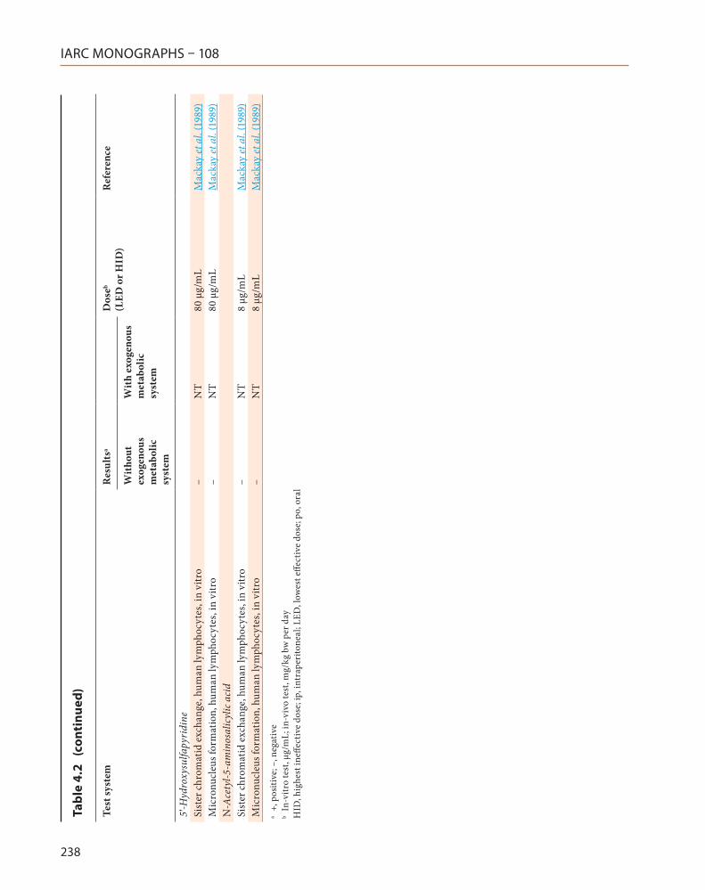
IARC MONOGRAPHS – 108
238
Test
syst
emR
esul
tsa
Dos
eb (L
ED o
r HID
)R
efer
ence
Wit
hout
ex
ogen
ous
met
abol
ic
syst
em
Wit
h ex
ogen
ous
met
abol
ic
syst
em
5′-H
ydro
xysu
lfapy
ridi
neSi
ster
chr
omat
id e
xcha
nge,
hum
an ly
mph
ocyt
es, i
n vi
tro
–N
T80
μg/
mL
Mac
kay
et a
l. (1
989)
Mic
ronu
cleu
s for
mat
ion,
hum
an ly
mph
ocyt
es, i
n vi
tro
–N
T80
μg/
mL
Mac
kay
et a
l. (1
989)
N-A
cety
l-5-a
min
osal
icyl
ic a
cid
Sist
er c
hrom
atid
exc
hang
e, h
uman
lym
phoc
ytes
, in
vitr
o–
NT
8 μg
/mL
Mac
kay
et a
l. (1
989)
Mic
ronu
cleu
s for
mat
ion,
hum
an ly
mph
ocyt
es, i
n vi
tro
–N
T8
μg/m
LM
acka
y et
al.
(198
9)a
+, p
ositi
ve; –
, neg
ativ
e b
In-v
itro
test
, μg/
mL;
in-v
ivo
test
, mg/
kg b
w p
er d
ayH
ID, h
ighe
st in
effec
tive
dose
; ip,
intr
aper
itone
al; L
ED, l
owes
t effe
ctiv
e do
se; p
o, o
ral
Tabl
e 4.
2 (
cont
inue
d)
Sulfasalazine
239
has been reported in human lymphocytes or Chinese hamster ovary cells in vitro (Mackay et al., 1989; Witt et al., 1992b). In vivo, no increase in the frequency of micronucleated polychro-matic erythrocytes was observed in the bone marrow of male mice treated with 5-ASA (dose range, 125–250 mg/kg bw per day for 3 days) by intraperitoneal injection (Witt et al., 1992b).
(b) Sulfapyridine
Sulfapyridine has been reported to induce sister chromatid exchange in Chinese hamster ovary cells and cultured human lymphocytes in the absence of metabolic activation (Mackay et al., 1989; Witt et al., 1992b); no increase in the frequency of sister chromatid exchange was noted in the presence of metabolic activation in Chinese hamster ovary cells (Witt et al., 1992b). Sulfapyridine did not induce chromosomal aber-ration in Chinese hamster ovary cells, with or without metabolic activation (Witt et al., 1992b). Mackay et al. (1989) reported that sulfapyri-dine did not induce micronucleus formation in cultured human lymphocytes in the absence of metabolic activation, at concentrations that reached 400 µg/mL. Sulfapyridine induced a strong, dose-related increase in the frequency of micronucleated polychromatic erythrocytes when administered either as multiple intraperi-toneal injections (Witt et al., 1992b) or by gavage (Witt et al., 1992a). As with sulfasalazine, the majority of micronucleated erythrocytes induced by sulfapyridine in mice were shown to contain kinetochores (Witt et al., 1992a), implying that sulfapyridine-induced micronucleus formation resulted from failure of mitotic chromosomal segregation, rather than chromosome breakage.
(c) Metabolites of 5-ASA and sulfapyridine
Mackay et al. (1989) also tested four acetylated and/or hydroxylated metabolites of sulfapyridine and 5-ASA for their ability to induce sister chromatid exchange and micronucleus formation in cultured human
lymphocytes. N4-acetylsulfapyridine was capable of inducing both sister chromatid exchange and micronucleus formation, while N4-acetyl-5′-hydroxysulfapyridine only induced sister chro-matid exchange. 5′-Hydroxysulfapyridine and N4-acetyl-5-aminosalicylic acid did not induce either sister chromatid exchange or micronu-cleus formation at the concentrations tested.
4.3 Other mechanistic data relevant to carcinogenicity
4.3.1 Adverse effects
In humans, sulfasalazine is associated with a wide range of adverse side-effects that include agranulocytosis (Kaufman et al., 1996), hepa-totoxicity (de Abajo et al., 2004; Jobanputra et al., 2008), nephrotoxicity (Gisbert et al., 2007), neurotoxicity (Liedorp et al., 2008), and pulmo-nary toxicity (Parry et al., 2002). Sulfasalazine is also associated with reversible infertility in men and in male experimental animals (O’Moráin et al., 1984). Reactions to sulfasalazine may result from an idiosyncratic delayed-type hypersensi-tivity reaction that may affect internal organs in variable ways (Jobanputra et al., 2008).
Case reports of serious hepatotoxicity asso-ciated with sulfasalazine are frequent and occur predominantly within the first month of starting therapy; the pattern of liver injury can be hepa-tocellular or cholestatic, and may lead to liver failure. Serious hepatotoxicity, which could be a part of the DRESS (drug rash, eosinophilia and systemic symptoms) syndrome is described in approximately 0.1% of users, but the estimated incidence is higher (0.4%) in patients with inflammatory arthritis (de Abajo et al., 2004; Jobanputra et al., 2008).
Renal toxicity associated with sulfasalazine treatment may be irreversible. Although the sulfapyridine moiety is thought to be respon-sible for most of the adverse effects of sulfasala-zine, several case reports in patients with IBD

IARC MONOGRAPHS – 108
240
indicate that renal toxicity in humans may occur from treatment with both sulfasalazine and 5-ASA (Gisbert et al., 2007). Clinically, 5-ASA-associated nephrotoxicity is typically expressed as interstitial nephritis, glomerulo-nephritis, nephritic syndrome, and acute renal failure (Barbour & Williams, 1990; Birketvedt et al., 2000; Augusto et al., 2009). The incidence of clinically restrictive renal impairment has been estimated at < 1 per 500 patients (World et al., 1996). The mechanism is unclear, although both a delayed cell-mediated response, and a dose-de-pendent effect have been considered (Corrigan & Stevens, 2000). 5-ASA-related nephrotoxicity appeared to be dose-related in female rats given a single intravenous injection of the sodium salt of 5-ASA at doses of up to 5.7 mmol/kg bw (Calder et al., 1972); however, dose-dependency may require the administration of doses much higher than those given to humans. Of note, 5-ASA combines the structural features of a salicy-late and a phenacetin, both of which have well documented nephrotoxic potential (Corrigan & Stevens, 2000).
It has been hypothesized that oxidative stress may be a factor in sulfasalazine-induced renal and hepatic injury. Treatment-related alterations in the levels of biomarkers of oxidative stress were detected in kidney and liver tissues of male Sprague-Dawley rats given sulfasalazine as daily oral doses at 0, 300, or 600 mg/kg bw for 14 days. At the highest dose, there were significant decreases in the activities of renal and hepatic superoxide dismutase, and significant increases in catalase activity, thiobarbituric acid-reactive substances, and in the oxidized/reduced glutathione ratio (Linares et al., 2009).
Sulfasalazine can cause haemolytic anaemia (Das et al., 1973; Mechanick, 1985) and methae-moglobinaemia (Miller et al., 1971; Kater, 1974; Azad Khan et al., 1983). In a group of 50 patients receiving sulfasalazine at 2.5 g per day as mainte-nance therapy for ulcerative colitis, approximately 40% had elevated levels of methaemoglobin
(Pounder et al., 1975). Although sulfonamide-in-duced haemolysis can be severe in patients with glucose-6-phosphate dehydrogenase deficiency, this study showed that sulfasalazine-induced erythrocyte damage also occurred in patients with normal levels of this enzyme (Pounder et al., 1975).
The role of metabolites in sulfasalazine-me-diated toxicity was investigated in vitro, using human erythrocytes and mononuclear leuko-cytes as target cells in the presence of human liver microsomes; methaemoglobin formation and cytotoxicity were selected as toxicity end-points. In addition to sulfasalazine, the study included the metabolites 5-ASA, sulfapyridine, and 5′-hydroxysulfapyridine (Pirmohamed et al., 1991). Bioactivation by human liver microsomes that are dependent on reduced nicotinamide adenine dinucleotide phosphate (NADPH) to a species that caused methaemoglobinaemia and cytotoxicity was only observed with sulfapyri-dine. Chromatographic analysis demonstrated that sulfapyridine was converted to a short-lived intermediate (t1/2, 8.1 minutes at pH 7.4) with elution characteristics identical to those of synthetic sulfapyridine hydroxylamine. This hydroxylamine (10–500 µM) caused a concen-tration-dependent increase in both methae-moglobinaemia (2.9–24.4%) and cytotoxicity in the absence of a microsomal system; neither sulfasalazine nor any of the other test metabolites had such effects. When the microsomal incuba-tions were conducted in the presence of micro-molar concentrations of reducing agents (e.g. ascorbic acid, glutathione, or N-acetylcysteine), sulfapyridine-induced cytotoxity was decreased in mononuclear leukocytes, but there was no effect upon the levels of methaemoglobinaemia. This suggested that sulfapyridine hydroxy-lamine could readily penetrate erythrocytes, where it may undergo redox cycling to nitroso-sulfapyridine, the species ultimately respon-sible for the production of methaemoglobin. These observations further suggested that
Sulfasalazine
241
N-hydroxylation of sulfapyridine may account for some of the adverse effects associated with sulfasalazine (Pirmohamed et al., 1991). [Of note, N-hydroxylation is thought to play a key role in the bioactivation of aromatic amine carcinogens, such as 4-aminobiphenyl (IARC, 2012).]
4.3.2 Effects upon folate pathways
Sulfasalazine has been shown to inhibit the activity of dihydrofolate reductase, methylene-tetrahydrofolate reductase, and serine transhy-droxymethylase, and also the cellular uptake of folate (Selhub et al., 1978; Jansen et al., 2004; Urquhart et al., 2010).
4.3.3 Urolithiasis
An increased incidence of transitional cell papilloma of the urinary bladder in male rats treated with sulfasalazine has been correlated (P < 0.01) with increased incidences of concre-tions (calculi) in the urinary bladder (NTP, 1997a; see also Section 3). In a subsequent study, there was decreased incidence of urinary hyperplasia in male rats subjected to caloric restriction and treated with sulfasalazine, and little evidence of urinary bladder concretion, compared with rats fed ad libitum (NTP, 1997b; see also Section 3). [These data suggested that chronic inflamma-tion associated with urolithiasis may be a factor in sulfasalazine-induced carcinogenesis of the bladder in male rats.]
4.4 Susceptibility
The adverse effects of sulfasalazine have been linked to sulfapyridine (Das et al., 1973). This metabolite, which is well absorbed from the colon, is inactivated by NAT2-mediated N-acetylation. NAT2 polymorphisms have been associated with different susceptibilities to the adverse effects of sulfasalazine. People with the slow-acetylator genotype have higher serum concentrations of free sulfapyridine and lower concentrations of
acetylated sulfapyridine than fast acetylators (see also Section 4.1.1), and appear more likely to experience toxic symptoms when treated with equivalent doses of sulfasalazine (Das & Dubin, 1976; Azad Khan et al., 1983; Ricart et al., 2002; Tanaka et al., 2002; Kumagai et al., 2004; Chen et al., 2007; Soejima et al., 2008).
4.5 Mechanistic considerations
Sulfasalazine was reported to act as a co-car-cinogen at a dose of 60 mg/kg bw per day in the 1,2-dimethylhydrazine model of colon carcino-genesis in rats. In the same study, 5-ASA, the active pharmacophore unit of sulfasalazine, acted as a co-carcinogen at a dose of 30 mg/kg bw per day, but not at 60 mg/kg bw per day, which suggested that 5-ASA exerts a protective effect on the colon mucosa, provided a sufficient amount of the compound reaches the colon (Davis et al., 1992). On the basis of early proposals that localized tissue folate deficiency may account for carcinogenesis (Lashner et al., 1989), it was hypothesized that sulfasalazine may be co-car-cinogenic due to its anti-folate characteristics. Sulfasalazine inhibited dihydrofolate reductase, methylenetetrahydrofolate reductase, and serine transhydroxymethylase, and also the cellular uptake of folate (Selhub et al., 1978; Jansen et al., 2004; Urquhart et al., 2010). Reduced levels of S-adenosylmethionine or 5,10-methylenetet-rahydrofolate, required for thymidine synthesis, might account for the effect; however, colonic cells may not be completely dependent on blood stream nutrients (Meenan, 1993). In the rat, colonic bacterial folate is incorporated in the hepatic folate pool (Rong et al., 1991), and this could counteract sulfasalazine-induced folate depletion (Meenan, 1993). In patients with ulcer-ative colitis, folate concentrations measured in colonic epithelial cells obtained from endoscopic colon biopsies were not decreased in sulfasala-zine-treated patients compared with controls; this contrasted with serum concentrations of
IARC MONOGRAPHS – 108
242
folate, which were reduced in patients receiving sulfasalazine (Meenan et al., 1996). These data suggested that the potentially protective effects of folate supplementation against colorectal carcinogenesis in patients with ulcerative colitis were not due to correction of localized folate deficiency.
Two-year studies in male and female F344/N rats given sulfasalazine by gavage indicated some evidence for carcinogenic activity on the basis of increased incidences of transitional cell papil-loma of the urinary bladder, and clear evidence for carcinogenic activity in male and female B6C3F1 mice on the basis of increased incidences of hepatocellular adenoma and hepatocellular carcinoma (NTP, 1997a; see also Section 3). The data on mutagenicity of sulfasalazine and its metabolite, sulfapyridine, suggested that the parent drug and the metabolite are predomi-nantly aneugens (Bishop et al., 1990; Witt et al., 1992a, b). Increased frequencies of micronucleus formation and sister chromatid exchange in patients with IBD receiving sulfasalazine have been reported, but confounding factors were apparent in the study (Erskine et al., 1984).
Folate deficiency was considered as a possible explanation for the induction of micronucleus formation by sulfasalazine in vivo. However, patients reported to have an elevated frequency of sister chromatid exchange and micronucleus formation had serum folate concentrations that were at the low end of the normal range, and the observation of reticulocytosis in a 90-day study in mice suggested an erythropoietic effect not characteristic of folate deficiency (Bishop et al., 1990).
The increased incidence of transitional cell papilloma of the urinary bladder in male rats treated orally with sulfasalazine was correlated with increased incidence of concretions (calculi) in the urinary bladder (NTP, 1997a). Chronic inflammation associated with urolithiasis may be a factor in sulfasalazine-induced carcinogen-esis of the bladder in male rats.
5. Summary of Data Reported
5.1 Exposure dataSulfasalazine is a synthetic aminosalicylate
used as an oral anti-inflammatory drug. The most common use of sulfasalazine is for the treatment of autoimmune arthritis. Prescriptions have been stable over the past decade, with global sales of US$ 222 million in 2012. Environmental contamination with sulfasalazine in ground-water has been noted, but exposure is likely to be predominantly through use as a medication.
5.2 Human carcinogenicity dataThe available studies of exposure to sulfasala-
zine included a surveillance study, two cohort studies, three nested case–control studies, and three case–control studies on cancer of the colorectum among patients with inflammatory bowel disease or ulcerative colitis. These studies were hampered in their ability to evaluate the association between exposure to sulfasalazine and risk of cancer of the colorectum by the small numbers of exposed cases, imprecise risk estimates, and little information on exposure or the dose or duration of sulfasalazine use. There were also concerns about selection bias in some studies based on clinical populations.
The best designed studies were a nested case–control study from a large cohort from the General Practice Research Database in the United Kingdom, and a large population-based case–control study among patients with inflam-matory bowel disease in the USA that evaluated exposure–response relationships. Conflicting findings were reported across studies, with four studies reporting relative risks of less than unity, two studies reporting estimates close to unity, and two studies reporting a relative risk of greater than unity. Most of these relative risks were not statistically significant. In the studies that evaluated dose–response relationships, no clear patterns were observed.

Sulfasalazine
243
5.3 Animal carcinogenicity data
In one study in male and female mice given sulfasalazine by gavage, there was a signifi-cant increase in the incidence of hepatocellular adenoma, and of hepatocellular adenoma or carcinoma (combined) in both sexes; there was also an increase in the incidence of hepatocellular carcinoma in females.
In a study of dietary restriction in male mice given diets containing sulfasalazine ad libitum, there were significant increases in the incidence of hepatocellular adenoma, and hepatocellular adenoma or carcinoma (combined) in exposed mice compared with the controls fed ad libitum and the weight-matched controls. In contrast, the incidence of hepatocellular tumours in dietary-re-stricted mice was significantly decreased after 2 years in the group exposed to sulfasalazine, and was similar to that in non-treated mice after 3 years.
In one study in male and female rats given sulfasalazine by gavage, there was a significant increase in the incidence of transitional cell papilloma of the urinary bladder in males; there was also a non-significant increase in the inci-dences of rare transitional cell papilloma of the kidney and of rare transitional cell papilloma of the urinary bladder in female rats.
In a study of dietary restriction in male rats, the incidence of transitional cell papilloma of the urinary bladder was significantly greater in rats receiving sulfasalazine than in controls fed ad libitum. No significant increase in the incidence of transitional cell papilloma of the urinary bladder was observed in rats subjected to dietary restriction and given sulfasalazine.
In a co-carcinogenicity study in male rats, sulfasalazine increased the total number and multiplicity of 1,2-dimethylhydrazine-induced intestinal tumours.
5.4 Mechanistic and other relevant data
The sulfasalazine molecule contains a 5-amino salicylic acid moiety linked by an azo bond to a sulfapyridine moiety. Cleavage of the azo bond by bacterial azoreductases in the colon releases two pharmacologically active compounds: 5-aminosalicylic acid and sulfapyri-dine. Sulfapyridine is absorbed, and N-acetylated by the highly polymorphic N-acetyl transferase 2 (NAT2), resulting in considerable inter-in-dividual variation in the pharmacokinetics of sulfasalazine.
Sulfasalazine is not mutagenic in standard bacterial assays for gene mutation, with or without exogenous metabolic activation. Tests for chro-mosomal damage in vitro after treatment with sulfasalazine were generally negative, although sporadic positive results have been reported. Likewise, no increases in the frequency of chro-mosomal aberration were observed in male mice or rats treated with sulfasalazine. Positive results were consistently obtained in assays for micro-nucleus formation in male and female mice in vivo when multiple treatments with sulfasala-zine were given; the results suggested that these results were primarily due to aneuploidy events rather than chromosome breakage.
Sulfasalazine inhibits the activity of dihy-drofolate reductase, methylenetetrahydrofolate reductase, and serine transhydroxymethylase, and also the cellular uptake of folate. However, folate deficiency does not appear to account for the effects of sulfasalazine in humans and mice.
The sulfasalazine metabolite sulfapyridine has been shown to undergo N-hydroxylation when incubated with human liver microsomes in the presence of NADPH. N-Hydroxylation is known to account for the bioactivation of carcinogenic aromatic amines to DNA-binding species, and such a pathway would be consistent with the target organs associated with sulfasala-zine-induced carcinogenicity in rats. However, no
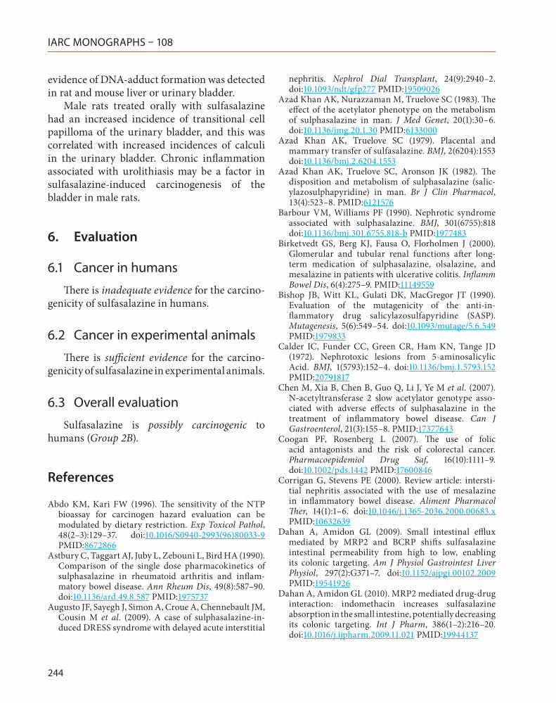
IARC MONOGRAPHS – 108
244
evidence of DNA-adduct formation was detected in rat and mouse liver or urinary bladder.
Male rats treated orally with sulfasalazine had an increased incidence of transitional cell papilloma of the urinary bladder, and this was correlated with increased incidences of calculi in the urinary bladder. Chronic inflammation associated with urolithiasis may be a factor in sulfasalazine-induced carcinogenesis of the bladder in male rats.
6. Evaluation
6.1 Cancer in humans
There is inadequate evidence for the carcino-genicity of sulfasalazine in humans.
6.2 Cancer in experimental animals
There is sufficient evidence for the carcino-genicity of sulfasalazine in experimental animals.
6.3 Overall evaluation
Sulfasalazine is possibly carcinogenic to humans (Group 2B).
References
Abdo KM, Kari FW (1996). The sensitivity of the NTP bioassay for carcinogen hazard evaluation can be modulated by dietary restriction. Exp Toxicol Pathol, 48(2–3):129–37. doi:10.1016/S0940-2993(96)80033-9 PMID:8672866
Astbury C, Taggart AJ, Juby L, Zebouni L, Bird HA (1990). Comparison of the single dose pharmacokinetics of sulphasalazine in rheumatoid arthritis and inflam-matory bowel disease. Ann Rheum Dis, 49(8):587–90. doi:10.1136/ard.49.8.587 PMID:1975737
Augusto JF, Sayegh J, Simon A, Croue A, Chennebault JM, Cousin M et al. (2009). A case of sulphasalazine-in-duced DRESS syndrome with delayed acute interstitial
nephritis. Nephrol Dial Transplant, 24(9):2940–2. doi:10.1093/ndt/gfp277 PMID:19509026
Azad Khan AK, Nurazzaman M, Truelove SC (1983). The effect of the acetylator phenotype on the metabolism of sulphasalazine in man. J Med Genet, 20(1):30–6. doi:10.1136/jmg.20.1.30 PMID:6133000
Azad Khan AK, Truelove SC (1979). Placental and mammary transfer of sulfasalazine. BMJ, 2(6204):1553 doi:10.1136/bmj.2.6204.1553
Azad Khan AK, Truelove SC, Aronson JK (1982). The disposition and metabolism of sulphasalazine (salic-ylazosulphapyridine) in man. Br J Clin Pharmacol, 13(4):523–8. PMID:6121576
Barbour VM, Williams PF (1990). Nephrotic syndrome associated with sulphasalazine. BMJ, 301(6755):818 doi:10.1136/bmj.301.6755.818-b PMID:1977483
Birketvedt GS, Berg KJ, Fausa O, Florholmen J (2000). Glomerular and tubular renal functions after long-term medication of sulphasalazine, olsalazine, and mesalazine in patients with ulcerative colitis. Inflamm Bowel Dis, 6(4):275–9. PMID:11149559
Bishop JB, Witt KL, Gulati DK, MacGregor JT (1990). Evaluation of the mutagenicity of the anti-in-flammatory drug salicylazosulfapyridine (SASP). Mutagenesis, 5(6):549–54. doi:10.1093/mutage/5.6.549 PMID:1979833
Calder IC, Funder CC, Green CR, Ham KN, Tange JD (1972). Nephrotoxic lesions from 5-aminosalicylic Acid. BMJ, 1(5793):152–4. doi:10.1136/bmj.1.5793.152 PMID:20791817
Chen M, Xia B, Chen B, Guo Q, Li J, Ye M et al. (2007). N-acetyltransferase 2 slow acetylator genotype asso-ciated with adverse effects of sulphasalazine in the treatment of inflammatory bowel disease. Can J Gastroenterol, 21(3):155–8. PMID:17377643
Coogan PF, Rosenberg L (2007). The use of folic acid antagonists and the risk of colorectal cancer. Pharmacoepidemiol Drug Saf, 16(10):1111–9. doi:10.1002/pds.1442 PMID:17600846
Corrigan G, Stevens PE (2000). Review article: intersti-tial nephritis associated with the use of mesalazine in inflammatory bowel disease. Aliment Pharmacol Ther, 14(1):1–6. doi:10.1046/j.1365-2036.2000.00683.x PMID:10632639
Dahan A, Amidon GL (2009). Small intestinal efflux mediated by MRP2 and BCRP shifts sulfasalazine intestinal permeability from high to low, enabling its colonic targeting. Am J Physiol Gastrointest Liver Physiol, 297(2):G371–7. doi:10.1152/ajpgi.00102.2009 PMID:19541926
Dahan A, Amidon GL (2010). MRP2 mediated drug-drug interaction: indomethacin increases sulfasalazine absorption in the small intestine, potentially decreasing its colonic targeting. Int J Pharm, 386(1–2):216–20. doi:10.1016/j.ijpharm.2009.11.021 PMID:19944137
Sulfasalazine
245
Das KM, Dubin R (1976). Clinical pharmacokinetics of sulphasalazine. Clin Pharmacokinet, 1(6):406–25. doi:10.2165/00003088-197601060-00002 PMID:15752
Das KM, Eastwood MA, McManus JPA, Sircus W (1973). Adverse reactions during salicylazosulfapyridine therapy and the relation with drug metabolism and acetylator phenotype. N Engl J Med, 289(10):491–5. doi:10.1056/NEJM197309062891001 PMID:4146729
Davis AE, Patterson F, Crouch R (1992). The effect of therapeutic drugs used in inflammatory bowel disease on the incidence and growth of colonic cancer in the dimethylhydrazine rat model. Br J Cancer, 66(5):777–80. doi:10.1038/bjc.1992.359 PMID:1358165
de Abajo FJ, Montero D, Madurga M, García Rodríguez LA (2004). Acute and clinically relevant drug-induced liver injury: a population based case-control study. Br J Clin Pharmacol, 58(1):71–80. doi:10.1111/j.1365-2125.2004.02133.x PMID:15206996
Diculescu M, Ciocîrlan M, Ciocîrlan M, Piţigoi D, Becheanu G, Croitoru A et al. (2003). Folic acid and sulfasalazine for colorectal carcinoma chemopre-vention in patients with ulcerative colitis: the old and new evidence. Rom J Gastroenterol, 12(4):283–6. PMID:14726972
Dyson JK, Rutter MD (2012). Colorectal cancer in inflam-matory bowel disease: what is the real magnitude of the risk? World J Gastroenterol, 18(29):3839–48. doi:10.3748/wjg.v18.i29.3839 PMID:22876036
Eaden J, Abrams K, Ekbom A, Jackson E, Mayberry J (2000). Colorectal cancer prevention in ulcerative colitis: a case-control study. Aliment Pharmacol Ther, 14(2):145–53. doi:10.1046/j.1365-2036.2000.00698.x PMID:10651654
Elmasry MS, Blagbrough IS, Rowan MG, Saleh HM, Kheir AA, Rogers PJ (2011). Quantitative HPLC analysis of mebeverine, mesalazine, sulphasalazine and dispers-ible aspirin stored in a Venalink monitored dosage system with co-prescribed medicines. J Pharm Biomed Anal, 54(4):646–52. doi:10.1016/j.jpba.2010.10.002 PMID:21106317
eMC; electronic Medicines Compendium (2013). Sulfasalazine. Available from: http://www.medicines.org.uk, accessed 30 June 2014.
Erskine IA, Mackay JM, Fox DP (1984). Monitoring patients on long-term drug therapy for genotoxic effects. Basic Life Sci, 29:Pt B: 895–905. PMID:6152153
Esbjörner E, Järnerot G, Wranne L (1987). Sulphasalazine and sulphapyridine serum levels in children to mothers treated with sulphasalazine during preg-nancy and lactation. Acta Paediatr Scand, 76(1):137–42. doi:10.1111/j.1651-2227.1987.tb10430.x PMID:2882643
European Pharmacopoeia (2008). Sulfasalazine European Pharmacopoeia Council of Europe, 2:2514–2516.Available from: http://www.edqm.eu/en/edqm-homepage-628.html, accessed 1 July 2014.
Fatta D, Achilleos A, Nikolaou A, Meriç S (2007). Analytical methods for tracing pharmaceutical resi-dues in water and wastewater. Trends Analyt Chem, 26(6):515–33. doi:10.1016/j.trac.2007.02.001
FDA (2013). Sulfasalazine. Silver Spring (MD): US Food and Drug Administration, Drugs@FDA. Available from: http://www.accessdata.fda.gov/scripts/cder/drugsatfda/index.cfm, accessed 30 June 2014.
Font G, Juan-García A, Picó Y (2007). Pressurized liquid extraction combined with capillary electrophore-sis-mass spectrometry as an improved methodology for the determination of sulfonamide residues in meat. J Chromatogr A, 1159(1–2):233–41. doi:10.1016/j.chroma.2007.03.062 PMID:17433345
Fukino K, Hashimoto M et al. (2007). Analysis between clinical efficacy of salazosulfapyridine and plasma concentrations of sulfapyridine in patients with rheu-matoid arthritis. Jpn. J. Ther. Drug Monit, 24:113–121.
Gisbert JP, González-Lama Y, Maté J (2007). 5-Aminosalicylates and renal function in inflammatory bowel disease: a systematic review. Inflamm Bowel Dis, 13(5):629–38. doi:10.1002/ibd.20099 PMID:17243140
Gu G-Z, Xia H-M, Pang ZQ, Liu ZY, Jiang XG, Chen J (2011). Determination of sulphasalazine and its main metabolite sulphapyridine and 5-aminosalicylic acid in human plasma by liquid chromatography/tandem mass spectrometry and its application to a pharmacokinetic study. J Chromatogr B Analyt Technol Biomed Life Sci, 879(5–6):449–56. doi:10.1016/j.jchromb.2010.12.034 PMID:21251889
Hanauer SB (2004). Review article: aminosalicylates in inflammatory bowel disease. Aliment Pharmacol Ther, 20(s4):Suppl 4: 60–5. doi:10.1111/j.1365-2036.2004.02048.x PMID:15352896
IARC (2012). Pharmaceuticals. Volume 100 A. A review of human carcinogens. IARC Monogr Eval Carcinog Risks Hum, 100:Pt A: 1–401. PMID:23189749
Iatropoulos MJ, Williams GM, Abdo KM, Kari FW, Hart RW (1997). Mechanistic studies on genotoxicity and carcinogenicity of salicylazosulfapyridine an anti-in-flammatory medicine. Exp Toxicol Pathol, 49(1–2):15–28. doi:10.1016/S0940-2993(97)80052-8 PMID:9085070
IMS Health (2012a). National Disease and Therapeutic Index (NDTI). Plymouth Meeting, Pennsylvania: IMS Health
IMS Health (2012b) Multinational Integrated Data Analysis (MIDAS). Plymouth Meeting, Pennsylvania: IMS Health
IMS Health (2012c) National Prescription Audit Plus (NPA). Plymouth Meeting, Pennsylvania: IMS Health
Jacoby HI (2000). Gastrointestinal Agents. Kirk-Othmer Encyclopedia of Chemical Technology. John Wiley & Sons, Inc. Available from: http://onlinelibrary.wiley.com/doi/10.1002/0471238961.0701192010010315.a01.pub2/abstract;jsessionid=B9EC963BD4DA13B08CE09E6F28B43EA7.f03t04, accessed 30 June 2014.
IARC MONOGRAPHS – 108
246
Jansen G, van der Heijden J, Oerlemans R, Lems WF, Ifergan I, Scheper RJ et al. (2004). Sulfasalazine is a potent inhibitor of the reduced folate carrier: implica-tions for combination therapies with methotrexate in rheumatoid arthritis. Arthritis Rheum, 50(7):2130–9. doi:10.1002/art.20375 PMID:15248210
Järnerot G, Into-Malmberg MB (1979). Sulphasalazine treatment during breast feeding. Scand J Gastroenterol, 14(7):869–71. doi:10.3109/00365527909181418 PMID:44005
Järnerot G, Into-Malmberg MB, Esbjörner E (1981). Placental transfer of sulphasalazine and sulphapyri-dine and some of its metabolites. Scand J Gastroenterol, 16(5):693–7. doi:10.3109/00365528109182032 PMID:6119765
Jess T, Loftus EV Jr, Velayos FS, Winther KV, Tremaine WJ, Zinsmeister AR et al. (2007). Risk factors for colorectal neoplasia in inflammatory bowel disease: a nested case-control study from Copenhagen county, Denmark and Olmsted county, Minnesota. Am J Gastroenterol, 102(4):829–36. doi:10.1111/j.1572-0241.2007.01070.x PMID:17222314
Jobanputra P, Amarasena R, Maggs F, Homer D, Bowman S, Rankin E et al. (2008). Hepatotoxicity associated with sulfasalazine in inflammatory arthritis: A case series from a local surveillance of serious adverse events. BMC Musculoskelet Disord, 9(1):48 doi:10.1186/1471-2474-9-48 PMID:18405372
Kater F (1974). Hemolysis, formation of Heinz bodies and of methemoglobin under therapy with Azulfidine. Med Klin, 69(11):466–8. [German] PMID:4151648
Kaufman DW, Kelly JP, Jurgelon JM, Anderson T, Issaragrisil S, Wiholm BE et al. (1996). Drugs in the aetiology of agranulocytosis and aplastic anaemia. Eur J Haematol Suppl, 60:suppl: 23–30. PMID:8987237
Kita T, Sakaeda T, Adachi S, Sakai T, Aoyama N, Hatanaka H et al. (2001). N-Acetyltransferase 2 geno-type correlates with sulfasalazine pharmacokinetics after multiple dosing in healthy Japanese subjects. Biol Pharm Bull, 24(10):1176–80. doi:10.1248/bpb.24.1176 PMID:11642327
Kuhn UD, Anschütz M, Schmücker K, Schug BS, Hippius M, Blume HH (2010). Phenotyping with sulfasala-zine - time dependence and relation to NAT2 phar-macogenetics. Int J Clin Pharmacol Ther, 48(1):1–10. doi:10.5414/CPP48001 PMID:20040334
Kumagai S, Komada F, Kita T, Morinobu A, Ozaki S, Ishida H et al. (2004). N-acetyltransferase 2 genotype-related efficacy of sulfasalazine in patients with rheuma-toid arthritis. Pharm Res, 21(2):324–9. doi:10.1023/B:PHAM.0000016246.84974.ec PMID:15032315
Kusuhara H, Furuie H, Inano A, Sunagawa A, Yamada S, Wu C et al. (2012). Pharmacokinetic interaction study of sulphasalazine in healthy subjects and the impact of curcumin as an in vivo inhibitor of BCRP.
Br J Pharmacol, 166(6):1793–803. doi:10.1111/j.1476-5381.2012.01887.x PMID:22300367
Lakatos PL, Lakatos L (2008). Risk for colorectal cancer in ulcerative colitis: changes, causes and management strategies. World J Gastroenterol, 14(25):3937–47. doi:10.3748/wjg.14.3937 PMID:18609676
Lashner BA, Heidenreich PA, Su GL, Kane SV, Hanauer SB (1989). Effect of folate supplementation on the incidence of dysplasia and cancer in chronic ulcer-ative colitis. A case-control study. Gastroenterology, 97(2):255–9. PMID:2568304
Lee HJ, Zhang H, Orlovich DA, Fawcett JP (2012). The influence of probiotic treatment on sulfasalazine metabolism in rat. Xenobiotica, 42(8):791–7. doi:10.3109/00498254.2012.660508 PMID:22348441
Lide DR, editor (2005). CRC Handbook of Chemistry and Physics. 86th ed. Boca Raton (FL), USA: CRC Press.
Liedorp M, Voskuyl AE, Van Oosten BW (2008). Axonal neuropathy with prolonged sulphasalazine use. Clin Exp Rheumatol, 26(4):671–2. PMID:18799104
Linares V, Alonso V, Albina ML, Bellés M, Sirvent JJ, Domingo JL et al. (2009). Lipid peroxidation and anti-oxidant status in kidney and liver of rats treated with sulfasalazine. Toxicology, 256(3):152–6. doi:10.1016/j.tox.2008.11.010 PMID:19071188
Lindberg BU, Broomé U, Persson B (2001). Proximal colorectal dysplasia or cancer in ulcerative colitis. The impact of primary sclerosing cholangitis and sulfasala-zine: results from a 20-year surveillance study. Dis Colon Rectum, 44(1):77–85. doi:10.1007/BF02234825 PMID:11805567
Ma JJ, Liu CG, Li JH, Cao XM, Sun SL, Yao X (2009). Effects of NAT2 polymorphism on SASP pharmacokinetics in Chinese population. Clin Chim Acta, 407(1–2):30–5. doi:10.1016/j.cca.2009.06.025 PMID:19560446
Mackay JM, Fox DP, Brunt PW, Hawksworth GM, Brown JE (1989). In vitro induction of chromosome damage by sulphasalazine in human lymphocytes. Mutat Res, 222(1):27–36. doi:10.1016/0165-1218(89)90032-3 PMID:2563144
McDonnell JP, Klaus F (1976). Sulfasalazine. Analytical Profiles of Drug Substances, 5:515–32. doi:10.1016/S0099-5428(08)60329-9
McGirt LY, Vasagar K, Gober LM, Saini SS, Beck LA (2006). Successful treatment of recalcitrant chronic idiopathic urticaria with sulfasalazine. Arch Dermatol, 142(10):1337–42. doi:10.1001/archderm.142.10.1337 PMID:17043190
Mechanick JI (1985). Coombs’ positive hemolytic anemia following sulfasalazine therapy in ulcerative colitis: case reports, review, and discussion of pathogenesis. Mt Sinai J Med, 52(8):667–70. PMID:2867466
Meenan J (1993). Co-carcinogenic effect of sulphasalazine. Br J Cancer, 68(5):1043–4. doi:10.1038/bjc.1993.477 PMID:8105863
Sulfasalazine
247
Meenan J, O’Hallinan E, Scott J, Weir DG (1996). Influence of sulfasalazine and olsalamine on colonic epithelial cell folate content in patients with ulcerative colitis. Inflamm Bowel Dis, 2:163–7. PMID:23282560
Miller B, Engelhart A, Goebel KM, Schmidt J, Martini GA (1971). [Methemoglobin synthesis during azulfi-dine therapy]. Verh Dtsch Ges Inn Med, 77:1326–30. PMID:4405361
Mitelman F, Strömbeck B, Ursing B (1980). No cytogenic effect of metronidazole. Lancet, 1(8180):1249–50. doi:10.1016/S0140-6736(80)91706-7 PMID:6104060
Mitelman F, Strömbeck B, Ursing B, Nordle O, Hartley-Asp B (1982). Metronidazole exhibits no clastogenic activity in a double-blind cross-over study on Crohn’s patients. Hereditas, 96(2):279–86. doi:10.1111/j.1601-5223.1982.tb00859.x PMID:6125490
Mohamed R, Hammel Y-A, LeBreton MH, Tabet JC, Jullien L, Guy PA (2007). Evaluation of atmospheric pressure ionization interfaces for quantitative measurement of sulfonamides in honey using isotope dilution liquid chromatography coupled with tandem mass spectrom-etry techniques. J Chromatogr A, 1160(1–2):194–205. doi:10.1016/j.chroma.2007.05.071 PMID:17560585
Moody GA, Jayanthi V, Probert CS, Mac Kay H, Mayberry JF (1996). Long-term therapy with sulphasalazine protects against colorectal cancer in ulcerative colitis: a retro-spective study of colorectal cancer risk and compliance with treatment in Leicestershire. Eur J Gastroenterol Hepatol, 8(12):1179–83. doi:10.1097/00042737-199612000-00009 PMID:8980937
NTP (1997a). NTP Toxicology and Carcinogenesis Studies of Salicylazosulfapyridine (CAS No. 599–79–1) in F344/N Rats and B6C3F1 Mice (savage studies). Natl Toxicol Program Tech Rep Ser, 457:1–327. PMID:12587019
NTP (1997b). Effect of dietary restriction on toxicology and carcinogenesis studies in F344/N rats and B6C3F1 mice. Natl Toxicol Program Tech Rep Ser, 460:1–414. PMID:12587016
Novacek A et al. (1991). Method of producing 2-hydroxy-5-[[4-[(2-pyridinylamino)sulfonyl]phenyl]azo]benzoic acid [sulfasalazine Available from: http://www.jazdlifesciences.com/pharmatech/company/United-States-Biological/Sulfasalazine-2-Hydroxy-5-4-2-pyridinylaminosulfonylphenylazobenzoic-AcidSulfasalazine-Azulfidine-Colo-Pleon-Salaz.htm?supplierId=30001720&productId=540654
O’Moráin C, Smethurst P, Doré CJ, Levi AJ (1984). Reversible male infertility due to sulphasalazine: studies in man and rat. Gut, 25(10):1078–84. doi:10.1136/gut.25.10.1078 PMID:6148293
O’Neil MJ (2001). The Merck Index. 13th ed. Whitehouse Station (NJ): Merck & Co., Inc.
OEHHA; Office of Environmental Health Hazard Assessment (2013). Chemicals known to the state to cause cancer or reproductive toxicity. Available
from: http://oehha.ca.gov/prop65/prop65_list/files/P65single052413.pdf, accessed 30 June 2014.
Parry SD, Barbatzas C, Peel ET, Barton JR (2002). Sulphasalazine and lung toxicity. Eur Respir J, 19(4):756–64. doi:10.1183/09031936.02.00267402 PMID:11999006
Pastor-Navarro N, Gallego-Iglesias E, Maquieira A, Puchades R (2007). Immunochemical method for sulfasalazine determination in human plasma. Anal Chim Acta, 583(2):377–83. doi:10.1016/j.aca.2006.10.028 PMID:17386570
Peppercorn MA, Goldman P (1972). The role of intestinal bacteria in the metabolism of salicylazosulfapyridine. J Pharmacol Exp Ther, 181(3):555–62. PMID:4402374
Pinczowski D, Ekbom A, Baron J, Yuen J, Adami HO (1994). Risk factors for colorectal cancer in patients with ulcer-ative colitis: a case-control study. Gastroenterology, 107(1):117–20. PMID:7912678
Pirmohamed M, Coleman MD, Hussain F, Breckenridge AM, Park BK (1991). Direct and metabolism-dependent toxicity of sulphasalazine and its principal metabolites towards human erythrocytes and leucocytes. Br J Clin Pharmacol, 32(3):303–10. doi:10.1111/j.1365-2125.1991.tb03903.x PMID:1685664
Porter RS, Kaplan JL (2013). The Merck manuals: Health care professionals. Whitehouse Station (NJ): Merck & Co., Inc. Available from: http://www.merckmanuals.com/professional/index.html, accessed 30 June 2014.
Pounder RE, Craven ER, Henthorn JS, Bannatyne JM (1975). Red cell abnormalities associated with sulphasalazine maintenance therapy for ulcerative colitis. Gut, 16(3):181–5. doi:10.1136/gut.16.3.181 PMID:235486
Ricart E, Taylor WR, Loftus EV, O’Kane D, Weinshilboum RM, Tremaine WJ et al. (2002). N-acetyltransferase 1 and 2 genotypes do not predict response or toxicity to treatment with mesalamine and sulfasalazine in patients with ulcerative colitis. Am J Gastroenterol, 97(7):1763–8. doi:10.1111/j.1572-0241.2002.05838.x PMID:12135032
Rong N, Selhub J, Goldin BR, Rosenberg IH (1991). Bacterially synthesized folate in rat large intestine is incorporated into host tissue folyl polyglutamates. J Nutr, 121(12):1955–9. PMID:1941259
Rosenbaum SE (2011). Basic pharmacokinetics and phar-macodynamics: An integrated textbook and computer simulations. Available from: http://www.theannals.com/content/46/2/305.full, accessed 5 September 2014.
Rubin DT, LoSavio A, Yadron N, Huo D, Hanauer SB (2006). Aminosalicylate therapy in the prevention of dysplasia and colorectal cancer in ulcerative colitis. Clin Gastroenterol Hepatol, 4(11):1346–50. doi:10.1016/j.cgh.2006.08.014 PMID:17059900
Rutter M, Saunders B, Wilkinson K, Rumbles S, Schofield G, Kamm M et al. (2004). Severity of inflammation is a risk factor for colorectal neoplasia in ulcerative
IARC MONOGRAPHS – 108
248
colitis. Gastroenterology, 126(2):451–9. doi:10.1053/j.gastro.2003.11.010 PMID:14762782
Schröder H, Campbell DE (1972). Absorption, metabo-lism, and excretion of salicylazosulfapyridine in man. Clin Pharmacol Ther, 13(4):539–51. PMID:4402886
Schröder H, Evans DA (1972). Acetylator phenotype and adverse effects of sulphasalazine in healthy subjects. Gut, 13(4):278–84. doi:10.1136/gut.13.4.278 PMID:4402420
SciFinder (2013). SciFinder databases: A division of the American Chemical Society. Available from: https://www.cas.org/products/scifinder, accessed 30 June 2014.
Selhub J, Dhar GJ, Rosenberg IH (1978). Inhibition of folate enzymes by sulfasalazine. J Clin Invest, 61(1):221–4. doi:10.1172/JCI108921 PMID:22555
Soejima M, Kawaguchi Y, Hara M, Kamatani N (2008). Prospective study of the association between NAT2 gene haplotypes and severe adverse events with sulfasalazine therapy in patients with rheumatoid arthritis. J Rheumatol, 35(4):724 PMID:18398952
Stretch GL, Campbell BJ, Dwarakanath AD, Yaqoob M, Stevenson A, Morris AI et al. (1996). 5-amino sali-cylic acid absorption and metabolism in ulcerative colitis patients receiving maintenance sulphasalazine, olsalazine or mesalazine. Aliment Pharmacol Ther, 10(6):941–7. doi:10.1046/j.1365-2036.1996.85257000.x PMID:8971292
Taggart AJ, McDermott BJ, Roberts SD (1992). The effect of age and acetylator phenotype on the pharmacokinetics of sulfasalazine in patients with rheumatoid arthritis. Clin Pharmacokinet, 23(4):311–20. doi:10.2165/00003088-199223040-00006 PMID:1356683
Tanaka E, Taniguchi A, Urano W, Nakajima H, Matsuda Y, Kitamura Y et al. (2002). Adverse effects of sulfasala-zine in patients with rheumatoid arthritis are associated with diplotype configuration at the N-acetyltransferase 2 gene. J Rheumatol, 29(12):2492–9. PMID:12465141
Tanigawara Y, Kita T, Aoyama N, Gobara M, Komada F, Sakai T et al. (2002). N-Acetyltransferase 2 geno-type-related sulfapyridine acetylation and its adverse events. Biol Pharm Bull, 25(8):1058–62. doi:10.1248/bpb.25.1058 PMID:12186410
Terdiman JP, Steinbuch M, Blumentals WA, Ullman TA, Rubin DT (2007). 5-Aminosalicylic acid therapy and the risk of colorectal cancer among patients with inflammatory bowel disease. Inflamm Bowel Dis, 13(4):367–71. doi:10.1002/ibd.20074 PMID:17206695
Tett SE (1993). Clinical pharmacokinetics of slow-acting antirheumatic drugs. Clin Pharmacokinet, 25(5):392–407. doi:10.2165/00003088-199325050-00005 PMID:7904547
Tokui K, Asai Y, Arakawa T, Matsumoto T, Nabeshima T (2002). Comparative absorption of 5-aminosalicylic acid (5-ASA) after administration of a 5-ASA enema and salazosulfapyridine (SASP) after an SASP suppository
in Japanese volunteers. Biol Pharm Bull, 25(2):264–7. doi:10.1248/bpb.25.264 PMID:11853180
Tomaru A, Morimoto N, Morishita M, Takayama K, Fujita T, Maeda K et al. (2013). Studies on the intestinal absorption characteristics of sulfasalazine, a breast cancer resistance protein (BCRP) substrate. Drug Metab Pharmacokinet, 28(1):71–4. doi:10.2133/dmpk.DMPK-12-NT-024 PMID:22785334
Tuckwell R, Revitt DM, Garelick H, Jones H, Ellis B (2011). Sources and pathways for pharmaceuticals in the urban water environment. In: 12th International Conference on Urban Drainage, 11–16 September 2011, Porto Alegre, Brazil.
Urquhart BL, Gregor JC, Chande N, Knauer MJ, Tirona RG, Kim RB (2010). The human proton-coupled folate transporter (hPCFT): modulation of intes-tinal expression and function by drugs. Am J Physiol Gastrointest Liver Physiol, 298(2):G248–54. doi:10.1152/ajpgi.00224.2009 PMID:19762432
Urquhart BL, Ware JA, Tirona RG, Ho RH, Leake BF, Schwarz UI et al. (2008). Breast cancer resistance protein (ABCG2) and drug disposition: intestinal expression, polymorphisms and sulfasalazine as an in vivo probe. Pharmacogenet Genomics, 18(5):439–48. doi:10.1097/FPC.0b013e3282f974dc PMID:18408567
US Pharmacopeia (2007). Sulfasalazine. Report No. USP30–NF25. Rockville (MD), USA: The United States Pharmacopoieal Convention.
US Pharmacopeia (2013). Sulfasalazine. Report No. USP36. Rockville (MD), USA: The United States Pharmacopoieal Convention.
van der Heijden J, de Jong MC, Dijkmans BA, Lems WF, Oerlemans R, Kathmann I et al. (2004). Development of sulfasalazine resistance in human T cells induces expression of the multidrug resistance transporter ABCG2 (BCRP) and augmented production of TNFalpha. Ann Rheum Dis, 63(2):138–43. doi:10.1136/ard.2002.005249 PMID:14722201
van Staa TP, Card T, Logan RF, Leufkens HG (2005). 5-Aminosalicylate use and colorectal cancer risk in inflammatory bowel disease: a large epidemiological study. Gut, 54(11):1573–8. doi:10.1136/gut.2005.070896 PMID:15994215
Voogd CE, Van der Stel JJ, Jacobs JJ (1980). On the muta-genic action of some enzyme immunoassay substrates. J Immunol Methods, 36(1):55–61. doi:10.1016/0022-1759(80)90093-9 PMID:7009752
WHO (2007). Cumulative List No. 14 of INN in Latin, English, French, Spanish, Arabic, Chinese and Russian (in alphabetical order of the Latin name), with supple-mentary information. Geneva, Switzerland: World Health Organization. Available from: http://www.vpb.gov.lt/files/539.pdf, accessed 30 June 2014.
Witt KL, Bishop JB, McFee AF, Kumaroo V (1992b). Induction of chromosomal damage in mammalian cells in vitro and in vivo by sulfapyridine or 5-aminosalicylic
Sulfasalazine
249
acid. Mutat Res, 283(1):59–64. doi:10.1016/0165-7992(92)90122-X PMID:1380664
Witt KL, Gudi R, Bishop JB (1992a). Induction of kineto-chore positive and negative micronuclei in mouse bone marrow cells by salicylazosulfapyridine and sulfapyri-dine. Mutat Res, 283(1):53–7. doi:10.1016/0165-7992(92)90121-W PMID:1380663
World MJ, Stevens PE, Ashton MA, Rainford DJ (1996). Mesalazine-associated interstitial nephritis. Nephrol Dial Transplant, 11(4):614–21. doi:10.1093/oxfordjournals.ndt.a027349 PMID:8671848
Yamasaki Y, Ieiri I, Kusuhara H, Sasaki T, Kimura M, Tabuchi H et al. (2008). Pharmacogenetic character-ization of sulfasalazine disposition based on NAT2 and ABCG2 (BCRP) gene polymorphisms in humans. Clin Pharmacol Ther, 84(1):95–103. doi:10.1038/sj.clpt.6100459 PMID:18167504
Zaher H, Khan AA, Palandra J, Brayman TG, Yu L, Ware JA (2006). Breast cancer resistance protein (Bcrp/abcg2) is a major determinant of sulfasalazine absorption and elimination in the mouse. Mol Pharm, 3(1):55–61. doi:10.1021/mp050113v PMID:16686369
Zeiger E, Anderson B, Haworth S, Lawlor T, Mortelmans K (1988). Salmonella mutagenicity tests: IV. Results from the testing of 300 chemicals. Environ Mol Mutagen, 11(S12):Suppl 12: 1–157. doi:10.1002/em.2850110602 PMID:3277844
Zheng W, Winter SM, Mayersohn M, Bishop JB, Sipes IG (1993). Toxicokinetics of sulfasalazine (salicylazosul-fapyridine) and its metabolites in B6C3F1 mice. Drug Metab Dispos, 21(6):1091–7. PMID:7905389