Embed Size (px)
Citation preview
1
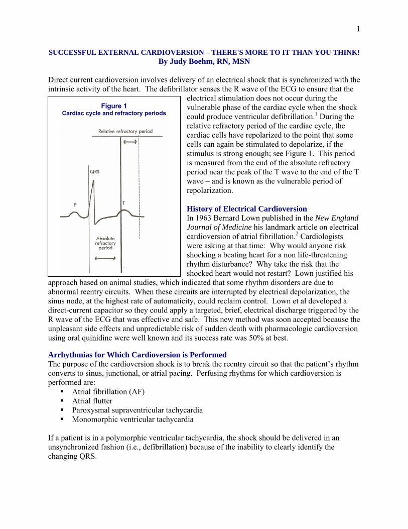
Figure 1 Cardiac cycle and refractory periods
SUCCESSFUL EXTERNAL CARDIOVERSION – THERE'S MORE TO IT THAN YOU THINK! By Judy Boehm, RN, MSN
Direct current cardioversion involves delivery of an electrical shock that is synchronized with the intrinsic activity of the heart. The defibrillator senses the R wave of the ECG to ensure that the
electrical stimulation does not occur during the vulnerable phase of the cardiac cycle when the shock could produce ventricular defibrillation.1 During the relative refractory period of the cardiac cycle, the cardiac cells have repolarized to the point that some cells can again be stimulated to depolarize, if the stimulus is strong enough; see Figure 1. This period is measured from the end of the absolute refractory period near the peak of the T wave to the end of the T wave – and is known as the vulnerable period of repolarization. History of Electrical Cardioversion In 1963 Bernard Lown published in the New England Journal of Medicine his landmark article on electrical cardioversion of atrial fibrillation.2 Cardiologists were asking at that time: Why would anyone risk shocking a beating heart for a non life-threatening rhythm disturbance? Why take the risk that the shocked heart would not restart? Lown justified his
approach based on animal studies, which indicated that some rhythm disorders are due to abnormal reentry circuits. When these circuits are interrupted by electrical depolarization, the sinus node, at the highest rate of automaticity, could reclaim control. Lown et al developed a direct-current capacitor so they could apply a targeted, brief, electrical discharge triggered by the R wave of the ECG that was effective and safe. This new method was soon accepted because the unpleasant side effects and unpredictable risk of sudden death with pharmacologic cardioversion using oral quinidine were well known and its success rate was 50% at best. Arrhythmias for Which Cardioversion is Performed The purpose of the cardioversion shock is to break the reentry circuit so that the patient’s rhythm converts to sinus, junctional, or atrial pacing. Perfusing rhythms for which cardioversion is performed are:
Atrial fibrillation (AF) Atrial flutter Paroxysmal supraventricular tachycardia Monomorphic ventricular tachycardia
If a patient is in a polymorphic ventricular tachycardia, the shock should be delivered in an unsynchronized fashion (i.e., defibrillation) because of the inability to clearly identify the changing QRS.
2
Emergency vs Elective Cardioversion Cardioversion is performed emergently if the patient has hypotension, respiratory insufficiency, altered mental status, or ongoing chest pain as a result of the tachycardia. The American Heart Association also recommends that for a heart rate of >150/minute immediate cardioversion be given, though a brief trial of medications may ensue. 3 Emergency cardioversion is often performed at the bedside with adequate support related to personnel, monitoring, and emergency equipment. Otherwise cardioversion is considered elective, and often scheduled to be performed in the Post Anesthesia Care Unit or other specialty unit. A brief trial of antiarrhythmics may first be tried to achieve pharmacologic cardioversion. Success of Cardioversion The success of cardioversion is related to several factors:
The waveform used to deliver the current The patient’s weight Transthoracic impedance The duration of the atrial fibrillation
Scientific evidence has shown that a biphasic shock is more successful at converting AF compared to a damped sine wave monophasic shock at lower total energy levels with less peak current and fewer shocks – especially for patients whose impedance is greater than 70 ohms (Ω). Each of the major defibrillator companies has published studies comparing their conversion rates for AF with monophasic vs. biphasic waveforms.
1 Mittal, S. et al. Transthoracic cardioversion of atrial fibrillation, comparison of rectilinear biphasic versus damped sine wave monophasic shocks. Circulation 2000;101:1282-1287 (ZOLL).
2 Niebauer, M.J. et al. Comparison of the rectilinear biphasic waveform with the monophasic damped sine waveform for external cardioversion of atrial fibrillation and flutter. American Journal of Cardiology 2004;93:1495-1499 (ZOLL).
3 Page, R.L. et al. Biphasic versus monophasic shock waveform for conversion of atrial fibrillation. Journal of American College of Cardiology 2002;39:1956-1963 (Philips Medical Systems).
4 Koster, R.W. et al. Clinical trial summary: External cardioversion of atrial fibrillation Resuscitation 2000;45(1):S52 (Medtronic Physio-Control).
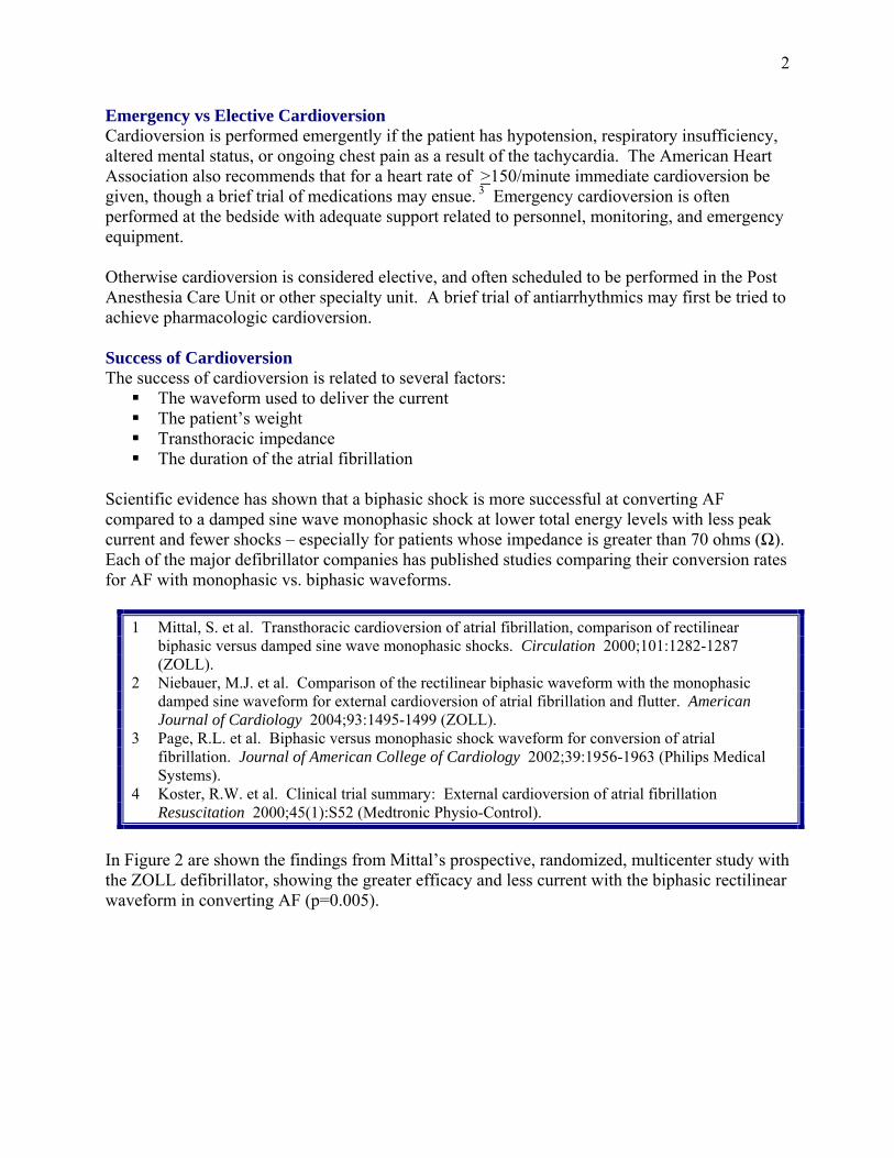
In Figure 2 are shown the findings from Mittal’s prospective, randomized, multicenter study with the ZOLL defibrillator, showing the greater efficacy and less current with the biphasic rectilinear waveform in converting AF (p=0.005).
3
Figure 2 Cumulative cardioversion efficacy of monophasic and biphasic shocks with ZOLL defibrillators
21%
44%
68%
79%
68%
85%
91%94%
0%
10%
20%
30%
40%
50%
60%
70%
80%
90%
100%
100 J 200J 300J 360J 70J 120J 150J 170J
Car
diov
ersi
on E
ffica
cy
Neal compared the efficacy of the biphasic rectilinear waveform (BRL) in the ZOLL M Series® with the biphasic truncated exponential waveform (BTE) in the Physio-Control Lifepak 12 for the conversion of AF to sinus rhythm, and found no significant difference.4 Shocks were delivered in a step-up fashion beginning with 50 joules to 102 patients randomly assigned to either the BRL or BTE waveform. 99% of the patients were successfully converted to sinus rhythm. Studies have also shown that the higher the patient’s weight, the less chance of success with cardioversion. Shock success is less if a transthoracic impedance is > 70 Ω. The longer the duration of the atrial fibrillation, the less the chance of conversion of the rhythm and the more frequent the recurrence. It has been found when using ZOLL defibrillators that the use of monophasic and biphasic waveforms is equally successful for conversion of atrial flutter. The literature does not provide a uniform definition of failed cardioversion of AF, with concepts ranging from inability to interrupt the fibrillatory mechanism at all, to immediate re-initiation of AF (generally defined as within 2 minutes), to multiple hours free of AF before going back into the arrhythmia.
Monophasic Biphasic
4
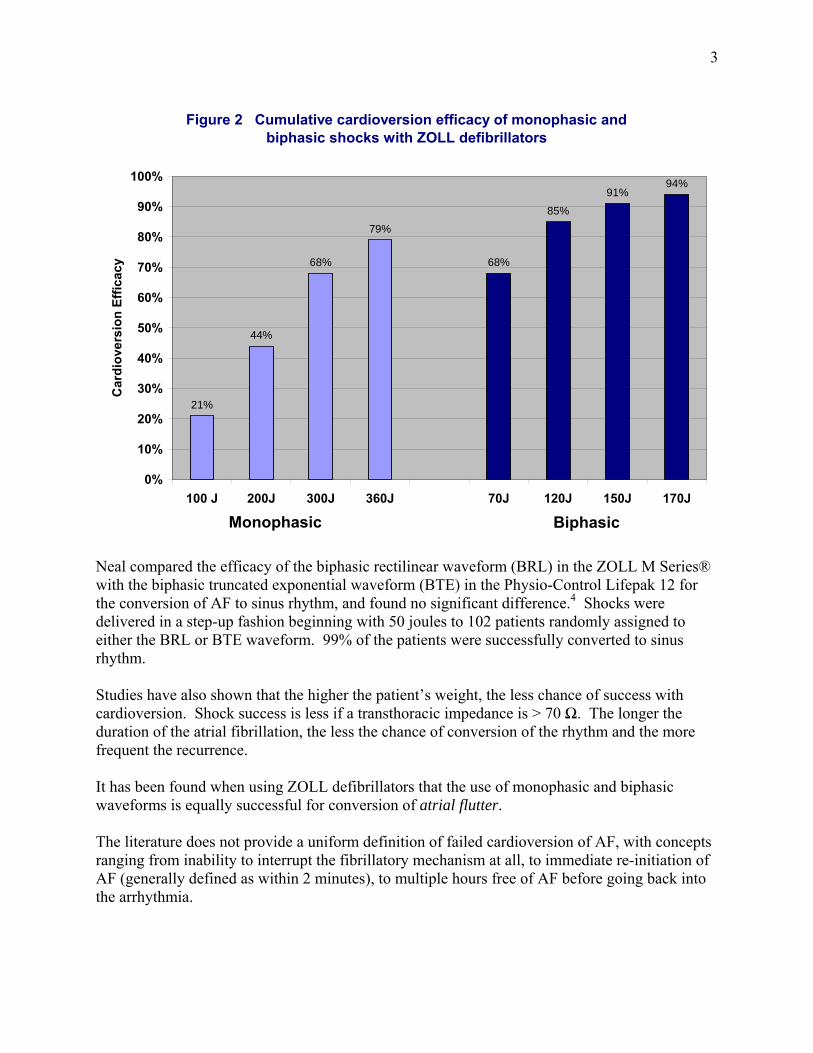
Working with Impedance to Improve Success of Cardioversion For increasing levels of transthoracic impedance, proportionately lower values of current density will flow through the patient’s myocardium for any given amount of energy administered – and thus the success of cardioversion will decrease. Biphasic defibrillators now measure impedance or resistance between the defibrillation electrodes immediately before the delivery of the shock and adjust the waveform to optimize current delivery. See Figure 3 for how biphasic waveforms adjust based on transthoracic impedance. ZOLL ensures a constant current during the 6 millisecond first phase of their Rectilinear Biphasic Waveform™ (RBW) by automatically adjusting the internal resistance of the defibrillator circuit. With the biphasic truncated exponential waveform (BTE) used in other defibrillators, the total duration of the shock is prolonged to deliver sufficient current with increasing impedance. This recent advancement in defibrillator technology has lead to improved success of cardioversion.
Figure 3 The difference in waveform morphology between BTE (Physio-Control) and BRL (ZOLL) at low and high impedance levels
Other factors that have been noted over the years to lower transthoracic impedance and improve cardioversion success are:
Shaving/clipping the hair underneath the electrodes The size of the electrode Pressing down on the paddles with 25 lb pressure
5
Pressing down on the anterior disposable defibrillation electrode Delivering high energy shocks (e.g., 720 joules) Using a salt-containing paste as the coupling agent with paddles Choosing a position for the electrodes in which the shock passes through the part of the
heart where the arrhythmia originates Delivering the shock at end expiration Administering an antiarrhythmic medication prior to the procedure Waiting 3 minutes after a first shock before delivering the second shock
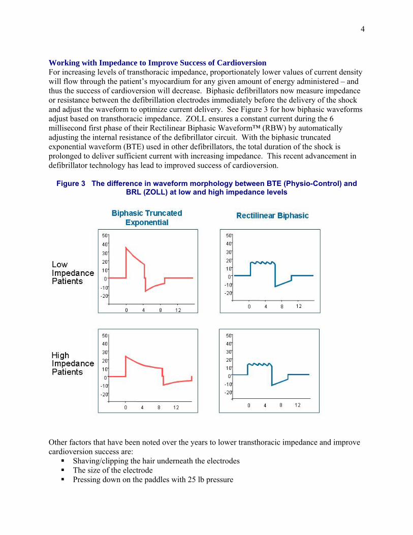
A study at the University of Iowa showed that shaving the chest of hirsute persons decreased by 35% the transthoracic impedance measured when disposable electrodes were put in place; see Figure 4. It was hypothesized that in hirsute individuals the high transthoracic impedance resulted from entrapped air (a poor conductor of electricity) when there was not good skin-electrode contact.5
Figure 4 Effect of shaving on transthoracic impedance with disposable electrodes
0
20
40
60
80
100
120
140
160
180
Hirsute - Before Shaving Hirsute - After Shaving
Tran
stho
raci
c Im
peda
nce
(Ω)
p <.01
When using paddles Sado found that the transthoracic impedance increased by 7.1% if hair is not shaved in hirsute individuals prior to defibrillation.6 The larger the defibrillator electrode, the lower the impedance. If the electrodes are too large, the impedance is low but the current density is not great enough for cardioversion. If electrodes are too small, not only is the impedance higher, but the current is concentrated and myocardial damage is more likely. The optimal size of the electrode for adults is 12-13 cm diameter, which
6
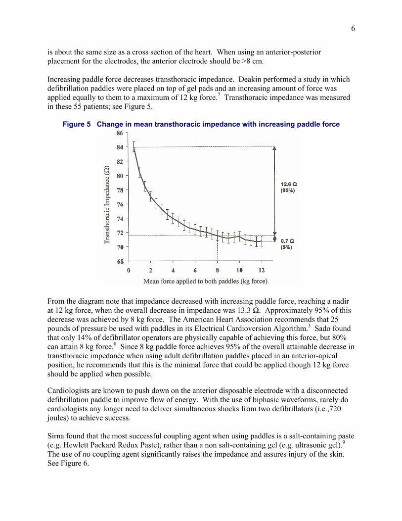
is about the same size as a cross section of the heart. When using an anterior-posterior placement for the electrodes, the anterior electrode should be >8 cm. Increasing paddle force decreases transthoracic impedance. Deakin performed a study in which defibrillation paddles were placed on top of gel pads and an increasing amount of force was applied equally to them to a maximum of 12 kg force.7 Transthoracic impedance was measured in these 55 patients; see Figure 5.
Figure 5 Change in mean transthoracic impedance with increasing paddle force
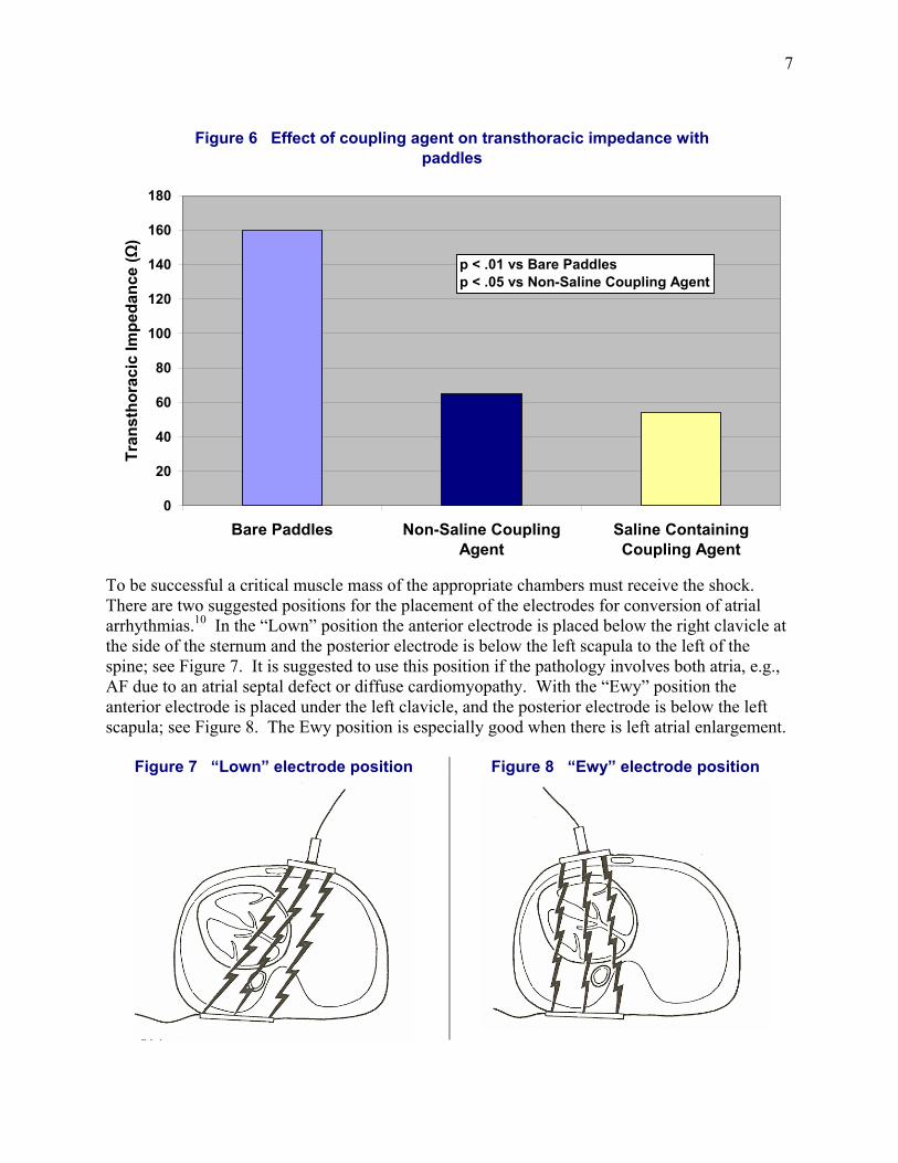
From the diagram note that impedance decreased with increasing paddle force, reaching a nadir at 12 kg force, when the overall decrease in impedance was 13.3 Ω. Approximately 95% of this decrease was achieved by 8 kg force. The American Heart Association recommends that 25 pounds of pressure be used with paddles in its Electrical Cardioversion Algorithm.3 Sado found that only 14% of defibrillator operators are physically capable of achieving this force, but 80% can attain 8 kg force.8 Since 8 kg paddle force achieves 95% of the overall attainable decrease in transthoracic impedance when using adult defibrillation paddles placed in an anterior-apical position, he recommends that this is the minimal force that could be applied though 12 kg force should be applied when possible. Cardiologists are known to push down on the anterior disposable electrode with a disconnected defibrillation paddle to improve flow of energy. With the use of biphasic waveforms, rarely do cardiologists any longer need to deliver simultaneous shocks from two defibrillators (i.e.,720 joules) to achieve success. Sirna found that the most successful coupling agent when using paddles is a salt-containing paste (e.g. Hewlett Packard Redux Paste), rather than a non salt-containing gel (e.g. ultrasonic gel).9 The use of no coupling agent significantly raises the impedance and assures injury of the skin. See Figure 6.
7
Figure 6 Effect of coupling agent on transthoracic impedance with paddles
0
20
40
60
80
100
120
140
160
180
Bare Paddles Non-Saline CouplingAgent
Saline ContainingCoupling Agent
Tran
stho
raci
c Im
peda
nce
(Ω)
p < .01 vs Bare Paddlesp < .05 vs Non-Saline Coupling Agent
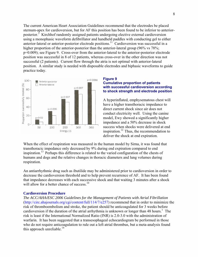
To be successful a critical muscle mass of the appropriate chambers must receive the shock. There are two suggested positions for the placement of the electrodes for conversion of atrial arrhythmias.10 In the “Lown” position the anterior electrode is placed below the right clavicle at the side of the sternum and the posterior electrode is below the left scapula to the left of the spine; see Figure 7. It is suggested to use this position if the pathology involves both atria, e.g., AF due to an atrial septal defect or diffuse cardiomyopathy. With the “Ewy” position the anterior electrode is placed under the left clavicle, and the posterior electrode is below the left scapula; see Figure 8. The Ewy position is especially good when there is left atrial enlargement.
Figure 7 “Lown” electrode position Figure 8 “Ewy” electrode position
8
The current American Heart Association Guidelines recommend that the electrodes be placed sternum-apex for cardioversion, but for AF this position has been found to be inferior to anterior-posterior.3 Kirchhof randomly assigned patients undergoing elective external cardioversion using a monophasic waveform defibrillator and handheld paddles with conducting gel to either anterior-lateral or anterior-posterior electrode positions.11 Cardioversion was successful in a higher proportion of the anterior-posterior than the anterior-lateral group (96% vs 78%; p=0.009); see Figure 9. Cross-over from the anterior-lateral to the anterior-posterior electrode position was successful in 8 of 12 patients, whereas cross-over in the other direction was not successful (2 patients). Current flow through the atria is not optimal with anterior-lateral position. A similar study is needed with disposable electrodes and biphasic waveforms to guide practice today.
Figure 9 Cumulative proportion of patients with successful cardioversion according to shock strength and electrode position A hyperinflated, emphysematous chest will have a higher transthoracic impedance to direct current shock since air does not conduct electricity well. Using the canine model, Ewy showed a significantly higher impedance and a 50% decrease in shock success when shocks were delivered at end inspiration.12 Thus, the recommendation to deliver the shock at end expiration.
When the effect of respiration was measured in the human model by Sirna, it was found that transthoracic impedance only decreased by 9% during end expiration compared to end inspiration.13 Perhaps this difference is related to the varied configuration of the chests of humans and dogs and the relative changes in thoracic diameters and lung volumes during respiration. An antiarrhythmic drug such as ibutilide may be administered prior to cardioversion in order to decrease the cardioversion threshold and to help prevent recurrence of AF. It has been found that impedance decreases with each successive shock and that waiting 3 minutes after a shock will allow for a better chance of success.10 Cardioversion Procedure The ACC/AHA/ESC 2006 Guidelines for the Management of Patients with Atrial Fibrillation (http://circ.ahajournals.org/cgi/content/full/114/7/e257) recommend that in order to minimize the risk of thromboembolism and stroke, the patient should be anticoagulated for 3 weeks before cardioversion if the duration of the atrial arrhythmia is unknown or longer than 48 hours.1 The risk is least if the International Normalized Ratio (INR) is 2.0-3.0 with the administration of warfarin. It has been suggested that a transesophageal echocardiogram be performed in those who do not require anticoagulation to rule out a left atrial thrombus, but a meta-analysis found this approach unreliable.14
9
When acute AF produces hemodynamic instability in the form of angina pectoris, MI, shock or pulmonary edema, immediate cardioversion should not be delayed to deliver therapeutic anticoagulation, but intravenous unfractionated heparin or subcutaneous injection of a low-molecular-weight heparin should be initiated before cardioversion. Cardioversion should be performed with the patient under adequate general anesthesia in a fasting state. Short-acting anesthetic drugs or agents that produce conscious sedation are preferred to enable rapid recovery after the procedure. Devices for measurement of pulse oximetry and non-invasive blood pressure should be in place on the patient. Suction should be set up and tested to make sure that it is ready for use. A patent IV line should be in place. Intubation equipment should be at the bedside and emergency equipment should be readily available. Defibrillation electrodes should be placed on the patient using one of the anterior-posterior positions described above. Apply the back electrode first to prevent buckling of the front electrode. Electrodes should always be placed on clean, dry intact skin with nothing underneath them. Remove any underlying ECG electrodes and transdermal medication patch, and wipe off residual gel or medication. When an implantable medical device is located in an area where the electrode would normally be placed, position the electrode at least 1 inch away from the device. Excessive hair should be removed. Clipping of hair is preferred to shaving since it produces less skin abrasions and post shock discomfort. The electrodes should be rolled onto the skin in order to eliminate air pockets and thus decrease impedance. ZOLL’s pro·padz® electrodes have been designed with liquid gel as the coupling agent to decrease skin damage with cardioversion (http://www.zoll.com/product_resource.aspx?id=767). Figure 10 ZOLL pro·padz® electrodes
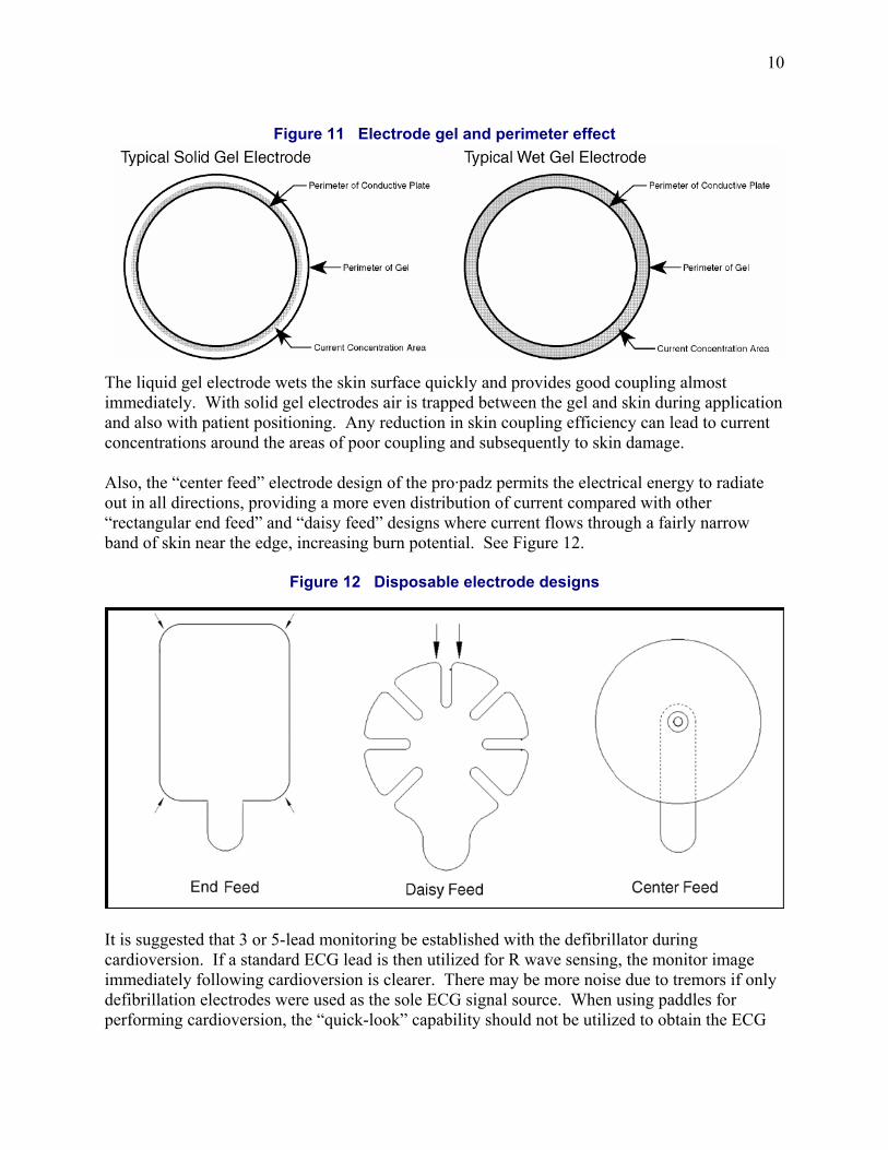
Disposable electrodes with high viscosity solid gels have relatively poor lateral conductivity and typically higher current concentration at the perimeter of the plate, which increases the possibility of skin damage. Lower viscosity wet gel electrodes yield a much wider band of current concentration extending from the edge of the conductive plate to the edge of the gel area and, therefore, significantly reduce the incidence of skin damage. See Figure 11.
10
Figure 11 Electrode gel and perimeter effect
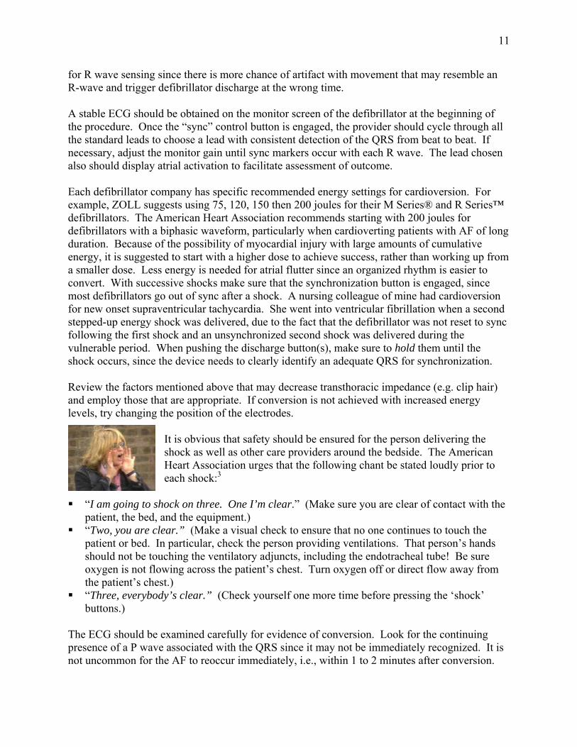
The liquid gel electrode wets the skin surface quickly and provides good coupling almost immediately. With solid gel electrodes air is trapped between the gel and skin during application and also with patient positioning. Any reduction in skin coupling efficiency can lead to current concentrations around the areas of poor coupling and subsequently to skin damage. Also, the “center feed” electrode design of the pro·padz permits the electrical energy to radiate out in all directions, providing a more even distribution of current compared with other “rectangular end feed” and “daisy feed” designs where current flows through a fairly narrow band of skin near the edge, increasing burn potential. See Figure 12.
Figure 12 Disposable electrode designs
It is suggested that 3 or 5-lead monitoring be established with the defibrillator during cardioversion. If a standard ECG lead is then utilized for R wave sensing, the monitor image immediately following cardioversion is clearer. There may be more noise due to tremors if only defibrillation electrodes were used as the sole ECG signal source. When using paddles for performing cardioversion, the “quick-look” capability should not be utilized to obtain the ECG
11
for R wave sensing since there is more chance of artifact with movement that may resemble an R-wave and trigger defibrillator discharge at the wrong time. A stable ECG should be obtained on the monitor screen of the defibrillator at the beginning of the procedure. Once the “sync” control button is engaged, the provider should cycle through all the standard leads to choose a lead with consistent detection of the QRS from beat to beat. If necessary, adjust the monitor gain until sync markers occur with each R wave. The lead chosen also should display atrial activation to facilitate assessment of outcome. Each defibrillator company has specific recommended energy settings for cardioversion. For example, ZOLL suggests using 75, 120, 150 then 200 joules for their M Series® and R Series™ defibrillators. The American Heart Association recommends starting with 200 joules for defibrillators with a biphasic waveform, particularly when cardioverting patients with AF of long duration. Because of the possibility of myocardial injury with large amounts of cumulative energy, it is suggested to start with a higher dose to achieve success, rather than working up from a smaller dose. Less energy is needed for atrial flutter since an organized rhythm is easier to convert. With successive shocks make sure that the synchronization button is engaged, since most defibrillators go out of sync after a shock. A nursing colleague of mine had cardioversion for new onset supraventricular tachycardia. She went into ventricular fibrillation when a second stepped-up energy shock was delivered, due to the fact that the defibrillator was not reset to sync following the first shock and an unsynchronized second shock was delivered during the vulnerable period. When pushing the discharge button(s), make sure to hold them until the shock occurs, since the device needs to clearly identify an adequate QRS for synchronization. Review the factors mentioned above that may decrease transthoracic impedance (e.g. clip hair) and employ those that are appropriate. If conversion is not achieved with increased energy levels, try changing the position of the electrodes.
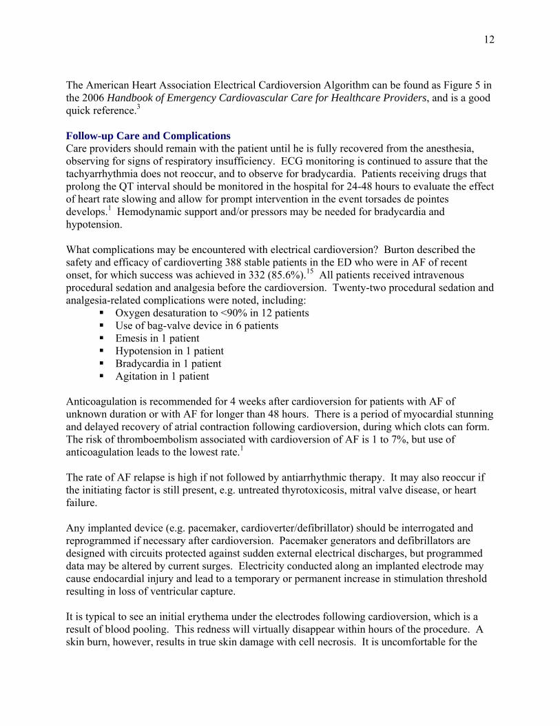
It is obvious that safety should be ensured for the person delivering the shock as well as other care providers around the bedside. The American Heart Association urges that the following chant be stated loudly prior to each shock:3
“I am going to shock on three. One I’m clear.” (Make sure you are clear of contact with the patient, the bed, and the equipment.)
“Two, you are clear.” (Make a visual check to ensure that no one continues to touch the patient or bed. In particular, check the person providing ventilations. That person’s hands should not be touching the ventilatory adjuncts, including the endotracheal tube! Be sure oxygen is not flowing across the patient’s chest. Turn oxygen off or direct flow away from the patient’s chest.)
“Three, everybody’s clear.” (Check yourself one more time before pressing the ‘shock’ buttons.)
The ECG should be examined carefully for evidence of conversion. Look for the continuing presence of a P wave associated with the QRS since it may not be immediately recognized. It is not uncommon for the AF to reoccur immediately, i.e., within 1 to 2 minutes after conversion.
12
The American Heart Association Electrical Cardioversion Algorithm can be found as Figure 5 in the 2006 Handbook of Emergency Cardiovascular Care for Healthcare Providers, and is a good quick reference.3 Follow-up Care and Complications Care providers should remain with the patient until he is fully recovered from the anesthesia, observing for signs of respiratory insufficiency. ECG monitoring is continued to assure that the tachyarrhythmia does not reoccur, and to observe for bradycardia. Patients receiving drugs that prolong the QT interval should be monitored in the hospital for 24-48 hours to evaluate the effect of heart rate slowing and allow for prompt intervention in the event torsades de pointes develops.1 Hemodynamic support and/or pressors may be needed for bradycardia and hypotension. What complications may be encountered with electrical cardioversion? Burton described the safety and efficacy of cardioverting 388 stable patients in the ED who were in AF of recent onset, for which success was achieved in 332 (85.6%).15 All patients received intravenous procedural sedation and analgesia before the cardioversion. Twenty-two procedural sedation and analgesia-related complications were noted, including:
Oxygen desaturation to <90% in 12 patients Use of bag-valve device in 6 patients Emesis in 1 patient Hypotension in 1 patient Bradycardia in 1 patient Agitation in 1 patient
Anticoagulation is recommended for 4 weeks after cardioversion for patients with AF of unknown duration or with AF for longer than 48 hours. There is a period of myocardial stunning and delayed recovery of atrial contraction following cardioversion, during which clots can form. The risk of thromboembolism associated with cardioversion of AF is 1 to 7%, but use of anticoagulation leads to the lowest rate.1 The rate of AF relapse is high if not followed by antiarrhythmic therapy. It may also reoccur if the initiating factor is still present, e.g. untreated thyrotoxicosis, mitral valve disease, or heart failure. Any implanted device (e.g. pacemaker, cardioverter/defibrillator) should be interrogated and reprogrammed if necessary after cardioversion. Pacemaker generators and defibrillators are designed with circuits protected against sudden external electrical discharges, but programmed data may be altered by current surges. Electricity conducted along an implanted electrode may cause endocardial injury and lead to a temporary or permanent increase in stimulation threshold resulting in loss of ventricular capture. It is typical to see an initial erythema under the electrodes following cardioversion, which is a result of blood pooling. This redness will virtually disappear within hours of the procedure. A skin burn, however, results in true skin damage with cell necrosis. It is uncomfortable for the

13
patient and shows as scabbing, blisters, and redness. Skin erythema and post shock discomfort (pain and itching) at the electrode sites increase with cumulative energy and a higher number of shocks. See the results below of Neal’s study of randomly assigning 101 patients to cardioversion with either a Rectilinear Biphasic Waveform (ZOLL M Series) or biphasic truncated exponential waveform (Physio-Control Lifepak 12P) using self-adhesive pads.4 Shocks were delivered in a step-up fashion starting with 50 joules. Skin redness was reported as:
No redness 54 patients (53%) Mild redness 44 patients (44%) Severe redness 1 patient (1%)
Figure 13 Correlation between skin appearance and cumulative energy, independent of
biphasic waveform group (p = 0.0004), and correlation between skin appearance and number of shocks (p <0.0001)
Figure 14 Correlation between comfort and total delivered energy, independent of biphasic waveform group (p = 0.0003), and correlation between comfort
and number of shocks (p = 0.0004)

14
Pain was reported as: No pain 58 patients (57%) Mild itching 20 patients (20%) Mild pain and moderate itching 16 patients (16%) Moderate pain and severe itch 3 patients (3%)
With the use of the biphasic waveform these dermal effects have been minimized when compared with monophasic shocks, somewhat due to lower cumulative energy and lower total number of shocks needed for cardioversion. Ambler found that patients who received a biphasic waveform for elective DC cardioversion of atrial arrhythmias experienced less pain as measured by the visual analogue scale at 2 and 24 hours after the procedure, compared with patients who received a monophasic waveform.16 There was also a reduction in the erythema index measured with a DermaSpectrometer (measures reflectance from the skin of red and green light) at the edge of the sternal pad sites in those patients who received a biphasic waveform. Conclusion Cardioversion is considered a safe, effective procedure for converting reentry supraventricular tachyarrhythmias and perfusing monophasic ventricular tachycardia. To be most successful at cardioversion, prep the skin appropriately and use wet gel electrodes placed in an anterior-posterior position. On the biphasic defibrillator select a lead with consistent synchronization of the R wave and good visualization of atrial activity. Deliver the shock at end expiration remembering to push and hold the discharge buttons. Now that disposable electrodes are often being used, the difficulty incurred by the usual operator in applying 25 pounds of pressure to the paddles and remaining safely away from the patient’s body has become less of an issue. Dermal injury is greatly lessened now with use of the biphasic waveform and wet gel electrodes having a center feed. Currently there is a debate about the true benefit of cardioversion of AF. The therapeutic goal of cardioversion is long-term maintenance of sinus rhythm with accompanying symptomatic and hemodynamic benefit along with reduced clinical thromboembolism. Even with aggressive antiarrhythmic therapy, electrical cardioversion is associated with recurrence rates that may exceed 50% at one year. Would it be better to just accept AF and control the rate? Recent data from the randomized Atrial Fibrillation Follow-up Investigation of Rhythm Management (AFFIRM)18 and the Rate Control vs. Electrical Cardioversion for persistent AF (RACE)19 trials have raised questions about the underlying value of cardioversion and maintenance of sinus rhythm. Results of both studies showed similar thromboembolism rates and quality-of-life measures among groups who were aggressively maintained in sinus rhythm (with use of antiarrhythmics and repeated DC cardioversions) and those who were managed with rate control of AF and long-term warfarin. The risk of serious ventricular arrhythmia (torsades de pointes) was low (12 of 2030 or 0.8%) in the rhythm control arm of the AFFIRM trial, but still important. The main difference between the two treatment arms in this study was that those in the rhythm control arm consumed more healthcare time and resources (for treatments such as DC cardioversion, pacemaker implantation, etc). So there is more to electrical cardioversion than you might have originally thought! It is hoped that you will use this scientific evidence to achieve conversion of the tachyarrhythmia and a safe, comfortable procedure for the patient.
15
References 1 ACC/AHA/ESC 2006 Guidelines for the Management of Patients with Atrial Fibrillation. Circulation
2006;114:700-752. 2 Lown, B. et al. “Cardioversion” of atrial fibrillation. New England Journal of Medicine
1963;269:325-331. 3 Field, J.M., Hazinski, M.F., & Gilmore, D. [Eds]. 2006 Handbook of Emergency Cardiovascular Care
for Healthcare Providers. American Heart Association. 4 Neal, S., Ngarmukos, T., Lessard, D. & Rosenthal, L. Comparison of the efficacy and safety of two
biphasic defibrillator waveforms for the conversion of atrial fibrillation to sinus rhythm. American Journal of Cardiology 2003;92:810-814.
5 Bissing, J.W. & Kerber, R.E. Effect of shaving the chest of hirsute subjects on transthoracic impedance to self-adhesive defibrillation electrode pads. American Journal of Cardiology 2000;86:587-589.
6 Sado, D.M. et al. Comparison of the effects of removal of chest hair with not doing so before external defibrillation on transthoracic impedance. American Journal of Cardiology 2004;93:98-100.
7 Deakin, C.D. et al. Determining the optimal paddle force for external defibrillation. American Journal of Cardiology 2002;90:812-813.
8 Sado, D., Deakin, C., & Petley, G. Are European Resuscitation Council recommendations for paddle force achievable during defibrillation? Resuscitation 2001;3:287-290.
9 Sirna, S.J. et al. Factors affecting transthoracic impedance during electrical cardioversion. American Journal of Cardiology 1988;62:1048-1052.
10 Ewy, G.A. The optimal technique for electrical cardioversion of atrial fibrillation. Clinical Cardiology 1994;17:79-84.
11 Kirchhof, P. et al. Anterior-posterior versus anterior-lateral electrode positions for external cardioversion of atrial fibrillation: A randomized trial. Lancet 2002;360:1275-1279.
12 Ewy, G.A. et al. Influence of ventilation phase on transthoracic impedance and defibrillation effectiveness. Critical Care Medicine 1980;8:164-166.
13 Sirna, S.J. et al. Factors affecting transthoracic impedance during electrical cardioversion. American Journal of Cardiology 1988;62:1048-1052.
14 Moreyra, E., Finkelhor, R.S., & Cebul, R.D. Limitations of transesophageal echocardiography in the risk assessment of patients before non anticoagulated cardioversion from atrial fibrillation and atrial flutter: An analysis of pooled trials. American Heart Journal 1995;129:71-75.
15 Burton, J.H. et al. Electrical cardioversion of emergency department patients with atrial fibrillation. Annals of Emergency Medicine 2004;44:20-30.
16 Ambler, J.J.S. & Deakin, C.D. A randomised controlled trial of the effect of biphasic or monophasic waveform on the incidence and severity of cutaneous burns following external direct current cardioversion. Resuscitation 2006;71:293-300.
17 Manning, W.J. & Zimetbaum, P.J. Direct current cardioversion of atrial fibrillation – the next 40 years. Mayo Clinic Proceedings 2002;77:895-896.
18 Wyse, D.G. et al. A comparison of rate control and rhythm control in patients with atrial fibrillation. New England Journal of Medicine 2002;347:1825-1833.
19 Van Gelder, I.C. et al. A comparison of rate control and rhythm control in patients with recurrent persistent atrial fibrillation. New England Journal of Medicine 2002;347:1834-1840.
![High-energy external defibrillation and transcutaneous ...quire external defibrillation or cardioversion [1]. The feasibility of in-bore defibrillation has been demon-strated in a](https://img.dokumen.tips/doc/110x75/60a040fa5ed69b1bff53b63d/high-energy-external-defibrillation-and-transcutaneous-quire-external-defibrillation.jpg)


![Check List Cardioversion I.gallastegi[1]](https://img.dokumen.tips/doc/110x75/55cf8e57550346703b912349/check-list-cardioversion-igallastegi1.jpg)