Embed Size (px)
Citation preview
on July 26, 2018http://rspb.royalsocietypublishing.org/Downloaded from
rspb.royalsocietypublishing.org
ResearchCite this article: Mao C-A et al. 2016
Substituting mouse transcription factor Pou4f2
with a sea urchin orthologue restores retinal
ganglion cell development. Proc. R. Soc. B 283:
20152978.
http://dx.doi.org/10.1098/rspb.2015.2978
Received: 12 December 2015
Accepted: 10 February 2016
Subject Areas:developmental biology, evolution, neuroscience
Keywords:retinal ganglion cells, retinal development,
transcription factor evolution, Pou4f2/Brn3b,
echinoderm photoreceptors
Author for correspondence:William H. Klein
e-mail: [email protected]
†Both first authors played separate and major
roles in this study. Each is equally worthy of
being listed as first author.
Electronic supplementary material is available
at http://dx.doi.org/10.1098/rspb.2015.2978 or
via http://rspb.royalsocietypublishing.org.
& 2016 The Authors. Published by the Royal Society under the terms of the Creative Commons AttributionLicense http://creativecommons.org/licenses/by/4.0/, which permits unrestricted use, provided the originalauthor and source are credited.Substituting mouse transcription factorPou4f2 with a sea urchin orthologuerestores retinal ganglion cell development
Chai-An Mao1,†, Cavit Agca1,†, Julie A. Mocko-Strand2, Jing Wang2,Esther Ullrich-Luter3, Ping Pan1, Steven W. Wang1, Maria Ina Arnone4,Laura J. Frishman2 and William H. Klein1
1Department of Systems Biology, Unit 0950, The University of Texas MD Anderson Cancer Center,Houston, TX 77030, USA2College of Optometry, University of Houston, Houston, TX 77204, USA3Museum fur Naturkunde, Berlin 10115, Germany4Biology and Evolution of Marine Organisms, Stazione Zoologica Anton Dohrn, Naples 80121, Italy
C-AM, 0000-0002-9700-6964; CA, 0000-0001-8472-7052
Pou domain transcription factor Pou4f2 is essential for the development of reti-
nal ganglion cells (RGCs) in the vertebrate retina. A distant orthologue of
Pou4f2 exists in the genome of the sea urchin (class Echinoidea) Strongylocen-trotus purpuratus (SpPou4f1/2), yet the photosensory structure of sea urchins is
strikingly different from that of the mammalian retina. Sea urchins have no
obvious eyes, but have photoreceptors clustered around their tube feet disc.
The mechanisms that are associated with the development and function of
photoreception in sea urchins are largely unexplored. As an initial approach
to better understand the sea urchin photosensory structure and relate it to
the mammalian retina, we asked whether SpPou4f1/2 could support RGC
development in the absence of Pou4f2. To answer this question, we replaced
genomic Pou4f2 with an SpPou4f1/2 cDNA. In Pou4f2-null mice, retinas expres-
sing SpPou4f1/2 were outwardly identical to those of wild-type mice.
SpPou4f1/2 retinas exhibited dark-adapted electroretinogram scotopic
threshold responses, indicating functionally active RGCs. During retinal
development, SpPou4f1/2 activated RGC-specific genes and in S. purpuratus,SpPou4f2 was expressed in photoreceptor cells of tube feet in a pattern dis-
tinct from Opsin4 and Pax6. Our results suggest that SpPou4f1/2 and
Pou4f2 share conserved components of a gene network for photosensory devel-
opment and they maintain their conserved intrinsic functions despite vast
morphological differences in mouse and sea urchin photosensory structures.
1. IntroductionThe deep conservation of regulatory genes for eye development has amply
demonstrated an underlying framework for eye diversification [1,2]. However,
the developmental and evolutionary mechanisms that led to this remarkable
diversity remain vague. Although changes in gene regulatory networks are
probably to be the drivers of eye diversification, very little is known about
the level at which conserved gene networks might have diverged to produce
different structures and functions [2]. A potentially informative but largely
unexplored phylum for investigating eye development and evolution is echino-
derms. Echinoderms are basal deuterostomes that develop in ways similar to
chordates but are distinctly different in their adult body plan. Unlike all
other deuterostomes, echinoderms lack an obvious anterior–posterior axis.
Instead, they exhibit highly derived body plans that are organized along a
radial axis [3]. Notably, echinoderms lack any structures that even remotely
wild-type allele 10.3 KB
BamH1 Met
left arm (1.8 KB)
SpPou4f1/2 Aq- PGK-Neo
Aq- PGK-Neo
SpPou4f1/2
MC1-TK-pA
SpPou4f1/2
targeting construct
knock-in alleleMet
Met
not 1
5¢UTR 3¢UTR
3¢UTR
3¢UTR
5¢UTR
5¢UTR
HA
HA
HA
right arm (3.2 KB)
p1 p2
p3 p4FRT
FRT
FRT
FRT
FRT
7.2 KB
BamH1
74 MmPou3f2
MmPou2f3
MmPou2f2
MmPou5f2
MmPou4f3
MmPou4f1
MmPou4f2
MmPou6f1
MmPou6f2
XtPou4f2
DrPou4f2
SpPou4f
SpPou6f
CiPou4f
MmPou5f1/Oct3-4
MmPou2f1/Oct1
MmPou3f3
MmPou3f4
MmPou3f1/oct-6
SpBm1/2/4
SpOct1
8888
100
52
96 100
71
99
70100
10076
65
100
7895
100BamH1
Rosa 26FLPeR
Pou4f2SpPou4f1/2 allele
BamH1
probe
Stop (TAG)
(b)(a)
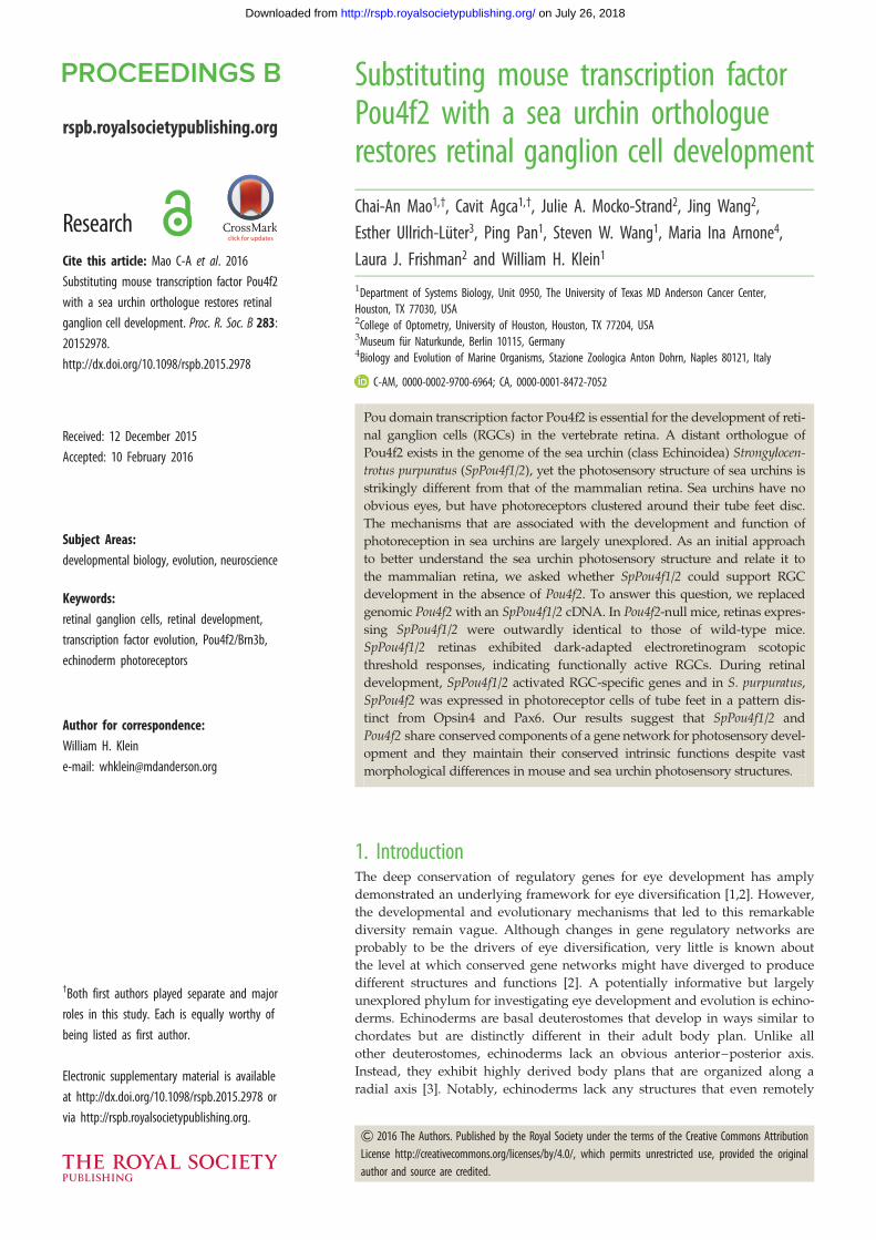
Figure 1. Sequence alignment of SpPou4f1/2 and Pou4f and generation of the Pou4f2SpPou4f1/2 KI allele. (a) Generation of Pou4f2SpPou4f1/2 KI allele, FRT indicates FLPrecombinase sites to remove the Neo selection cassette (brown box) by a Rosa26-FLPeR mouse line. SpPou4f1/2 cDNA sequences (green box) were fused in frame toa HA-epitope tag (yellow box). The dark purple thick lines indicate the site of recombination into a construct carrying the Pou4f2 coding region and upstreamregulatory regon. TK (orange box) indicates the TK cassette used for negative selection. The BamHI site was used for Southern genotyping, p1 and p2 are PCRprimers to detect wild-type mouse Pou4f2 allele, and p3 and p4 are primers to detect knock in SpPou4f1/2 allele. (b) Phylogeny analysis of SpPou4f1/2 and Pou4fgenes from other organisms. The tree was constructed with MEGA software v. 5.2. using neighbour-joining method with 1000 bootstrap repeats. Ci, Cionaintestinalis; Dr, Danio rerio; Mm, Mus musculus; Sp, Strogylocentrotus purpuratus; Xt, Xenopus tropicalis.
rspb.royalsocietypublishing.orgProc.R.Soc.B
283:20152978
2
on July 26, 2018http://rspb.royalsocietypublishing.org/Downloaded from
resemble a vertebrate eye. Nevertheless, many behavioural pat-
terns in adult sea urchins are attributable to highly developed
photoreception [4].
We recently addressed the question of whether adult
echinoderms have distinct photosensitive neurons that are
separate from the diffuse surface-wide neural network
already known to exist [5–7]. Using probes that were Strongy-locentrotus purpuratus orthologues of mouse genes expressed
in the retina [8], Agca et al. [9] and Ullrich-Luter et al. [10]
showed that small groups of photoreceptor neurons were
clustered around the periphery of the tube feet disc. This con-
clusion was based on the fact that many genes expressed in
the mouse retina had homologues that were expressed in
the tube feet neurons. The genes included retinal-expressed
transcription factors [8–12] and an orthologue of opsin,
Opsin4, which had signature features of a light-transducing
opsin [8–11,13]. Many of the transcription factors have
important roles in the developing mouse retina. Of particular
note, the S. purpuratus orthologue of Pou4f2, SpPou4f1/2, has a
91% and 96.6% sequence match with mouse Pou4f2 in its
Pou-specific and Pou-homeodomains, respectively. However,
it is highly divergent outside of these domains (electronic
supplementary material, figure S1a). Phylogenetic analysis
of Pou-class genes showed that SpPou4f1/2 clustered with
the Pou4f2 family over these domains [8]. We have shown
that SpPou4f1/2 is expressed in the tube feet [9].
Although the results of Agca et al. [9] and Ullrich-Luter et al.[10] are correlative, they suggest the presence of functional
photoreceptor neurons in the tube feet. However, the function
of SpPou4f1/2 in the tube feet is unknown. Given the technical
difficulties in directly determining the function of SpPou4f1/2 in
the adult sea urchin tube feet, we chose a more feasible, albeit
less direct, approach using a mouse knock-in (KI) strategy. We
asked whether SpPou4f1/2 could function in the context of the
developing mouse retina. Pou4f2 is essential for retinal ganglion
cell (RGC) differentiation and survival [14–16]. In the
developing retina, Pou4f2 expression is restricted to newly dif-
ferentiated RGCs, and its expression is maintained throughout
adult life. If SpPou4f1/2 functionally replaced Pou4f2, it would
suggest that SpPou4f1/2 binds to and activates a similar set
of genes in tube feet neurons, despite more than 540 Myr of
divergence from the common ancestor of sea urchins and mice.
As demonstrated further in the article, our experiments
in both mice and sea urchins support this hypothesis
and indicate that conservation of Pou-specific and Pou-
homeodomains in SpPou4f1/2 is, to high degree, sufficient
to support RGC development.
2. Material and methods(a) Pou4 class protein sequence analysisWe performed protein sequence analysis with Megalign software
(DNASTAR, Madison, WI, USA) using the ClustalW multiple
sequence alignment method using four protein sequences:
mouse Pou4f1, Pou4f2, Pou4f3 and S. purpuratus SpPou4f1/2.
Phylogeny tree was constructed with MEGA5 software [17]
using neighbour-joining method with 1000 bootstrap repeats.
Amino acid sequences from multiple POU families were used
to construct a phylogeny tree [18]. Sequences from Ciona intesti-nalis, Danio rerio, Mus Musculus, Strogylocentrotus purpuratus and
Xenopus tropicalis were used for comparison.
(b) Generation of Pou4f2SpPou4f1/2 knock-in constructThe SpPou4f1/2 KI allele was generated by replacing mouse
Pou4f2 with an S. purpuratus SpPou4f1/2 cDNA sequence using
recombineering. Three copies of the HA epitope tag were
inserted in a frame downstream of the SpPou4f1/2 sequence.
A neomycin cassette was inserted and was flanked by two
flip-recombinase target (FRT) sites. A BamHI site was introduced
downstream of the second FRT site for subsequent Southern
genotyping (figure 1a).
rspb.royalsocietypublishing.orgProc.R.Soc.B
283:20152978
3
on July 26, 2018http://rspb.royalsocietypublishing.org/Downloaded from
(c) Generation and genotyping of Pou4f2SpPou4f1/2/Z
and Pou4f2SpPou4f1/2/SpPou4f1/2 miceThe targeting construct was used to electroporate G4 129�C57BL/6 F1 hybrid embryonic stem cells. Positive clones were
selected by Southern blotting. The wild-type allele yielded a
10.3-kb band, whereas the KI allele yielded a 7.2-kb band (elec-
tronic supplementary material, figure S1b). The SpPou4f1/2 KI
mouse was generated by blastocyst injection. High-percentage chi-
meras were bred with C57BL/6 mice to generate heterozygous
Pou4f2SpPou4f1/2/þ KI progenies. The FRT-flanked Neo cassette in
the KI progenies was further removed by a Rosa26-FLPeRmouse line to produce Pou4f2SpPou4f1/2/þ mice [19]. Pou4f2þ/Z
and Pou4f2þ/AP control mice were described previously [16].
Pou4f2SpPou4f1/2/þmice were then crossed with Pou4f2þ/Z mice to gen-
erate Pou4f2SpPou4f1/2/Z mice. Homozygous Pou4f2SpPou4f1/2/SpPou4f1/2
mice were obtained by intercrossing Pou4f2SpPou4f1/2/þ mice.
PCR was used to genotype the wild-type Pou4f2 allele and
the SpPou4f1/2 KI allele (figure 1a). PCR primers for the Pou4f2wild-type allele were p1: 50-TCTGGAAGCCTACTTCGCCA and
p2: 50-CCGGTTCACAATCTCTCTGA. Primers to detect the
SpPou4f1/2 KI allele were p3: 50-ATGAATATGAAGGAGCATGT
and p4: 50-TAGTTGGTGTCGTTCTTGAT.
(d) Isolation and processing of embryos, embryonicretinas, and adult eyes, optic nerves and retinas
Embryos or adult eyes with optic nerves were harvested from
different stages and processed in different ways. For histological
analysis, tissues were fixed overnight in 4% paraformaldehyde
(PFA) and 3% glutaradehyde in phosphate-buffered saline
(PBS), subjected to PBS washing and methanol dehydration,
and finally embedded in paraffin for sectioning. For immuno-
labelling of sections, tissues were fixed in 4% PFA for 30 min,
washed three times with PBS, and embedded in optimal cutting
temperature compound (Fisher Scientific).
(e) Histology, immunolabelling and TUNEL assaysof mouse samples
Haematoxylin and eosin staining was described previously [20].
Immunofluorescence staining of paraffin sections or cryosections
and flat-mount staining was carried out to detect RGC axons as
previously described [20]. Primary antibodies used were mouse
anti-HA (Cell Signaling, 1 : 500; Cat. 2367S), goat anti-Pou4f2/
Brn3 (Santa Cruz, 1 : 100; Cat. sc6026), mouse anti-Pou4f1/
Brn3a (Chemicon, 1 : 400; Cat. MAB1585), mouse anti-SMI32
(Covance, 1 : 1000; Cat. SMI-32R), mouse anti-neurofilament-L
(NF-L) (Invitrogen, 1 : 1000; Cat. 13-0400), chicken anti-b-galacto-
sidase (AbCam, 1 : 2000; Cat. 9361), rabbit anti-melanopsin/
Opn4 (Advanced Targeting Systems, 1 : 1000; Cat. N39), mouse
anti-Islet1 (Isl-1) (DSHB, 1 : 500; Cat. 39.3F7) and rabbit
anti-Tbr2/Eomes (AbCam, 1 : 1000; Cat. ab23345). The Alexa-
conjugated secondary antibodies used in this study were
obtained from Molecular Probes and were used at 1 : 500
dilutions. DAPI (1 mg ml21, Vector Lab) was used to stain the
nuclei. TUNEL assays on embryonic retinas were performed
using an in situ cell death detection kit (Roche Applied Science)
following the manufacturer’s instructions. Images were acquired
on an Olympus FV1000 confocal laser-scanning microscope.
( f ) Electroretinogram recordings(i) SubjectsSubjects were 2.5- to 3.5-month-old mice with the following
genotypes: Pou4f2þ/þ (n ¼ 4), Pou4f2SpPou4f1/2/SpPou4f1/2 (n ¼ 4)
and Pou4f22/2 (n ¼ 4).
(ii) Electroretinogram recordingsMice were dark-adapted overnight and preparations for recording
were all performed under dim red illumination (l . 650 nm) as
previously described in [21] and the electronic supplementary
material. Stimuli were provided from light-emitting diodes
(lmax ¼ 462 nm) over a range of time-integrated flash illuminances
(stimulus strengths) from 26.7 to 2.3 log scotopic (sc) cd-s m22.
The inter-flash interval was adjusted to allow the electroretinogram
(ERG) response to return to baseline between flashes.
(iii) Data analysisAmplitudes (microvolts) of a-waves were measured on the
leading edge of the wave, at a fixed time (7 ms) after the brief
flash, which was close to the peak amplitude for the strongest
stimuli. Amplitudes of b-waves were measured between the
a-wave trough and the b-wave peak after applying a low-pass
60 Hz filter to remove oscillatory potentials (the electronic
supplementary material).
(g) Expression of SpPou4f1/2, Pax6 and Opsin4in Strongylocentrotus purpuratus tube foot
(i) Tube feet preparationsTube feet were dissected from live S. purpuratus and immediately
fixed in 4% PFA in PBS at room temperature for 2–4 h. After
washing the collected tube feet several times in PBS, we trans-
ferred them to 100% methanol and stored them at 2208C until
experimental processing.
(ii) In situ hybridization and immunohistochemistrySpPou4f1/2 probes were generated by cloning full-length
SpPou4f1/2 cDNA into pIRES-hrGPF-2a vector (Agilent). The
Pax6 probe has been described in Ullrich-Luter et al. [10]. Both
antisense- and sense-digoxigenin-labelled SpPou4f1/2 probes
were obtained using a digoxigenin-RNA labelling kit (Roche),
following the manufacturer’s instructions by using 1 mg of linear-
ized plasmids. The Pax6 RNA probe was similarly prepared
using unlabelled ribonucleotides and was subsequently labelled
with 2, 4-dinitrophenyl using a Label-it kit (Mirus) following the
manufacturer instructions. Whole-mount in situ hybridization
and immunostaining against Sp-Opsin4 followed the protocol
of Ullrich-Luter et al. [10]. Two-colour in situ staining was per-
formed as described in Cole et al. [22]. After staining, samples
were mounted in glycerol and analysed on a Leica TCS SP2
confocal laser-scanning microscope.
3. Results(a) Characterization of mature retinas in Pou4f2SpPou4f1/2
miceWe inserted a full-length SpPou4f1/2 cDNA strand containing
an in-frame human influenza haemagglutinin (HA) epitope
tag into the Pou4f2 locus (figure 1a). Heterozygous
SpPou4f1/2 mice (Pou4f2SpPou4f1/2/þ) were bred to a Pou4f2-
null mouse line with lacZ inserted into the Pou4f2 locus
(Pou4fZ/Z) to generate Pou4f2SpPou4f1/2/Z offspring (electronic
supplementary material, figure S1b). Pou4f2SpPou4f1/2/SpPou4f1/2
mice were generated by intercrossing (Pou4f2SpPou4f1/2/þ)
heterozygotes (electronic supplementary material, figure
S1b). Pou4f2þ/Z and Pou4f2Z/Z mice served as positive and
negative controls, respectively. All the mouse lines were
viable and fertile. In our initial experiments, we found no
qualitative or quantitative differences in the phenotypes of
Pou4f2Z/+
P60 40 mm P60 P60
Pou4f2SpPou4f1/2/Z Pou4f2Z/Z(b)(a) (c)
SMI32SMI32 SMI32100 mmP60 P60 P60
Pou4f2Z/+ Pou4f2SpPou4f1/2/Z Pou4f2Z/Z
NF-LNF-L NF-LP60 P60 P60
Pou4f2Z/+ Pou4f2SpPou4f1/2/Z Pou4f2Z/Z(h)(g) (i)
(d ) (e) ( f )
100 mm
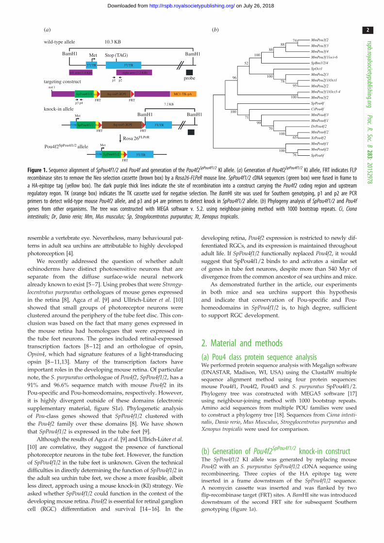
Figure 2. Restoration of RGCs in Pou4f2SpPou4f1/2/Z retinas. (a – c) H&E staining of retinas from mice at P60. (d – f ) Immunofluorence staining of retinas from mice atP60 by anti-neurofilament heavy chain (SMI-32) antibody (green). (g – i) Immunofluorence staining of retinas from mice at P60 by anti-neurofilament ligh chain(NF-L) antibody (green). (a,d,g) Pou4f2Z/þ control retinas. (b,e,h) Pou4f2SpPou4f1/2/Z retinas. (c,f,i) Pou4f2Z/Z retinas.
rspb.royalsocietypublishing.orgProc.R.Soc.B
283:20152978
4
on July 26, 2018http://rspb.royalsocietypublishing.org/Downloaded from
Pou4f2SpPou4f1/2/Z and Pou4f2SpPou4f1/2/SpPou4f1/2 mice. Therefore,
we used Pou4f2SpPou4f1/2/Z or Pou4f2SpPou4f1/2/AP mice for most
of the reported experiments, except that the RGC anterograde
tracing and ERG analysis were conducted using wild-type(WT), Pou4f2SpPou4f1/2/SpPou4f1/2 and Pou4f2Z/Z mice.
The Pou4f2SpPou4f1/2 KI targeting construct contained a PGK-neo-pA cassette and flanking FRT sites (figure 1a) and SpPou4f1/2expression could not be detected by immunostaining using an
anti-HA antibody (electronic supplementary material, figure
S1e and data not shown). We therefore removed the PGK-Neo-pA cassette by breeding the Pou4f2SpPou4f1/2 allele to a
Rosa26-FLPeR mouse line. Expression from the SpPou4f1/2allele in E14.5 retinas of Pou4f2SpPou4f1/2/þ mice, detected by
immunostaining with the anti-HA antibody (electronic sup-
plementary material, figure S1c), was spatially comparable
with that of endogenous Pou4f2 expression (electronic sup-
plementary material, figure S1d). However, expression levels
in Pou4f2SpPou4f1/2/þ retinas were substantially lower than those
in the Pou4f2HA/þ retinas using an anti-HA Pou4f2 antibody
(electronic supplementary material, figure S1f,g), suggesting
that SpPou4f1/2 was less stable than endogenous Pou4f2.
We first determined whether SpPou4f1/2 could restore
RGCs in mature adult retinas in the absence of Pou4f2. Ret-
inas of Pou4f2Z/Z mice at 60 days of age (P60) lacked
approximately 70% of their RGCs compared with the retinas
of wild-type mice (Pou4f2þ/Z) (figure 2a,c) [15,16]. This
depletion of RGCs resulted in a thinner retina (figure 2a,c).
The retinas of Pou4f2SpPou4f1/2/þ mice were normal, indicating
that the SpPou4f2-KI allele had no dominant phenotype (data
not shown). Most importantly, Pou4f2SpPou4f1/2/Z retinas were
not notably different from Pou4f2SpPou4f1/2/þ or Pou4f2þ/Z ret-
inas (data not shown). SpPou4f1/2-expressing retinas
appeared to have a normal ganglion cell layer with a full
complement of RGCs (figure 2b). This result suggests that
the Pou4f2SpPou4f1/2 allele was able to replace Pou4f2’s function
in forming RGCs during retinal development and in
maintaining the survival of RGCs in adult retinas.
Immunostaining of flat-mounted P60 Pou4f2SpPou4f1/2/Z
retinas with either SMI32 or NF-L antibodies showed the
presence of many well-bundled axons emanating from
SpPou4f1/2-expressing RGCs, whereas in Pou4f2Z/Z there
was little detectable staining (figure 2d– i). The number of
axons emitted from Pou4f2SpPou4f1/2/Z RGCs was substantially
greater than the number observed in Pou4f2Z/Z mice
(figure 2e,f and h,i), and their appearance was not qualitat-
ively different than those of Pou4f2þ/Z axons (figure 2d,eand g,h). These results suggested that SpPou4f1/2 expression
rescued the phenotype generated by the absence of Pou4f2.
Moreover, optic nerves of Pou4f2SpPou4f1/2/SpPou4f1/2 mice were
indistinguishable in thickness and appearance from those of
wild-type mice whereas Pou4f2Z/Z mice had only a thin
sheath largely devoid of axon fibres (cf. arrowheads in the
electronic supplementary material, figure S2a–c).
To further pursue the properties of SpPou4f1/2-expressing
axons, we traced their path into the brain with alkaline phos-
phatase (AP) using a Pou4f2-AP allele. Pou4f2-AP mice were
bred to Pou4f2þ/SpPou4f1/2 mice and retinorecepient regions
in the brain were stained for AP activity. Pou4f2þ/AP and
Pou4f2z/+
E15
E15
E15
200 µm
200 µm
200 µm
Pou4f2z/zPou4f2SpPou4f1/2/Z
Pou4f2z/+ Pou4f2z/zPou4f2SpPou4f1/2/Z
Pou4f2z/+ Pou4f2z/zPou4f2SpPou4f1/2/Z
TUNELTUNEL TUNEL
Pou4f1
Tbr2Tbr2 Tbr2
Pou4f1 Pou4f1
(a) (b) (c)
(d) (e) ( f )
(g) (h) (i)
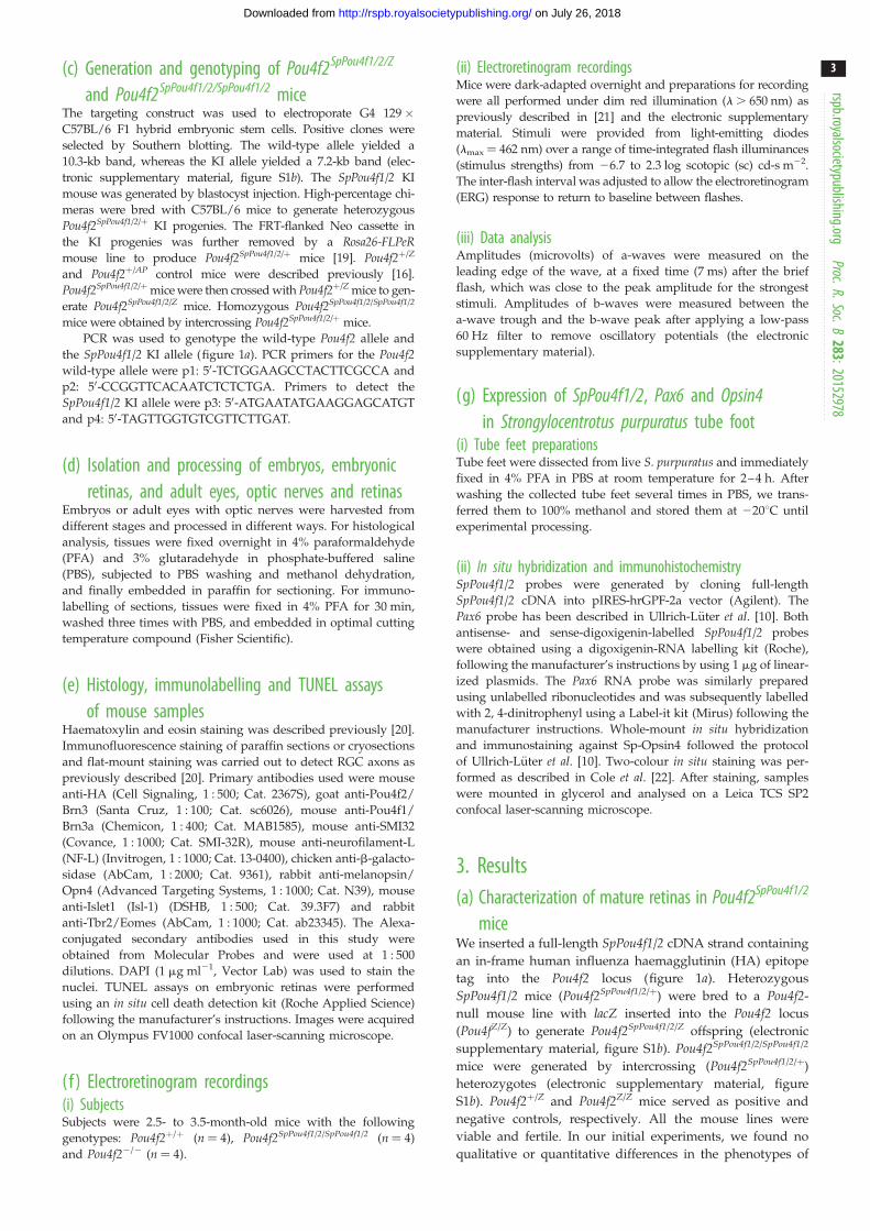
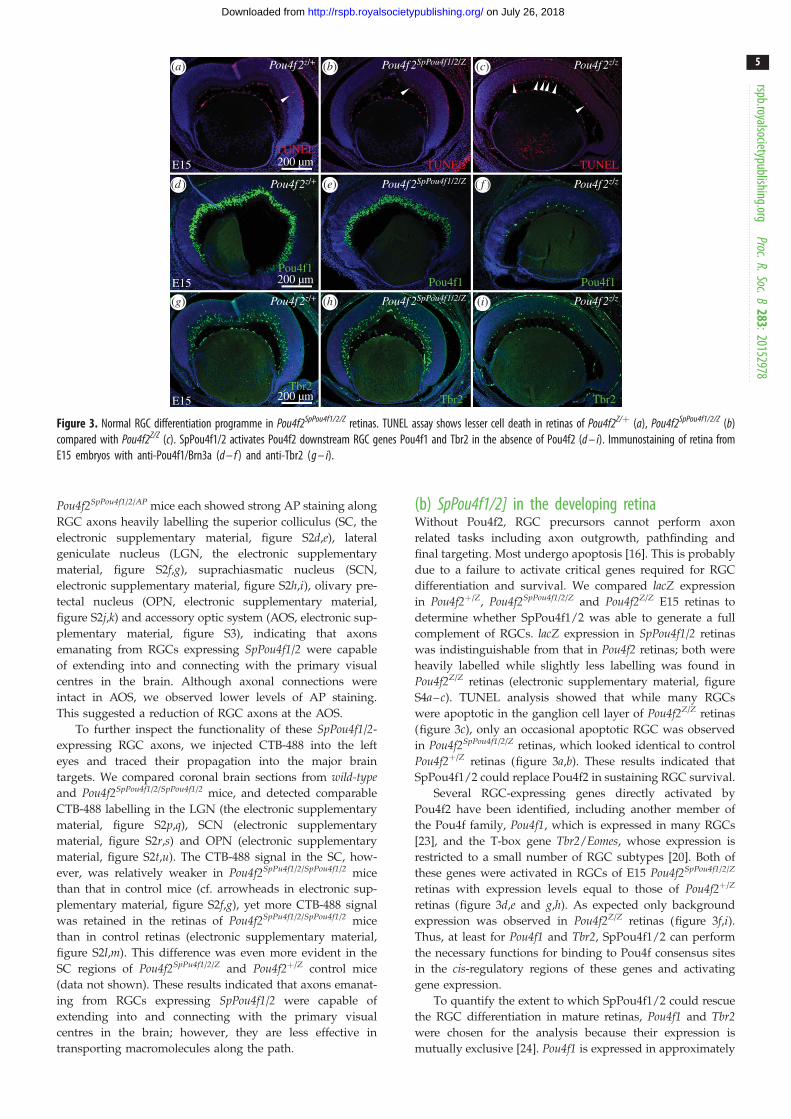
Figure 3. Normal RGC differentiation programme in Pou4f2SpPou4f1/2/Z retinas. TUNEL assay shows lesser cell death in retinas of Pou4f2Z/þ (a), Pou4f2SpPou4f1/2/Z (b)compared with Pou4f2Z/Z (c). SpPou4f1/2 activates Pou4f2 downstream RGC genes Pou4f1 and Tbr2 in the absence of Pou4f2 (d – i). Immunostaining of retina fromE15 embryos with anti-Pou4f1/Brn3a (d – f ) and anti-Tbr2 (g – i).
rspb.royalsocietypublishing.orgProc.R.Soc.B
283:20152978
5
on July 26, 2018http://rspb.royalsocietypublishing.org/Downloaded from
Pou4f2SpPou4f1/2/AP mice each showed strong AP staining along
RGC axons heavily labelling the superior colliculus (SC, the
electronic supplementary material, figure S2d,e), lateral
geniculate nucleus (LGN, the electronic supplementary
material, figure S2f,g), suprachiasmatic nucleus (SCN,
electronic supplementary material, figure S2h,i), olivary pre-
tectal nucleus (OPN, electronic supplementary material,
figure S2j,k) and accessory optic system (AOS, electronic sup-
plementary material, figure S3), indicating that axons
emanating from RGCs expressing SpPou4f1/2 were capable
of extending into and connecting with the primary visual
centres in the brain. Although axonal connections were
intact in AOS, we observed lower levels of AP staining.
This suggested a reduction of RGC axons at the AOS.
To further inspect the functionality of these SpPou4f1/2-
expressing RGC axons, we injected CTB-488 into the left
eyes and traced their propagation into the major brain
targets. We compared coronal brain sections from wild-typeand Pou4f2SpPou4f1/2/SpPou4f1/2 mice, and detected comparable
CTB-488 labelling in the LGN (the electronic supplementary
material, figure S2p,q), SCN (electronic supplementary
material, figure S2r,s) and OPN (electronic supplementary
material, figure S2t,u). The CTB-488 signal in the SC, how-
ever, was relatively weaker in Pou4f2SpPu4f1/2/SpPou4f1/2 mice
than that in control mice (cf. arrowheads in electronic sup-
plementary material, figure S2f,g), yet more CTB-488 signal
was retained in the retinas of Pou4f2SpPu4f1/2/SpPou4f1/2 mice
than in control retinas (electronic supplementary material,
figure S2l,m). This difference was even more evident in the
SC regions of Pou4f2SpPu4f1/2/Z and Pou4f2þ/Z control mice
(data not shown). These results indicated that axons emanat-
ing from RGCs expressing SpPou4f1/2 were capable of
extending into and connecting with the primary visual
centres in the brain; however, they are less effective in
transporting macromolecules along the path.
(b) SpPou4f1/2] in the developing retinaWithout Pou4f2, RGC precursors cannot perform axon
related tasks including axon outgrowth, pathfinding and
final targeting. Most undergo apoptosis [16]. This is probably
due to a failure to activate critical genes required for RGC
differentiation and survival. We compared lacZ expression
in Pou4f2þ/Z, Pou4f2SpPou4f1/2/Z and Pou4f2Z/Z E15 retinas to
determine whether SpPou4f1/2 was able to generate a full
complement of RGCs. lacZ expression in SpPou4f1/2 retinas
was indistinguishable from that in Pou4f2 retinas; both were
heavily labelled while slightly less labelling was found in
Pou4f2Z/Z retinas (electronic supplementary material, figure
S4a–c). TUNEL analysis showed that while many RGCs
were apoptotic in the ganglion cell layer of Pou4f2Z/Z retinas
(figure 3c), only an occasional apoptotic RGC was observed
in Pou4f2SpPou4f1/2/Z retinas, which looked identical to control
Pou4f2þ/Z retinas (figure 3a,b). These results indicated that
SpPou4f1/2 could replace Pou4f2 in sustaining RGC survival.
Several RGC-expressing genes directly activated by
Pou4f2 have been identified, including another member of
the Pou4f family, Pou4f1, which is expressed in many RGCs
[23], and the T-box gene Tbr2/Eomes, whose expression is
restricted to a small number of RGC subtypes [20]. Both of
these genes were activated in RGCs of E15 Pou4f2SpPou4f1/2/Z
retinas with expression levels equal to those of Pou4f2þ/Z
retinas (figure 3d,e and g,h). As expected only background
expression was observed in Pou4f2Z/Z retinas (figure 3f,i).Thus, at least for Pou4f1 and Tbr2, SpPou4f1/2 can perform
the necessary functions for binding to Pou4f consensus sites
in the cis-regulatory regions of these genes and activating
gene expression.
To quantify the extent to which SpPou4f1/2 could rescue
the RGC differentiation in mature retinas, Pou4f1 and Tbr2were chosen for the analysis because their expression is
mutually exclusive [24]. Pou4f1 is expressed in approximately
flash strength(log sc cd-s m–2)
1.4
0.4
–1.4
–4.7
–5.6
–6.8
+/+ SP/SP –/–
pSTR
nSTR
0 200 400 600 0 200 400 600 0 200 400 600time after the flash (ms)
pSTR and b-wave nSTR a-wave at 7 ms
1000 0 0
–100
–200
–300
–400 +/+–/–SP/SP
–500
–5
–10
–15
–20
–25
–30
–35
100
10
1
ER
G a
mpl
itude
(µV
)
pSTR
–7 –6 –5 –4 –3 –2 –1 –1 0 1 2–7 –6 –5 –40 1 2flash strength (log sc cd-s m–2)
(a)
(b) (c) (d)
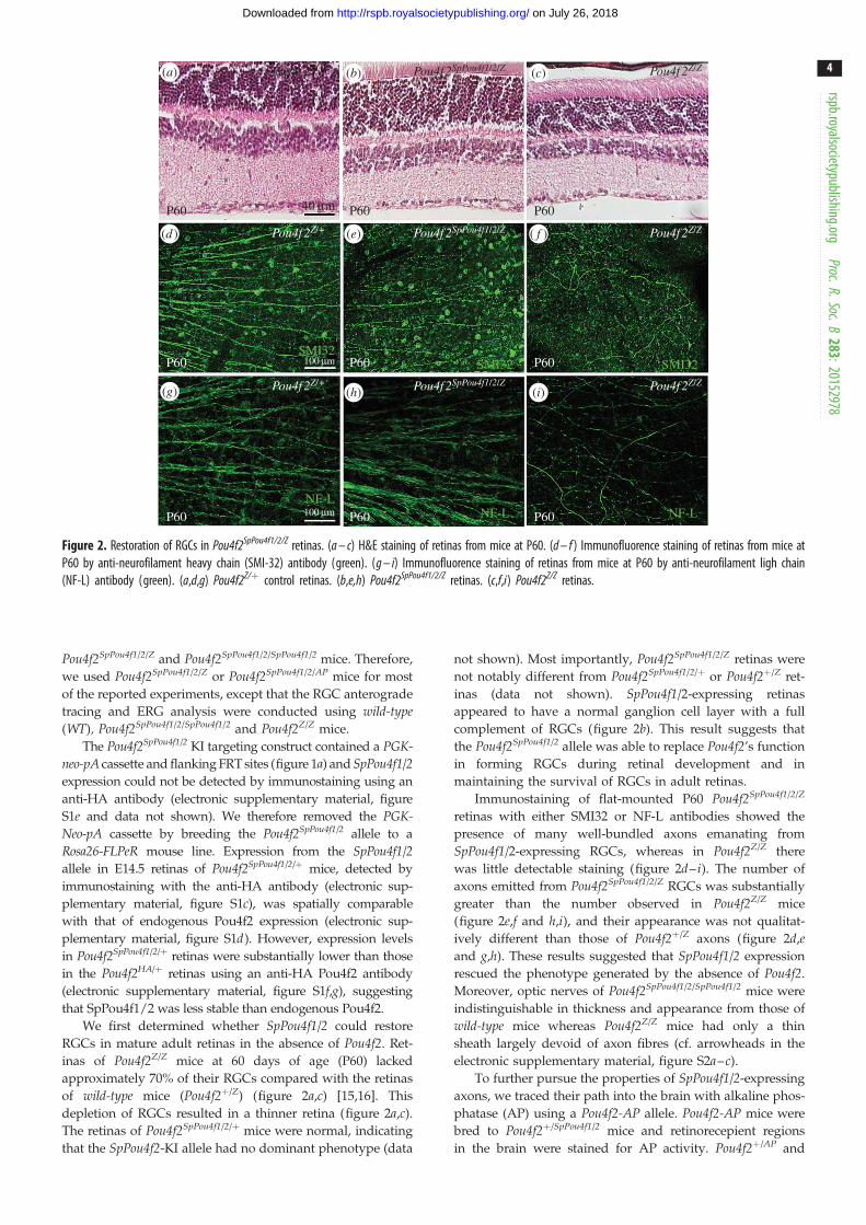
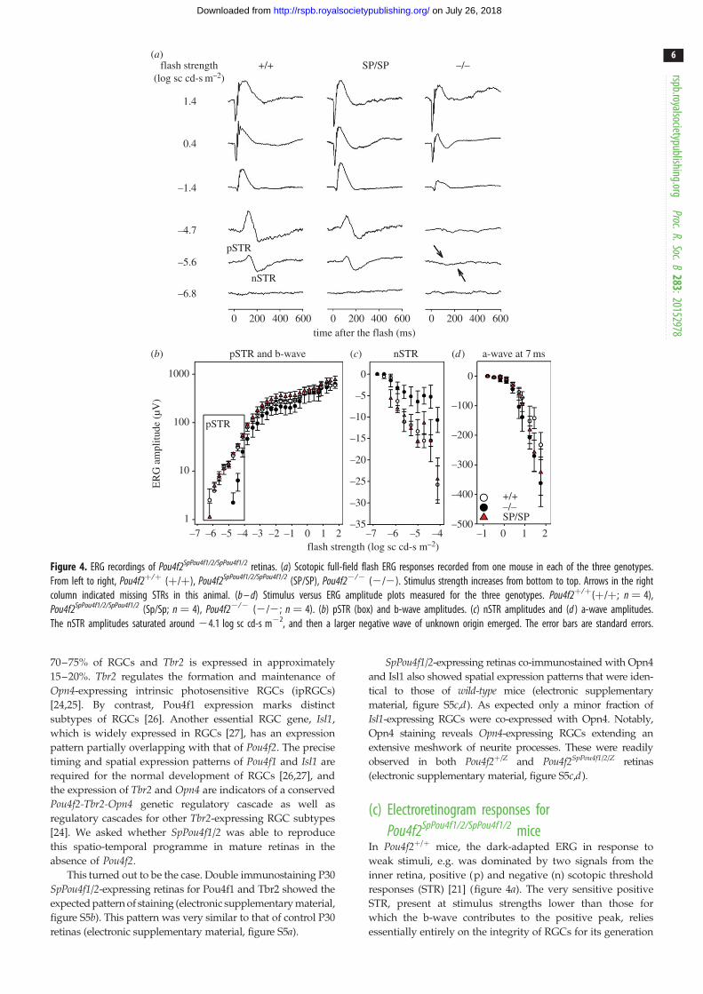
Figure 4. ERG recordings of Pou4f2SpPou4f1/2/SpPou4f1/2 retinas. (a) Scotopic full-field flash ERG responses recorded from one mouse in each of the three genotypes.From left to right, Pou4f2þ/þ (þ/þ), Pou4f2SpPou4f1/2/SpPou4f1/2 (SP/SP), Pou4f22/2 (2/2). Stimulus strength increases from bottom to top. Arrows in the rightcolumn indicated missing STRs in this animal. (b – d) Stimulus versus ERG amplitude plots measured for the three genotypes. Pou4f2þ/þ(þ/þ; n ¼ 4),Pou4f2SpPou4f1/2/SpPou4f1/2 (Sp/Sp; n ¼ 4), Pou4f22/2 (2/2; n ¼ 4). (b) pSTR (box) and b-wave amplitudes. (c) nSTR amplitudes and (d ) a-wave amplitudes.The nSTR amplitudes saturated around 24.1 log sc cd-s m22, and then a larger negative wave of unknown origin emerged. The error bars are standard errors.
rspb.royalsocietypublishing.orgProc.R.Soc.B
283:20152978
6
on July 26, 2018http://rspb.royalsocietypublishing.org/Downloaded from
70–75% of RGCs and Tbr2 is expressed in approximately
15–20%. Tbr2 regulates the formation and maintenance of
Opn4-expressing intrinsic photosensitive RGCs (ipRGCs)
[24,25]. By contrast, Pou4f1 expression marks distinct
subtypes of RGCs [26]. Another essential RGC gene, Isl1,
which is widely expressed in RGCs [27], has an expression
pattern partially overlapping with that of Pou4f2. The precise
timing and spatial expression patterns of Pou4f1 and Isl1 are
required for the normal development of RGCs [26,27], and
the expression of Tbr2 and Opn4 are indicators of a conserved
Pou4f2-Tbr2-Opn4 genetic regulatory cascade as well as
regulatory cascades for other Tbr2-expressing RGC subtypes
[24]. We asked whether SpPou4f1/2 was able to reproduce
this spatio-temporal programme in mature retinas in the
absence of Pou4f2.
This turned out to be the case. Double immunostaining P30
SpPou4f1/2-expressing retinas for Pou4f1 and Tbr2 showed the
expected pattern of staining (electronic supplementary material,
figure S5b). This pattern was very similar to that of control P30
retinas (electronic supplementary material, figure S5a).
SpPou4f1/2-expressing retinas co-immunostained with Opn4
and Isl1 also showed spatial expression patterns that were iden-
tical to those of wild-type mice (electronic supplementary
material, figure S5c,d). As expected only a minor fraction of
Isl1-expressing RGCs were co-expressed with Opn4. Notably,
Opn4 staining reveals Opn4-expressing RGCs extending an
extensive meshwork of neurite processes. These were readily
observed in both Pou4f2þ/Z and Pou4f2SpPou4f1/2/Z retinas
(electronic supplementary material, figure S5c,d).
(c) Electroretinogram responses forPou4f2SpPou4f1/2/SpPou4f1/2 mice
In Pou4f2þ/þ mice, the dark-adapted ERG in response to
weak stimuli, e.g. was dominated by two signals from the
inner retina, positive (p) and negative (n) scotopic threshold
responses (STR) [21] (figure 4a). The very sensitive positive
STR, present at stimulus strengths lower than those for
which the b-wave contributes to the positive peak, relies
essentially entirely on the integrity of RGCs for its generation
rspb.royalsocietypublishing.orgProc.R.Soc.B
283:20152978
7
on July 26, 2018http://rspb.royalsocietypublishing.org/Downloaded from
[21,28]. The negative STR amplitude is also impacted by the
loss of RGCs, but the reported extent of loss has varied
[28]. Pou4f22/2 mice lacked both waves originating from
the inner retina that were present in the other genotypes
(figure 4a).
The ERG responses to the weakest stimuli in
Pou4f2SpPou4f1/2/SpPou4f1/2 mice (figure 4a) were much more
similar to those in Pou4f2þ/þ mice than to those in
Pou4f22/2 mice, indicating the presence of RGC function. In
all three groups, the ERG responses to stronger stimuli, e.g.
21.4 log sc cd-s m22, were similar: the b-wave, thought to
be generated by rod-driven bipolar cells increased in ampli-
tude with stimulus strength, and for the strongest stimuli,
e.g. 1.4 log sc cd-s m22, a negative-going a-wave, which
reflects photoreceptor currents, was present at the beginning
of the response [29].
To quantify the ERG responses, stimulus versus ERG
amplitude plots were constructed based on measurements
of the ERG waves, pSTR and b-wave, measured at the peak
(figure 4b) and trough of the response (figure 4c), respect-
ively. Figure 4d shows the amplitude measured at 7 ms
after the flash on the leading edge of the a-wave. As predicted
by the ERG traces (figure 4a; electronic supplementary
material, table S1), the pSTR amplitudes were significantly
lower in the Pou4f22/2 mice than in the other groups. For
pSTR, amplitudes for the different groups were compared
only in the boxed region in the pSTR/b-wave plot. The
nSTR amplitudes of the Pou4f22/2 mice were also signifi-
cantly lower than in Pou4f2SpPou4f1/2/SpPou4f1/2 mice, and just
missed being significantly lower the Pou4f2þ/þ mice, whereas
p- and nSTRs in Pou4f2þ/þ and Pou4f2SpPou4f1/2/SpPou4f1/ mice
were not significantly different from each another. No other
ERG amplitude measures were significantly different across
the groups, although the b-wave amplitudes for the
Pou4f22/2 mice tended to be lower than those for the other
groups. Implicit times for measured responses also did not
differ significantly across groups.
(d) SpPou4f1/2 expression in Strongylocentrotuspurpuratus tube feet
Our results suggest that SpPou4f1/2 performs similar regulatory
functions to those of Pou4f2 in photoreceptor neurons of
S. purpuratus tube feet. It is also likely that SpPou4f1/2 is regu-
lated directly or indirectly by the S. purpuratus orthologue of
Pax6, as Pax6 is expressed in neuronal cells of the tube feet
disc [9,10]. Our expectation was that SpPou4f1/2 would be
expressed in tube feet photoreceptor cells and its expression
would overlap with that of both Pax6 and the sea urchin photo-
pigment Opsin4 [10]. Accordingly, we performed in situhybridization with adult S. purpuratus tube feet using
SpPou4f1/2 and Pax6 RNA probes and immunolabelling
using an anti-Opsin4 antibody [10].
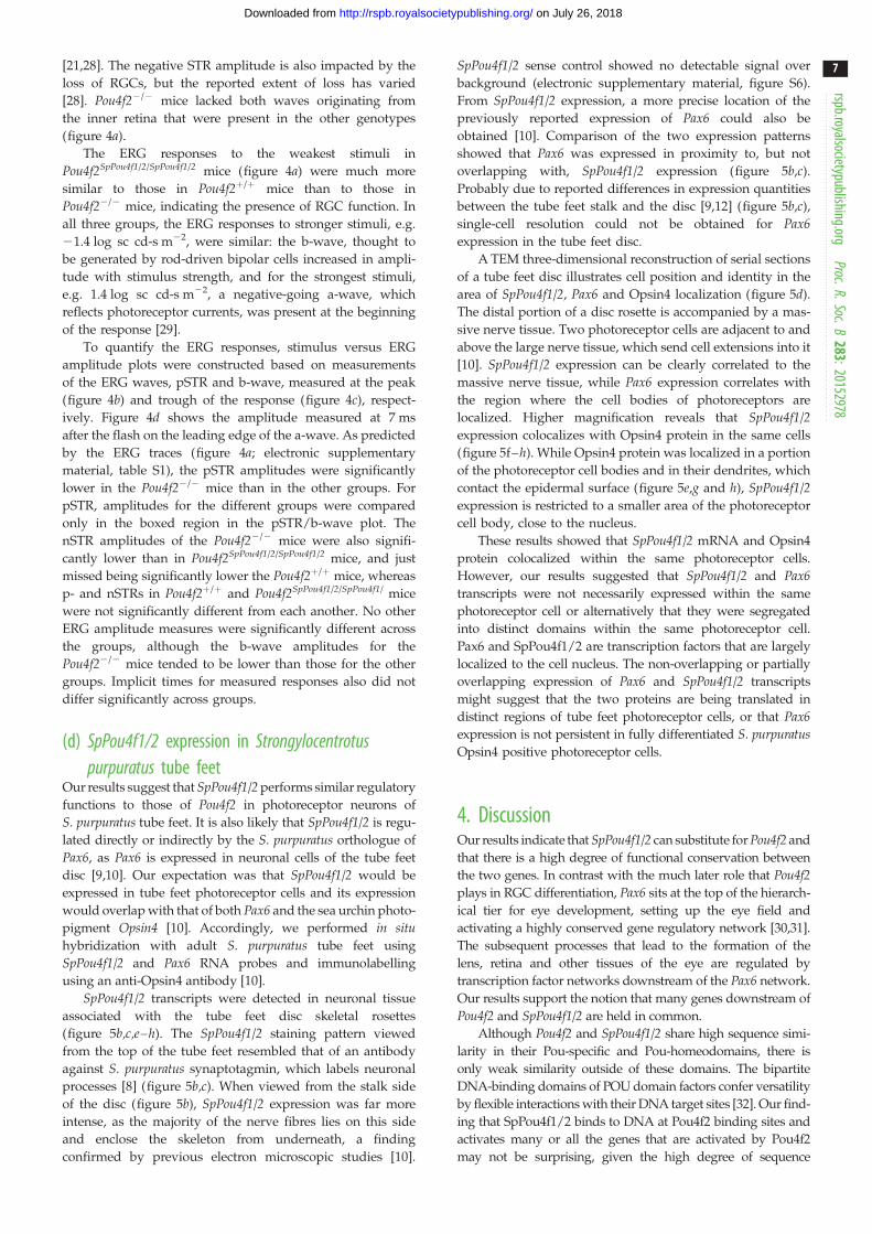
SpPou4f1/2 transcripts were detected in neuronal tissue
associated with the tube feet disc skeletal rosettes
(figure 5b,c,e–h). The SpPou4f1/2 staining pattern viewed
from the top of the tube feet resembled that of an antibody
against S. purpuratus synaptotagmin, which labels neuronal
processes [8] (figure 5b,c). When viewed from the stalk side
of the disc (figure 5b), SpPou4f1/2 expression was far more
intense, as the majority of the nerve fibres lies on this side
and enclose the skeleton from underneath, a finding
confirmed by previous electron microscopic studies [10].
SpPou4f1/2 sense control showed no detectable signal over
background (electronic supplementary material, figure S6).
From SpPou4f1/2 expression, a more precise location of the
previously reported expression of Pax6 could also be
obtained [10]. Comparison of the two expression patterns
showed that Pax6 was expressed in proximity to, but not
overlapping with, SpPou4f1/2 expression (figure 5b,c).
Probably due to reported differences in expression quantities
between the tube feet stalk and the disc [9,12] (figure 5b,c),
single-cell resolution could not be obtained for Pax6expression in the tube feet disc.
A TEM three-dimensional reconstruction of serial sections
of a tube feet disc illustrates cell position and identity in the
area of SpPou4f1/2, Pax6 and Opsin4 localization (figure 5d).
The distal portion of a disc rosette is accompanied by a mas-
sive nerve tissue. Two photoreceptor cells are adjacent to and
above the large nerve tissue, which send cell extensions into it
[10]. SpPou4f1/2 expression can be clearly correlated to the
massive nerve tissue, while Pax6 expression correlates with
the region where the cell bodies of photoreceptors are
localized. Higher magnification reveals that SpPou4f1/2expression colocalizes with Opsin4 protein in the same cells
(figure 5f–h). While Opsin4 protein was localized in a portion
of the photoreceptor cell bodies and in their dendrites, which
contact the epidermal surface (figure 5e,g and h), SpPou4f1/2expression is restricted to a smaller area of the photoreceptor
cell body, close to the nucleus.
These results showed that SpPou4f1/2 mRNA and Opsin4
protein colocalized within the same photoreceptor cells.
However, our results suggested that SpPou4f1/2 and Pax6transcripts were not necessarily expressed within the same
photoreceptor cell or alternatively that they were segregated
into distinct domains within the same photoreceptor cell.
Pax6 and SpPou4f1/2 are transcription factors that are largely
localized to the cell nucleus. The non-overlapping or partially
overlapping expression of Pax6 and SpPou4f1/2 transcripts
might suggest that the two proteins are being translated in
distinct regions of tube feet photoreceptor cells, or that Pax6expression is not persistent in fully differentiated S. purpuratusOpsin4 positive photoreceptor cells.
4. DiscussionOur results indicate that SpPou4f1/2 can substitute for Pou4f2 and
that there is a high degree of functional conservation between
the two genes. In contrast with the much later role that Pou4f2plays in RGC differentiation, Pax6 sits at the top of the hierarch-
ical tier for eye development, setting up the eye field and
activating a highly conserved gene regulatory network [30,31].
The subsequent processes that lead to the formation of the
lens, retina and other tissues of the eye are regulated by
transcription factor networks downstream of the Pax6 network.
Our results support the notion that many genes downstream of
Pou4f2 and SpPou4f1/2 are held in common.
Although Pou4f2 and SpPou4f1/2 share high sequence simi-
larity in their Pou-specific and Pou-homeodomains, there is
only weak similarity outside of these domains. The bipartite
DNA-binding domains of POU domain factors confer versatility
by flexible interactions with their DNA target sites [32]. Our find-
ing that SpPou4f1/2 binds to DNA at Pou4f2 binding sites and
activates many or all the genes that are activated by Pou4f2
may not be surprising, given the high degree of sequence
tfd
drn
tfs
ros
ga
tfn
ske
ros
ros
(a) (b)
(c)
( f ) (g) (h)
(d) (e)
(b¢)
(b¢¢)
Figure 5. Co-expression analysis of SpPou4f1/2, SpPax6 and SpOpsin4 in S. purpuratus tube feet. (a) Schematic view of a sea urchin tube feet displaying themorphology of tube feet stalk (tfs), disc (tfd) and rosette (ros); the nervous system (green) is represented by tube feet nerve (tfn), ganglion (ga) and discring nerve (drn). Sp-Opsin4 positive photoreceptor cells (red) are located at the rim of the disc and in a depression of the skeleton (ske) at the base of thetube feet. (b,c,e,f,g,h) Z-stack projection of tube feet disc. (b) Bottom view (seen from the stalk), (c,e,f,g,h) top view, showing SpPou4f1/2 and SpPax6 mRNAexpression and Sp-Opsin4 protein in disc photoreceptors. The whole tube feet disc is shown in (b) while (c), (e – g) display only a quadrant of it. SpPou4f1/2mRNA is shown in yellow/green (b) or green (c,f – h), SpPax6 mRNA in red (b) or purple (c), SpOpsin4 protein in red (c,e,g and h). (b) SpPou4f1/2 mRNA expressionassociated with skeletal rosettes, SpPax6 mRNA in stalk and disc. (b0 and b00) are details of (b) as indicated. (c) Expression of SpPou4f1/2 corresponds to neuronaltissue and axonal projection area of photoreceptors. SpPax6 expression in the area between skeletal rosette elements correlates with localization of photoreceptor cellbodies and SpOpsin4 protein in the apical part of the photoreceptor cells. (d ) Three-dimensional reconstruction of serial TEM sections clarifies cell position andidentity in SpPou4f1/2, SpPax6 and SpOpsin4 expression regions. Skeletal rosette (grey) with associated nerve tissue (yellow) and two photoreceptors ( purple andblue). (e – g) Different overlays showing coexpression of SpPou4f1/2 mRNA and SpOpsin4 protein in the apical region of photoreceptors. (h) A twofold zoom of thearea indicated by the rectangle in (e – g) showing a single photoreceptor (arrow shows nucleus).
rspb.royalsocietypublishing.orgProc.R.Soc.B
283:20152978
8
on July 26, 2018http://rspb.royalsocietypublishing.org/Downloaded from
similarity in the DNA-binding domains of the two proteins.
However, POU domain transcription factors must interact with
complex transcriptional machineries in order to function [33].
Several proteins are known to interact with POU domain factors
at sequences mapping outside of the bipartite DNA-binding
domains [33,34]. These interactions provide further functional
specificity to target gene selection. SpPou4f1/2 is probably to
interact with a multitude of proteins in tube feet photosensory
neurons. The observed functional equivalence of Pou4f2 and
SpPou4f1/2 implies that their interactions with co-activators,
co-repressors and other components of the transcriptional
machinery are also functionally conserved. Given the high
degree of sequence divergence of Pou4f2 and SpPou4f1 outside
of their Pou-specific and Pou-homeodomain, interactions with
other proteins are likely to be confined to these conserved
domains. However, the lack of sequence similarity outside the
Pou-specific and Pou-homeodomains does not preclude a con-
served role for at least some amino acids in these regions. We
think it is unlikely that a Pou4f class protein with sequences
chosen at random outside the conserved domains would
properly fold into a functional protein.
Our experiments support the view of functional equival-
ence of the Pou4f factors. While many genes expressed in
photosensitive neurons in the tube feet and RGCs in the
retina are likely to be held in common, there are likely to
be genes specifically required for the specialized neurons of
each species. Tube feet-specific genes might have acquired
consensus Pou4f DNA-binding sites at some point during
rspb.royalsocietypublishing.orgProc.R.Soc.B
283:20152978
9
on July 26, 2018http://rspb.royalsocietypublishing.org/Downloaded from
echinoderm evolution. The corresponding orthologous genes
in the mouse genome would not have Pou4f DNA-binding
sites and would not be expressed in RGCs. Conversely, for
genes specifically expressed in RGC differentiation, there
would be a set of corresponding orthologous genes in the
S. purpuratus genome that would not have SpPou4f1/2
DNA-binding sites.
Low levels of SpPou4f1/2 expression in the developing retina
were not surprising as its expression is under the control of the
relatively weak Pou4f2 promoter and intron 1 was deleted in the
KI allele. Nevertheless, the low expression of SpPou4f1/2 was
sufficient to form fully functional RGCs, even in the complete
absence of Pou4f2. Pou4f2Z/Z mice are genetically null and
Pou4f2 protein cannot be detected in mutant retinas [15,16].
However, Pou4f2þ/Z mice are phenotypically wild-type. As
Pou4f2SpPou4f1/2/Z expression levels are significantly lower than
Pou4f2þ/Z expression in the developing retina, the threshold
level for Pou4f2 necessary to function in RGC development is
probably to be substantially lower.
Ethics. All experimental and animal care procedures adhered to theAssociation for Research in Vision and Ophthalmology statementfor the Use of Animals in Ophthalmic and Vision Research wereapproved by the Institutional Animal Care and Use Committee atThe University of Texas MD Anderson Cancer Center and the
University of Houston and followed the United States PublicHealth Service Policy on Humane Care and Use of LaboratoryAnimals.
Data accessibility. The Pou4f2SpPou4f1/2 mouse line generated in this studyis freely available to all investigators upon request as stipulated inMD Anderson’s Memorandum Transfer Agreement policies.
Authors’ contributions. The authors have made the following declarationsabout their contributions: conceived and designed the experiments:C.A., C.-A.M., L.J.F., M.I.A., W.H.K. Performed the experiments:C.A., C.-A.M., S.W.W., P.P., J.W., J.A.M.-S., E.U.-L. Analysed thedata: C.A., C.-A.M., S.W.W., E.U.-L., L.J.F., J.W., J.A.M.-S., M.I.A.,W.H.K. Wrote the paper: W.H.K., L.J.F., C.A., C.-A.M., S.W.W.,M.I.A., E.U.-L.
Competing interests. We have no competing interests.
Funding. Support for this study came from grants from the NationalEye Institute (EY011930, EY019015, and EY010608-139005 toW.H.K., EY07551 to L.J.F. and EY024376 to C.-A.M.), from theRobert A. Welch Foundation (G-0010) to W.H.K., from a NationalCancer Institute Cancer Center Support Grant (CA016672) to theMD Anderson Cancer Center (Ronald DePinho, PI) and from VeluxFoundation (695) to C.A.
Acknowledgements. We are grateful to Jan Parker-Thornburg and theGenetically Engineered Mouse Facility (GEMF) for assistance in gen-eration of the Pou4f2SpPou4f1/2 mice. We also acknowledge the assistantof The DNA Analysis Facility and Research Animal Support Facility.We also thank Dr Xiuqian Mu (University at Buffalo, The StateUniversity of New York) for fruitful discussions at the initial phaseof the project.
References
1. Gehring WJ. 2002 The genetic control of eyedevelopment and its implications for the evolutionof the various eye-types. Int. J. Dev. Biol. 46,65 – 73.
2. Peter IS, Davidson EH. 2011 Evolution of generegulatory networks controlling body plandevelopment. Cell 144, 970 – 985. (doi:10.1016/j.cell.2011.02.017)
3. Popodi E, Raff RA. 2001 Hox genes in a pentameralanimal. BioEssays 23, 211 – 214. (doi:10.1002/1521-1878(200103)23:3,211::AID-BIES1030.3.0.CO;2-6)
4. Millott N. 1954 Sensitivity to light and the reactionsto changes in light intensity of the echinoid, Diademaantillarum Philippi. Phil. Trans. R. Soc. Lond. B 238,187 – 202. (doi:10.1098/rstb.1954.0009)
5. Blevins E, Johnsen S. 2004 Spatial vision in theechinoid genus Echinometra. J. Exp. Biol. 207,4249 – 4253. (doi:10.1242/jeb.01286)
6. Millott N. 1966 Coordination of spine movementsin echinoids. In Physiology of Echinodermata,pp. 187 – 220. New York, NY: Interscience.
7. Yoshida. 1966 Photosensitivity. In Physiology ofEchinodermata (ed. RA Boolootian), pp. 435 – 464.New York, NY: Interscience.
8. BurkeRD, et al. 2006 A genomic view of the seaurchin nervous system. Dev. Biol. 300, 434 – 460.(doi:10.1016/j.ydbio.2006.08.007)
9. Agca C, Elhajj MC, Klein WH, Venuti JM. 2011Neurosensory and neuromuscular organization intube feet of the sea urchin Strongylocentrotuspurpuratus. J. Comp. Neurol. 519, 3566 – 3579.(doi:10.1002/cne.22724)
10. Ullrich-Luter EM, Dupont S, Arboleda E, Hausen H,Arnone MI. 2011 Unique system of photoreceptors
in sea urchin tube feet. Proc. Natl Acad. Sci. USA108, 8367 – 8372. (doi:10.1073/pnas.1018495108)
11. Lesser MP, Carleton KL, Bottger SA, Barry TM,Walker CW. 2011 Sea urchin tube feet arephotosensory organs that express a rhabdomeric-like opsin and PAX6. Proc. R. Soc. B 278,3371 – 3379. (doi:10.1098/rspb.2011.0336)
12. Czerny T, Busslinger M. 1995 DNA-binding andtransactivation properties of Pax-6: three aminoacids in the paired domain are responsible for thedifferent sequence recognition of Pax-6 and BSAP(Pax-5). Mol. Cell Biol. 15, 2858 – 2871. (doi:10.1128/MCB.15.5.2858)
13. Raible F, Tessmar-Raible K, Arboleda E, Kaller T,Bork P, Arendt D, Arnone MI. 2006 Opsins andclusters of sensory G-protein-coupled receptors inthe sea urchin genome. Dev. Biol. 300, 461 – 475.(doi:10.1016/j.ydbio.2006.08.070)
14. Erkman L et al. 1996 Role of transcription factorsBrn-3.1 and Brn-3.2 in auditory and visual systemdevelopment. Nature 381, 603 – 606. (doi:10.1038/381603a0)
15. Gan L, Xiang M, Zhou L, Wagner DS, Klein WH,Nathans J. 1996 POU domain factor Brn-3b isrequired for the development of a large set ofretinal ganglion cells. Proc. Natl Acad. Sci. USA 93,3920 – 3925. (doi:10.1073/pnas.93.9.3920)
16. Gan L, Wang SW, Huang Z, Klein WH. 1999 POUdomain factor Brn-3b is essential for retinalganglion cell differentiation and survival but notfor initial cell fate specification. Dev. Biol. 210,469 – 480. (doi:10.1006/dbio.1999.9280)
17. Tamura K, Peterson D, Peterson N, Stecher G, Nei M,Kumar S. 2011 MEGA5: molecular evolutionary
genetics analysis using maximum likelihood,evolutionary distance, and maximum parsimonymethods. Mol. Biol. Evol. 28, 2731 – 2739. (doi:10.1093/molbev/msr121)
18. Gold DA, Gates RD, Jacobs DK. 2014 The earlyexpansion and evolutionary dynamics of POU classgenes. Mol. Biol. Evol. 31, 3136 – 3147. (doi:10.1093/molbev/msu243)
19. Farley FW, Soriano P, Steffen LS, Dymecki SM. 2000Widespread recombinase expression using FLPeR(Flipper) mice. Genesis 28, 106 – 110. (doi:10.1002/1526-968X(200011/12)28:3/4,106::AID-GENE30.3.0.CO;2-T)
20. Mao CA, Kiyama T, Pan P, Furuta Y, HadjantonakisAK, Klein WH. 2008 Eomesodermin, a target geneof Pou4f2, is required for retinal ganglion cell andoptic nerve development in the mouse.Development 135, 271 – 280. (doi:10.1242/dev.009688)
21. Saszik SM, Robson JG, Frishman LJ. 2002 Thescotopic threshold response of the dark-adaptedelectroretinogram of the mouse. J. Physiol. 543,899 – 916. (doi:10.1113/jphysiol.2002.019703)
22. Cole AG, Rizzo F, Martinez P, Fernandez-Serra M,Arnone MI. 2009 Two ParaHox genes, SpLox andSpCdx, interact to partition the posterior endodermin the formation of a functional gut. Development136, 541 – 549. (doi:10.1242/dev.029959)
23. Trieu M, Rhee JM, Fedtsova N, Turner EE. 1999Autoregulatory sequences are revealed by complexstability screening of the mouse Brn-3.0 locus.J. Neurosci. 19, 6549 – 6558.
24. Sweeney NT, Tierney H, Feldheim DA. 2014 Tbr2 isrequired to generate a neural circuit mediating the

rspb.royalsocietypublishing.orgProc.R.Soc.B
283
10
on July 26, 2018http://rspb.royalsocietypublishing.org/Downloaded from
pupillary light reflex. J. Neurosci. 34, 5447 – 5453.(doi:10.1523/JNEUROSCI.0035-14.2014)
25. Mao CA, Li H, Zhang Z, Kiyama T, Panda S, Hattar S,Ribelayga CP, Mills SL, Wang SW. 2014 T-boxtranscription regulator Tbr2 is essential for theformation and maintenance of Opn4/melanopsin-expressing intrinsically photosensitive retinalganglion cells. J. Neurosci. 34, 13 083 – 13 095.(doi:10.1523/JNEUROSCI.1027-14.2014)
26. Badea TC, Cahill H, Ecker J, Hattar S, Nathans J.2009 Distinct roles of transcription factors Brn3a andBrn3b in controlling the development, morphology,and function of retinal ganglion cells. Neuron 61,852 – 864. (doi:10.1016/j.neuron.2009.01.020)
27. Mu X, Fu X, Beremand PD, Thomas TL, Klein WH.2008 Gene regulation logic in retinal ganglion celldevelopment: Isl1 defines a critical branch distinct
from but overlapping with Pou4f2. Proc. Natl Acad.Sci. USA 105, 6942 – 6947. (doi:10.1073/pnas.0802627105)
28. Smith BJ, Wang X, Chauhan BC, Cote PD, TremblayF. 2014 Contribution of retinal ganglion cells to themouse electroretinogram. Doc. Ophthalmol. 128,155 – 168. (doi:10.1007/s10633-014-9433-2)
29. Robson JG, Frishman LJ. 2014 The rod-drivena-wave of the dark-adapted mammalianelectroretinogram. Prog. Retinal Eye Res. 39, 1 – 22.(doi:10.1016/j.preteyeres.2013.12.003)
30. Treisman JE. 1999 A conserved blueprint for theeye? BioEssays 21, 843 – 850. (doi:10.1002/(SICI)1521-1878(199910)21:10,843::AID-BIES6.3.0.CO;2-J)
31. Silver SJ, Rebay I. 2005 Signaling circuitriesin development: insights from the retinal
determination gene network. Development 132,3 – 13. (doi:10.1242/dev.01539)
32. Phillips K, Luisi B. 2000 The virtuoso ofversatility: POU proteins that flex to fit. J. Mol.Biol. 302, 1023 – 1039. (doi:10.1006/jmbi.2000.4107)
33. Andersen B, Rosenfeld MG. 2001 POU domainfactors in the neuroendocrine system: lessons fromdevelopmental biology provide insights into humandisease. Endocr. Rev. 22, 2 – 35. (doi:10.1210/edrv.22.1.0421)
34. Gonzalez MM, Carlberg C. 2002 Cross-repression,a functional consequence of the physicalinteraction of non-liganded nuclear receptors andPOU domain transcription factors. J. Biol. Chem.277, 18 501 – 18 509. (doi:10.1074/jbc.M200205200)
:
201 52978


![Integration, transcription, a mouse · 2005-04-22 · shock response eventually subsides, andnormal transcription and translation resume [reviewed by Ashburner and Bonner (1)]. Thefunction](https://img.dokumen.tips/doc/110x75/5f467f96e0bd590c76116f12/integration-transcription-a-mouse-2005-04-22-shock-response-eventually-subsides.jpg)


![Neuronal Transcription Factors Induce Conversion of Human ... · induces mouse fibroblasts to become functional neurons [12]. Other transcription factors, such as Ngn2 or Dlx1, are](https://img.dokumen.tips/doc/110x75/60418e3cb320ed3248628b47/neuronal-transcription-factors-induce-conversion-of-human-induces-mouse-fibroblasts.jpg)