Embed Size (px)
Citation preview
Title:Submucosal tunneling endoscopic resection for anunusually sized esophageal submucosal tumor protrudinginto the mediastinum
Authors:Shilan Zhang, Xiao Du, Xiaoyu Tang, Deliang Liu
DOI: 10.17235/reed.2019.5750/2018Link: PubMed (Epub ahead of print)
Please cite this article as:Zhang Shilan, Du Xiao, Tang Xiaoyu, Liu Deliang .Submucosal tunneling endoscopic resection for anunusually sized esophageal submucosal tumor protrudinginto the mediastinum. Rev Esp Enferm Dig 2019. doi:10.17235/reed.2019.5750/2018.
This is a PDF file of an unedited manuscript that has been accepted for publication. As a service to ourcustomers we are providing this early version of the manuscript. The manuscript will undergocopyediting, typesetting, and review of the resulting proof before it is published in its final form.Please note that during the production process errors may be discovered which could affect thecontent, and all legal disclaimers that apply to the journal pertain.
IPD 5750
Submucosal tunneling endoscopic resection for an unusually sized esophageal
submucosal tumor protruding into the mediastinum
Shilan-Zhang1, Xiao Du2, Xiaoyu Tang1 and Deliang Liu1
Departments of 1Gastroenterology and 2Cardiovascular Medicine. The Second
Xiangya Hospital of Central South University. Changsha, Hunan. China
Correspondence: Deliang Liu
e-mail: [email protected]
Author contributions
Shilan-Zhang contributed to the drafting of the manuscript. Xiao Du and Xiaoyu Tang
collected the writing material. Deliang Liu contributed to the conception of the study
and conducted the procedure.
CASE REPORT
A 50-year-old female came to our hospital with a six-month history of upper
abdominal discomfort. An upper endoscopy detected a protruding lesion that
measured 3.0 × 2.0 cm at around 35-38 cm from the incisors located on the posterior
wall. Endoscopic ultrasonography revealed a homogeneous hyperechoic mass
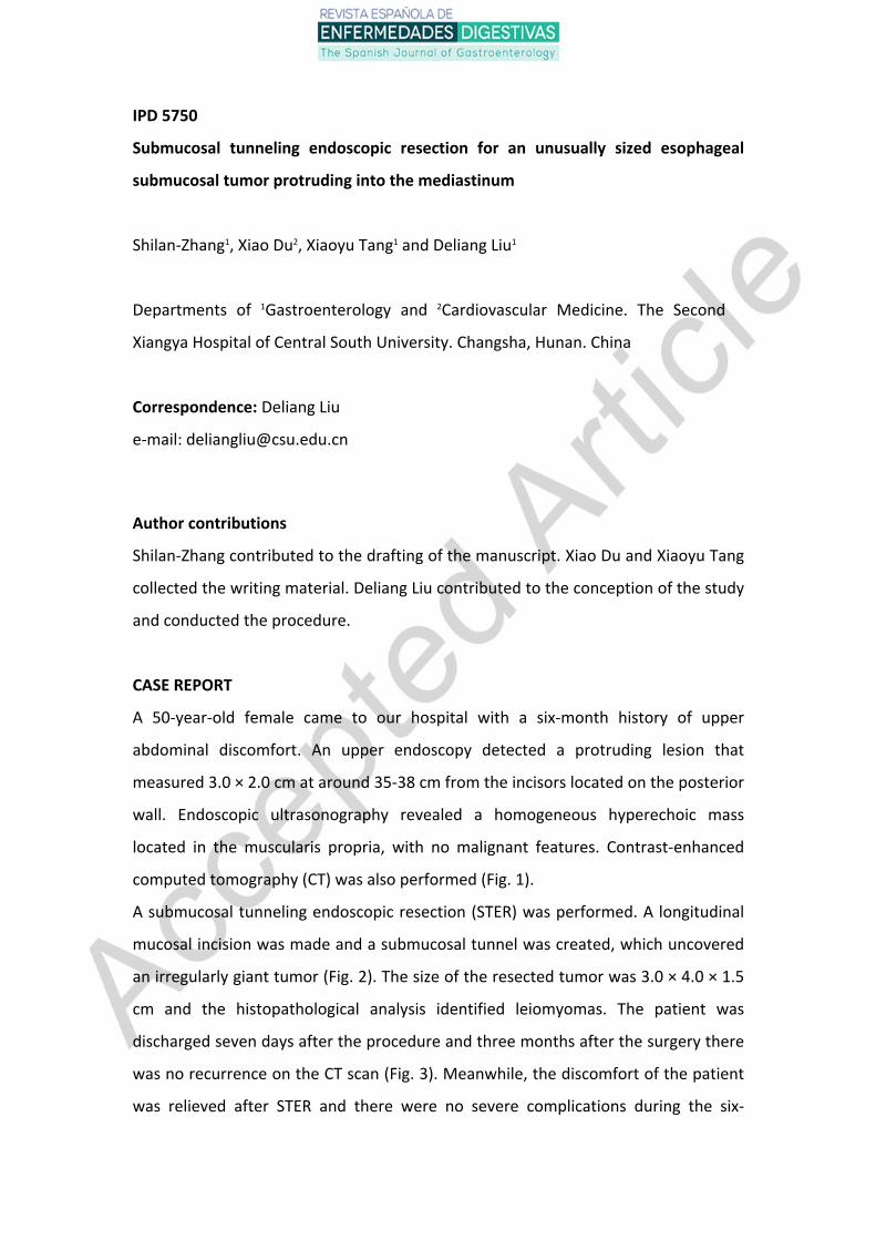
located in the muscularis propria, with no malignant features. Contrast-enhanced
computed tomography (CT) was also performed (Fig. 1).
A submucosal tunneling endoscopic resection (STER) was performed. A longitudinal
mucosal incision was made and a submucosal tunnel was created, which uncovered
an irregularly giant tumor (Fig. 2). The size of the resected tumor was 3.0 × 4.0 × 1.5
cm and the histopathological analysis identified leiomyomas. The patient was
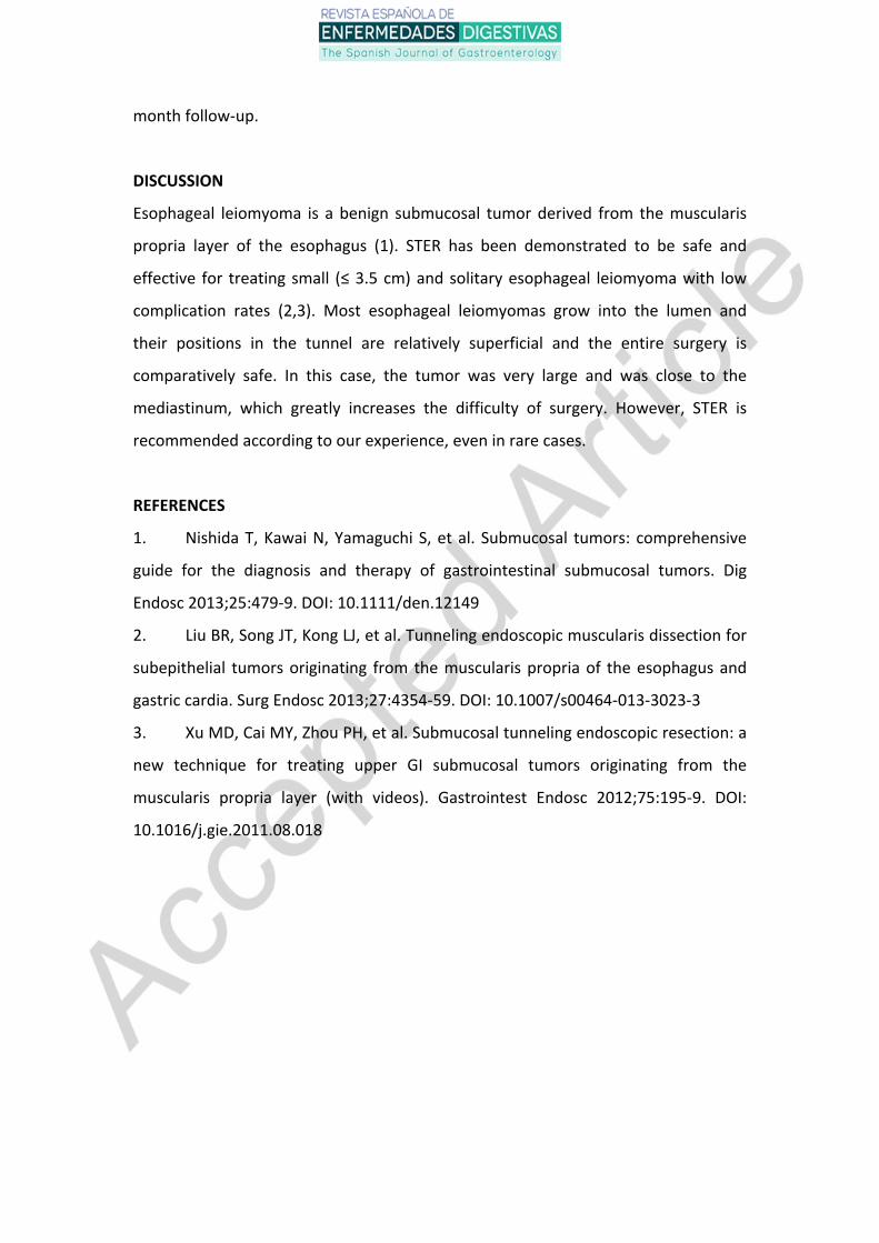
discharged seven days after the procedure and three months after the surgery there
was no recurrence on the CT scan (Fig. 3). Meanwhile, the discomfort of the patient
was relieved after STER and there were no severe complications during the six-
month follow-up.
DISCUSSION
Esophageal leiomyoma is a benign submucosal tumor derived from the muscularis
propria layer of the esophagus (1). STER has been demonstrated to be safe and
effective for treating small (≤ 3.5 cm) and solitary esophageal leiomyoma with low
complication rates (2,3). Most esophageal leiomyomas grow into the lumen and
their positions in the tunnel are relatively superficial and the entire surgery is
comparatively safe. In this case, the tumor was very large and was close to the
mediastinum, which greatly increases the difficulty of surgery. However, STER is
recommended according to our experience, even in rare cases.
REFERENCES
1. Nishida T, Kawai N, Yamaguchi S, et al. Submucosal tumors: comprehensive
guide for the diagnosis and therapy of gastrointestinal submucosal tumors. Dig
Endosc 2013;25:479-9. DOI: 10.1111/den.12149
2. Liu BR, Song JT, Kong LJ, et al. Tunneling endoscopic muscularis dissection for
subepithelial tumors originating from the muscularis propria of the esophagus and
gastric cardia. Surg Endosc 2013;27:4354-59. DOI: 10.1007/s00464-013-3023-3
3. Xu MD, Cai MY, Zhou PH, et al. Submucosal tunneling endoscopic resection: a
new technique for treating upper GI submucosal tumors originating from the
muscularis propria layer (with videos). Gastrointest Endosc 2012;75:195-9. DOI:
10.1016/j.gie.2011.08.018

Fig. 1. Computerized tomography (CT) revealed a 2.1 × 1.9 cm hypoechoic lesion in
the lower-esophagus and protruded to the mediastinal without invading the
surrounding nerves and blood vessels.
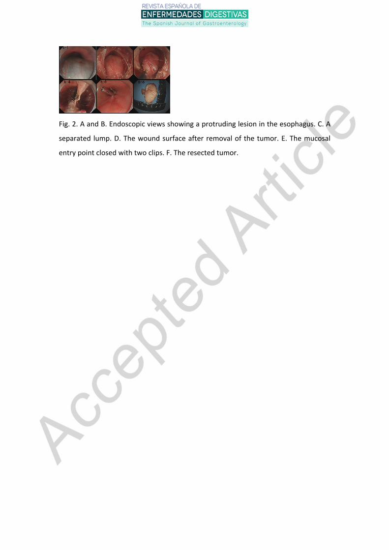
Fig. 2. A and B. Endoscopic views showing a protruding lesion in the esophagus. C. A
separated lump. D. The wound surface after removal of the tumor. E. The mucosal
entry point closed with two clips. F. The resected tumor.
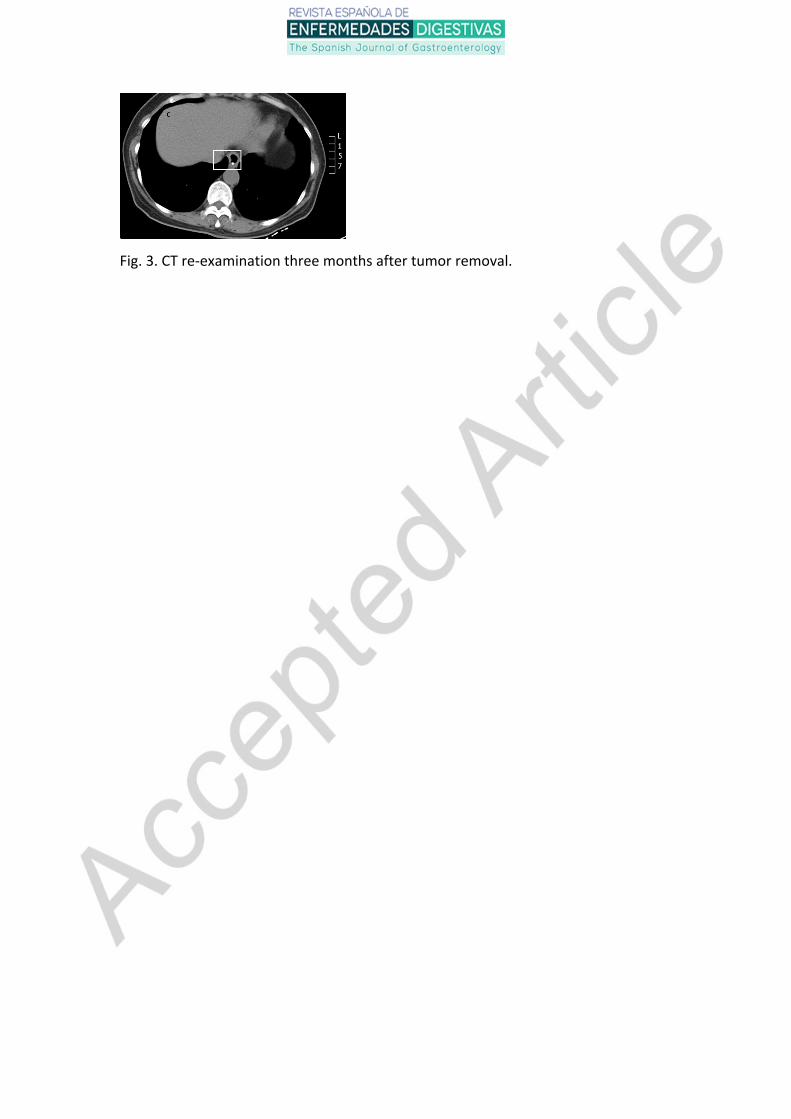
Fig. 3. CT re-examination three months after tumor removal.