Embed Size (px)
Citation preview

J Euk. M i c r u h d 43(6). 1996 pp 498-504 0 1996 by the Society of Promroologists
Subgenus Systematics of Acanthamoeba: Four Nuclear 18s rDNA Sequence Types
REBECCA J. GAST,I DOLENA R. LEDEE,2 PAUL A. FUERST and THOMAS J. BYERS3 Department of Molecular Genetics, The Ohio State University, 484 West 12th Avenue, Columbus, Ohio 43210-1292
ABSTRACT. Classification of Acanthamoeba at the subgenus level has been problematic, but increasing reports of Acantharnoeba as an opportunistic human pathogen have generated an interest in finding a more consistent basis for classification. Thus, we are developing a classification scheme based on RNA gene sequences. This first report is based on analysis of complete sequences of nuclear small ribosomal subunit RNA genes (Rns) from 18 strains. Sequence variation was localized in 12 highly variable regions. Four distinct sequence types were identified based on parsimony and distance analyses. Three were obtained from single strains: Type TI from Acanthamoeba crrslellanii V006, T2 from Acanthamueha pa1e.rtinensi.r Reich, and T3 from Acanthamoeba griflni S-7. T4, the fourth sequence type, included 15 isolates classified as A. castellanii, Acanthamoeba polyphagu, Acanthamoebu rhysodes, or Acanrhamoeba sp.. and included all 10 Acunthamoeba keratitis isolates. Interstrain sequence differences within T4 were 0%-4.3%, whereas differences among sequence types were 6%-12%. Branching orders obtained by parsimony and distance analyses were inconsistent with the current classification of T4 strains and provided further evidence of a need to reevaluate criteria for classification in this genus. Based on this report and others in preparation, we propose that Rns sequence types provide the consistent quantititive basis for classification that is needed.
Supplementary key words. Keratitis, pathogenic amoeba, ribosomal RNA genes, ssu rDNA.
CAhJTHAMOEBA is a genus of ubiquitous free-living amoe- A bae that are opportunistic pathogens of humans and other animal species [22, 301. In humans, the most common disease is Acanthamoeba keratitis, a painful sight-threatening eye infection, but acanthamoebae also cause granulomatous amebic encephalitis, a fatal brain infection, and are known to infect other organs in AIDS patients [18, 37, 381. It is unknown whether the different types of infection are caused by different subgroups of the genus or whether all acanthamoebae are capable of causing disease in the eyes, in the central nervous system, and in other organ tissues. An answer to this question depends on a reliable method of clas- sifying isolates.
Classification of Acanthamoeba at the subgenus level has been particularly problematic, but molecular diversity within the genus is large and offers hope for development of a more consistent taxonomy. Daggett et al. [ 1 13, in a previous cladistic study, identified 15 different isozyme electrophoretic zymo- grams (phenotypes) in the genus. These authors found numer- ous inconsistencies between their data and the existing isolate identifications. These inconsistencies could be due partly to problems with interpretation of zymograms, but most likely are due primarily to the absence of consistent criteria for classifi- cation of isolates. Johnson et al. [23] concluded that rRNA se- quence divergence within the genus Acanthamoeba is roughly similar to that differentiating vertebrates and invertebrates. A large rDNA sequence diversity also has been found in the data reported here.
In addition to detection, diagnosis and classification, interest in the function and evolution of processes in Acanthamoeba will undoubtedly be stimulated by recent discoveries, which include the complete sequence of the mitochondrial genome of Acanthamoeba castellanii Neff [4], the identification of the first cases of mitochondrial tRNA editing [27, 281 and evidence for the possible horizontal transfer of introns between algal chlo- roplasts and Acanthamoeba mitochondria [29, 351. Selection of the most appropriate Acanthamoeba species for study would be important. Our ribosomal DNA data and Rns types would be
I Current address: Department of Biology, Woods Hole Oceanograph- ic Insrurirurion, 324 Redjield Building, Woods Hole, Massachusetts 02543.
Current address: Department of Ophthalmology, Universiv of Penn- sylvania, Srellar Chance Labs, 422 Curie Boulevard, Philadelphia, Prnnsvlvania 19/04.
To whom correspondence should be addressed. Telephone: 614-292- 5963, Fax: 6 14-292-4466, Email: [email protected]
useful for distinguishing between closely or distantly related isolates.
Cyst size and structure have been used to divide species of Acanthamoeba into morphological Groups I, I1 and 111 [36]. Further study of intrageneric relationships has been based on criteria such as isoenzyme patterns [l I, 12, 211, physiological characteristics [9], immunological reactivities [ 131, mitochon- drial DNA restriction fragment length polymorphisms (mt- RFLP) [2, 10, 17, 411, and partial sequences of nuclear small subunit rRNA [23]. In spite of these various approaches, in- consistencies in classification persist. Of those approaches that have been tried, the use of rRNA gene sequences seems most likely to succeed. In this report, we describe the sites and extent of interstrain sequence variation in nuclear small ribosomal sub- unit RNA genes (Rns). We identify four distinct sequence types and propose using them as units of subgenus classification. Most Acanthamoeba keratitis isolates cluster in a single se- quence type. Subsequent reports will describe additional Rns types and present analyses of those nuclear or mitochondrial rDNA sequences.
MATERIAL AND METHODS Strains and culture conditions. The terms “strain” and “iso-
late” are used interchangeably throughout this report. Acan- thamoeba cultures were grown axenically as monolayers in Coming 25 cm2 culture flasks at 27” C-30” C in OGM [6]. Table 1 lists the strains used and their sources. The Ma isolate, ob- tained from Dr. E. Willaert, was identified as Acanthamoeba polyphaga based upon morphology, but later was identified as A. castellanii by reactivity with hyperimmune sera [30]. A soil isolate from Japan with an mtWLP pattern identical to Ma was identified as A. polyphaga JACK2 [41]. Reactivity with hyper- immune sera was used to identify Acanthamoeba sp. 88-2-27 as A. castellanii or A. polyphaga or Acanthamoeba rhysodes and Acanthamoeba sp. 88-2-37 as A. castellanii or A. polypha- ga (Osato, M., pers. commun.).
Gene nomenclature. Lonergan and Gray [29] have adopted names for Acanthamoeba mitochondrial rRNA genes that are based on nomenclature for Schizosaccharomyces pombe [25]. We propose the same mnemonics for the nuclear genes, but with the recommendation of the Commission on Plant Gene Nomenclature that nuclear genes can be designated by using upper case for the first letter of the mnemonic and organelle genes can be designated with lower case for the first letter [8]. The mitochondrial large and small subunit RNA genes have been named rnl and rns. Therefore, we propose that the cor- responding large and small subunit nuclear genes should be
498

CAST ET AL.-A CANTHAMOEBA NUCLEAR rDNA SEQUENCES 499
Table 1. Acunthunzoeha isolates analyzed.
Strains ATCC no. GenBank no.* Source
Group I1 A. ca.stellunii
Castellani 50374 U074 1 3 Yeast culture (London, UK) Ma 50370 U07414 Keratitis (New York, NY) Neff 50373 U074 16 Soil (Pacific Grove, CA) CDC:098 1 :V006b 50494 U07400 Brain (Atlanta, GA) CDC:0184:V014b 50492 U07401 Keratitis (India) CDC:0786:V042b 50493 U07403 Keratitis (IL) CDC:0180: Ih 5049 1 U07405 Lung (Pittsburg, PA)
A. griffin; S-7 3073 I U07412 Shallow beach (New London, CT) A. polyphugu
JACIS2' 50372 U074 15 Soil (Japan) 73- 1 - 1 6d 5037 1 U07407 Keratitis (Houston, TX) CDC:0885:V029b 50495 U07402 Keratitis (Boston, MA)
85-6- I I 6d 50368 U07406 Keratitis (Houston, TX) A. rhysodes
Singh - U074 17 -
Group 111 A. pulestinensis
Reich 30870 U07411 Soil (Israel)
Group Not Identified Aunthamoebu sp.
82- 12-324d - U07408 Keratitis (Houston, TX) 88-2-27d 50369 U07409 Keratitis (Houston, TX) 88-2-37d 50497 U074 10 Keratitis (Houston, TX) CDC:0688:V125b - U07404 Keratitis (Los Angeles, CA)
GenBank accession numbers for Rns sequences. Gift from Dr. Govinda Visvesvara, Atlanta, GA. Gift from Dr. Takuro Endo. Tokyo, Japan. Gift from Dr. Michael Osato, Houston, TX.
named RnZ and Rns. The nuclear large and small subunit rRNA genes in Acanthamoeba also have been referred to as the 26s and 18s rRNA genes, respectively.
DNA and RNA isolation, PCR amplification and DNA se- quencing. Amoebae were harvested from confluent cultures (-1 X lob amoebae) by low speed centrifugation (- 1000 g , 5- 10 min). The cell pellet was either resuspended in 500 p1 of lysis buffer (200 kg Proteinase K, 0.2% sodium dodecyl sulfate, 10 mM TrisHCl pH 7.4, 10 mM NaC1, 10 mh4 EDTA [3]) and incubated at 60" C for 2 h, or resuspended in 500 ~1 of UNSET (8 M urea, 2% sodium dodecyl sulfate, 0.15 M NaCl, 0.001 M EDTA, 0.1 M Tris.HC1 pH 7.5 [20]). The lysate was phenol- chloroform extracted twice and the nucleic acid precipitated by ethanol. The dried nucleic acid pellet was resuspended in 40 k1 sterile, double distilled water. PCR amplification [32] of the complete Rns gene used 1 p1 of whole cell DNA, oligonucle- otide primers complementary to the 5' and 3' ends of the gene (SSU1, SSU2) [16, 401, and a standard amplification program (30 cycles; 1 min, 94" C; 2 min, 45" C; 3 min, 72" C). PCR amplification of shorter segments of Rns used internal primers to conserved Rns sequences [40].
PCR products representing complete Rns genes were cloned into pBluescriptSK(-) (Stratagene, La Jolla, CA) or M13 (Boeh- ringer Mannheim, Indianapolis, IN) for double stranded or sin- gle stranded dideoxy sequencing and to preserve the product for future reference. Direct double stranded dideoxy sequencing (dsCycle Sequencing Kit; GibcoBRL, Gaithersburg, MD) of band isolated PCR products was also used to obtain sequence rapidly. At least 85% of each molecule was sequenced on both strands. The remainder, which was in conserved regions at the ends, was sequenced on a single strand at least twice.
Alignment and phylogenetic analysis. Acanthamoeba se- quences were deposited in GenBank [l] (Table 1) and aligned sequences are available upon request from TJB or RJG. Amoe- ba strains were deposited at the American Type Culture Col- lection (Rockville, MD) (Table 1). All sequences were aligned using the Eyeball Sequence Editor (ESEE v. 1.09e [7]) and alignments were based on primary and secondary structure con- servation [31]. The 5' and 3' terminal 23 bases are primer spec- ified for all of the Acanthamoeba sequences and were not in- cluded in any of the analyses.
When sequences for the 18 Acanthamoeba strains were aligned to each other, 2050 unambiguously aligned sites were identified; 46 of these sites were considered informative for the phylogenetic analysis because they were ditypic (at least two isolates shared a base that differed from the rest at that posi- tion). The number of alignable sites increased to 2163 if se- quences for V006, Reich and S-7 were removed, but only 33 were considered informative. All sites that could be aligned unambiguously were used to analyze branch order within the genus Acanthamoeba. Bootstrapped parsimony using heuristic search algorithms with rearrangement by tree bisection-recon- nection (PAUP 3.0 [34]) and neighbor-joining distance analysis with Kimura 2-parameter correction (DNADIST and NEIGH- BOR, PHYLIP [14]) were used for phylogenetic reconstruc- tions. Dissimilarity values were calculated as a percentage of mismatched bases in pairwise comparisons of sequences with- out the removal of unique gaps or ambiguous positions. Hence, they are not evolutionary distances. Evolutionary distances were caiculated based upon the dataset where all ambiguous and gap positions were removed. Dissimilarity values are used to emphasize the amount of sequence variability between iso-
500 J. EUK. MICROBIOL., VOL. 43, NO. 6, NOVEMBER-DECEMBER 1996
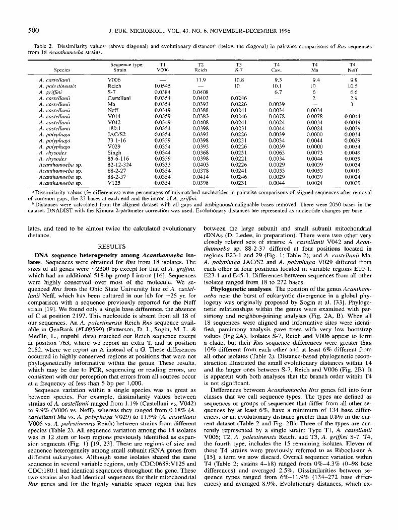
Table 2. Dissimilarity valuesd (above diagonal) and evolutionary distancesb (below the diagonal) in pairwise comparisons of Rns sequences from 18 Acarzrhamoeba strains.
Sequence type: TI T2 T3 T4 T4 T4 Species Strain V006 Reich s-I Cast. Ma Neff
A. castellanii V006 - 11.9 10.8 9.3 9.4 9.9 A. palestinensis Reich 0.0545 - 10 10.1 10 10.5 A. RriSJini s-7 0.0384 0.0408 - 6.7 6 6.6
2 2.9 A. castellanii Castellani 0.0354 0.0403 0.0246 -
A. castellanii Ma 0.0354 0.0393 0.0226 0.0039 - A. castellanii Neff 0.0349 0.0388 0.0241 0.0034 0.0034 -
A. castellanii V014 0.0359 0.0383 0.0246 0.0078 0.0078 0.0044 A. castellanii V042 0.0349 0.0408 0.0241 0.0024 0.0034 0.00 19 A. castellanii 180: 1 0.0354 0.0398 0.023 1 0.0044 0.0024 0.0039 A. polyphaga JACiS2 0.0354 0.0393 0.0226 0.0039 0.0000 0.0034 A. polyphaga 73-1-16 0.0339 0.0398 0.023 1 0.0034 0.0044 0.0029 A. polyphaga V029 0.0354 0.0393 0.0226 0.0039 0.0000 0.0034 A . rhysodes Singh 0.0344 0.0368 0.025 1 0.0063 0.0073 0.0049 A. rhysodes 85-6-116 0.0339 0.0398 0.0221 0.0034 0.0044 0.0039 Acanthamoeba sp. 82-12-324 0.0333 0.0403 0.0226 0.0029 0.0039 0.0034 Acantharnoeba sp. 88-2-27 0.0354 0.0378 0.0241 0.0053 0.0053 0.0019 Acantharnoeba sp. 88-2-37 0.0354 0.0414 0.0246 0.0029 0.0039 0.0024 Acanthamoeba sp. V125 0.0354 0.0398 0.023 1 0.0044 0.0024 0.0039
3
a Dissimilarity values (9% differences) were percentages of mismatched nucleotides in pairwise comparisons of aligned sequences after removal
Distances were calculated from the aligned dataset with all gaps and ambiguouslunalignable bases removed. There were 2050 bases in the of common gaps, the 23 bases at each end and the intron of A. grifJini.
dataset. DNADIST with the Kimura 2-parameter correction was used. Evolutionary distances are represented as nucleotide changes per base.
lates, and tend to be almost twice the calculated evolutionary distance.
RESULTS DNA sequence heterogeneity among Acanthamoeba iso-
lates. Sequences were obtained for Rns from 18 isolates. The sizes of all genes were -2300 bp except for that of A. griflni, which had an additional 518-bp group I intron [16]. Sequences were highly conserved over most of the molecule. We se- quenced Rns from the Ohio State University line of A. castel- lanii Neff, which has been cultured in our lab for -25 yr, for comparison with a sequence previously reported for the Neff strain [ 191. We found only a single base difference, the absence of C at position 2197. This nucleotide is absent from all 18 of our sequences. An A. palestinensis Reich Rns sequence avail- able in GenBank (#L09599) (Patterson, D. J., Sogin, M. L. & Medlin, L., unpubl. data) matched our Reich sequence except at position 763, where we report an extra T, and at position 2182, where we report an A instead of a G. These differences occurred in highly conserved regions at positions that were not phylogenetically informative within the genus. These results, which may be due to PCR, sequencing or reading errors, are consistent with our perception that errors from all sources occur at a frequency of less than 5 bp per 1,000.
Sequence variation within a single species was as great as between species. For example, dissimilarity values between strains of A. castellanii ranged from 1.1 % (Castellani vs. V042) to 9.9% (V006 vs. Neff), whereas they ranged from 0.18% (A. castellanii Ma vs. A. polyphaga V029) to 11.9% (A. castellanii V006 vs. A. pulestinensis Reich) between strains from different species (Table 2). All sequence variation among the 18 isolates was in 12 stem or loop regions previously identified as expan- sion segments (Fig. 1) [19, 231. These are regions of size and sequence heterogeneity among small subunit rRNA genes from different eukaryotes. Although some isolates shared the same sequence in several variable regions, only CDC:0688:V125 and CDC: 180: 1 had identical sequences throughout the gene. These two strains also had identical sequences for their mitochondrial Rns genes and for the highly variable spacer region that lies
between the large subunit and small subunit mitochondrial rDNAs (D. Ledee, in preparation). There were two other very closely related sets of strains: A. castellanii V042 and Acan- thamoeba sp. 88-2-37 differed at four positions located in regions E23-1 and 29 (Fig. 1; Table 2); and A. castellunii Ma, A. polyphaga JAClS2 and A. polyphaga V029 differed from each other at four positions located in variable regions E10-1, E23- 1 and E45- 1. Differences between sequences from all other isolates ranged from 18 to 272 bases.
Phylogenetic analyses. The position of the genus Acantham- oeba near the burst of eukaryotic divergence in a global phy- logeny was originally proposed by Sogin et al. [33]. Phyloge- netic relationships within the genus were examined with par- simony and neighbor-joining analyses (Fig. 2A, B). When all 18 sequences were aligned and informative sites were identi- fied, parsimony analysis gave trees with very low bootstrap values (Fig.2A). Isolates S-7, Reich and V006 appear to form a clade, but their Rns sequence differences were greater than 10% different from each other and at least 6% different from all other isolates (Table 2). Distance-based phylogenetic recon- struction illustrated the small evolutionary distances within T4 and the larger ones between S-7, Reich and V006 (Fig. 2B). It is apparent with both analyses that the branch order within T4 is not significant.
Differences between Acanthamoebu Rns genes fell into four classes that we call sequence types. The types are defined as sequences or groups of sequences that differ from all other se- quences by at least 6%, have a minimum of 134 base differ- ences, or an evolutionary distance greater than 0.8% in the cur- rent dataset (Table 2 and Fig. 2B). Three of the types are cur- rently represented by a single strain: Type T1, A. castellanii V006; T2, A. palestinensis Reich; and T3, A. gri'ni S-7. T4, the fourth type, includes the 15 remaining isolates. Eleven of these T4 strains were previously referred to as Ribocluster A [15], a term we now discard. Overall sequence variation within T4 (Table 2; strains 4-18) ranged from 0%-4.3% (0-98 base differences) and averaged 2.5%. Dissimilarities between se- quence types ranged from 6%-ll .9% (134-272 base differ- ences) and averaged 8.9%. Evolutionary distances, which ex-

GAST ET AL.-ACANTHAMOEBA NUCLEAR rDNA SEQUENCES 50 1
Table 2. Extended.
T4 T4 T4 T4 T4 T4 ~4 T4 T4 T4 T4 T4 V014 V042 180 JAC 16 V029 Sineh 116 324 21 37 V125
9.6 10.7 7 3.8 3.8 3.6
0.0063 0.0063 0.0078 0.0044 0.0078 0.0063 0.0044 0.0049 0.0024 0.0058 0.0063
-
9.3 10.4 6.7 1.1 1.8 2.6 3.3
0.0039 0.0034 0.0019 0.0034 0.0049 0.0029 0.0024 0.0039 0.0005 0.0039
-
9.8 10.5 6.7 2.7 1.8 3.4 4.3 2.4
0.0024 0.0049 0.0024 0.0068 0.0039 0.0044 0.0039 0.0044 0.0000
-
9.4
6.1 1.9 0.2 3.1 3.8 1.8 1.9
10
-
0.0044 0.0000 0.0073 0.0044 0.0039 0.0053 0.0039 0.0024
9.6 10.3 6.1 1.1 2.4 2.9 3.4 1.3 2.5 2.4
0.0044 0.0058 0.0010 0.0005 0.0049 0.0015 0.0049
-
- 9.5 9.8 8.9 9.2 9.6 9.3 9.8
10. I 10 10.3 10.3 10.8 10.4 10.5 6 7.2 6.6 6.7 6.8 6.9 6.7 I .9 3.2 1.5 0.89 3.6 1.3 2.7 0.2 3.9 2.3 2 3.3 1.8 1.8 3. I 3.7 3.1 3.1 3.2 2.8 3.4 3.8 3.4 3.2 3.4 0.8 3.2 4.3 1.7 3.1 1.1 1 .5 3 0.2 2.4 2 4 2.4 2.8 3.9 2.4 0 0.2 3.9 2.3 2 3.4 1.8 1.9 2.3 3.5 1.5 1.9 3.4 1.2 2.5 - 3.9 2.3 1.9 3.4 1.7 2
0.0073 - 3.1 3.4 3.1 3.2 4 0.0044 0.0058 __ 1.2 3.3 1.1 2.4 0.0039 0.0063 0.0005 - 3.5 1.4 2.8 0.0053 0.0039 0.0049 0.0053 - 3.1 3.9
2.4 0.0039 0.0053 0.0024 0.0019 0.0044 -
0.0024 0.0068 0.0039 0.0044 0.0039 0.0044 -
cluded differences in the highly variable and unalignable regions, ranged from 0.0-0.8 within T4 (0-16 unambiguous base differences) and from 2.2-5.4 between isolates from dif- ferent sequence types (45-1 1 1 unambiguous base differences).
DISCUSSION Phylogeny and taxonomy based on Rns. In this study we
have examined Rns gene sequences from 13 Acanthamoeba Group I1 strains (four species), one Group I11 strain (one spe- cies) and four unclassified isolates. Seventeen of the sequences determined from the 18 isolates differed from each other by at least four nucleotides. The Rns gene sequences were identical for Acanthamoeba sp. V125, isolated from an eye infection in California in 1988, and A. castellanii 180:1, isolated from a lung infection in Pennsylvania in 1980. Their identity was con- firmed by sequencing the same genes from two separate ship- ments of the strains.
T4 strains have a worldwide distiribution with isolates from Asia, Europe and North America. Recent discovery of addi- tional strains of A. grijjini [26] indicates that T3 also has a worldwide distribution. Data presented here indicate that T4 includes representatives of three different species, A. castellanii, A. polyphaga and A. rhysodes (Fig. 2). It includes all 10 of the
194 335 985 721 892, 1856
keratitis isolates included in our study, as well as A. castellanii Neff, the single strain that has been used for the great majority of all previous cellular and molecular studies.
Parsimony and distance analyses failed to resolve details of branching in T4, confirming inconsistencies in prior species classifications. A. castellanii Ma differs from five other isolates of this species (excluding V006) by 41-85 nucleotides, but dif- fers from A. polyphaga V029 and A. polyphaga JAC/S2 by only four nucleotides each (Table 2). Likewise, A. castellanii Cas- tellani differs from the five other isolates of A. castellanii by 62-89 nucleotides, but differs from A. rhysodes 85-6-116 by only 33 nucleotides. The two A. rhysodes isolates also differed from each other by 69 nucleotides, well within the range of differences seen for isolates of the other T4 species. The am- biguous identification of Acanthamoeba sp. 88-2-27 (A. castel- lanii, A. polyphaga, or A. rhysodes ) and Acanthamoeba sp. 88-2-37 (A. castellanii or A. polyphaga ) based on reactivity with hyperimmune sera (Osato, M., pers. commun.) actually is conistent with the Rns data in that they both are clearly mem- bers of T4.
Although the ambiguity in assigning species names to T4 strains suggests a need for reclassification of some isolates, no reliable way to subdivide all members of T4 has yet been found
1258 1654 1920 2138
I I I 4 3 E45-1 49
I E23-1 I E23-5 I) 7 29
8/9 L O O P E18-1 E23-8'/ E23-9' E23-7 L O O P
Fig. 1. Distribution of the 12 variable regions along the Rns gene of Acanthumorba. The location of variable regions is indicated by numbers below the figure that identify small subunit rRNA stems or loops predicted by Neefs et al. [31]. Numbers above the figure indicate the base pair where the stems and loops start in our sequence for A. castellanii Neff (GenBank #U07416).

502
A
J. EUK. MICROBIOL., VOL. 43, NO. 6 , NOVEMBER-DECEMBER 1996
B r A . castellanii Castellani - h A. castellanii Castellmi 1
I A. rhysodes 85-6-116 * A . polyphaga 73-1-16 *
1 -Acan&humeba sp. 82-12-324* I
I A. castellanii 180* 1 69?'= Acanthameba sp. V125* A . polyphaga JAClS2 A. castellanii Ma * I T4
, 861-1 8 9 r
L A . polyphaga V029 * Acanthameba sp. 88-2-27* A. castellanii V014* A. castellanii Neff A. castellanii V042* Acanthumeba sp. 88-2-37 * A. rhysodes Singh
56 A. castellanii VOO6 * T1 A. griffni S-7 T3 A. palestinemis Reich T2
A. rhysodes 85-6-1 16 * Acanthameba sp. 82-12-324 * A. polyphaga 73-1-16 *
A . castellanii VM2+ Acanthameba sp. 88-2-37 * A. castellanii 180 * Acanthamoeba sp. V125 * A. castellanii Ma* A. polyphaga VO29 * A. polyphaga JAClS2
A. castellanii Neff A. castellanii V014*
Acanthameba sp. 88-2-27 *
T4
I - A. rhysodes Singh A. castellanii V006 * T1
A. griffini S-7 T3 A. palestinensis Reich "2
1% divergence
Fig. 2. A. Parsimony tree for Rns sequences from 18 Acanthamoeha isolates. The tree was constructed using 500 bootstrap replications of heuristic searches with tree bisection-reconnection rearrangements. This tree is unrooted. Bootstrap values below 50% were not shown. B. Distance tree for Rns sequences from 18 Acanthamoeba isolates. This tree is unrooted. The evolutionary distance matrix (Table 2, lower triangle) was calculated using Kimura 2-parameter correction and the tree was constructed using neighbor-joining. The scale bar represents the evolutionary distance equivalent to 1%. (*), Human disease isolate. (Tl-T4), Strains with differing Rns sequence types as described in the text.
and no quantitative boundaries for species have been deter- mined. A reasonable possibility would be to assign all T4 strains to a single species, but preliminary evidence suggests that sequence variation in this group of isolates may be several times higher than the interstrain variation of named species in other sequence types (Shroeder-Diedrich, J., DRL, Stothard, D., & TJB, unpubl. observ.). Although our Rns sequence data do not provide a reliable subdivision of T4, other molecular approaches might be more successful. In 1996, an international cooperative project to coordinate various molecular approaches to the systematics of Acanthamoeba was initiated at the Sixth International Conference on Amoebae in Adelaide, Australia. Thus, it would be premature to rename T4 strains at this time. Whenever possible, however, new isolates should be typed on the basis of Rns sequences for comparison with other typing methods. Identification of the sequence type could be achieved by sequencing Rns genes, as described here, by use of Rns- specific PCR (Lehmann, 0. J., Keys, M. J.,Green, S. M., Kil- vington, S., McGill, A. R., Elkington, l? J. & Watt, l? J., 1995. Early diagnosis of Acanthamoeba keratitis with the polymerase chain reaction. Abstract. Invest. Ophthalmol. Vis. Sci., 36: S182), or in situ probes [15].
Rns sequence types and isozyme patterns. The most ex- tensive phylogenetic analysis of Acanthamoeba was based on isoenzyme electrophoretic patterns for 7 1 isolates including 15 identified species [ 1 11. This study identified 15 Acanthamoeba lineages, but noted inconsistencies between the various lineages and the classification of strains. Eight of the 15 lineages con- tained more than one species and several species occurred in multiple lineages. In some cases, the isozyme patterns linked strains that were clearly morphologically distinct. The authors assumed this result most likely was due to problems of strains classified based primarily on morphological criteria. It is not
clear, however, whether the problem is with the other criteria or with their application by different taxonomists.
Isoenzyme patterns can be very helpful for classification pur- poses, but problems in their use with Acanthamoeba have been noted. Jacobson and Band [ 2 11 discovered a large heterogeneity in patterns obtained from environmental isolates of A. polypha- ga. Although this might be attributed to difficulty in identifying Acanthamoeba species, they also reported that patterns changed when environmental isolates were grown axenically under lab- oratory selection. If this type of change was due to changes in gene expression or post-translational processing of the en- zymes, rather than to selective replication of strains in a mixed culture, it could be problematic for isoenzyme-based classifi- cation. The use of stable DNA sequences as a basis for clas- sification should eliminate this type of problem.
Rns sequence types and mitochondria1 restriction frag- ment length polymorphisms. Analyses in our lab of genomic mitochondrial DNA RFLP (mtRFLP) [2, 51 indicated a high degree of variation among Acanthamoeba isolates. However, more recently, several labs have been able to cluster strains based on identical mtRFLP [ 17, 24,411. Unfortunately, the pub- lished literature indicates few strains that have been examined both with mtRFLP and Rns sequences. When we compared Rns sequences from the Japanese isolate A. polyphaga JAC/S2 and the North American isolate A. castellanii Ma, which have iden- tical mtRFLP [41], we found they differed by only four nucle- otide pairs (Table 2 ) . More extensive studies suggest similar close relationships between mtRFLP and R m sequence types in other strains (RJG, DRL, Yagita, K. & Endo, T., unpubl. ob- serv.). This relationship should be examined further since RFLPs are generally easier to obtain than sequences and might be an alternative approach to recognize the clusters of strains identified by sequence types. One disadvantage of using mt-

GAST ET AL.-ACANTHAMOEBA NUCLEAR rDNA SEQUENCES 503
RFLP to identify isolates, however, is that relatively large num- bers of amoebae are required for an analysis.
18s rRNA sequences. Johnson et al. [23] were the first to use ribosomal nucleic acid sequences to study the phylogeny of the genus Acanthamoeba. They used reverse transcriptase to directly sequence three segments of 18s rRNA transcribed from seven isolates representing all three morphological groups. Their analysis suggested that the sequence divergence in Acan- thamoeba was comparable to that between vertebrates and in- vertebrates. Although our data are inconsistent with some of their reported sequences, the authors are correct in reporting a relatively high degree of 18s rRNA sequence diversity within the genus. For general use, however, DNA sequencing is much easier than direct RNA sequencing.
Pathogenicity. The pathogenicity of an Acanthamoeba iso- late cannot be determined from DNA sequence information alone. However, as we and others have noted [2, 17, 41 1. close phylogenetic relatedness can be one useful characteristic in the evaluation of an isolate’s pathogenic potential. We suggest, for example, that the environmental isolate A. polyphaga JAC/S2 should be considered a potential human pathogen. The very close relationship of this isolate to the human eye isolates A. castellanii Ma and A. polyphaga V029 is indicated by the Rns data presented here (Table 2 and Fig. 2) and by sequences de- termined in our lab for a number of additional genes (DRL, unpubl. data). The most closely related strains in this study are V12.5, an eye isolate, and 180:1, a lung isolate. So far, we have been unable to find any genetic differences between them. Al- though these are different isolates, their genetic similarity is the best evidence available that any isolate might be capable of infecting more than one target tissue.
Vodkin et al. [39] designed PCR primers that appeared to distinguish pathogenic and nonpathogenic Acanthamoeba iso- lates. Their primers appeared to support amplification of a DNA fragment from nonpathogenic, but not from pathogenic acan- thamoebae. The sequencing that we have done for this study reveals, however, that one of their primers is complementary to a highly variable region in the genomic DNA and that its ability to promote amplification is unrelated to pathogenicity. For ex- ample, the nonpathogen-specific PCR product could be ob- tained from the human disease isolate A. rhysodes 8.5-6- 1 16. We cannot rule out the possibility that all isolates of Acantham- oeha are potential human patho . However, in this study and others to be reported later, in ch we have examined Rns sequences from more than SO strains, the large majority of the human disease isolates have T4 sequences. Thus, it seems rea- sonable that all T4 strains should be considered potential patho- gens.
ACKNOWLEDGMENTS
This work was supported by National Institutes of Health/ National Eye Institute Grant No. EY09073 to TJB and PAE Portions were submitted to The Ohio State University by RJG in partial fulfillment of requirements for the Ph.D. degree. We thank Drs. Govinda Visvesvara, Michael Osato and Takuro Endo for providing isolates from their culture collections, Dr. David Caron for making computer facilities at Woods Hole Oceanographic Institution available to RJG, and Dr. Diane Sto- thard for help with tree construction.
LITERATURE CITED
1. Bilofsky, H. S. & Burks, C. 1988. The GenBank genetic sequence data bank. Nucl. Acids Rex, 16:1861-1864.
2. Bogler, S. A,, Zarley, C. D., Burianek, L. L., Fuerst, P. A. & Byers, T. J. 1983. Interstrain mitochondrial DNA polymorphism detected in
Acanthamoebu by restriction endonuclease analysis. Mol. Biochem. Parasitol., 8: 145-163.
3. Burg, J. L., Grover, C. M., Pouletty, I? & Boothroyd, J . C. 1989. Direct and sensitive detection of a pathogenic protozoan Toxoplasma gondii by polymerase chain reaction. J . Clin. Microbiol., 27: 1787-1 792.
4. Burger, G . , Plante, I., Lonergan, K. M. & Gray, M. W. 1995. The mitochondrial DNA of the amoeboid protozoan, Acanthamoeba castel- lanii: complete sequence, gene content and genome organization. J . Mol. Biol., 245:522-537.
5. Byers, T. J., Hugo, E. R., Stewart, V. J. 1990. Genes of Acan- thamoeba: DNA, RNA and protein sequences (a review). J. Prorozool., 37:175-25S.
6. Byers, T. J., Akins, R. A., Maynard, B. J . , Lefken, R. A. & Martin, S. M. 1980. Rapid growth of Acanthamoeba in defined media; induc- tion of encystment by glucose-acetate starvation. J . Prorozool., 27:2 16- 219.
7. Cabot, E. L. & Beckenbach, A. T. 1989. Simultaneous editing of multiple nucleic acid and protein sequences with ESEE. Comput. Appl. Biosci., 5: 233-234.
8. Commision on Plant Gene Nomenclature. 1994. Nomenclature of sequenced plant genes. Plant. Mol. Biol. Reptr., 12:S81.
9. Costas, M. & Griffiths, A. J. 1986. Physiological characterization of Acanthamoeba strains. J. Protozool., 33:304-309.
10. Costas, M., Edwards, S. W., Lloyd, D., Griffiths, A. J. & Turner, G . 1983. Restriction enzyme analysis of mitochondria1 DNA of mem- bers of the genus Acunthamoeba as an aid in taxonomy. FEMS Micro- biol. Lett., 17:231-234.
11. Daggett, I?-M., Lipscomb, D. S., Thomas, K. & Nerad, T. A. 1985. A molecular approach to the phylogeny of Acanthamoeba. Bio- svstems, 18:399-405.
12. De Jonckheere, J. E 1983. Isoenzyme and total protein analysis by agarose isoelectric focusing, and taxonomy of the genus Acanrham- oeba. J . Prorozool., 30:701-706.
13. Epstein, R. J., Wilson, L. A,, Visvesvara, G. S., & Plounde, E. G. 1986. Rapid diagnosis of Acanthamoeba keratitis from corneal scraping using indirect fluorescent antibody staining. Arch. Opthalmol., 104: 13 18-321.
14. Felsenstein, J. 1989. PHYLIP-phylogeny inference package, vers. 3.2. Cladisrics, 5:164-166.
15. Gast, R. J. & Byers, T. J. 1995. Genus- and subgenus-specific oligonucleotide probes for Acanthamoeba. Mol. Biochem. Parasitol., 71:255-260.
16. Gast, R. J. , Fuerst, I? A. & Byers, T J. 1994. Discovery of group I introns in the nuclear small subunit ribosomal RNA genes of Acan- rhamoeba. Nucl. Acids Res., 22:592-596.
17. Gautom, R. K., Lory, S., Seyedirashti, S., Bergeron, D. L. & Fritsche, T. R. 1994. Mitochondria1 DNA fingerprinting of Acantham- oeba spp. isolated from clinical and environmental sources. J. Clin. Microbiol., 32: 1070-073.
18. Gullet, J . , Mills, J., Hadley, K., Podemski, B., Pitts, L. & Gelber, R. 1979. Disseminated granulomatous Acanthamoeba infection pre- senting as an unusual skin lesion. Am. J . Med., 67:891-896.
19. Gunderson, J. H. & Sogin, M. L. 1986. Length variation in eukaryotic rRNAs: small subunit rRNAs from the protists Acantham- oeba castellanii and Euglena gracilis. Gene, 44:63-70.
20. Hugo, E. R., Stewart, V. J., Gast, R. J. & Byers, T. J. 1992. Purification of amoeba mtDNA using the UNSET procedure. In: Soldo, A. T. & Lee, J . J . (ed.), Protocols in Protozoology. Allen Press, Law- rence, Kansas. P. D-7.1,
21. Jacobson, L. M. & Band, R. N. 1987. Genetic heterogeneity in a natural population of Acanthamoeba polyphaga from soil, an isoen- zyme analysis. J . Protozool., 34:83-86.
22. John, D. T. 1993. Opportunistically pathogenic free-living ame- bae. In: Kreier, J. (ed.), Parasitic Protozoa, 2d ed. Academic Press, San Diego. 1: 143-246.
23. Johnson, A. M., Fielke, R., Christy, I? E., Robinson, B. & Bav- erstock, I? R. 1990. Small subunit ribosomal RNA evolution in the genus Acanthamoeba. J. Gen. Microbiol., 136: 1689-1698.
24. Kilvington, S., Beeching, J. R., & White, D. G. 1991. Differ- entiation of Acanthamoeba strains from infected corneas and the envi- ronment by using restriction endonuclease digestion of whole-cell DNA. J . Clin. Microbiol., 29:310-314.

504 J. EUK. MICROBIOL., VOL. 43, NO. 6, NOVEMBER-DECEMBER 1996
25. Kohli, J. 1987. Genetic nomenclature and gene list of the fission yeast Schizosaccharomyces pombe. Curr. Genet., 11:575-589.
26. Ledee, D. R., Hay, J., Byers, T. J., Seal, D. V. & Kirkness, C. M. 1996. Acanthamoeba grifjni: molecular characterization of a new corneal pathogen. Invest. Ophthalmol. Vis. Sci., 37:544-550.
27. Lonergan, K. M. & Gray, M. W. 1993a. Editing of transfer RNAs in Acanthamoeba castellanii mitochondria. Science, 259:812- 816.
28. Lonergan, K. M. & Gray, M. W. 1993b. Predicted editing of additional transfer RNAs in Acanthamoeba castellanii mitochondria. Nucl. Acids Res., 2114402.
29. Lonergan, K. M. & Gray, M. W. 1994. The ribosomal gene region in Acanthamoeba castellanii mitochondrial DNA. A case of evo- lutionary transfer of introns between mitochondria and plastids? J. Mol. Biol., 239:476-499.
30. Ma, P., Willaert, E., Juechter, K. B. & Stevens, A. R. 1981. A case of keratitis due to Acanthamoeba in New York, New York, and features of 10 cases. J. Infect. Dis., 1431662-667.
31. Neefs, J.-M., Van de Peer, Y. D., De Rijk, P., Chapelle, S . & De Wachter, R. 1993. Compilation of small ribosomal subunit RNA struc- tures. Nucl. Acids Res., 21:3025-3049.
32. Saiki, R., Walsh, I? S . , Levenson, C. & Erlich, H. 1989. Genetic analysis of amplified DNA with immobilized sequence-specific oligo- nucleotide probes. Proc. Nut. Acad. Sci. USA, 86:6230-6234.
33. Sogin, M. L., Elwood, H. J. & Gunderson, J. H. 1986. Evolu- tionary diversity of eukaryotic small-subunit rRNA genes. Proc. Nut. Acad. Sci. USA, 83:1383-1387.
34. Swofford, D. L. 1990. PAUP: Phylogenetic Analysis Using Par- simony, Vers. 3.0. Illinois Natural History Survey, Champaign, Illinois.
35. Tunnel, M., C6te, V., Otis, C., Mercier, J. I?, Gray, M. W., Lo- nergan, K. M. & Lemieux. C. 1995. Evolutionary transfer of ORF- containing group I introns between different cellular compartments (chloroplast and mitochondrion). Mol. Biol. Evol., 12:533-545.
36. Visvesvara, G. S. 1991. Classification of Acanthamoeba. Rev. Infect. Dis., 13(S5):S369-S372.
37. Visvesvara, G. S. 1993. Epidemiology of infections with free- living amebas and laboratory diagnosis of Microsporidiosis. M I . Sinai J. Med., 60:283-288.
38. Visvesvara, G. S. & Stehr-Green, J.K. 1990. Epidemiology of free-living ameba infections. J. Protozool., 37:258-33S.
39. Vodkin, M. H., Howe, D. K., Visvesvara, G. S. & McLaughlin, G. L. 1992. Identification of Acanthamoeba at the generic and specific levels using the polymerase chain reaction. J . Protozool., 39:378-385.
40. Weekers, I? H. H., Gast, R. J., Fuerst, P. A. & Byers, T. J. 1994. Sequence variations in small-subunit ribosomal RNAs of Hartmannella vermiformis and their phylogenetic implications. Mol. Biol. Evol., 11:
41. Yagita, K. & Endo, T. 1990. Restriction enzyme analysis of mitochondrial DNA of Acantharnoeba strains in Japan. J. Prorozool., 371570-575.
684-690
Received 12-20-95, 6- 7-96; accepted 8-9-96.
13th Seminar on Amebiasis January 29-31, 1997 Mexico City, MCxico
For more information, contact: Dr. Adolfo Martinez-Palomo
CINVESTAV-IPN, Aptdo. Postal 14-700 07000 MCxico
FAX: 525 747 7107