Embed Size (px)
Citation preview
TÜRK JİNEKOLOJİK ONKOLOJİ DERGİSİ2021-1, Sayfa 35-40Olgu Sunumu
Struma Ovarii In Our Series: A Retrospective Study
Kliniğimizde Struma Ovarii Olguları: Retrospektif bir İnceleme
Didar Gursoy1, Sibel Hakverdı2, Esin Dogan2, Tumay Ozgur2, Ilke Evrim Secinti1
Geliş Tarihi: 13/10/2020 Kabul Tarihi: 02/02/2021
1Assistant Professor, Hatay Mustafa Kemal University, Faculty of Medicine, Department of Pathology
2Professor, Hatay Mustafa Kemal University, Faculty of Medicine, Department of Pathology
2Professor, Hatay Mustafa Kemal University, Faculty of Medicine, Department of Pathology
2Professor, Hatay Mustafa Kemal University, Faculty of Medicine, Department of Pathology
1Assistant Professor, Hatay Mustafa Kemal University, Faculty of Medicine, Department of Pathology
Corresponding Author: Didar Gursoyİletişim: Hatay Mustafa Kemal University, Faculty of Medicine, Department of PathologyTel: 0505 271 85 09E-posta: [email protected]
ÖZETObjective: To evaluate clinical and pathological features of struma ovarii cases retrospectively.
Material and Methods: The present study included 8 patients reported with pathology of struma ovarii in the medical records. Archive slides were reviewed for diagnostic confirmation, regarding patterns and accompanying thyroid pathologies. Extraovarian dissemination of thyroid tissue, presence of accompanying other ovarian pathologies and that how many cases were composed of pure thyroid tissues were investigated. The clinical features were determined by screening the patient files. Data such tumor size and lateralization were obtained from the pathology reports.
Results: The age of the patients ranged between 45-85 years. The most common hospital admission complaint was palpable mass. Five patients had right ovarian struma ovarii. The tumor diamater ranged between 9-15 cm. Tumor was limited by ovary in all the cases. All the struma ovarii cases demonstrated follicular pattern in the microscopic examination. No thyroid-associated malignancy was encountered. Tumor was composed of pure thyroid tissue in 6 cases. We assessed two cases as proliferative struma ovarii. Recurrence was detected during follow-ups in none of the cases.
Coclusion: Struma ovarii is the most commonly seen ovarian monodermal teratoma and mostly benign. The presence of malignancy associated with thyroid should be eliminated by a careful histopathological examination in order not to hinder the chance of additional treatment.
Key words: struma ovarii, monodermal teratoma, germ cell tumor
ABSTRACTAmaç: Struma ovarii olgularının klinik ve patolojik özelliklerini retrospektif olarak değerlendirmek
Gereç-Yöntem: Bu çalışmaya tıbbi kayıtlarda struma ovarii tanısı olan 8 hasta dahil edildi. Arşiv preparatları tanı konfirmasyonu, izlenen paternler ve eşlik eden tiroid patolojileri için gözden geçirildi. Tiroid dokusunun over dışı yayılımı, eşlik eden diğer over patolojilerinin varlığı ve kaç tümörün saf tiroid dokusundan oluştuğu araştırıldı. Klinik özellikler hasta dosyaları taranarak belirlendi. Tümör boyutu ve lateralizasyon gibi veriler patoloji raporlarından elde edildi.
Bulgular: Hastaların yaşı 45-85 arasında değişmekteydi. En sık hastaneye başvuru şikayeti ele gelen kitleydi. Beş hastada sağ overde struma ovarii saptadık. Tümör çapları 9-15 cm arasında değişmekteydi. Tüm olgularda tümör overler ile sınırlıydı. Tüm struma ovarii olguları mikroskobik incelemede foliküler patern gösterdi. Altı olguda tümör sadece tiroid dokusundan oluşuyordu. Tiroidle ilişkili malignite ile karşılaşılmadı. İki olguyu proliferatif struma ovarii olarak değerlendirdik. Hiçbir olguda takiplerde nüks saptanmadı.
Sonuç: Struma ovarii en sık görülen overyan monodermal teratomudur ve çoğunlukla benigndir. Tiroid ile ilişkili malignite varlığı, ek tedavi şansını engellememek için dikkatli bir histopatolojik inceleme ile ekarte edilmelidir.
Anahtar Kelimeler: Struma ovari, monodermal teratom, germ hücreli tümör

36 • Struma Ovarii In Our Series: A Retrospective Stud
INTRODUCTİON:Germ cell tumors make up approximately 15-20% of all ovarian tumors and a major part of germ cell tumors are teratomas (1). Struma ovarii (SO) is the most common ovarian monodermal teratoma (2) and represents 0.5-1% and 2-5% of all ovarian tumors and teratomas, respectively (3, 4). Besides normal-appearing thyroid follicles with microscopically varying sizes, different histological patterns also have been identified. These include diffuse, trabecular and pseudotubular patterns as well as small clusters of tumor cells (2). Thyroid tissue may be found in 5-20% of the mature teratomas, however, the presence of thyroid tissue more than ≥50% of overall tumor is required to be termed as SO. Also the presence of thyroid tissue below 50% with accompanying thyroid-associated malignancy is adequate to diagnose SO (5, 6, 7). SO cases are mostly benign while malignancy rate is 5-10% (4, 6, 8). Malignant SO makes up 0.01% of all ovarian tumors (8). A case with characteristics of thyroid carcinomas is diagnosed to be malignant SO (9). The most common malignancy is papillary thyroid carcinoma (PTC) followed by follicular carcinoma (FC) as the most common second malignancy (4, 5). Roth et al. have identified “highly differentiated follicular carcinoma of ovarian origin (HDFCO)” which is the extraovarian dissemination of normal-appearing thyroid tissue and this entity was previously known as peritoneal strumosis (5). SO may be detected in all age groups, particularly between 4th-6th decades of the lifespan (1, 8).
We have aimed to retrospectively analyze the struma ovarii cases diagnosed in our hospital.
MATERIALS-METHOD:The present study included 8 patients reported with pathology of SO between 2006-2017in the medical records saved in Medical Faculty Hospital of Hatay Mustafa Kemal University. Archive slides were reviewed for diagnostic confirmation. The diagnosis was based on the presence of tumor tissue comprising of ≥50% thyroid tissue or <50% thyroid tissue accompanied with thyroid-associated malignancy (5). The slides were analyzed in terms of patterns (follicular/diffuse/trabecular/pseudotubular/small clusters of tumor cells) and accompanying thyroid pathologies (tumoral/non-tumoral) if present. The diagnosis of papillary thyroid carcinoma (PTC) was based on the nuclear characteristics of PTC while the diagnosis of follicular carcinoma required the presence of lymphovascular or capsular invasion without nuclear characteristics of PTC. The diagnostic term “proliferative SO” was used in the presence of tightly packed thyroid follicles or papillary structures without nuclear characteristics of PTC. We have evaluated the cases regarding the presence of extraovarian dissemination of thyroid tissue and accompanying other ovarian pathologies as well as that how many cases included thyroid tissues. Data such as patient age, patient complaint for hospital admission, thyroid hormone levels, CA125 levels, radiological features of the tumors (solid/cystic), type of the performed surgery and presence of recurrence were reviewed by screening the patient files. Data such tumor size and lateralization (right/left ovary?) were obtained from the pathology reports.
The present study was approved by Hatay Mustafa Kemal University Board of Ethics on Noninvasive
Table 1. The Clinical features of TumorsCase No
Age Symptoms Radiology Surgical Procedure
Tumor Location
Tumor Size (cm)
1 46 None NRR TAH+BSO Left 102 74 Pelvic mass Heterogenous mass TAH+BSO Right 113 74 None NRR Right USO Right 114 54 Pelvic mass Cystic mass TAH+BSO Left 145 64 Abdominal pain NRR TAH+BSO Left 126 45 Vaginal bleeding Cystic mass TAH+BSO Right 107 85 Pelvic mass Heterogenous mass TAH+BSO Right 158 46 Pelvic mass Cystic mass TAH+Right USO Right 9
NRR: no radiologic report
Kliniğimizde Struma Ovarii Olguları: Retrospektif bir İnceleme 37
Clinical Human Studies Ethics Committee (Dated: 04.06.2020 - Approval No: 02).
RESULTS:We analyzed totally 8 cases of struma ovarii. The ages of the patients ranged between 45-85 years while mean age±standard deviation was 61±15.48 years. Palpable mass, abdominal pain and vaginal bleeding were the baseline complaints in 4, 1 and 1 patients for admission to the clinic, respectively. No complaint was reported in two patients. Thyroid hormone levels of the patients before surgery were unavailable in the medical records; thyroid function tests were postoperatively performed in only one patient and resulted slightly elevated T4 level. CA125 value of 1 patient could be reached from the medical records and that was within normal limits. The ultrasonographies (USG) of the patients revealed multiloculated cystic mass in the adnexal field of three patients while two patients had heterogeneous masses. USG examinations of the three patients could not be obtained. The surgical procedures such as total abdominal hysterectomy+bilateral salpingo-oophorectomy (TAH+BSO), TAH+right salpingo-oophorectomy (USO) and right USO were performed in 6, 1 and 1 patients, respectively. Five patients (62.5%) had right ovarian SO while left ovarian SO was detected
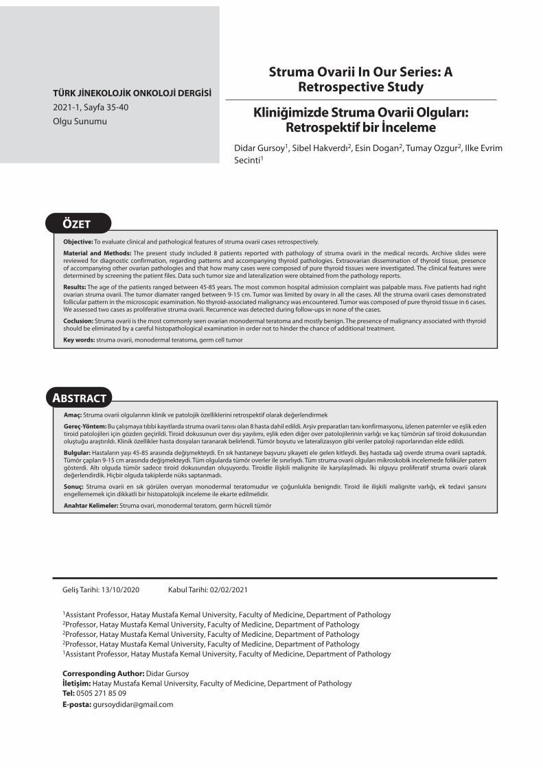
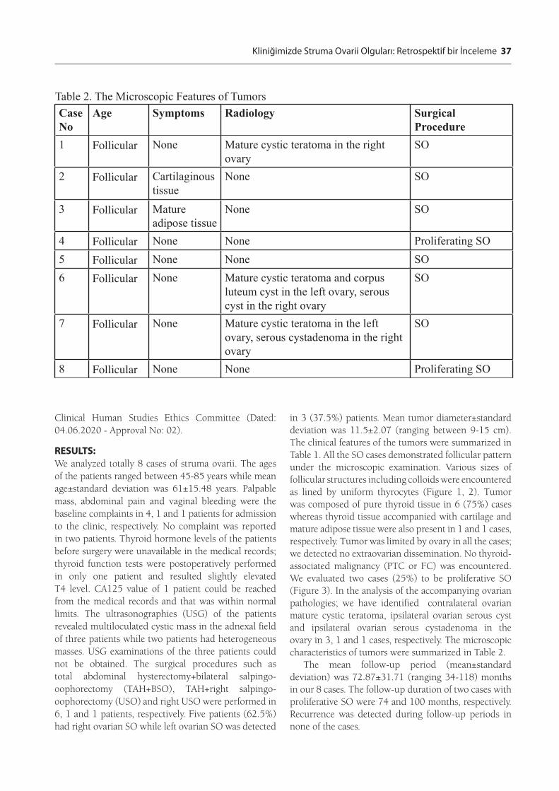
in 3 (37.5%) patients. Mean tumor diameter±standard deviation was 11.5±2.07 (ranging between 9-15 cm). The clinical features of the tumors were summarized in Table 1. All the SO cases demonstrated follicular pattern under the microscopic examination. Various sizes of follicular structures including colloids were encountered as lined by uniform thyrocytes (Figure 1, 2). Tumor was composed of pure thyroid tissue in 6 (75%) cases whereas thyroid tissue accompanied with cartilage and mature adipose tissue were also present in 1 and 1 cases, respectively. Tumor was limited by ovary in all the cases; we detected no extraovarian dissemination. No thyroid-associated malignancy (PTC or FC) was encountered. We evaluated two cases (25%) to be proliferative SO (Figure 3). In the analysis of the accompanying ovarian pathologies; we have identified contralateral ovarian mature cystic teratoma, ipsilateral ovarian serous cyst and ipsilateral ovarian serous cystadenoma in the ovary in 3, 1 and 1 cases, respectively. The microscopic characteristics of tumors were summarized in Table 2.
The mean follow-up period (mean±standard deviation) was 72.87±31.71 (ranging 34-118) months in our 8 cases. The follow-up duration of two cases with proliferative SO were 74 and 100 months, respectively. Recurrence was detected during follow-up periods in none of the cases.
Table 2. The Microscopic Features of TumorsCase No
Age Symptoms Radiology Surgical Procedure
1 Follicular None Mature cystic teratoma in the right ovary
SO
2 Follicular Cartilaginous tissue
None SO
3 Follicular Mature adipose tissue
None SO
4 Follicular None None Proliferating SO5 Follicular None None SO6 Follicular None Mature cystic teratoma and corpus
luteum cyst in the left ovary, serous cyst in the right ovary
SO
7 Follicular None Mature cystic teratoma in the left ovary, serous cystadenoma in the right ovary
SO
8 Follicular None None Proliferating SO

38 Struma Ovarii In Our Series: A Retrospective Study
DISCUSSION:SO is defined as the ovarian goitre that completely or predominantly includes thyroid tissue (≥50%). This diagnostic term also involves the mature teratoma cases accompanied with thyroid-associated malignancy (5). It has been first identified in 1889 by Boettlin (9,10). SO may be detected in all age goups, particularly between 4th-6th decades of the lifespan (1,8,9). It is usually observed unilaterally accompanying contralateral mature cystic teratoma and cystadenoma may be present in some cases (11, 12). All of our cases were also unilateral and tumor was localized in the right ovary in 62.5% of the cases. In our study, we have analyzed the ovaries with respect to accompanying pathologies. We have encountered contralateral ovarian mature cystic teratoma, ipsilateral ovarian serous cyst and ipsilateral ovarian serous cystadenoma in the ovary of 3, 1 and 1 cases, respectively. Age of the patients ranged between 45-85 years in our study. That age range was higher compared with the studies in literature.
The patients mostly apply to the clinic with the complaint of unilateral palpable mass, whereas some patients may have acute abdomen, vaginal bleeding or menstrual irregularity (4, 13). On the other hand, 40% of the patients are asymptomatic (13). Ascites or pleural effusion (Pseudo-Meigs Syndrome) may be detected in some patients, laboratory examination may manifest hyperthyroidism and elevated levels of serum CA125 (6, 11). CA125 has a low clinical importance since it may increase in both benign and malignant SO cases. Increased serum CA125 level is due to presence of ascites rather than tumor in the patients with SO (14). The symptoms and findings of thyrotoxicosis
were determined in only 5-8% of the cases despite dominance of thyroid tissue (1, 7). In our case series, the most common cause for hospital admission was unilateral palpable mass consistently with the literature. None of our cases indicated a symptom of hyperthyroidism, because, thyroid function tests were not performed preoperatively. CA125 value of 1 patient could be reached in the medical records and found within normal limits.
Malignant SO cases are encountered in the 5th-6th decades of the lifespan. The patients are mostly asymptomatic, the most common admission cause is unilateral palpable mass (3, 4). The findings of hyperthyroidism such as weight loss, fatigue, heat intolerance, tremor and tachycardia are rare (9). The clinical picture of ascites and pleural effusion is more commonly associated with benign SO, however, it may be observed in also malignant SO cases, although rare (13). Also, we have analyzed our case series with respect to accompanying malignancy, none of our cases manifested a finding in favor of malignancy.
SO cases are usually macroscopically cystic, they may include also solid fields. Their outer surface is usually smooth and section surface appears shiny due to inclusion of thyroid tissue (6, 12). Microscopic images show various sizes of follicles that include colloids and are lined by a single-row epithelium. The use of diagnostic term “proliferative SO” is recommended in the presence of crowded and tightly packed thyroid follicles without malignant characteristics (12). Different histological patterns of SO also have been described. Those include diffuse, trabecular and pseudotubular patterns as well as small clusters of tumor cells. Follicular
Figure 1: Various sizes of follicular structures including colloids (H+E, x40)
Figure 2: Various sizes of follicular structures including colloids (H+E, x100)

Kliniğimizde Struma Ovarii Olguları: Retrospektif bir İnceleme 39
pattern is a differentiated form. Other patterns do not include colloids and follicular formation and are the dedifferentiated forms because of these characteristics (2). All the cases in our series had a follicular (micro or macro) pattern and two (25%) of our cases had proliferative SO.
The most common malignancy which develops associated with SO is PTC followed by FC (1,4,5). The fact whether diagnostic criteria for PTC and FC which develop in the thyroid gland are valid in the diagnosis of the tumors developing associated with SO is controversial (1,5,15). The rarity of these lesions and absence of an uniform criterion create difficulty in diagnosis of these lesions (3, 12). Nevertheless, many studies have defended the diagnostic validity of the diagnosis based on the histopathological criteria for the thyroid carcinoma that develops in the thyroid gland (16). Approximately 5-23% of the malignant SO cases metastasize; the most frequent metastatic target is abdominal cavity, however, metastases may be encountered in also liver, lung, brain, bones and contralateral ovary (1, 17). On the other side, HDFCO is a rarely seen thyroid-type carcinoma developing associated with SO and characterized by the presence of benign-appearing thyroid follicles that spread over the ovaries. It is biologically malignant because of its metastatic potential (18). All genetic anomalies specific for cancer types developing from thyroid gland have been identified also for the thyroid cancers developing associated with ovarian teratomas. These genetic anomalies include the point mutations on BRAF, HRAS and NRAS, loss of heterozygosity in the PTEN gene region and RET/PTC rearrangements (13).
The treatment of malignant SO is controversial. Radical surgery is necessary in the postmenopausal
women or women without fertility expectancy. On the other hand, conservative treatment should be performed in the women who expect pregnancy; however, this treatment option is possible only if the disease is unilateral in the absence of invasion or metastasis (1). Radioactive iodine treatment is recommended after surgery in the patients with a tumor larger than 4 cm in diameter, extraovarian dissemination and aggressive histological features. Thyroidectomy should be performed prior to this treatment in these patients (19). Thyroidectomy is performed to confirm histological normality of thyroid tissue and increase the efficacy of radioactive iodine treatment (17, 20). The target of radioactive iodine treatment is ablation of thyroid remnants and destruction of the metastatic foci (1). The prognosis of benign SO and non-metastatic malignant SO is good. Thyroglobulin is used as a tumor marker in the follow-up of malignant cases (6). Surgery was applied to all cases in our series; no additional treatment was applied while no malignancy was detected. Recurrence was detected during follow-up period in none of the cases.
CONCLUSION:Struma ovarii cases are the most commonly seen ovarian monodermal teratomas and mostly benign. Thyroid tissue may be found in 5-20% of the mature teratomas, however, thyroid tissue constitute more than ≥50% of overall tumor to be termed as SO. The presence of thyroid tissue below 50% is adequate to diagnose SO if accompanied with thyroid-associated malignancy. The presence of malignancy associated with thyroid should be ruled out by a meticulous examination to preserve the option of an additional treatment.
ACKNOWLEDGMENTThis study was presented as a poster presentation at 29th European Congress of Pathology.
Conflict of interest: No conflict of interest was declared by the authors.
Financial Disclosure: The authors declare that this study has received no financial support.
References1. Gonet, A., Slusarczyk, R., Gasior-Perczak, D., Kowa-
lik, A., Kopczyski, J., & Kowalska, A. (2020). Papillary Thyroid Cancer in a Struma Ovarii in a 17-Year-Old Nul-liparous Patient: A case Report. Diagnostics, 10(1), 45.
2. Osakabe, M., Fukagawa, T., Fukagawa, D., Sugimoto, R., Uesugi, N., Ishida, K., ... & Sugai, T. (2017). Case Report Struma ovarii with unique histological features: a case re-port. Int J Clin Exp Pathol, 10(11), 11230-11233.
3. Yasutake, N., Noguchi, H., Ibayashi, Y., Nakamura, H., Figure 3: Tightly packed thyroid follicles (H+E, x40)

40 Struma Ovarii In Our Series: A Retrospective Study
Tateishi, K., Yuki, K., ... & Fujita, T. (2018). The smallest reported malignant struma ovarii: a case report. Case re-ports in oncology, 11(3), 693-698.
4. Hassan, S. A., Akhtar, A., Falah, N. U., & Sheikh, F. N. (2019). Malignant Thyroid-type Papillary Neoplasm in Struma Ovarii: A Case Report. Cureus, 11(12).
5. Wei, S., Baloch, Z. W., & LiVolsi, V. A. (2015). Pathology of struma ovarii: a report of 96 cases. Endocrine patho-logy, 26(4), 342-348.
6. Singh, P., Lath, N., Shekhar, S., Goyal, M., Gothwal, M., Yadav, G., & Khera, P. (2018). Struma ovarii: A report of three cases and literature review. Journal of Mid-life He-alth, 9(4), 225.
7. Kim, D., Cho, H. C., Park, J. W., Lee, W. A., Kim, Y. M., Chung, P. S., ... & Park, C. H. (2009). Struma ovarii and peritoneal strumosis with thyrotoxicosis.
8. Frysak, Z., Schovanek, J., Halenka, M., Metelkova, I., Duskova, M., & Karasek, D. (2016). Ovarian gOiter as a rare cause Of hyperthyrOidism. Acta Endocrinologica (Bucharest), 12(3), 335.
9. Moayerifar, M., Koohmanaee, S., Moayerifar, M., Nakhoc-hari, A. M., Rad, A. H., & Dalili, S. (2018). Malignant struma ovarii in an 11-year-old girl. Journal of Pediatric Surgery Case Reports, 29, 1-4.
10. Yoo, S. C., Chang, K. H., Lyu, M. O., Chang, S. J., Ryu, H. S., & Kim, H. S. (2008). Clinical characteristics of struma ovarii. Journal of gynecologic oncology, 19(2), 135-138.
11. Qiao, P. F., Gao, Y., & Niu, G. M. (2015). Struma ovarii accompanied with mature cystic teratoma of the other ovary: A case report and literature review. Oncology let-ters, 9(5), 2053-2055.
12. Kruti P. Maniar and Russell Vang. Germ Cell Tumors of the Ovary. Kurman R (eds). Blaustein’s Pathology of the
Female Genital Tract, 7th edition. New York, Springer-
Verlag, 2019:1047-1124.
13. Wolff, E. F., Hughes, M., Merino, M. J., Reynolds, J. C.,
Davis, J. L., Cochran, C. S., & Celi, F. S. (2010). Expres-
sion of benign and malignant thyroid tissue in ovarian
teratomas and the importance of multimodal manage-
ment as illustrated by a BRAF-positive follicular variant
of papillary thyroid cancer. Thyroid, 20(9), 981-987.
14. Dujardin, M. I., Sekhri, P., & Turnbull, L. W. (2014).
Struma ovarii: role of imaging?. Insights into imaging,
5(1), 41-51.
15. Robboy, S. J., Shaco-Levy, R., Peng, R. Y., Snyder, M. J.,
Donahue, J., Bentley, R. C., ... & Young, R. H. (2009). Ma-
lignant struma ovarii: an analysis of 88 cases, including
27 with extraovarian dissemination. International Journal
of Gynecological Pathology, 28(5), 405-422.
16. Devaney, K., Snyder, R., Norris, H. J., & Tavassoli, F. A.
(1993). Proliferative and histologically malignant struma
ovarii: a clinicopathologic study of 54 cases. Internatio-
nal journal of gynecological pathology: official journal of
the International Society of Gynecological Pathologists,
12(4), 333-343.
17. DeSimone, C. P., Lele, S. M., & Modesitt, S. C. (2003).
Malignant struma ovarii: a case report and analysis of ca-
ses reported in the literature with focus on survival and
I131 therapy. Gynecologic oncology, 89(3), 543-548.
18. Dobi, A., Kim, S. A., Zhang, M., Iames, E., & Lamzabi, I.
(2020). Highly differentiated.


![Malignant Struma ovarii in a 30-year old nulliparous patient... · Struma ovarii is a monodermal germ cell tumor first de-scribed by R. Boëttlin in 1889 [1]. It represents 2–3%](https://img.dokumen.tips/doc/110x75/608e9bef6e3ef169014ed01c/malignant-struma-ovarii-in-a-30-year-old-nulliparous-patient-struma-ovarii.jpg)


![Papillary thyroid cancer located in malignant struma ... · found in struma ovarii, and papillary carcinoma is the most common [14–16]. Immunohistochemical staining with Tg, HBME-1,](https://img.dokumen.tips/doc/110x75/5e1bc0f33beaf31e675deab1/papillary-thyroid-cancer-located-in-malignant-struma-found-in-struma-ovarii.jpg)