Embed Size (px)
Citation preview
STRUCTURE AND FUNCTION OF AN ACETYLCHOLINERECEPTOR
JOERG KISTLER, ROBERT M. STROUD, MICHAEL W. KLYMKOWSKY, ROGER A. LALANCETTE, ANDROBERT H. FAIRCLOUGHDepartment ofBiochemistry and Biophysics, University of California, San Francisco, California 94143U.S.A.
ABSTRACT Structural analysis of an acetylcholine receptor from Torpedo californica leads to a three-dimensionalmodel in which a "monomeric" receptor is shown to contain subunits arranged around a central ionophoretic channel,which in turn traverses the entire 1 10 A length of the molecule. The receptor extends - 15 A on the cytoplasmic side, 55A on the synaptic side of the membrane. The a-bungarotoxin/agonist binding site is found to be -55 A from theentrance to the central gated ion channel. A hypothesis for the mechanism of AcChR is presented which takes intoaccount the structural and kinetic data, which isagonist-induced structure change in AcChR.
INTRODUCTION
The release of neurotransmitters from nerve terminalsinduces a selective increase in permeability to cations andsubsequent depolarization of the electric potential acrossthe postsynaptic membrane. This excitable membrane hasbeen extensively characterized at the electrophysiological,cellular, kinetic, biochemical, and structural levels. Mostsuch characterization pertains to nicotinic receptors foracetylcholine (AcCh) in vertebrate neuromuscular junc-tions and in the electrocytes of Torpedo (a marine elasmo-branch) or Electrophorus (a fresh water teleost) (reviews,1-4).We present here our recent results on the structural
analysis of acetylcholine receptor (AcChR) in Torpedocalifornica electrocyte membranes. These and relatedresults are discussed with reference to possible mechanisticconsequences, focusing on a testable hypothesis forAcChR action.
AcChR-rich membrane fractions are routinely obtainedby differential centrifugation of homogenized electricorgan and subsequent separation on density gradients (5).Radioactively labeled neurotoxins such as a-bungarotoxin,which bind extremely tightly but noncovalently to AcChRwith dissociation constants in the range 10-' -10-' M atroom temperature (6, 7), serve as a marker through theisolation procedure; agonist-dependent cation flux can bemeasured for microsac preparations to ensure functionalintegrity of AcChR in vitro (8, 9). Under reducing condi-tions SDS polyacrylamide gel electrophoresis of such prep-arations shows six major bands with apparent mol wts of40,000 (a), 43,000, 50,000 (d), 60,000 (y), 65,000 (6), and
Please address correspondence to Dr. Stroud.
BIOPHYS. J. Biophysical Society - 0006-3495/82/01/371/13 $1.00Volume 37 January 1982 371-383
testable, and which serves as a focus for future studies on the
90,000 (10). Exposure to pH 11 for 1 h at 200C removesthe 43,000 and 90,000 mol wt polypeptides from theAcChR-rich membranes without loss of agonist inducedfluxing ability (10, 11). These chains are thereforepresumed to be peripheral membrane proteins. Function-ally intact AcChR vesicles which contain only a, fp, y, and6 subunit types have been reconstituted from AcChReither solubilized in octylglucoside (12) or in the presenceof exogenous lipids and cholate (13, 14). The subunitcomposition of monomeric AcChR has been establishedindependently in several laboratories ( 15, 16) and found tobe a2f3-y6.' In vivo, however, AcChR occurs as a dimercross-linked via a disulfide between 6 subunits (17, 18),though both the monomeric and dimeric forms showidentical ligand binding affinity and are equally active in22Na-flux assays (19). Thus, the physiological relevance ofdimerization is as yet unidentified.
Evidence in favor of discrete ionophoretic channels inthe postsynaptic membrane was first obtained from thenoise-analysis of agonist-induced current fluctuationsmeasured for motor endplates under voltage-clamp condi-tions (20, 21). More recently, with improved techniqueselementary single channel events have been directlyobserved as all-or-none, rectangular, pulse-like wave formsof constant amplitude (22). Agonist, temperature, andmembrane potential determine the average channel life-time to be typically 1-3 ms. At 200C - I04 Na+ ions flowinto the cell during the opening of a single channel,induced by the binding of AcCh. The ionophoreticchannel conducts ions as large as dimethyldiethanolam-monium and must therefore be at least 6.5 A Diam (23)throughout its entire length in the "open channel" state.
'First proposed by Reynolds and Karlin (47).
371
Which subunit contains the agonist binding site andtherefore acts as the primary effector of the proteinconformational change associated with the opening of thetransmembrane channel? Using a variety of affinity labelsand neurotoxins, the AcChR agonist (or antagonist) bind-ing sites have been located on the two a subunits (review,4). Following reduction with dithiothreitol, one a subunitof the T. californica AcChR can be affinity labeled with4-(N-maleimido)-benzyltrimethyl-ammonium (MBTA)or with bromoacetylcholine (BAC), either of which reactcovalently with a single sulfhydryl and block 50% of theavailable toxin binding sites (24, 25). Delegeane andMcNamee (26) claim that after one AcCh site is blockedby alkylation with MBTA 22Na-flux is still induced bybinding of carbamylcholine to the second site. HoweverLindstrom et al. (14) showed, using reconstituted AcChRvesicles, that alkylation with MBTA in fact removed allagonist dependent fluxing ability. Both results indicatethat ligand binding to one particular a-subunit (and notthe other) leads to channel opening. In unmodified AcChRit is possible that agonist binding to both sites is moreeffective than simply to the one site (27). The Hill coeffi-cient for agonist-induced ion permeability response is1.97 ± 0.062 thus two agonists are required for activa-tion.3 4
AcCHR IS FUNNEL-SHAPED: ALLSUBUNITS SPAN THE BILAYER
The transmembrane nature of the AcChR complex hasbeen independently demonstrated by x-ray diffractionstudies and by use of analytical probes such as proteases oranti-AcChR-specific antibodies. Analysis of continuousx-ray scattering profiles recorded from pellets of AcChR-rich membrane preparations revealed an asymmetricdistribution of protein perpendicular to the lipid bilayer,and showed that the protein extends 55 A on one side ofthe membrane and 15 A on the other (28). In conjunctionwith the in-plane dimensions of AcChR from electronmicroscopy and the measured density of receptors per unitarea, this analysis provided a low-resolution, cylindricallyaveraged structure of AcChR (29).A sharp reflection of spacing 5.1 A is oriented exclu-
sively perpendicular to the membrane plane. This is theexpected angle for scattering from a-helices alone, and
2Neubig R. R., and J. B. Cohen. 1980. Permeability control by choliner-gic receptors in Torpedo postsynaptic membranes. Agonist dose-responserelations measured at second and millisecond time scales. Biochemistry.19:2770-2779.
3Cash D. J., and G. P. Hess. 1980. Molecular mechanisms of acetylcho-line receptor-controlled ion translocation across cell membranes. Proc.Natl. Acad. Sci. U.S.A. 77:842-846.
4Sine S. M., P. Taylor. 1980. The relationship between agonist occupa-tion and the permeability response of the cholinergic receptor revealed bybound cobra a-toxin. J. Biol. Chem. 255:10144-10156.
circular dichroism suggests that 34% of AcChR is a-helical (30). The sharpness of the x-ray reflection indicatesthat the a-helices in AcChR are on average 80 A long; itsorientation implies that essentially all a-helices are almostperpendicular to the membrane, and this is strong evidencethat some (at least two) or all of the subunits arethemselves elongated perpendicular to the membraneplane.The funnel-shaped structure of AcChR is directly
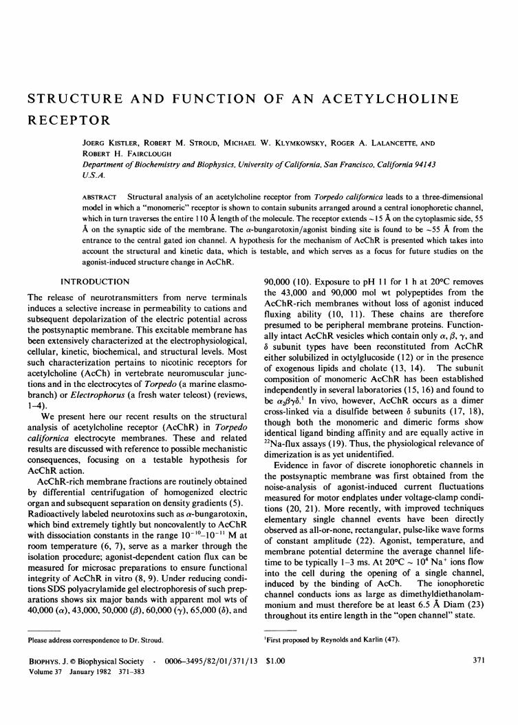
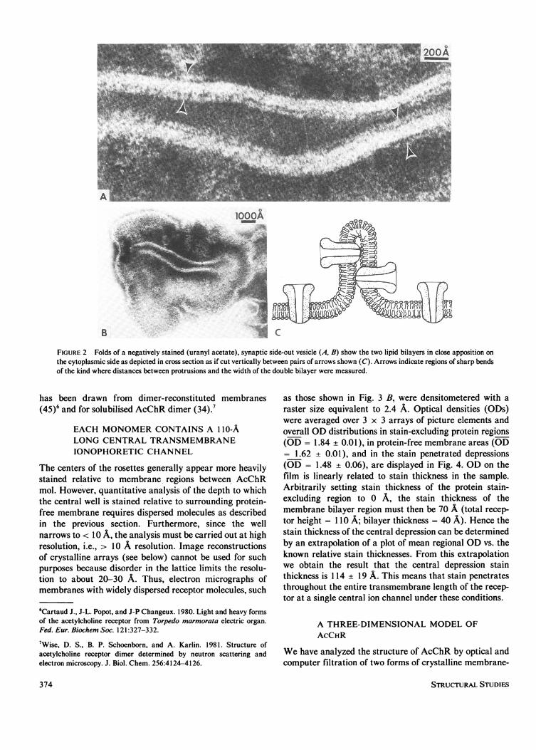
visualized in side view by negative stain electron micros-copy at the edge of a folded-over membrane vesicle (Fig.1). Proof that these structures are membrane-boundAcChR oligomers was obtained by immuno-electronmicroscopy, since we were able to label them with anti-AcChR antibodies coupled to -200 A-sized colloidal goldspheres (29). Furthermore, experiments in which AcChRwas treated first with a-bungarotoxin and then with anti-toxin antibodies attached to gold beads established thatthe protein protrusion of 55 A was on the extracellular,i.e., synaptic, side of the membrane. The smaller protru-sion on the cytoplasmic surface was more difficult tovisualize by electron microscopy. However, as membranesdry down onto the carbon support film, folds are oftenformed. A small portion of these are sharp, and unambigu-ously show the extracellular 55 A protrusion of AcChRmolecules on the outer surface of the folded doublemembrane (Fig. 2). The minimum overall width of suchfolded double membranes measures -200 A, close to twicethe 110 A length of the AcChR oligomer determined byx-ray diffraction analysis (slight shrinkage is likely to beassociated with specimen dehydration for electron micros-copy). The central double bilayer thickness averages 86 A.As this value is close to twice the 40 A thickness of thesingle bilayer (28), there must be little of the proteinprotruding from the cytoplasmic surface. This observationis in agreement with the asymmetric protein profilecomputed from the x-ray diffraction data. The transmem-brane nature of AcChR has further been demonstrated byimmuno-electron microscopy: antibodies raised againstsolubilized AcChR and coupled with ferritin bind to boththe synaptic and the cytoplasmic sides of open membranevesicles (31, 32).We now ask whether all five subunits span the bilayer or
whether there are components which are entirely buried inthe membrane while others are accessible from one sideonly. As each subunit type present in membrane-boundAcChR is accessible to degradation by exogenousproteases (33-35) they must all protrude into the aqueousphase and none is entirely buried within the lipid bilayer.This conclusion is further substantiated by the finding thatall four subunit species can be iodinated in the presence oflactoperoxidase (36). The fact that all subunits naturallyoccur in their glycosylated form implies their exposure onthe synaptic membrane side (37, 38). Photoactivablereagents that partition into the lipid phase of the
STRUCTURAL STUDIES372
B
FIGURE 1 View of a uranyl acetate-stained synaptic edge of AcChR membrane vesicle (A) and schematic representation of the 55 A-longfunnel-shaped protrusions of the receptor molecules (B). The central channel is filled with uranyl stain down to at least the level of the lipidbilayer. Modified from Fig. 4 in reference 29.
membrane label at least the /3, -y (39) and the a subunits(40); therefore, an exclusively peripheral location of these(and probably all) subunits is excluded. Combinationlabeling and proteolysis showed the transmembranenature of the 40, 50 and 66 kilodalton chains in T.marmarota.' Recently, Strader and Raftery (35) demon-strated that all AcChR subunit types can be proteolyti-cally cleaved from both the cytoplasmic and from thesynaptic side of the membrane. Thus current evidenceshows that all five subunits are elongated perpendicular tothe membrane, are in contact with the lipids for part oftheir length, and protrude on both sides of the bilayer.
PROOF THAT EACH AcCHR MONOMER ISA SINGLE INFUNDIBULIFORMSTRUCTURE
Electron micrographs of negatively stained AcChRmembranes (Fig. 3 A) reveal "rosette" structures 85 A
5Wennogle L. P., J-P Changeux. 1980. Transmembrane ?????????? ofproteins present in acetylcholine receptor-rich membranes from Torpedomarmorata studied by selective proteolysis. Eur. J. Biochem. 106:381-393.
Diam dispersed in the plane of the membranes (29,41-44). Each rosette is a projection perpendicular to themembrane surface of a single funnel-shaped structure.AcChR occurs in vivo as dimers of the five subunitcomplex; however, until recently direct evidence onwhether the infundibuliform structure corresponds to amonomeric or dimeric receptor complex has been lacking.
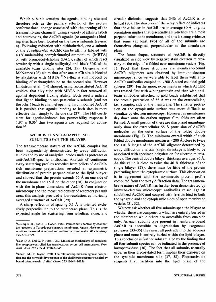
Standard membrane preparations rich in monomeric ordimeric AcChR appear indistinguishable from each otherbecause the molecules are so closely packed together.Membranes reconstituted from exogenous lipid and solu-bilized AcChR that was predominantly dimeric (79% byintegration of stained bands on SDS gels) revealed rosettesspaced much further apart: 69% of the rosettes are seen tobe dimerized and 31% are single rosettes (total counted,690). In a second approach, excessively base-treated (pH11.5 for 1 h at 200C) and sonicated membranes from thesame source (79% dimeric) also led to a portion ofmembranes with more dispersed AcChR mol (Fig. 3 B). Inthis case, 78% of rosettes were in pairs, 22% were single(total counted, 803). This unambiguously shows that eachinfundibuliform structure is one AcChR monomer. Thissame conclusion which we deduced in reference 28 and 29
KISTLER ET AL. Structural Analysis ofAcetylcholine Receptor 373
olOOOA
B
FIGURE 2 Folds of a negatively stained (uranyl acetate), synaptic side-out vesicle (A, B) show the two lipid bilayers in close apposition onthe cytoplasmic side as depicted in cross section as if cut vertically between pairs of arrows shown (C). Arrows indicate regions of sharp bendsof the kind where distances between protrusions and the width of the double bilayer were measured.
has been drawn from dimer-reconstituted membranes(45)6 and for solubilised AcChR dimer (34).7
EACH MONOMER CONTAINS A 1 io-ALONG CENTRAL TRANSMEMBRANEIONOPHORETIC CHANNEL
The centers of the rosettes generally appear more heavilystained relative to membrane regions between AcChRmol. However, quantitative analysis of the depth to whichthe central well is stained relative to surrounding protein-free membrane requires dispersed molecules as describedin the previous section. Furthermore, since the wellnarrows to < 10 A, the analysis must be carried out at highresolution, i.e., > 10 A resolution. Image reconstructionsof crystalline arrays (see below) cannot be used for suchpurposes because disorder in the lattice limits the resolu-tion to about 20-30 A. Thus, electron micrographs ofmembranes with widely dispersed receptor molecules, such
6Cartaud J., J-L. Popot, and J-P Changeux. 1980. Light and heavy formsof the acetylcholine receptor from Torpedo marmorata electric organ.Fed. Eur. Biochem Soc. 121:327-332.
'Wise, D. S., B. P. Schoenborn, and A. Karlin. 1981. Structure ofacetylcholine receptor dimer determined by neutron scattering andelectron microscopy. J. Biol. Chem. 256:4124-4126.
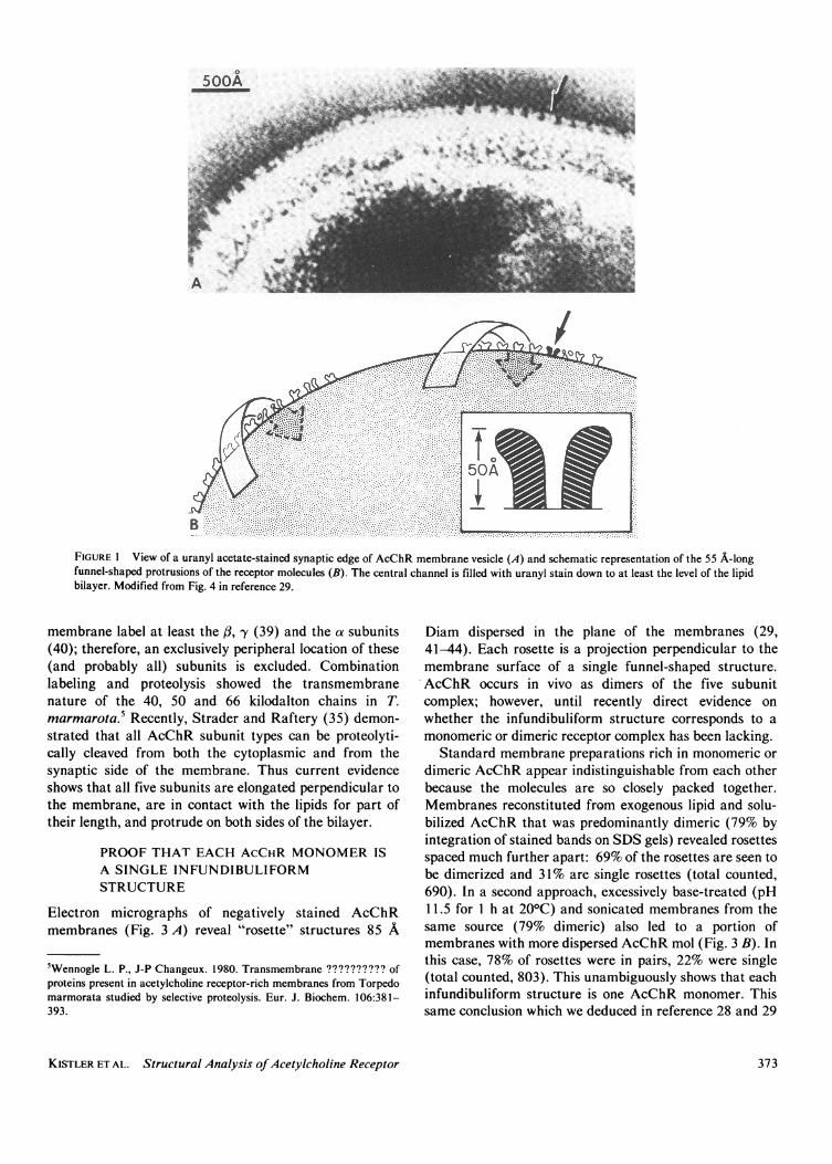
as those shown in Fig. 3 B, were densitometered with araster size equivalent to 2.4 A. Optical densities (ODs)were averaged over 3 x 3 arrays of picture elements andoverall OD distributions in stain-excluding protein regions(OD = 1.84 ± 0.01), in protein-free membrane areas (OD= 1.62 ± 0.01), and in the stain penetrated depressions(OD = 1.48 ± 0.06), are displayed in Fig. 4. OD on thefilm is linearly related to stain thickness in the sample.Arbitrarily setting stain thickness of the protein stain-excluding region to 0 A, the stain thickness of themembrane bilayer region must then be 70 A (total recep-tor height = 110 A; bilayer thickness = 40 A). Hence thestain thickness of the central depression can be determinedby an extrapolation of a plot of mean regional OD vs. theknown relative stain thicknesses. From this extrapolationwe obtain the result that the central depression stainthickness is 114 ± 19 A. This means that stain penetratesthroughout the entire transmembrane length of the recep-tor at a single central ion channel under these conditions.
A THREE-DIMENSIONAL MODEL OFAcCHR
We have analyzed the structure of AcChR by optical andcomputer filtration of two forms of crystalline membrane-
STRUCTURAL STUDIES374
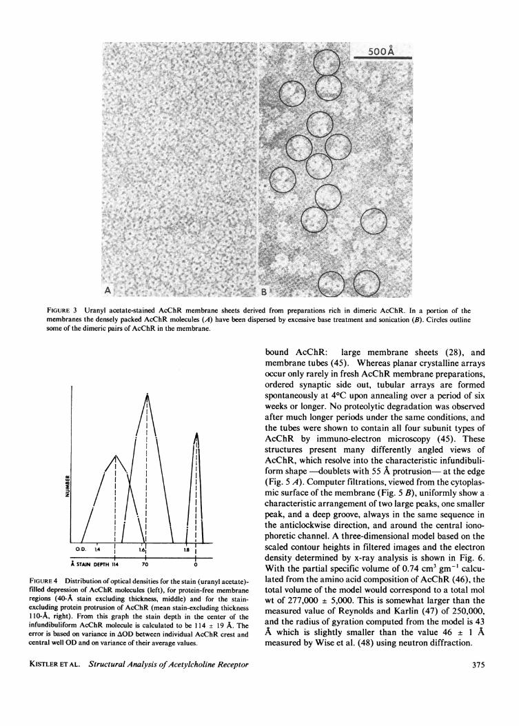
FIGURE 3 Uranyl acetate-stained AcChR membrane sheets derived from preparations rich in dimeric AcChR. In a portion of themembranes the densely packed AcChR molecules (A) have been dispersed by excessive base treatment and sonication (B). Circles outlinesome of the dimeric pairs of AcChR in the membrane.
0
z
FIGURE 4 Distribution of optical densities for the stain (uranyl acetate)-filled depression of AcChR molecules (left), for protein-free membraneregions (40-A stain excluding thickness, middle) and for the stain-excluding protein protrusion of AcChR (mean stain-excluding thickness110-A, right). From this graph the stain depth in the center of theinfundibuliform AcChR molecule is calculated to be 114 + 19 A. Theerror is based on variance in AOD between individual AcChR crest andcentral well OD and on variance of their average values.
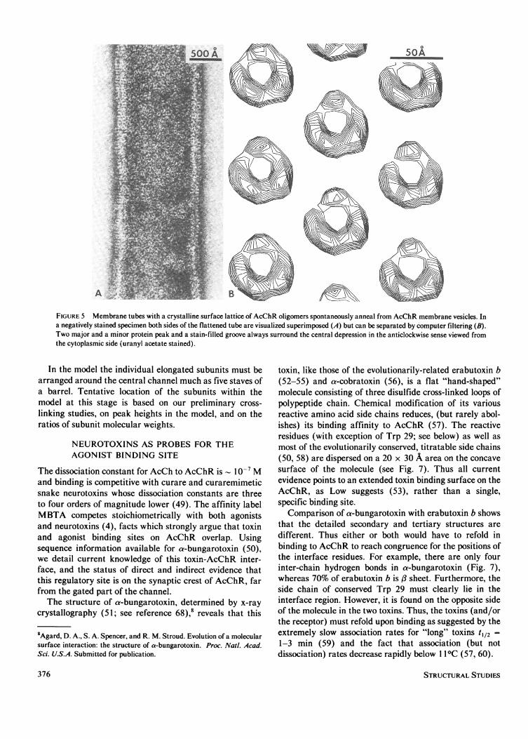
bound AcChR: large membrane sheets (28), andmembrane tubes (45). Whereas planar crystalline arraysoccur only rarely in fresh AcChR membrane preparations,ordered synaptic side out, tubular arrays are formedspontaneously at 40C upon annealing over a period of sixweeks or longer. No proteolytic degradation was observedafter much longer periods under the same conditions, andthe tubes were shown to contain all four subunit types ofAcChR by immuno-electron microscopy (45). Thesestructures present many differently angled views ofAcChR, which resolve into the characteristic infundibuli-form shape -doublets with 55 A protrusion- at the edge(Fig. 5 A). Computer filtrations, viewed from the cytoplas-mic surface of the membrane (Fig. 5 B), uniformly show acharacteristic arrangement of two large peaks, one smallerpeak, and a deep groove, always in the same sequence inthe anticlockwise direction, and around the central iono-phoretic channel. A three-dimensional model based on thescaled contour heights in filtered images and the electrondensity determined by x-ray analysis is shown in Fig. 6.With the partial specific volume of 0.74 cm3 gm-' calcu-lated from the amino acid composition of AcChR (46), thetotal volume of the model would correspond to a total molwt of 277,000 + 5,000. This is somewhat larger than themeasured value of Reynolds and Karlin (47) of 250,000,and the radius of gyration computed from the model is 43A which is slightly smaller than the value 46 ± 1 Ameasured by Wise et al. (48) using neutron diffraction.
KISTLER ET AL. Structural Analysis ofAcetylcholine Receptor 375
50A
A
FIGURE 5 Membrane tubes with a crystalline surface lattice of AcChR oligomers spontaneously anneal from AcChR membrane vesicles. Ina negatively stained specimen both sides of the flattened tube are visualized superimposed (A) but can be separated by computer filtering (B).Two major and a minor protein peak and a stain-filled groove always surround the central depression in the anticlockwise sense viewed fromthe cytoplasmic side (uranyl acetate stained).
In the model the individual elongated subunits must bearranged around the central channel much as five staves ofa barrel. Tentative location of the subunits within themodel at this stage is based on our preliminary cross-linking studies, on peak heights in the model, and on theratios of subunit molecular weights.
NEUROTOXINS AS PROBES FOR THEAGONIST BINDING SITE
The dissociation constant for AcCh to AcChR is 10-' Mand binding is competitive with curare and curaremimeticsnake neurotoxins whose dissociation constants are threeto four orders of magnitude lower (49). The affinity labelMBTA competes stoichiometrically with both agonistsand neurotoxins (4), facts which strongly argue that toxinand agonist binding sites on AcChR overlap. Usingsequence information available for a-bungarotoxin (50),we detail current knowledge of this toxin-AcChR inter-face, and the status of direct and indirect evidence thatthis regulatory site is on the synaptic crest of AcChR, farfrom the gated part of the channel.The structure of a-bungarotoxin, determined by x-ray
crystallography (51; see reference 68),' reveals that this
8Agard, D. A., S. A. Spencer, and R. M. Stroud. Evolution of a molecularsurface interaction: the structure of a-bungarotoxin. Proc. Nati. Acad.Sci. U.S.A. Submitted for publication.
toxin, like those of the evolutionarily-related erabutoxin b(52-55) and a-cobratoxin (56), is a flat "hand-shaped"molecule consisting of three disulfide cross-linked loops ofpolypeptide chain. Chemical modification of its variousreactive amino acid side chains reduces, (but rarely abol-ishes) its binding affinity to AcChR (57). The reactiveresidues (with exception of Trp 29; see below) as well asmost of the evolutionarily conserved, titratable side chains(50, 58) are dispersed on a 20 x 30 A area on the concavesurface of the molecule (see Fig. 7). Thus all currentevidence points to an extended toxin binding surface on theAcChR, as Low suggests (53), rather than a single,specific binding site.
Comparison of a-bungarotoxin with erabutoxin b showsthat the detailed secondary and tertiary structures aredifferent. Thus either or both would have to refold inbinding to AcChR to reach congruence for the positions ofthe interface residues. For example, there are only fourinter-chain hydrogen bonds in a-bungarotoxin (Fig. 7),whereas 70% of erabutoxin b is f: sheet. Furthermore, theside chain of conserved Trp 29 must clearly lie in theinterface region. However, it is found on the opposite sideof the molecule in the two toxins. Thus, the toxins (and/orthe receptor) must refold upon binding as suggested by theextremely slow association rates for "long" toxins t112 =1-3 min (59) and the fact that association (but notdissociation) rates decrease rapidly below 11° C (57, 60).
STRUCTURAL STUDIES376
04 --85A-A-
LIPID
A
ION CHANNEL-
80A
a X
B
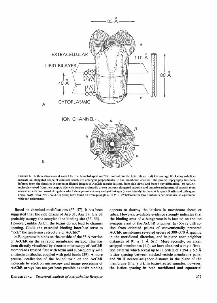
FIGURE 6 A three-dimensional model for the funnel-shaped AcChR molecule in the lipid bilayer. (A) On average 80 A-long a-helicesindicate an elongated shape of subunits which are arranged perpendicular to the membrane channel. The protein topography has beeninferred from the densities in computer filtered images of AcChR tubular lattices, from side views, and from x-ray diffraction. (B) AcChRmolecule viewed from the synaptic side with borders arbitrarily drawn between elongated subunits and tentative assignment of subunit typesconsistent with our cross-linking data which show prominent a-y and -y4 linkages (disuccinimidyl tartarte, 6 A span). Karlin and colleagues(Proc. Natl. Acad. Sci. U.S.A.; in press) have found an average angle of 1 130 + 330 between the two a subunits per monomer, in agreementwith our assignment.
Based on chemical modifications (53, 57), it has beensuggested that the side chains of Asp 31, Arg 37, Gly 38probably occupy the acetylcholine binding site (53, 55).However, unlike AcCh, the toxins do not lead to channelopening. Could the extended binding interface serve to"lock" the quaternary structure of AcChR?
a-Bungarotoxin binds on the outside of the 55 A portionof AcChR on the synaptic membrane surface. This hasbeen directly visualized by electron microscopy of AcChRmembranes treated first with toxin and subsequently withantitoxin antibodies coupled with gold beads (29). A moreprecise localization of the bound toxin on the AcChRmolecule by electron microscopy and image processing ofAcChR arrays has not yet been possible as toxin binding
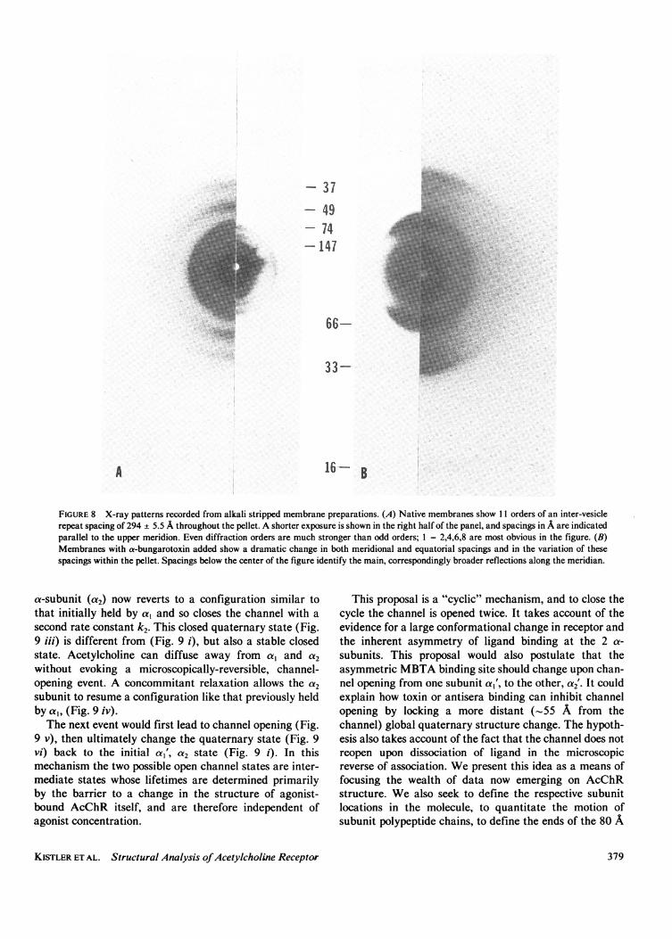
appears to destroy the lattices in membrane sheets ortubes. However, available evidence strongly indicates thatthe binding area of a-bungarotoxin is located on the topsynaptic crest of the AcChR oligomer. (a) X-ray diffrac-tion from oriented pellets of conventionally preparedAcChR membranes revealed orders of 300-370 A spacingin the meridional direction, and in-plane near neighbordistances of 91 ± 1 A (61). More recently, on alkalistripped membranes (1 1), we have obtained x-ray diffrac-tion patterns which reveal up to 11 orders of a 294 ± 5.5 Alattice spacing between stacked vesicle membrane pairs,and 90 A nearest-neighbor distance in the plane of themembrane (Fig. 8 A). In toxin-treated samples, however,the lattice spacing in both meridional and equatorial
KISTLER ET AL. Structural Analysis ofAcetylcholine Receptor 377
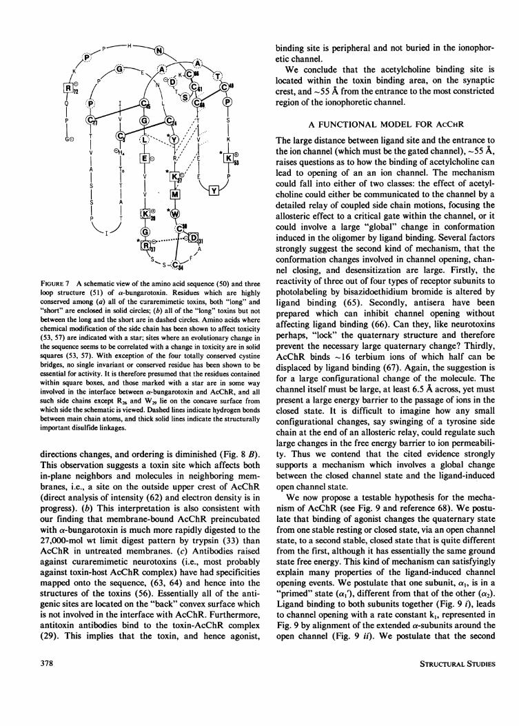
FIGURE 7 A schematic view of the amino acid sequence (50) and threeloop structure (51) of a-bungarotoxin. Residues which are highlyconserved among (a) all of the curaremimetic toxins, both "long" and"short" are enclosed in solid circles; (b) all of the "long" toxins but notbetween the long and the short are in dashed circles. Amino acids wherechemical modification of the side chain has been shown to affect toxicity(53, 57) are indicated with a star; sites where an evolutionary change inthe sequence seems to be correlated with a change in toxicity are in solidsquares (53, 57). With exception of the four totally conserved cystinebridges, no single invariant or conserved residue has been shown to beessential for activity. It is therefore presumed that the residues containedwithin square boxes, and those marked with a star are in some wayinvolved in the interface between a-bungarotoxin and AcChR, and allsuch side chains except R26 and W29 lie on the concave surface fromwhich side the schematic is viewed. Dashed lines indicate hydrogen bondsbetween main chain atoms, and thick solid lines indicate the structurallyimportant disulfide linkages.
directions changes, and ordering is diminished (Fig. 8 B).This observation suggests a toxin site which affects bothin-plane neighbors and molecules in neighboring mem-branes, i.e., a site on the outside upper crest of AcChR(direct analysis of intensity (62) and electron density is inprogress). (b) This interpretation is also consistent withour finding that membrane-bound AcChR preincubatedwith a-bungarotoxin is much more rapidly digested to the27,000-mol wt limit digest pattern by trypsin (33) thanAcChR in untreated membranes. (c) Antibodies raisedagainst curaremimetic neurotoxins (i.e., most probablyagainst toxin-host AcChR complex) have had specificitiesmapped onto the sequence, (63, 64) and hence into thestructures of the toxins (56). Essentially all of the anti-genic sites are located on the "back" convex surface whichis not involved in the interface with AcChR. Furthermore,antitoxin antibodies bind to the toxin-AcChR complex(29). This implies that the toxin, and hence agonist,
binding site is peripheral and not buried in the ionophor-etic channel.We conclude that the acetylcholine binding site is
located within the toxin binding area, on the synapticcrest, and -55 A from the entrance to the most constrictedregion of the ionophoretic channel.
A FUNCTIONAL MODEL FOR ACCHR
The large distance between ligand site and the entrance tothe ion channel (which must be the gated channel), - 55 A,raises questions as to how the binding of acetylcholine canlead to opening of an an ion channel. The mechanismcould fall into either of two classes: the effect of acetyl-choline could either be communicated to the channel by adetailed relay of coupled side chain motions, focusing theallosteric effect to a critical gate within the channel, or itcould involve a large "global" change in conformationinduced in the oligomer by ligand binding. Several factorsstrongly suggest the second kind of mechanism, that theconformation changes involved in channel opening, chan-nel closing, and desensitization are large. Firstly, thereactivity of three out of four types of receptor subunits tophotolabeling by bisazidoethidium bromide is altered byligand binding (65). Secondly, antisera have beenprepared which can inhibit channel opening withoutaffecting ligand binding (66). Can they, like neurotoxinsperhaps, "lock" the quaternary structure and thereforeprevent the necessary large quaternary change? Thirdly,AcChR binds -16 terbium ions of which half can bedisplaced by ligand binding (67). Again, the suggestion isfor a large configurational change of the molecule. Thechannel itself must be large, at least 6.5 A across, yet mustpresent a large energy barrier to the passage of ions in theclosed state. It is difficult to imagine how any smallconfigurational changes, say swinging of a tyrosine sidechain at the end of an allosteric relay, could regulate suchlarge changes in the free energy barrier to ion permeabili-ty. Thus we contend that the cited evidence stronglysupports a mechanism which involves a global changebetween the closed channel state and the ligand-inducedopen channel state.We now propose a testable hypothesis for the mecha-
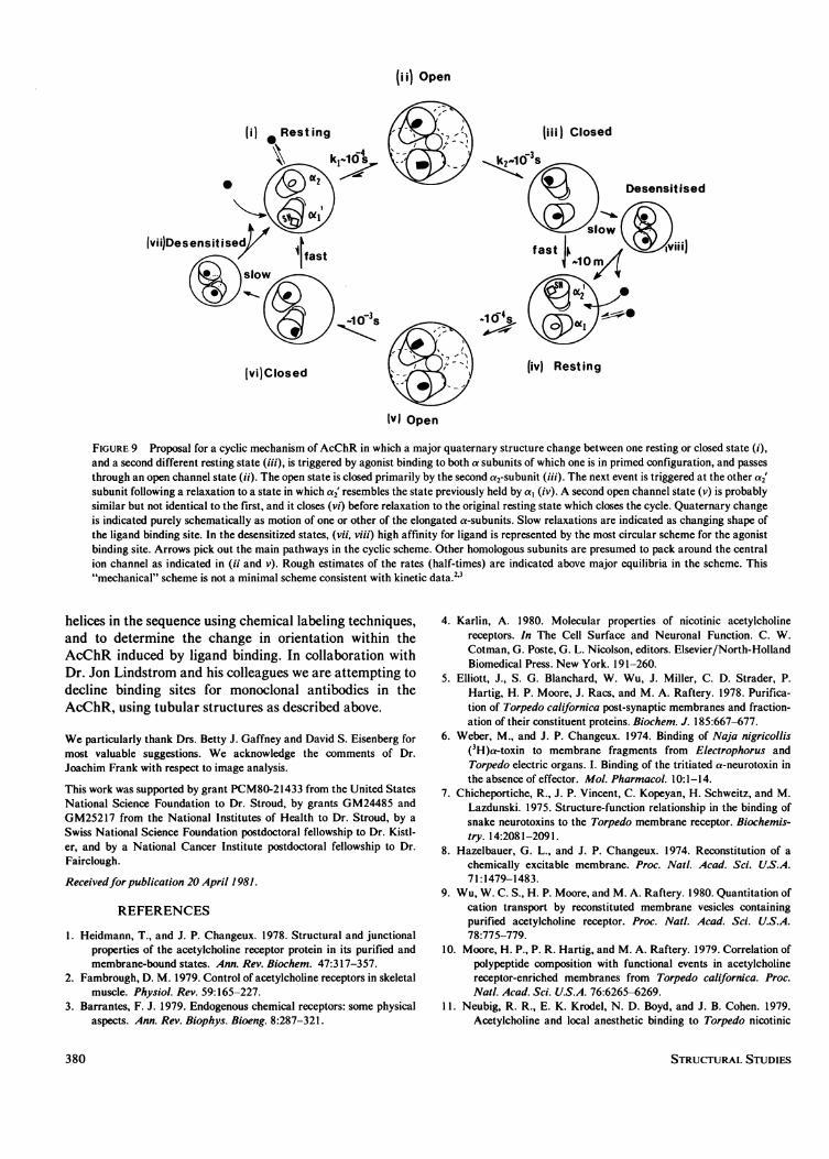
nism of AcChR (see Fig. 9 and reference 68). We postu-late that binding of agonist changes the quaternary statefrom one stable resting or closed state, via an open channelstate, to a second stable, closed state that is quite differentfrom the first, although it has essentially the same groundstate free energy. This kind of mechanism can satisfyinglyexplain many properties of the ligand-induced channelopening events. We postulate that one subunit, a,, is in a"primed" state (a,'), different from that of the other (a2).Ligand binding to both subunits together (Fig. 9 i), leadsto channel opening with a rate constant kj, represented inFig. 9 by alignment of the extended a-subunits around theopen channel (Fig. 9 ii). We postulate that the second
STRUCTURAL STUDIES378
- 37*- 49..- 74*.-147
66-
33-.
16-
FIGURE 8 X-ray patterns recorded from alkali stripped membrane preparations. (A) Native membranes show 11 orders of an inter-vesiclerepeat spacing of 294 + 5.5 A throughout the pellet. A shorter exposure is shown in the right half of the panel, and spacings in A are indicatedparallel to the upper meridion. Even diffraction orders are much stronger than odd orders; 1 = 2,4,6,8 are most obvious in the figure. (B)Membranes with a-bungarotoxin added show a dramatic change in both meridional and equatorial spacings and in the variation of thesespacings within the pellet. Spacings below the center of the figure identify the main, correspondingly broader reflections along the meridian.
a-subunit (a2) now reverts to a configuration similar tothat initially held by a, and so closes the channel with asecond rate constant k2. This closed quaternary state (Fig.9 iii) is different from (Fig. 9 i), but also a stable closedstate. Acetylcholine can diffuse away from a, and a2without evoking a microscopically-reversible, channel-opening event. A concommitant relaxation allows the a2subunit to resume a configuration like that previously heldby aI, (Fig. 9 iv).The next event would first lead to channel opening (Fig.
9 v), then ultimately change the quaternary state (Fig. 9vi) back to the initial a,', a2 state (Fig. 9 i). In thismechanism the two possible open channel states are inter-mediate states whose lifetimes are determined primarilyby the barrier to a change in the structure of agonist-bound AcChR itself, and are therefore independent ofagonist concentration.
This proposal is a "cyclic" mechanism, and to close thecycle the channel is opened twice. It takes account of theevidence for a large conformational change in receptor andthe inherent asymmetry of ligand binding at the 2 a-subunits. This proposal would also postulate that theasymmetric MBTA binding site should change upon chan-nel opening from one subunit a,', to the other, a2'. It couldexplain how toxin or antisera binding can inhibit channelopening by locking a more distant (-55 A from thechannel) global quaternary structure change. The hypoth-esis also takes account of the fact that the channel does notreopen upon dissociation of ligand in the microscopicreverse of association. We present this idea as a means offocusing the wealth of data now emerging on AcChRstructure. We also seek to define the respective subunitlocations in the molecule, to quantitate the motion ofsubunit polypeptide chains, to define the ends of the 80 A
KISTLER ET AL. Structural Analysis ofAcetylcholine Receptor 379
ii) Open
(i) *Resting ( (iii) Closed-4 -3
* / > <3;<Desensitised
fvii)Desensitise t 1fast fast | Q vii)
t ) ( t ~ ~ ~~~~~~3.1( >
-10s 0
(vi)Closed - (ivj Resting
MvJ Open
FIGURE 9 Proposal for a cyclic mechanism of AcChR in which a major quaternary structure change between one resting or closed state (i),and a second different resting state (iii), is triggered by agonist binding to both a subunits of which one is in primed configuration, and passesthrough an open channel state (ii). The open state is closed primarily by the second a2-subunit (iii). The next event is triggered at the other a2'subunit following a relaxation to a state in which a2' resembles the state previously held by a, (iv). A second open channel state (v) is probablysimilar but not identical to the first, and it closes (vi) before relaxation to the original resting state which closes the cycle. Quaternary changeis indicated purely schematically as motion of one or other of the elongated a-subunits. Slow relaxations are indicated as changing shape ofthe ligand binding site. In the desensitized states, (vii, viii) high affinity for ligand is represented by the most circular scheme for the agonistbinding site. Arrows pick out the main pathways in the cyclic scheme. Other homologous subunits are presumed to pack around the centralion channel as indicated in (ii and v). Rough estimates of the rates (half-times) are indicated above major equilibria in the scheme. This"mechanical" scheme is not a minimal scheme consistent with kinetic data.2'3
helices in the sequence using chemical labeling techniques,and to determine the change in orientation within theAcChR induced by ligand binding. In collaboration withDr. Jon Lindstrom and his colleagues we are attempting todecline binding sites for monoclonal antibodies in theAcChR, using tubular structures as described above.
We particularly thank Drs. Betty J. Gaffney and David S. Eisenberg formost valuable suggestions. We acknowledge the comments of Dr.Joachim Frank with respect to image analysis.
This work was supported by grant PCM80-21433 from the United StatesNational Science Foundation to Dr. Stroud, by grants GM24485 andGM25217 from the National Institutes of Health to Dr. Stroud, by aSwiss National Science Foundation postdoctoral fellowship to Dr. Kistl-er, and by a National Cancer Institute postdoctoral fellowship to Dr.Fairclough.
Receivedfor publication 20 April 1981.
REFERENCES
1. Heidmann, T., and J. P. Changeux. 1978. Structural and junctionalproperties of the acetylcholine receptor protein in its purified andmembrane-bound states. Ann. Rev. Biochem. 47:317-357.
2. Fambrough, D. M. 1979. Control of acetylcholine receptors in skeletalmuscle. Physiol. Rev. 59:165-227.
3. Barrantes, F. J. 1979. Endogenous chemical receptors: some physicalaspects. Ann. Rev. Biophys. Bioeng. 8:287-321.
4. Karlin, A. 1980. Molecular properties of nicotinic acetylcholinereceptors. In The Cell Surface and Neuronal Function. C. W.Cotman, G. Poste, G. L. Nicolson, editors. Elsevier/North-HollandBiomedical Press. New York. 191-260.
5. Elliott, J., S. G. Blanchard, W. Wu, J. Miller, C. D. Strader, P.Hartig, H. P. Moore, J. Racs, and M. A. Raftery. 1978. Purifica-tion of Torpedo californica post-synaptic membranes and fraction-ation of their constituent proteins. Biochem. J. 185:667-677.
6. Weber, M., and J. P. Changeux. 1974. Binding of Naja nigricollis(3H)a-toxin to membrane fragments from Electrophorus andTorpedo electric organs. I. Binding of the tritiated a-neurotoxin inthe absence of effector. Mol. Pharmacol. 10: 1- 14.
7. Chicheportiche, R., J. P. Vincent, C. Kopeyan, H. Schweitz, and M.Lazdunski. 1975. Structure-function relationship in the binding ofsnake neurotoxins to the Torpedo membrane receptor. Biochemis-try. 14:2081-2091.
8. Hazelbauer, G. L., and J. P. Changeux. 1974. Reconstitution of achemically excitable membrane. Proc. Nat!. Acad. Sci. U.S.A.71:1479-1483.
9. Wu, W. C. S., H. P. Moore, and M. A. Raftery. 1980. Quantitation ofcation transport by reconstituted membrane vesicles containingpurified acetylcholine receptor. Proc. Nat!. Acad. Sci. U.S.A.78:775-779.
10. Moore, H. P., P. R. Hartig, and M. A. Raftery. 1979. Correlation ofpolypeptide composition with functional events in acetylcholinereceptor-enriched membranes from Torpedo californica. Proc.Nat!. Acad. Sci. U.S.A. 76:6265-6269.
11. Neubig, R. R., E. K. Krodel, N. D. Boyd, and J. B. Cohen. 1979.Acetylcholine and local anesthetic binding to Torpedo nicotinic
380 STRUCTURAL STUDIES
postsynaptic membranes after removal of non-receptor peptides.Proc. Nat!. Acad. Sci. U.S.A. 76:690-694.
12. Gonzalez-Ros, J. M., A. Paraschos, and M. Martinez-Carrion. 1980.Reconstitution of functional membrane-bound acetylcholinereceptor from isolated Torpedo californica receptor protein andelectroplax lipids. Proc. Nat!. Acad. Sci. U.S.A. 77:1796-1800.
13. Epstein, M., and E. Racker. 1978. Reconstitution of carbamylcho-line dependent sodium ion flux and desensitization of the acetyl-choline receptor from Torpedo californica. J. Biol. Chem.253:6660-6662.
14. Lindstrom, J., R. Anholt, B. Einarson, A. Engel, M. Osame, and M.Montal. 1980. Purification of acetylcholine receptors, reconstitu-tion into lipid vesicles, and study of agonist-induced cation channelregulation. J. Biol. Chem. 255:8340-8350.
15. Lindstrom, J., J. Merlie, and G. Yogeeswaran. 1979. Biochemicalproperties of acetylcholine receptor subunits from Torpedo cali-fornica. Biochemistry. 18:4465-4470.
16. Raftery, M. A., M. W. Hunkapiller, C. D. Strader, and L. E. Hood.1980. Acetylcholine receptor: complex of homologous subunits.Science (Wash., D.C.). 208:1454-1457.
17. Chang, H. W., and E. Bock. 1977. Molecular forms of acetylcholinereceptor: effects of calcium ions and a sulfhydryl reagent on theoccurrence of oligomers. Biochemistry. 16:4513-4520.
18. Hamilton, S. L., M. McLaughlin, and A. Karlin. 1977. Disulfidebond cross-linked dimer in acetylcholine receptor from Torpedocalifornica. Biochem. Biophys. Res. Commun. 79:692-699.
19. Anholt, R., J. Lindstrom, and M. Montal. 1980. Functional equiva-lence of monomeric and dimeric forms of purified acetylcholinereceptors from Torpedo californica in reconstituted lipid vesicles.Eur. J. Biochem. 109:481-487.
20. Katz, B., and R. Miledi. 1972. The statistical nature of the acetyl-choline potential and its molecular components. J. Physiol.224:665-699.
21. Stevens, C. F. 1977. Study of membrane permeability changes byfluctuation analysis. Nature (Lond.). 270:391-396.
22. Neher, E., and B. Sakmann. 1976. Single-channel currents recordedfrom membrane of denervated frog muscle fibers. Nature (Lond.).260:799-801.
23. Maeno, T., C. Edwards, and M. Anraku. 1977. Permeability of theendplate membrane activated by acetylcholine to some organiccations. J. Neurobiol. 8:173-184.
24. Damle, V., and A. Karlin. 1978. Affinity labeling of one of twoa-neurotoxin binding sites in acetylcholine receptor from Torpedocalifornica. Biochemistry. 17:2039-2045.
25. Damle, V., M. McLaughlin, and A. Karlin. 1978. Bromoacetylcho-line as an affinity label of the acetylcholine receptor from Torpedocalifornica. Biochem. Biophys. Res. Commun. 84:845-851.
26. Delegeane, A. M., and M. G. McNamee. 1980. Independent activa-tion of the acetylcholine receptor from Torpedo californica at twosites. Biochemistry. 19:890-896.
27. Dionne, V. E., J. H. Steinbach, and C. F. Stevens. 1978. An analysisof the dose-response relationship at voltage-clamped frog neuro-muscular junctions. J. Physiol. 281:421-444.
28. Ross, M. J., M. W. Klymkowsky, D. A. Agard, and R. M. Stroud.1977. Structural studies of a membrane bound acetylcholinereceptor from Torpedo californica. J. Mol. Biol. 116:635-659.
29. Klymkowsky, M. W., and R. M. Stroud. 1979. Immunospecificidentification and three-dimensional structure of a membrane-bound acetylcholine receptor from Torpedo californica. J. Mol.Biol. 128:319-334.
30. Moore, W. M., L. A. Holladay, D. Puett, and R. N. Brady. 1974. Onthe conformation of the acetylcholine receptor protein fromTorpedo nobiliana. Fed. Eur. Biochem. Soc. Lett. 45:145-149.
31. Tarrab-Hazdai, R., B. Geiger, S. Fuchs, and A. Amsterdam. 1978.Localization of acetylcholine receptor in excitable membrane fromthe electric organ of Torpedo: evidence for exposure of receptor
antigenic sites on both sides of the membrane. Proc. Nati. Acad.Sci. U.S.A. 75:2497-2501.
32. Strader, C. D., J. P. Revel, and M. A. Raftery. 1979. Demonstrationof the transmembrane nature of the acetylcholine receptor bylabeling with anti-receptor antibodies. J. Cell Biol. 83:499-510.
33. Klymkowsky, M. W., J. E. Heuser, and R. M. Stroud. 1980.Protease effects on the structure of acetylcholine receptormembranes from Torpedo californica. J. Cell Biol. 85:823-838.
34. Lindstrom, J., W. Gullick, B. Conti-Tronconi, and M. Ellisman.1980. Proteolytic nicking of the acetylcholine receptor. Biochem-istry. 19:4791-4795.
35. Strader, C. D., and M. A. Raftery. 1980. Topographic studies ofTorpedo acetylcholine receptor subunits as a transmembranecomplex. Proc. Natl. Acad. Sci. U.S.A. 77:5807-5811.
36. Hartig, P. R., and M. A. Raftery. 1979. Acetylcholine receptortopology in sealed, oriented membrane vesicles. Biophys. J.25:192a (Abstr.).
37. Karlin, A., C. L. Weill, M. G. McNamee, and R. Valderrama. 1975.Facets of the structure of acetylcholine receptors from Electro-phorus and Torpedo. Cold Spring Harbor Symp. Quant. Biol.40:203-213.
38. Vandlen, R. L., W. C. S. Wu, J. C. Eisenach, and M. A. Raftery.1979. Studies of the composition of purified Torpedo californicaacetylcholine receptor and of its subunits. Biochemistry. 18:1845-1854.
39. Sator, V., J. M. Gonzalez-Ros, P. Calvo-Fernandez, and M. Marti-nez-Carrion. 1979. Pyrenesulfonyl azide: a marker of acetylcho-line receptor subunits in contact with membrane hydrophobicenvironment. Biochemistry. 18:1200-1206.
40. Bercovici, T., and C. Gitler. 1978. Iodonaphthyl azide, a reagent todetermine the penetration of proteins into the lipid bilayer ofbiological membranes. Biochemistry. 17:1484-1489.
41. Nickel, E., and L. T. Potter. 1973. Ultrastructure of isolatedmembranes of Torpedo electric tissue. Brain Res. 57:508-517.
42. Cartaud, J., E. L. Benedetti, J. B. Cohen, J. C. Meunier, and J. P.Changeux. 1973. Presence of a lattice structure in membranefragments rich in nicotinic receptor protein from the electric organof Torpedo marmorata. Fed. Eur. Biochem. Soc. 33:109-113.
43. Cartaud, J., E. L. Benedetti, A. Sobel, and J. P. Changeux. 1978. Amorphological study of the cholinergic receptor protein fromTorpedo marmorata in its membrane environment and in itsdetergent-extracted purified form. J. Cell Sci. 29:313-337.
44. Schiebler, W., and F. Hucho. 1978. Membranes rich in acetylcholinereceptor: characterization and reconstitution to excitable mem-branes from exogenous lipids. Eur. J. Biochem. 85:55-63.
45. Kistler, J., and R. M. Stroud, 1981. Crystalline arrays of membrane-bound acetylcholine receptor. Proc. Natl. Acad. Sci. U.S.A.78:3678-3682.
46. Changeux, J. P., L. Benedetti, J. P. Bourgeois, A. Brisson, J.Cartaud, P. Devaux, H. Griinhagen, M. Moreau, J. L. Popot, A.Sobel, and M. Weber. 1975. Some structural properties of thecholinergic receptor protein in its membrane environment relevantto its function as a pharmacological receptor. Cold Spring HarborSymp. Quant. Biol. 40:211-230.
47. Reynolds, J., and A. Karlin. 1978. Molecular weight in detergentsolution of acetylcholine receptor from Torpedo californica.Biochemistry. 17:2035-2038.
48. Wise, D. S., A. Karlin, and B. P. Schoenborn. 1979. An analysis bylow angle neutron scattering of the structure of the acetylcholinereceptor from Torpedo californica in detergent solution. Biophys.J. 28:473-496.
49. Weber, M., and J. P. Changeux. 1974. Binding of Naja nigricollis(3H) a-toxin to membrane fragments from Electrophorus andTorpedo electric organs. II. Effect of cholinergic agonists andantagonists on the binding of the tritiated a-neurotoxin. Mol.Pharmacol. 10: 15-34.
50. Mebs, D., K. Narita, S. Iwanaga, Y. Samejima, and C. Y. Lee.
KISTLER ET AL. Structural Analysis ofAcetylcholine Receptor 381

1972. Purification, properties and amino acid sequence of a-bungarotoxin from the venom Bungarus multicinctus. Hoppe-Seyler's Z. Physiol. Chem. 353:243-262.
51. Agard, D. A., and R. M. Stroud. 1981. a-bungarotoxin structurerevealed by a rapid method for averaging electron density ofnon-crystallographically, translationally-related molecules. ActaCryst. In press.
52. Low, B. W., H. S. Preston, A. Sato, L. S. Rosen, J. E. Searl, A. D.Rudko, and J. S. Richardson. 1976. Three dimensional structureof erabutoxin b neurotoxic protein: inhibitor of acetylcholinereceptor. Proc. Natl. Acad. Sci. U.S.A. 73:2991-2994.
53. Low, B. W. 1979. The three dimensional structure of postsynapticneurotoxins: consideration of structure and function. In Handbookof Experimental Pharmacology. C. Y. Lee, editor. 52:213-257.
54. Tsernoglou, D., and G. A. Petsko. 1976. The crystal structure of apostsynaptic neurotoxin from sea snake at 2.2 A resolution. Fed.Eur. Biochem. Soc. 68:1-4.
55. Tsernoglou, D., G. A. Petsko, and R. A. Hudson. 1978. Structureand function of snake venom curarimimetic neurotoxins. Mol.Pharmacol. 14:710-716.
56. Walkinshaw, M. D., W. Saenger, and A. Maelicke. 1980. Three-dimensional structure of the "long" neurotoxin from cobra venom.Proc. Natl. Acad. Sci. U.S.A. 77:2400-2404.
57. Karlsson, E. 1979. Chemistry of protein toxins in snake venoms InHandbook of Experimental Pharmacology. C. Y. Lee, editor.52:158-212.
58. Strydom, D. J. 1979. The evolution of toxins found in snake venoms,In Handbook of Experimental Pharmacology. C. Y. Lee, editor.52:258-275.
59. Banks, B. E. C., R. Miledi, and R. A. Shipolini. 1974. The primarysequences and neuromuscular effects of three neurotoxic polypep-
tide from the venom of Dendroaspis viridis. Eur. J. Biochem.45:457-468.
60. Lester, H. 1971. Cobratoxin's action on nicotinic acetylcholinereceptors. J. Gen. Physiol. 57:255.
61. Dupont, Y., J. B. Cohen, and J. P. Changeux. 1973. X-raydiffraction study of membrane fragments rich in acetylcholinereceptor protein prepared from the electric organ of Torpedomarmorata. Fed. Eur. Biochem. Soc. 40:130-133.
62. Stroud, R. M., and D. A. Agard. 1979. Structure determination ofasymmetric membrane profiles using an iterative Fourier method.Biophys. J. 25:495-512.
63. Menez, A., J. C. Boulain, and P. Fromageot. 1979. Attempts todefine the antigenic structure of Naja nigricollis a-toxin. Toxicon.17. Suppl. 1:123.
64. Tamiya, N., and T. Abe. 1979. Antigenicity determining amino acidresidues of erabutoxin b. Toxicon. 17. Suppl. 1 :186.
65. Witzemann, V., and M. Raftery. 1978. Ligand binding sites andsubunit interactions of Torpedo californica acetylcholine receptor.Biochemistry. 17:3593-3604.
66. Lindstrom, J., B. Einarson, and M. Francy. 1977. Acetylcholinereceptors and myasthenia gravis: the effect of antibodies to eelacetylcholine receptors on eel electric organ cells. In CellularNeurobiology. Z. Hall and R. Kelly, editors. 119-130.
67. Rubesamen, H., A. T. Eldefrawi, M. E. Eldefrawi, and G. Hess.1978. Characterization of the calcium-binding sites of the purifiedacetylcholine receptor and identification of the calcium-bindingsubunit. Biochemistry. 17:3818-3825.
68. Stroud, R. M. 1981. Structures of an acetylcholine receptor, ahypothesis for a dynamic mechanism of its action. In The SecondSUNYA Conversation in the Discipline Molecular Stereodynam-ics. R. H. Sarma, editor. In press.
DISCUSSIONSession Chairman: Donald M. Engelman Scribe: Adam W. Dalziel
McNAMEE: You said that toxin appears to destroy the lattice organi-zation. Do other ligands also destroy the lattice structure? For example,it would be interesting to look at AChR with covalently-bound bromoace-tylcholine.
STROUD: We don't know whether or not reagents such as bromoacetyl-choline break up the lattice.
McNAMEE: If a fairly large conformational change is associated withchannel opening and desensitization, could you detect it?
STROUD: Yes, at 30-A resolution we can still hope to detect changes ofa much smaller distance. A difference Fourier transform map can detectchanges of 6 A at 30 A resolution. In collaboration with SebastianDoniach, Robert Fairclough, and Keith Hodgson, we have investigatedconformational change of the receptor using anomolous x-ray scatteringfrom Terbium ions. George Hess has showed that these ions bind to thereceptor but are removed by the binding of ligand.
McNAMEE: George Hess has presented a detailed kinetic scheme forreceptor-mediated ion flux based on initial rate studies in eel vesicles(Hess et al. 1979. Nature [Lond.]. 282:329 -331). Your model is morecomplex and more speculative. Have you tried to reconcile your modelwith Hess's?
STROUD: Fig. 9 shows our proposal. It is not a minimal model for theaction of the receptor, but it was designed to deal with the question that if
ligand binding to the receptor leads to channel opening (in agreementwith the model of Hess, who has quantitated the rates of many of thesesteps), why does ligand diffusing away not lead to channel opening againin returning to the same starting structure? Channel opening is depen-dent on agonist type and concentration, while channel closing is indepen-dent of either. It therefore depends on the receptor complex. Our model isa cyclic scheme; half of this model would almost be equivalent to Hess'sscheme. Mark McNamee and George Hess now have evidence for twosteps in the desensitization of torpedo receptor (Walker et al. 1981.Biochem. Biophys. Res. Commun. 100:86 -98). One step is slow and oneis fast.
McNAMEE: Alkali extraction removes the 43,000-dalton protein. It hasbeen suggested that this protein is involved in receptor organization. Doesthe 43,000-dalton protein have any effect on the lattices you obtain?
KISTLER: The tubes with the crystalline AChR arrangement can onlybe obtained with preparations which have not been stripped with alkalinepH and so the 43,000-dalton protein is present in the preparations.However, we have not yet attempted to use anti-43K antibodies todetermine if the 43K protein is present in the tubes. If it is, then we wouldpresume that it is attached to the inside of the tubes since we know thatthe tubes are oriented with the synaptic side out.
STROUD: EPR studies by Philippe Devaux on the stripped andunstripped AChR membranes have been used to study the immobiliza-tion of the AChR. Devaux showed that the presence of the 43K proteinleads to relative immobilization of the receptor.
EISENBERG: Why is it that the doublets in Fig. 3 are not seen inprojection in Fig. 1?
382 STRUCTURAL STUDIES