Embed Size (px)
Citation preview
LETTERdoi:10.1038/nature12651
Structural basis for the modular recognition ofsingle-stranded RNA by PPR proteinsPing Yin1,2*, Quanxiu Li1,2*, Chuangye Yan2,3, Ying Liu1,2, Junjie Liu2,3, Feng Yu4, Zheng Wang5, Jiafu Long5, Jianhua He4,Hong-Wei Wang2,3, Jiawei Wang1,2, Jian-Kang Zhu6,7, Yigong Shi2,3 & Nieng Yan1,2
Pentatricopeptide repeat (PPR) proteins represent a large family ofsequence-specific RNA-binding proteins that are involved in mul-tiple aspects of RNA metabolism. PPR proteins, which are found inexceptionally large numbers in the mitochondria and chloroplastsof terrestrial plants1–5, recognize single-stranded RNA (ssRNA) in amodular fashion6–8. The maize chloroplast protein PPR10 binds totwo similar RNA sequences from the ATPI–ATPH and PSAJ–RPL33intergenic regions, referred to as ATPH and PSAJ, respectively9,10.By protecting the target RNA elements from 59 or 39 exonucleases,PPR10 defines the corresponding 59 and 39 messenger RNA termini9–11.Despite rigorous functional characterizations, the structural basisof sequence-specific ssRNA recognition by PPR proteins remains tobe elucidated. Here we report the crystal structures of PPR10 inRNA-free and RNA-bound states at resolutions of 2.85 and 2.45 A,respectively. In the absence of RNA binding, the nineteen repeats ofPPR10 are assembled into a right-handed superhelical spiral. PPR10forms an antiparallel, intertwined homodimer and exhibits conside-rable conformational changes upon binding to its target ssRNA, an18-nucleotide PSAJ element. Six nucleotides of PSAJ are specificallyrecognized by six corresponding PPR10 repeats following the pre-dicted code. The molecular basis for the specific and modular recog-nition of RNA bases A, G and U is revealed. The structural elucidationof RNA recognition by PPR proteins provides an important frame-work for potential biotechnological applications of PPR proteins inRNA-related research areas.
PPR proteins function in multiple aspects of organelle RNA meta-bolism, such as RNA splicing, editing, degradation and translation1–5.In plants, PPR mutants may cause embryonic lethality12–14, and a numberof PPR proteins act as restorers of fertility to overcome cytoplasmic malesterility15–19. In humans, mutations in the mitochondrial PPR proteinLRPPRC are associated with the French-Canadian-type Leigh syndromecharacterized by the deficiency in Complex IV20,21.
PPR proteins contain 2–30 tandem repeats, each typically compris-ing 35 amino acids that are organized into a hairpin of a-helices1,6,22,23.PPRs are divided into two classes: the P-class, whose members onlycomprise the 35-amino-acid repeats; and the PLS-class, which hasrepeats of 31–36 amino acids and extra domains at the carboxyl ter-minus3,12. Computational and biochemical analyses suggest that PPRproteins may recognize RNA in a modular fashion, but different fromthat of the RNA-binding PUF domain6,24. The putative RNA recog-nition code by PPR proteins derived from bioinformatic and biochem-ical analyses awaits structural corroboration2,6–8.
To elucidate the mechanism of specific RNA recognition by PPRproteins, we sought to determine the crystal structure of well-characterizedPPR proteins in complex with their target RNAs. The recombinantprotein of maize chloroplast PPR10, which belongs to the P-class, speci-fically binds to the 17-nucleotide (nt) (ATPH) and 18-nt (PSAJ) RNA
oligonucleotides (Extended Data Fig. 1a)10. We launched a systematiceffort to determine the structures of PPR10 in both RNA-free andRNA-bound states.
The crystal structure of the RNA-free PPR10 fragment (residues 61–786) containing quadruple Cys mutations (C256S/C279S/C430S/C449S)was determined at 2.85 A resolution. PPR10 forms a right-handed two-turn superhelical assembly, with 19 PPR motifs (residues 107–771) cappedby three short a-helices at the amino-terminal domain (NTD) and asingle a-helix at the C terminus (Fig. 1a). Capping motifs are known tocontribute to ligand specificity for repeat proteins such as TPR (tetra-tricopeptide repeat)25 and TALE (transcription activator-like effector)26,27.The function of the extra motifs in PPR10 remains to be determined.
The 35 amino acids in each PPR motif form a hairpin of a-helices,each containing four helical turns, followed by a five-residue loop (Fig. 1b).The two helices, designated helix a and helix b, are connected by a shortturn of two amino acids. Helices a and b of each repeat constitute theinner and outer layers of the superhelical assembly, respectively (Fig. 1a).In the crystals, there is one molecule of PPR10 in each asymmetric unit,yet two symmetry-related molecules are intertwined in an antiparallelfashion. The N terminus of one molecule is in close contact with the Cterminus of the other, yielding an overall appearance of an ellipsoidwith a polar axis of approximately 140 A and an equatorial diameter of70 A (Fig. 1c).
On the basis of the PPR10 structure, we defined the starting aminoacid of helix a as the first residue in a PPR motif (Fig. 1b and Extended DataFig. 1b). This definition results in a one-residue shift either forwards6,12
or backwards7,28 within each repeat compared to the previously describedboundary of a PPR motif (Extended Data Fig. 1c). With the new boun-dary assignment of a PPR motif, the residues that were predicted todetermine RNA binding specificity are all included in one structurallyintact motif. We hope that this structure-based demarcation of the PPRmotif will simplify future descriptions of PPR proteins.
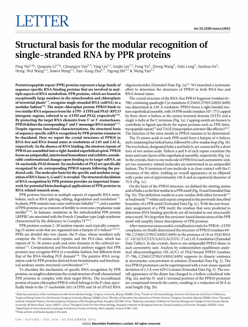
After numerous unsuccessful crystallization trials for PPR10–ATPHcomplexes, we finally determined the structure of PPR10 (residues 69–786, C256S/C279S/C430S/C449S) in the presence of 18-nt PSAJ RNA(59-GUAUUCUUUAAUUAUUUC-39) at 2.45 A resolution (ExtendedData Table1). In the crystals, there is one antiparallel PPR10 dimer ineach asymmetric unit. Analysis by sedimentation equilibrium analy-tical ultracentrifugation (SE-AUC) of PSAJ-bound PPR10 (residues37–786, C256S/C279S/C430S/C449S) supports its dimeric existenceat micromolar concentration in solution (Extended Data Fig. 2). Thetwo PPR10 protomers can be superimposed with a root-mean-squareddeviation of 1.3 A over 629 Ca atoms (Extended Data Fig. 3). The ove-rall appearance of the dimer has changed to a hollow cylindrical tube(Fig. 2a), and the N- and C-terminal portions of the PPR10 protomerare compressed towards the centre, resulting in a reduction of 20 A inaxial length (Fig. 2b).
*These authors contributed equally to this work.
1State Key Laboratory of Bio-membrane and Membrane Biotechnology, Tsinghua University, Beijing 100084, China. 2Center for Structural Biology, School of Life Sciences and School of Medicine,Tsinghua-Peking Center for Life Sciences, Tsinghua University, Beijing 100084, China. 3Ministry of Education Key Laboratory of Protein Science, Tsinghua University, Beijing 100084, China. 4ShanghaiInstitute of Applied Physics, Chinese Academy of Sciences, 239 Zhangheng Road, Shanghai 201204, China. 5State Key Laboratory of Medicinal Chemical Biology and College of Life Sciences, NankaiUniversity, 94 Weijin Road, Tianjin 300071, China. 6Shanghai Center for Plant Stress Biology, Shanghai Institutes for Biological Sciences, Chinese Academy of Sciences, Shanghai 200032, China.7Department of Horticulture and Landscape Architecture, Purdue University, West Lafayette, Indiana 47907, USA.
1 6 8 | N A T U R E | V O L 5 0 4 | 0 5 D E C E M B E R 2 0 1 3
Macmillan Publishers Limited. All rights reserved©2013
Following assignment of most amino acids of PPR10 into the elec-tron density map, strong electron densities indicative of RNA basesbecame clearly visible in the cavities on both ends of the cylindricaltube (Fig. 2c). Assignment of 18 and 14 nucleotides of the two boundRNA elements was validated by the anomalous signals of bromine(Br), which were collected for crystals of PPR10 bound to Br-labelledRNA oligonucleotides (Extended Data Fig. 4 and Extended Data Table2). The 59 and 39 portions of the ssRNA are specifically recognized bythe N-terminal repeats of one protomer and C-terminal repeats of theother. By contrast, the middle portion of the ssRNA, comprising nucle-otides U5 to A10, remains largely uncoordinated by PPR10 (Fig. 2dand Extended Data Fig. 5a, b).
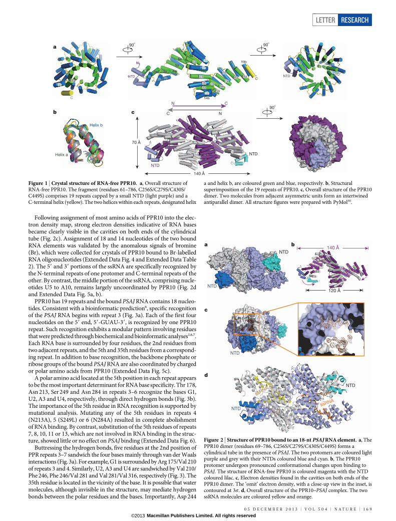
PPR10 has 19 repeats and the bound PSAJ RNA contains 18 nucleo-tides. Consistent with a bioinformatic prediction6, specific recognitionof the PSAJ RNA begins with repeat 3 (Fig. 3a). Each of the first fournucleotides on the 59 end, 59-GUAU-39, is recognized by one PPR10repeat. Such recognition exhibits a modular pattern involving residuesthat were predicted through biochemical and bioinformatic analyses2,6,7.Each RNA base is surrounded by four residues, the 2nd residues fromtwo adjacent repeats, and the 5th and 35th residues from a correspond-ing repeat. In addition to base recognition, the backbone phosphate orribose groups of the bound PSAJ RNA are also coordinated by chargedor polar amino acids from PPR10 (Extended Data Fig. 5c).
A polar amino acid located at the 5th position in each repeat appearsto be the most important determinant for RNA base specificity. Thr 178,Asn 213, Ser 249 and Asn 284 in repeats 3–6 recognize the bases G1,U2, A3 and U4, respectively, through direct hydrogen bonds (Fig. 3b).The importance of the 5th residue in RNA recognition is supported bymutational analysis. Mutating any of the 5th residues in repeats 4(N213A), 5 (S249L) or 6 (N284A) resulted in complete abolishmentof RNA binding. By contrast, substitution of the 5th residues of repeats7, 8, 10, 11 or 13, which are not involved in RNA binding in the struc-ture, showed little or no effect on PSAJ binding (Extended Data Fig. 6).
Buttressing the hydrogen bonds, five residues at the 2nd position ofPPR repeats 3–7 sandwich the four bases mainly through van der Waalsinteractions (Fig. 3a). For example, G1 is surrounded by Arg 175/Val 210of repeats 3 and 4. Similarly, U2, A3 and U4 are sandwiched by Val 210/Phe 246, Phe 246/Val 281 and Val 281/Val 316, respectively (Fig. 3). The35th residue is located in the vicinity of the base. It is possible that watermolecules, although invisible in the structure, may mediate hydrogenbonds between the polar residues and the bases. Importantly, Asp 244
140 Å
90º 90º
90º
70 Å
NTD
N C
C N
NTD
NTD
Helix a
Helix b
11a 2a 3a 4a
5a
6a
7a
9a
10a0a
12a2a
14a4a
14b4b
13a3a
15a5a16a6a
17a7a18a8a 19a9a1b
2b 3b
9b
10b0b
11b1b
13b3b
12b2b
17b7b
19b9b
NTDTD
N
C
1a 2a 3a 4a
5a
6a
7a
9a
10a
12a
14a
14b
13a
15a16a
17a18a 19a1b
2b 3b
9b
10b
11b
13b
12b
17b
19b
NTD
N
C
1a1a2a
1b 1a2a
1b
C
19a9a19b9b
19a19b
a
b c
Figure 1 | Crystal structure of RNA-free PPR10. a, Overall structure ofRNA-free PPR10. The fragment (residues 61–786, C256S/C279S/C430S/C449S) comprises 19 repeats capped by a small NTD (light purple) and aC-terminal helix (yellow). The two helices within each repeats, designated helix
a and helix b, are coloured green and blue, respectively. b, Structuralsuperimposition of the 19 repeats of PPR10. c, Overall structure of the PPR10dimer. Two molecules from adjacent asymmetric units form an intertwinedantiparallel dimer. All structure figures were prepared with PyMol30.
a b
c
d
NTD
NTD
NTD
NTD
ssRNA
ssRNA
NTD
120 Å
140 Å
Figure 2 | Structure of PPR10 bound to an 18-nt PSAJ RNA element. a, ThePPR10 dimer (residues 69–786, C256S/C279S/C430S/C449S) forms acylindrical tube in the presence of PSAJ. The two protomers are coloured lightpurple and grey with their NTDs coloured blue and cyan. b, The PPR10protomer undergoes pronounced conformational changes upon binding toPSAJ. The structure of RNA-free PPR10 is coloured magenta with the NTDcoloured lilac. c, Electron densities found in the cavities on both ends of thePPR10 dimer. The ‘omit’ electron density, with a close-up view in the inset, iscontoured at 3s. d, Overall structure of the PPR10–PSAJ complex. The twossRNA molecules are coloured yellow and orange.
LETTER RESEARCH
0 5 D E C E M B E R 2 0 1 3 | V O L 5 0 4 | N A T U R E | 1 6 9
Macmillan Publishers Limited. All rights reserved©2013
and Asp 314, the 35th residues in repeats 4 and 6, are respectively hydrogenbonded to Asn 213 and Asn 284, the 5th residues in the correspondingrepeats, and may help to stabilize their conformation for base recog-nition (Fig. 3b and Extended Data Fig. 6d).
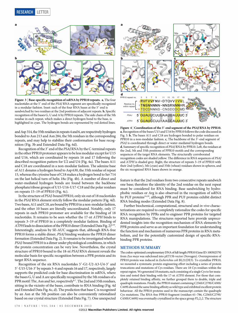
Recognition of the 39 end of the PSAJ RNA by the C-terminal repeatsin the other PPR10 protomer appears to be less modular except for U15and U16, which are coordinated by repeats 16 and 17 following thedescribed recognition pattern for U2 and U4 (Fig. 4a). The bases A11and C18 are coordinated in a non-modular fashion. The adenine baseof A11 donates a hydrogen bond to Asp 630, the 35th residue of repeat15, whereas the cytosine base of C18 makes a hydrogen bond to Ser 714on the last helical turn of helix 18a (Fig. 4b). A number of direct andwater-mediated hydrogen bonds are found between the backbonephosphate/ribose groups of U15-U16-U17-C18 and the polar residueson repeats 15–19 of PPR10 (Fig. 4c).
In the structure of PSAJ-bound PPR10, only six out of 18 nucleotidesin the PSAJ RNA element strictly follow the modular pattern (Fig. 4d).Two bases, A11 and C18, are bound by PPR10 in a non-modular fashion,and the other 10 bases are literally uncoordinated. Notably, only 17repeats in each PPR10 protomer are available for the binding of 18nucleotides. It remains to be seen whether the 17-nt ATPH binds torepeats 3–19 of PPR10 in a completely modular fashion. Binding ofATPH leads to dissociation of the PPR10 dimer (Extended Data Fig. 2)6,10.Interestingly, analysis by SE-AUC suggests that, although RNA-freePPR10 forms a stable dimer, PSAJ binding weakens the PPR10 dimerformation (Extended Data Fig. 2). It remains to be investigated whetherPSAJ-bound PPR10 is a dimer under physiological conditions, in whichthe protein concentration can be very low. Nevertheless, the crystalstructure of PPR10 bound to the 18-nt PSAJ RNA element reveals themolecular basis for specific recognition between a PPR protein and itstarget RNA sequence.
Recognition of the six RNA nucleotides 59-G1-U2-A3-U4-39 and59-U15-U16-39 by repeats 3–6 and repeats 16 and 17, respectively, largelysupports the predicted code for base discrimination in ssRNA, wherethe bases G, U and A are specifically recognized by the 5th residues of aPPR motif: Thr, Asn and Ser, respectively6,7. The 2nd and 35th residues,sitting in the vicinity of the bases, contribute to RNA binding (Fig. 4dand Extended Data Fig. 6c, d). The prediction that base C is recognizedby an Asn at the 5th position can also be conveniently rationalizedbased on our crystal structure (Extended Data Fig. 7). One unexpected
feature is that the 2nd residues from two consecutive repeats sandwichone base; therefore the identity of the 2nd residue on the next repeatmust be considered for RNA binding. Base sandwiching by hydro-phobic residues or Arg is also observed in the recognition of ssRNAby PUF proteins24,29, although PPR and PUF proteins exhibit distinctRNA binding modes (Extended Data Fig. 8).
Further biochemical, computational, structural and in vivo charac-terizations are required to completely rationalize the codes for specificRNA recognition by PPRs and to engineer PPR proteins for targetedRNA manipulations. The structures reported here provide unprece-dented insights into the recognition mechanism of RNA elements byPPR proteins and serve as an important foundation for understandingthe function and mechanism of numerous PPR proteins in RNA meta-bolism, and for the potentially customized design of specific-RNA-binding PPR proteins.
METHODS SUMMARYThe codon-optimized complementary DNA of full-length PPR10 (Gene ID: 100302579)from Zea mays was subcloned into pET15b vector (Novagen). Overexpression ofPPR10 protein was induced in Escherichia coli BL21(DE3). To crystallize PPR10,we mounted a systematic protein engineering effort including a series of proteintruncations and mutations of Cys residues. There are 18 Cys residues within therepeat region. We generated 18 mutants, each consisting of a single Cys to Ser muta-tion and tested their binding with the 17-nt ATPH element. For those that com-pletely retained binding affinity, we further grouped them to double, triple andquadruple mutations. Finally, the PPR10 mutant containing C256S/C279S/C430S/C449S showed the same binding affinity as wild type and exhibited excellent proteinbehaviour. All the PPR10 proteins used in the manuscript contain the quadrupleCys mutations. The RNA-free PPR10 fragment (residues 61–786, C256S/C279S/C430S/C449S) was eventually crystallized in the space group P21212. The structure
NTDNTD
1a
2a3a 4a
5a5′
3′
6a
R175175
R175175
V210210F246246 V281281
T178178
N213213
S249249V210210
F246246
D244244
3a
4a
4a
5a
5a
6a
F246246
V281281S279279
V210210
T208208
G1
5′
U2 A3U4
G1U2
A3
a
b
NTD
1a
2a3a 4a
5a5′
3′
6a
R175
R175
V210F246 V281
V316
T178
N213
S249V210
F246
D244
3a
4a
4a
5a
5a
6a
F246
V281S279
V210
T208
G1
5′
U2 A3U4
G1U2
A3
Figure 3 | Base-specific recognition of ssRNA by PPR10 repeats. a, The fournucleotides at the 59 end of the PSAJ RNA segment are specifically recognizedin a modular fashion. Inset: each of the four RNA bases at the 59 end issandwiched by two residues at the 2nd positions of adjacent repeats. b, Specificrecognition of the bases G, U and A by PPR10 repeats. The side chain of the 5thresidue in each repeat, which makes a direct hydrogen bond to the base, ishighlighted in cyan. The hydrogen bonds are represented by red dotted lines.
2
5
35
Residue no.within repeat
d
U1515
A1111
C1818
U1616
I632632
D666666
C701701V668668
V703703
19a9a
18a8a
17a7a16a6a15a5a
16a6a
16a6a
18a8a
15a5a
17a7a
18a8a
C1818
U1717U1616U1515
3′
N635635N671671
S714714
D630630
S604604
S636636 T672672 N675675
R742742
U15
A11
C18
U16
I632
D666
C701V668
V703
19a
18a
17a16a15a
16a
16a
18a
15a
17a
18a
C18
U17U16U15
3′
N635N671
S714
D630
S604
S636 T672 N675
R742
b c
′
′
PSAJ
ATPH
′
′
PPR10 repeat no. 3 5 7 9 11 13 15 17 19
A11A11
U1212
A1010
U9
U1313
A1414U1515
U1616
19a9a
18a8a
17a7a16a6a
15a5a
14a4a U1717
C1818
A11
U12
A10
U9
U13
A14U15
U16
19a
18a
17a16a
15a
14a U17
C18
a
Figure 4 | Coordination of the 39-end segment of the PSAJ RNA by PPR10.a, Recognition of the bases U15 and U16 by PPR10 follows the code discussed inFig. 3. b, The bases A11 and C18 are hydrogen bonded to polar residues onPPR10 in a non-modular fashion. c, The backbone of the 39-end segment ofPSAJ is coordinated through direct or water-mediated hydrogen bonds.d, Summary of specific recognition of PSAJ RNA by PPR10. Left, the residues atthe 2nd, 5th and 35th positions of PPR10 motifs and the correspondingsequences of the target RNA elements. The structurally corroboratedrecognition codes are shaded yellow. The difference in RNA sequences of PSAJand ATPH is shaded grey. Right, the structure of repeats 3–19 of PPR10 withtheir 2nd (yellow), 5th (cyan) and 35th (wheat) residues shown in spheres, andthe six recognized RNA bases shown in orange.
RESEARCH LETTER
1 7 0 | N A T U R E | V O L 5 0 4 | 0 5 D E C E M B E R 2 0 1 3
Macmillan Publishers Limited. All rights reserved©2013
was determined by selenium-based single-wavelength anomalous diffraction andrefined to 2.85 A resolution (Extended Data Table 1). In the effort to crystallizePPR10 in complex with its target RNA, despite numerous trials, most PPR10–ATPH complexes defied crystallization; for those that crystallized, X-ray diffrac-tion was consistently poor. We applied the same strategy to complexes betweenPPR10 and PSAJ. After screening more than 100,000 conditions, we were able tocrystallize the complex between PPR10 (residues 69–786, C256S/C279S/C430S/C449S) and the 18-nt PSAJ RNA (59-GUAUUCUUUAAUUAUUUC-39) in thespace group P43. These crystals diffract X-rays beyond 2.5 A. The structure wasdetermined by molecular replacement using successive segments of the RNA-freePPR10 structure, but not the entire molecule. We were able to assign all 18 nucleo-tides of one bound PSAJ RNA element, but only 14 of the other. For details of elec-trophoretic mobility shift assay and SE-AUC experiments, please refer to Methods.
Online Content Any additional Methods, Extended Data display items and SourceData are available in the online version of the paper; references unique to thesesections appear only in the online paper.
Received 28 May; accepted 12 September 2013.
Published online 27 October 2013.
1. Small, I. D. & Peeters, N. The PPR motif — a TPR-related motif prevalent in plantorganellar proteins. Trends Biochem. Sci. 25, 45–47 (2000).
2. Nakamura, T., Yagi, Y. & Kobayashi, K. Mechanistic insight into pentatricopeptiderepeat proteins as sequence-specific RNA-binding proteins for organellar RNAs inplants. Plant Cell Physiol. 53, 1171–1179 (2012).
3. Schmitz-Linneweber, C. & Small, I. Pentatricopeptide repeat proteins: a socket setfor organelle gene expression. Trends Plant Sci. 13, 663–670 (2008).
4. Fujii, S. & Small, I. The evolution of RNA editing and pentatricopeptide repeatgenes. New Phytol. 191, 37–47 (2011).
5. Kotera, E., Tasaka, M. & Shikanai, T. A pentatricopeptide repeat protein is essentialfor RNA editing in chloroplasts. Nature 433, 326–330 (2005).
6. Barkan, A. et al. A combinatorial amino acid code for RNA recognition bypentatricopeptide repeat proteins. PLoS Genet. 8, e1002910 (2012).
7. Yagi, Y., Hayashi, S., Kobayashi, K., Hirayama, T. & Nakamura, T. Elucidation of theRNA recognition code for pentatricopeptide repeat proteins involved in organelleRNA editing in plants. PLoS ONE 8, e57286 (2013).
8. Yagi, Y. et al. Pentatricopeptide repeat proteins involved in plant organellar RNAediting. RNA Biol. 10, 1236–1242 (2013).
9. Pfalz, J., Bayraktar, O. A., Prikryl, J. & Barkan, A. Site-specific binding of a PPRprotein defines and stabilizes 59 and 39 mRNA termini in chloroplasts. EMBO J. 28,2042–2052 (2009).
10. Prikryl, J., Rojas, M., Schuster, G. & Barkan, A. Mechanism of RNA stabilization andtranslational activation by a pentatricopeptide repeat protein. Proc. Natl Acad. Sci.USA 108, 415–420 (2011).
11. Zhelyazkova, P.et al.Protein-mediatedprotection as the predominant mechanismfor defining processed mRNA termini in land plant chloroplasts. Nucleic Acids Res.40, 3092–3105 (2012).
12. Lurin, C. et al. Genome-wide analysis of Arabidopsis pentatricopeptide repeatproteins reveals their essential role in organelle biogenesis. Plant Cell 16,2089–2103 (2004).
13. Cushing, D. A., Forsthoefel, N. R., Gestaut, D. R. & Vernon, D. M. Arabidopsis emb175andotherpprknockoutmutants reveal essential roles forpentatricopeptide repeat(PPR) proteins in plant embryogenesis. Planta 221, 424–436 (2005).
14. Khrouchtchova, A., Monde, R. A. & Barkan, A. A short PPR protein required for thesplicing of specific group II introns in angiosperm chloroplasts. RNA 18,1197–1209 (2012).
15. Bentolila, S., Alfonso, A. A. & Hanson, M. R. A pentatricopeptide repeat-containinggene restores fertility to cytoplasmic male-sterile plants. Proc. Natl Acad. Sci. USA99, 10887–10892 (2002).
16. Desloire, S. et al. Identification of the fertility restoration locus, Rfo, in radish, as amember of the pentatricopeptide-repeat protein family. EMBO Rep. 4, 588–594(2003).
17. Wang, Z. et al. Cytoplasmic male sterility of rice with boro II cytoplasm is caused bya cytotoxic peptide and is restored by two related PPR motif genes via distinctmodes of mRNA silencing. Plant Cell 18, 676–687 (2006).
18. Chase, C. D. Cytoplasmic male sterility: a window to the world of plantmitochondrial–nuclear interactions. Trends Genet. 23, 81–90 (2007).
19. Hu, J. et al. The rice pentatricopeptide repeat protein RF5 restores fertility in Hong-Lian cytoplasmic male-sterile lines via a complex with the glycine-rich proteinGRP162. Plant Cell 24, 109–122 (2012).
20. Mootha, V. K. et al. Identification of a gene causing human cytochrome c oxidasedeficiency by integrative genomics. Proc. Natl Acad. Sci. USA 100, 605–610(2003).
21. Ruzzenente, B. et al. LRPPRC is necessary for polyadenylation and coordination oftranslation of mitochondrial mRNAs. EMBO J. 31, 443–456 (2012).
22. Howard, M. J., Lim, W. H., Fierke, C. A. & Koutmos, M. Mitochondrial ribonucleaseP structure provides insight into the evolution of catalytic strategies forprecursor-tRNA 59 processing. Proc. Natl Acad. Sci. USA 109, 16149–16154(2012).
23. Ringel, R. et al. Structure of human mitochondrial RNA polymerase. Nature 478,269–273 (2011).
24. Wang, X., McLachlan, J., Zamore, P. D. & Hall, T. M. Modular recognition of RNA by ahuman pumilio-homology domain. Cell 110, 501–512 (2002).
25. Grove, T. Z., Cortajarena, A. L. & Regan, L. Ligand binding by repeat proteins:natural and designed. Curr. Opin. Struct. Biol. 18, 507–515 (2008).
26. Deng, D. et al. Structural basis for sequence-specific recognition of DNA by TALeffectors. Science 335, 720–723 (2012).
27. Gao, H., Wu, X., Chai, J. & Han, Z. Crystal structure of a TALE protein reveals anextended N-terminal DNA binding region. Cell Res. 22, 1716–1720 (2012).
28. Kobayashi, K. et al. Identification and characterization of the RNA binding surfaceof the pentatricopeptide repeat protein. NucleicAcids Res. 40, 2712–2723 (2012).
29. Filipovska, A. & Rackham, O. Modular recognition of nucleic acids by PUF, TALEand PPR proteins. Mol. Biosyst. 8, 699–708 (2012).
30. DeLano, W. L. The PyMOL Molecular Graphics System. http://www.pymol.org(2002).
Acknowledgements We thank X. Yu and Y. Chen at the Institute of Biophysics, ChineseAcademy ofSciences, for technical support.We thank K.Hasegawa and T. Kumasaka atthe SPring-8 beamline BL41XU for on-site assistance. This work was supported byfunds from the Ministry of Science and Technology (grant number 2011CB910501 forN.Y.), and Projects 91017011 (N.Y.), 31070644 (N.Y.), 31021002 (Y.S., N.Y., J.W.) and31200567 (P.Y.) of the National Natural Science Foundation of China. The research ofN.Y. was supported in part by an International Early Career Scientist grant from theHoward Hughes Medical Institute.
Author Contributions P.Y., Q.L., J.-K.Z., Y.S. and N.Y. designed all experiments. P.Y., Q.L.,C.Y., Y.L., J.L., F.Y., Z.W., J.L., J.H., H.-W.W., J.W. and N.Y. performed the experiments. Allauthors analysed the data and contributed to manuscript preparation. N.Y. wrote themanuscript.
Author Information The atomic coordinates and structure factors of RNA-free andRNA-bound PPR10 have been deposited in the Protein Data Bank (PDB) with theaccession codes 4M57 and 4M59, respectively. Reprints and permissions informationis available at www.nature.com/reprints. The authors declare no competing financialinterests. Readers are welcome to comment on the online version of the paper.Correspondence and requests for materials should be addressed to N.Y.([email protected]).
LETTER RESEARCH
0 5 D E C E M B E R 2 0 1 3 | V O L 5 0 4 | N A T U R E | 1 7 1
Macmillan Publishers Limited. All rights reserved©2013
METHODSProtein preparation. The codon-optimized complementary DNA of full-lengthPPR10 (Gene ID: 100302579) from Zea mays was subcloned into pET15b vector(Novagen). Overexpression of PPR10 protein was induced in E. coli BL21(DE3)with 0.2 mM isopropyl-b-D-thiogalactoside at an OD600 nm of 1.2. After growingfor 16 h at 16 uC, the cells were collected, homogenized in a buffer containing 25mM Tris-HCl, pH 8.0, and 150 mM NaCl. After sonication and centrifugation, thesupernatant was applied to Ni21 affinity resin (Ni-NTA, Qiagen) and further frac-tionated by ion-exchange chromatography (Source 15Q, GE Healthcare). ThePPR10 mutants were generated using two-step PCR and subcloned, overexpressedand purified in the same way as the wild-type protein.
A systematic protein engineering effort was mounted for crystallization of RNA-free and -bound PPR10. A series of protein truncations were tested without givingrise to crystals. There are 18 Cys residues within the repeat region. It is well knownthat the presence of surface Cys residues, which are subject to oxidation, may leadto protein heterogeneity and impede crystallization. We therefore generated 18mutants, each consisting of a single Cys to Ser mutation and tested their bindingwith the 17-nt ATPH element. For those that completely retained binding affinity,we further grouped them to double, triple and quadruple mutations. Finally, thePPR10 mutant containing C256S/C279S/C430S/C449S showed the same bindingaffinity as wild type and exhibited excellent protein behaviour. For consistency, allthe PPR10 proteins used in the manuscript contain the quadruple Cys mutations.
For the crystallization trials of RNA-free PPR10 (residues 61–786, C256S/C279S/C430S/C449S), the protein was concentrated and applied to gel filtration chromato-graphy (Superdex-200 10/30, GE Healthcare) in the buffer containing 25 mM Tris-HCl, pH 8.0, 150 mM NaCl and 10 mM dithiothreitol (DTT). Selenomethionine(Se-Met)-derived protein was purified similarly.
To obtain the crystals of protein–RNA complex, PPR10 (residues 69–786, C256S/C279S/C430S/C449S) was purified through Ni21 affinity resin (Ni-NTA, Qiagen),followed by heparin affinity column (HiPrep Heparin FF 16/10, GE Healthcare).The protein was then applied to gel filtration chromatography (Superdex-200 10/30,GE Healthcare). The buffer for gel filtration contained 25 mM Tris-HCl, pH 8.0,50 mM NaCl, 5 mM MgCl2 and 10 mM DTT. The peak fractions were incubatedwith target RNA oligonucleotides with a molar ratio of approximately 1:1.5 at 4 uCfor about 40 min before crystallization trials.Crystallization. Both RNA-free and RNA-bound PPR10 proteins were crystal-lized by hanging-drop vapour-diffusion method at 18 uC. PPR10 (residues 61–786,C256S/C279S/C430S/C449S), at a concentration of approximately 6.0 mg ml21,was mixed with an equal volume of reservoir solution containing 1.8–2.1 M sodiumformate, and 0.1 M Bis-Tris propane, pH 6.5. Plate-shaped crystals appeared over-night and grew to full size within 1–2 weeks. Se-Met-labelled protein was crystal-lized similarly.
To obtain crystals of protein–RNA complex, various combinations of proteinboundaries and RNA oligonucleotides (Takara) were examined. Because the firstvisible residue in the structure of RNA-free PPR10 starts at position 69, we investedmore effort into this construct. Finally, the protein (residues 69–786, C256S/C279S/C430S/C449S) and 18-nt RNA from the PSAJ–RPL33 intergenic region with thesequence 59-GUAUUCUUUAAUUAUUUC-39 (designated PSAJ RNA) gave riseto crystals in the reservoir solution containing 8–10% (w/v) polyethylene glycol3350, 8% Tacsimate, pH 6.0 (Hampton Research), and 0.1 M MES, pH 5.5.Data collection and structural determination. All data sets were collected at SSRFbeamline BL17U or SPring-8 beamline BL41XU and processed with the HKL2000packages31. Further processing was carried out with programs from the CCP4 suite32.Data collection and structure refinement statistics are summarized in ExtendedData Tables 1 and 2.
The RNA-free PPR10 structure was solved by single anomalous diffraction(SAD) of Se-Met using the program ShelxC/D/E33. Then a crude helical model
was manually built in the program Coot34. Using this partial model as input, theidentified Se atom positions were refined and phases were recalculated using theSAD experimental phasing module of the program Phaser35. With the improvedmap, the molecular boundary was unambiguously defined and one molecule wasfound in an asymmetry unit. The crude model was further rebuilt with Coot and refinedwith Phenix36. The sequence docking was aided by anomalous map of selenium.
Data sets collected from five crystals of the PPR10–RNA complex were mergedfor complete and better data. The structure of the PPR10–RNA complex was solvedby molecular replacement with the newly solved RNA-free structure as the searchmodel using the program Phaser35. To find the right solution, the structure of theRNA-free PPR10 protomer was divided into three consecutive segments. The assign-ment of RNA sequence was aided by the anomalous signal of bromine obtained forcrystals of PPR10 in complex with Br-labelled RNA oligonucleotides, where U4/U7/U15, U5/U7/U15 or U12 were substituted by 5-bromouracil (Extended DataTable 2). The structure was manually refined with Coot and Phenix iteratively(Extended Data Table 1).Electrophoretic mobility shift assay (EMSA). The ssRNA oligonucleotides wereradiolabelled at the 59 end with [c-32P] ATP (PerkinElmer) catalysed by T4 poly-nucleotide kinase (Takara). The sequences of ssRNA oligonucleotides used in EMSAare: PSAJ, 59-GUAUUCUUUAAUUAUUUC-39; and ATPH, 59-GUAUCCUUAACCAUUUC-39.
For EMSA, PPR10 (residues 37–786, C256S/C279S/C430S/C449S) and theother variants consisting of the indicated point mutations were incubated withapproximately 40 pM 32P-labelled probe in the final binding reactions containing40 mM Tris-HCl, pH 7.5, 100 mM NaCl, 4 mM DTT, 0.1 mg ml21 BSA, 5mg ml21
heparin and 10% glycerol at room temperature (22 uC) for 20 min. Reactions werethen resolved on 6% native acrylamide gels (37.5:1 for acrylamide:bisacrylamide)in 0.53 Tris-glycine buffer under an electric field of 15 V cm21 for 40 min. Vacuum-dried gels were visualized on a phosphor screen (Amersham Biosciences) with aTyphoon Trio Imager (Amersham Biosciences).SE-AUC. The oligomeric states of PPR10 (residues 37–786, C256S/C279S/C430S/C449S) with or without target RNA oligonucleotides in solution were investigatedby AUC experiments. SE-AUC experiments were performed in a BeckmanCoulter XL-I analytical ultracentrifuge using six-channel centrepieces. RNA-freePPR10, PSAJ-bound PPR10 and ATPH-bound PPR10 were in solutions contain-ing 25 mM Tris-HCl, pH 8.0, 150 mM NaCl and 2 mM DTT. The sequences ofRNA oligonucleotides were identical to those used in EMSA. Data were collectedby interference detection at 4 uC for all three protein concentrations (4mM, 6mMand 8mM) at different rotor speeds (6,000, 8,500 and 12,000 r.p.m.). The buffercomposition (density and viscosity) and protein partial specific volume (V-bar)were obtained using the SEDNTERP program (available through the BostonBiomedical Research Institute). The SE-AUC data were globally analysed usingthe Sedfit and Sedphat programs37 and were fitted to a monomer–dimer equilib-rium model to determine the dissociation constants (Kd) for the homodimers.
31. Otwinowski, Z. & Minor, W. Processing of X-ray diffraction data collected inoscillation mode. Methods Enzymol. 276, 307–326 (1997).
32. Collaborative Computational Project, Number 4. The CCP4 suite: programs forprotein crystallography. Acta Crystallogr. D 50, 760–763 (1994).
33. Schneider, T. R. & Sheldrick, G. M. Substructure solution with SHELXD. ActaCrystallogr. D 58, 1772–1779 (2002).
34. Emsley, P. & Cowtan, K. Coot: model-building tools for molecular graphics. ActaCrystallogr. D 60, 2126–2132 (2004).
35. McCoy, A. J. et al. Phaser crystallographic software. J. Appl. Crystallogr. 40,658–674 (2007).
36. Adams, P. D. et al. PHENIX: building new software for automated crystallographicstructure determination. Acta Crystallogr. D 58, 1948–1954 (2002).
37. Schuck, P. On the analysis of protein self-association by sedimentation velocityanalytical ultracentrifugation. Anal. Biochem. 320, 104–124 (2003).
RESEARCH LETTER
Macmillan Publishers Limited. All rights reserved©2013
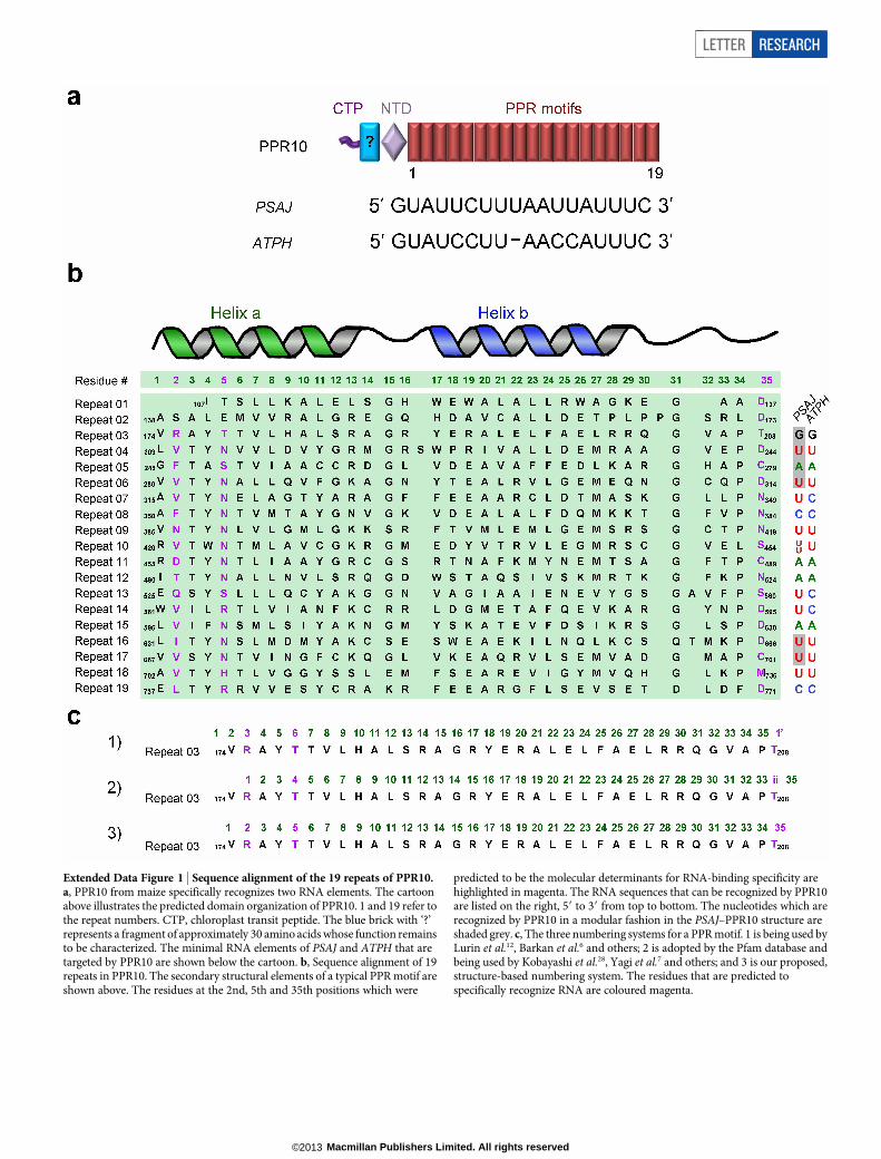
Extended Data Figure 1 | Sequence alignment of the 19 repeats of PPR10.a, PPR10 from maize specifically recognizes two RNA elements. The cartoonabove illustrates the predicted domain organization of PPR10. 1 and 19 refer tothe repeat numbers. CTP, chloroplast transit peptide. The blue brick with ‘?’represents a fragment of approximately 30 amino acids whose function remainsto be characterized. The minimal RNA elements of PSAJ and ATPH that aretargeted by PPR10 are shown below the cartoon. b, Sequence alignment of 19repeats in PPR10. The secondary structural elements of a typical PPR motif areshown above. The residues at the 2nd, 5th and 35th positions which were
predicted to be the molecular determinants for RNA-binding specificity arehighlighted in magenta. The RNA sequences that can be recognized by PPR10are listed on the right, 59 to 39 from top to bottom. The nucleotides which arerecognized by PPR10 in a modular fashion in the PSAJ–PPR10 structure areshaded grey. c, The three numbering systems for a PPR motif. 1 is being used byLurin et al.12, Barkan et al.6 and others; 2 is adopted by the Pfam database andbeing used by Kobayashi et al.28, Yagi et al.7 and others; and 3 is our proposed,structure-based numbering system. The residues that are predicted tospecifically recognize RNA are coloured magenta.
LETTER RESEARCH
Macmillan Publishers Limited. All rights reserved©2013
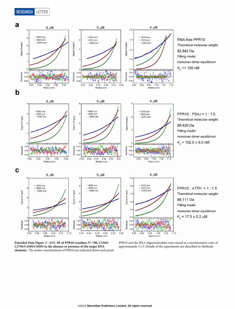
Extended Data Figure 2 | AUC-SE of PPR10 (residues 37–786, C256S/C279S/C430S/C449S) in the absence or presence of the target RNAelements. The molar concentrations of PPR10 are indicated above each panel.
PPR10 and the RNA oligonucleotides were mixed at a stoichiometric ratio ofapproximately 1:1.5. Details of the experiments are described in Methods.
RESEARCH LETTER
Macmillan Publishers Limited. All rights reserved©2013
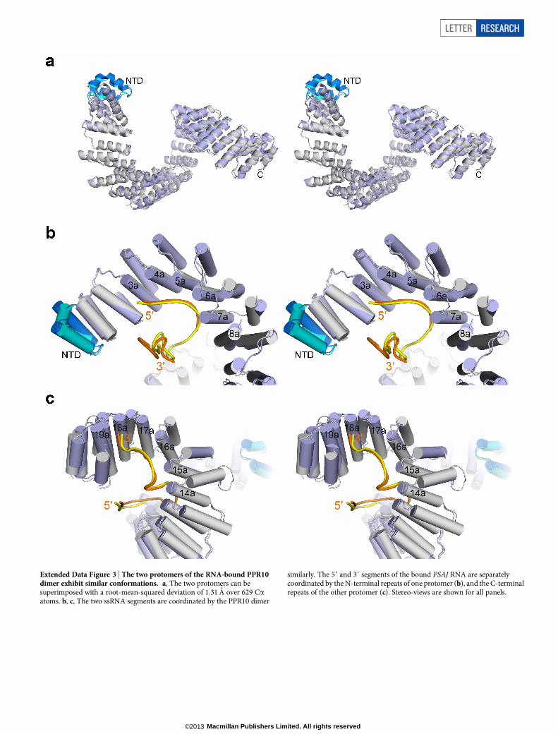
Extended Data Figure 3 | The two protomers of the RNA-bound PPR10dimer exhibit similar conformations. a, The two protomers can besuperimposed with a root-mean-squared deviation of 1.31 A over 629 Caatoms. b, c, The two ssRNA segments are coordinated by the PPR10 dimer
similarly. The 59 and 39 segments of the bound PSAJ RNA are separatelycoordinated by the N-terminal repeats of one protomer (b), and the C-terminalrepeats of the other protomer (c). Stereo-views are shown for all panels.
LETTER RESEARCH
Macmillan Publishers Limited. All rights reserved©2013
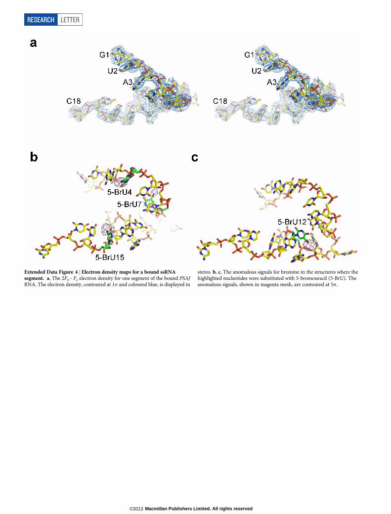
Extended Data Figure 4 | Electron density maps for a bound ssRNAsegment. a, The 2Fo – Fc electron density for one segment of the bound PSAJRNA. The electron density, contoured at 1s and coloured blue, is displayed in
stereo. b, c, The anomalous signals for bromine in the structures where thehighlighted nucleotides were substituted with 5-bromouracil (5-BrU). Theanomalous signals, shown in magenta mesh, are contoured at 5s.
RESEARCH LETTER
Macmillan Publishers Limited. All rights reserved©2013
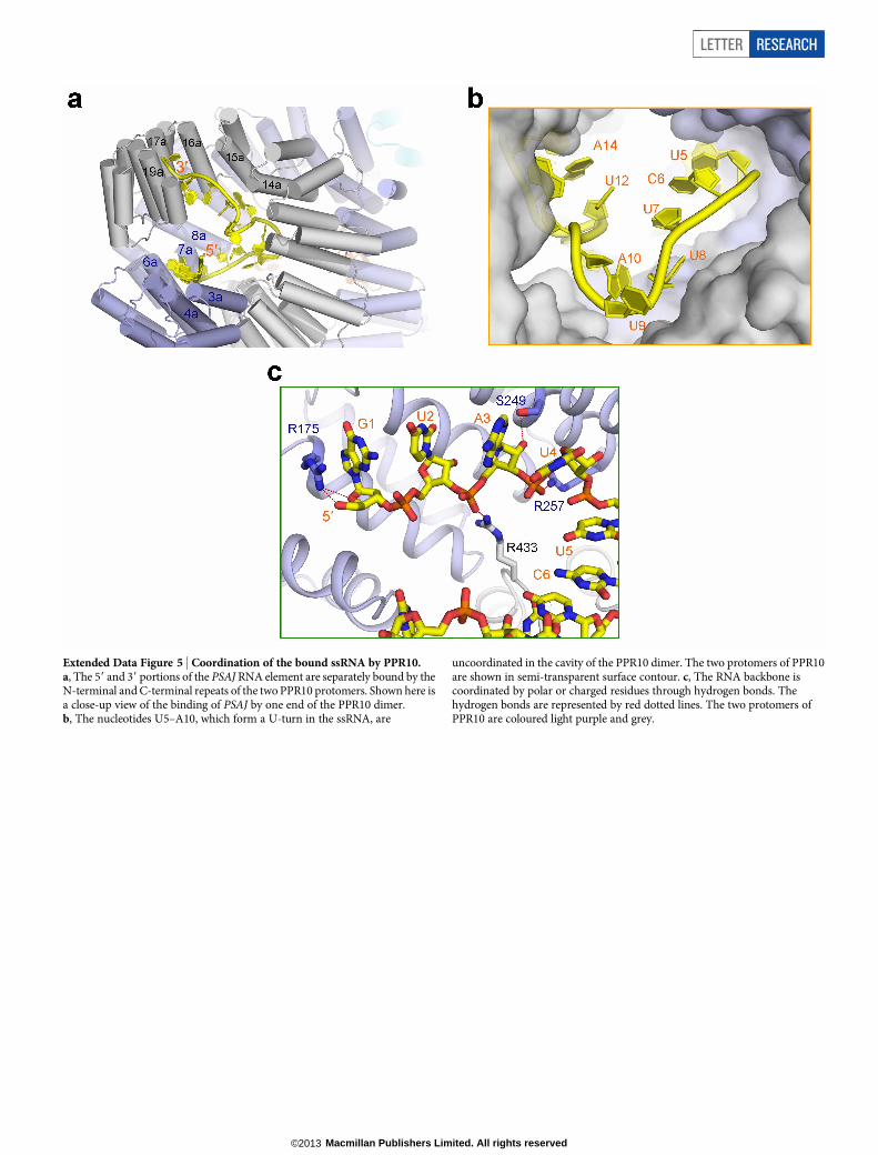
Extended Data Figure 5 | Coordination of the bound ssRNA by PPR10.a, The 59 and 39 portions of the PSAJ RNA element are separately bound by theN-terminal and C-terminal repeats of the two PPR10 protomers. Shown here isa close-up view of the binding of PSAJ by one end of the PPR10 dimer.b, The nucleotides U5–A10, which form a U-turn in the ssRNA, are
uncoordinated in the cavity of the PPR10 dimer. The two protomers of PPR10are shown in semi-transparent surface contour. c, The RNA backbone iscoordinated by polar or charged residues through hydrogen bonds. Thehydrogen bonds are represented by red dotted lines. The two protomers ofPPR10 are coloured light purple and grey.
LETTER RESEARCH
Macmillan Publishers Limited. All rights reserved©2013
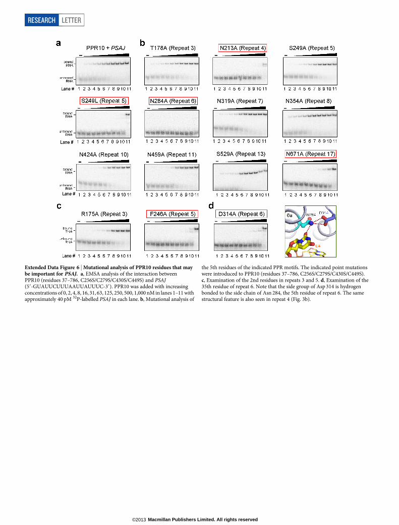
Extended Data Figure 6 | Mutational analysis of PPR10 residues that maybe important for PSAJ. a, EMSA analysis of the interaction betweenPPR10 (residues 37–786, C256S/C279S/C430S/C449S) and PSAJ(59-GUAUUCUUUAAUUAUUUC-39). PPR10 was added with increasingconcentrations of 0, 2, 4, 8, 16, 31, 63, 125, 250, 500, 1,000 nM in lanes 1–11 withapproximately 40 pM 32P-labelled PSAJ in each lane. b, Mutational analysis of
the 5th residues of the indicated PPR motifs. The indicated point mutationswere introduced to PPR10 (residues 37–786, C256S/C279S/C430S/C449S).c, Examination of the 2nd residues in repeats 3 and 5. d, Examination of the35th residue of repeat 6. Note that the side group of Asp 314 is hydrogenbonded to the side chain of Asn 284, the 5th residue of repeat 6. The samestructural feature is also seen in repeat 4 (Fig. 3b).
RESEARCH LETTER
Macmillan Publishers Limited. All rights reserved©2013
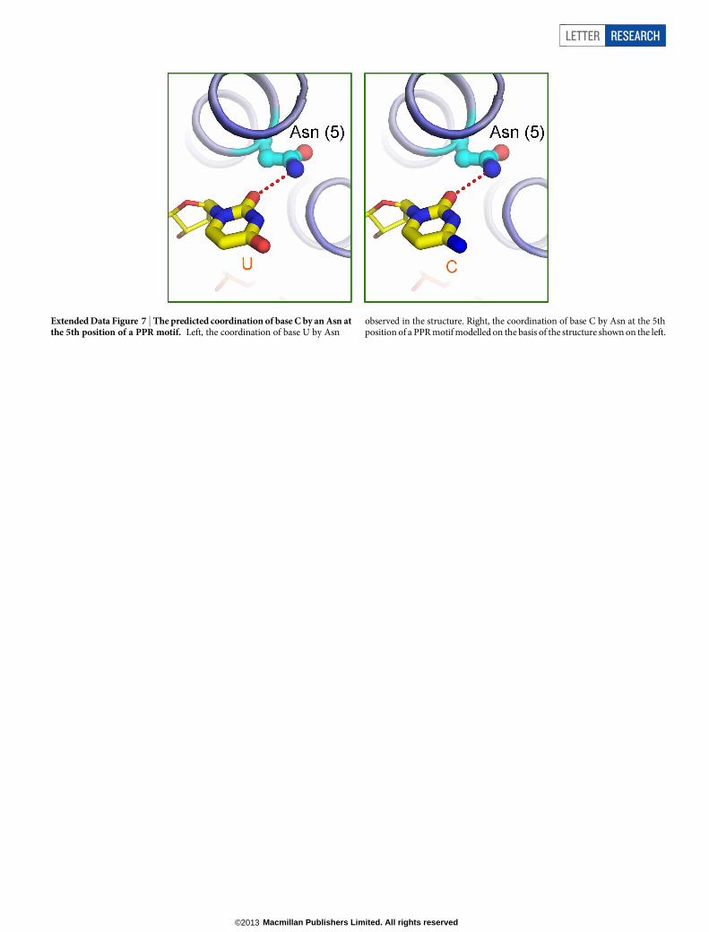
Extended Data Figure 7 | The predicted coordination of base C by an Asn atthe 5th position of a PPR motif. Left, the coordination of base U by Asn
observed in the structure. Right, the coordination of base C by Asn at the 5thposition of a PPR motif modelled on the basis of the structure shown on the left.
LETTER RESEARCH
Macmillan Publishers Limited. All rights reserved©2013
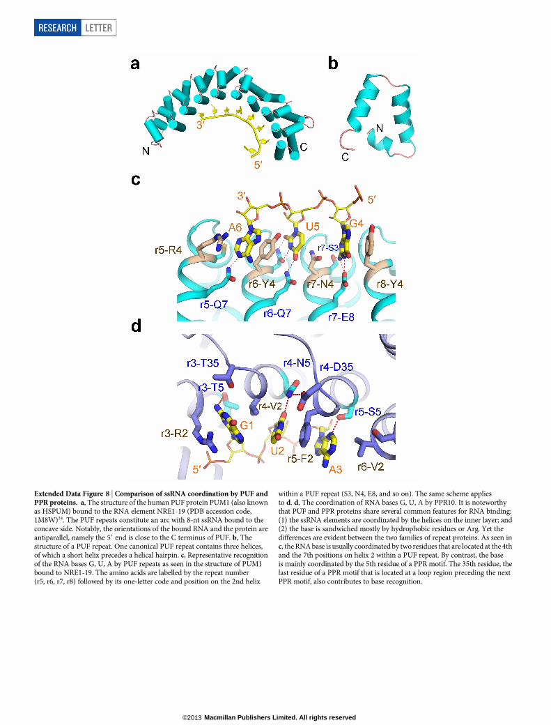
Extended Data Figure 8 | Comparison of ssRNA coordination by PUF andPPR proteins. a, The structure of the human PUF protein PUM1 (also knownas HSPUM) bound to the RNA element NRE1-19 (PDB accession code,1M8W)24. The PUF repeats constitute an arc with 8-nt ssRNA bound to theconcave side. Notably, the orientations of the bound RNA and the protein areantiparallel, namely the 59 end is close to the C terminus of PUF. b, Thestructure of a PUF repeat. One canonical PUF repeat contains three helices,of which a short helix precedes a helical hairpin. c, Representative recognitionof the RNA bases G, U, A by PUF repeats as seen in the structure of PUM1bound to NRE1-19. The amino acids are labelled by the repeat number(r5, r6, r7, r8) followed by its one-letter code and position on the 2nd helix
within a PUF repeat (S3, N4, E8, and so on). The same scheme appliesto d. d, The coordination of RNA bases G, U, A by PPR10. It is noteworthythat PUF and PPR proteins share several common features for RNA binding:(1) the ssRNA elements are coordinated by the helices on the inner layer; and(2) the base is sandwiched mostly by hydrophobic residues or Arg. Yet thedifferences are evident between the two families of repeat proteins. As seen inc, the RNA base is usually coordinated by two residues that are located at the 4thand the 7th positions on helix 2 within a PUF repeat. By contrast, the baseis mainly coordinated by the 5th residue of a PPR motif. The 35th residue, thelast residue of a PPR motif that is located at a loop region preceding the nextPPR motif, also contributes to base recognition.
RESEARCH LETTER
Macmillan Publishers Limited. All rights reserved©2013
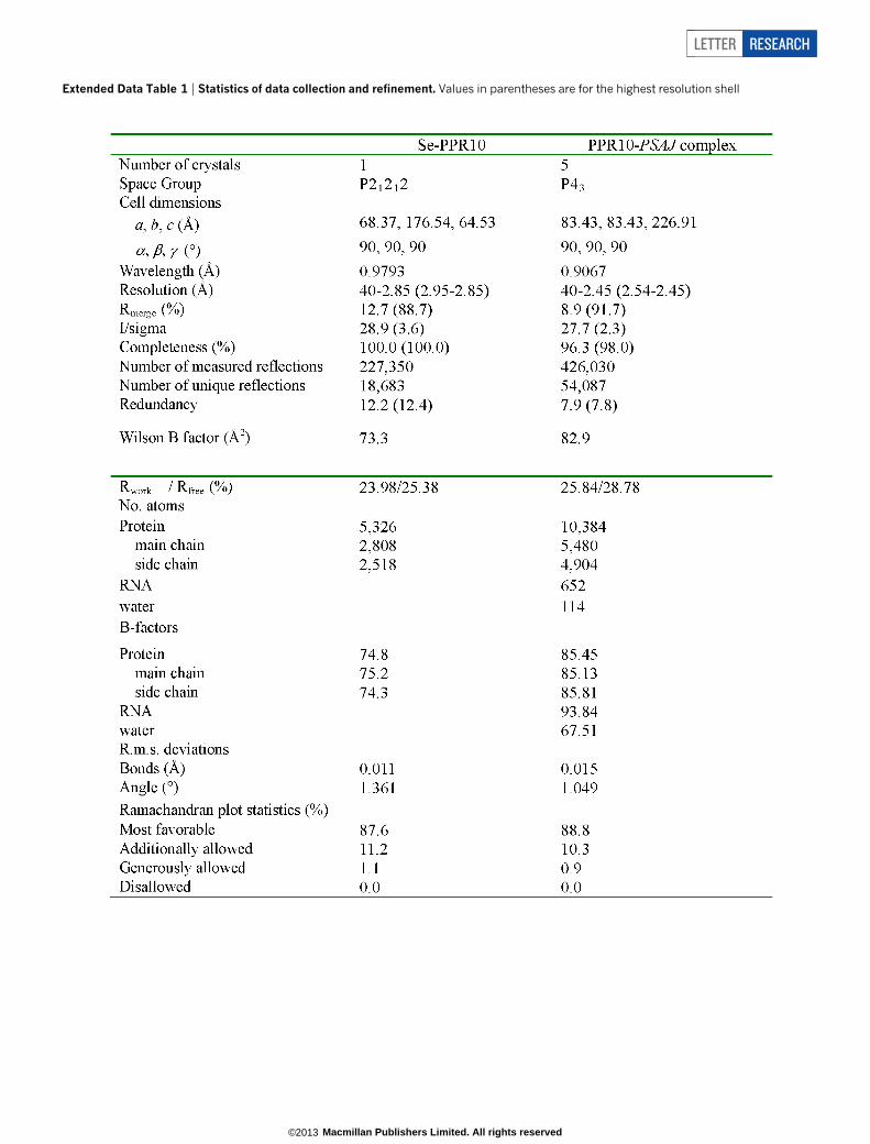
Extended Data Table 1 | Statistics of data collection and refinement. Values in parentheses are for the highest resolution shell
LETTER RESEARCH
Macmillan Publishers Limited. All rights reserved©2013
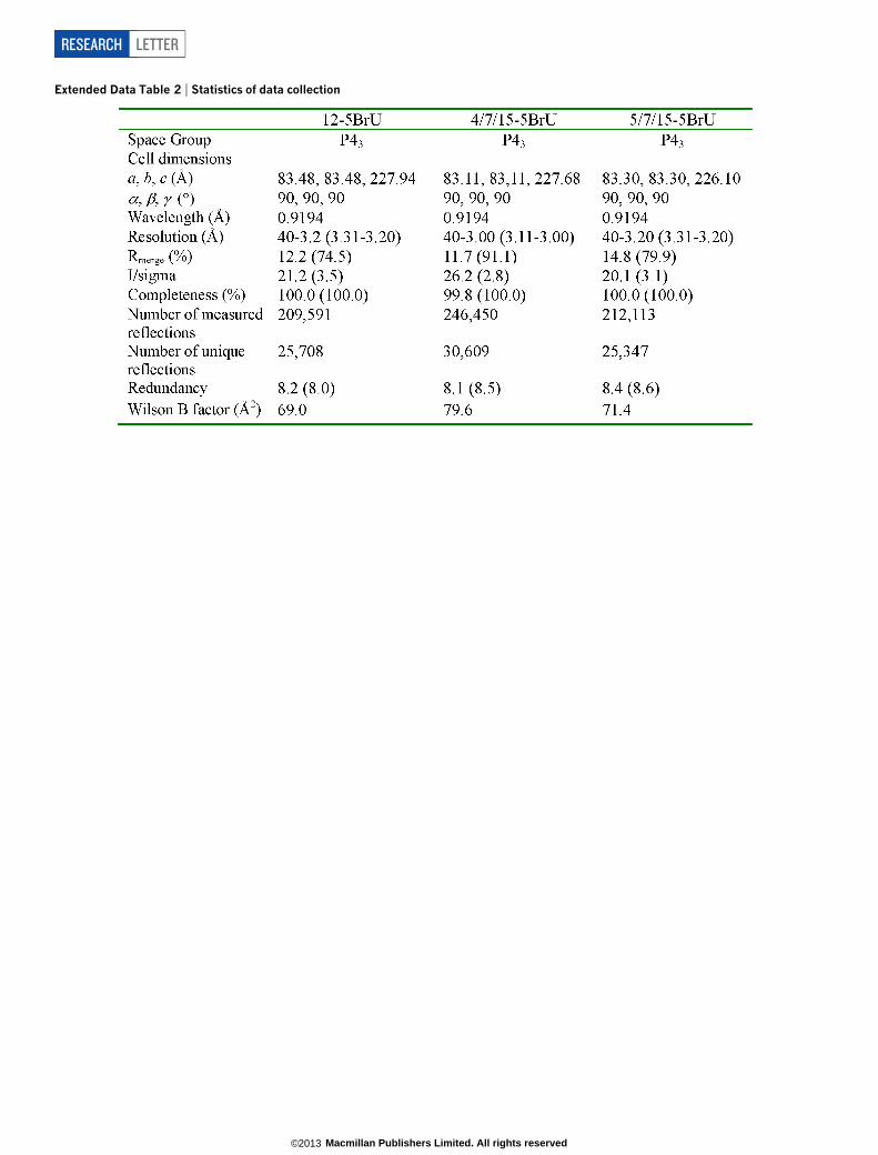
Extended Data Table 2 | Statistics of data collection
RESEARCH LETTER
Macmillan Publishers Limited. All rights reserved©2013