Embed Size (px)
Citation preview

University of Pennsylvania University of Pennsylvania
ScholarlyCommons ScholarlyCommons
Publicly Accessible Penn Dissertations
2013
Stress, Monoamines, and Cognitive Flexibility Stress, Monoamines, and Cognitive Flexibility
Kevin Snyder University of Pennsylvania, [email protected]
Follow this and additional works at: https://repository.upenn.edu/edissertations
Part of the Biology Commons, Neuroscience and Neurobiology Commons, and the Social and
Behavioral Sciences Commons
Recommended Citation Recommended Citation Snyder, Kevin, "Stress, Monoamines, and Cognitive Flexibility" (2013). Publicly Accessible Penn Dissertations. 928. https://repository.upenn.edu/edissertations/928
This paper is posted at ScholarlyCommons. https://repository.upenn.edu/edissertations/928 For more information, please contact [email protected].

Stress, Monoamines, and Cognitive Flexibility Stress, Monoamines, and Cognitive Flexibility
Abstract Abstract Stress has been implicated in psychiatric disorders that are characterized by impaired executive function, which is mediated by the prefrontal cortex (PFC). The stress-related neuropeptide, corticotropin-releasing factor (CRF) regulates monoamine systems that project to the PFC, including the locus coeruleus norepinephrine (LC-NE) system and the dorsal raphe-serotonin (DRN-5-HT) system. CRF actions on these systems may underlie cognitive symptoms of stress-related disorders. The age at which stress occurs can determine its impact, and adolescent stress has been linked to adult psychopathology. This dissertation explores the role of CRF in stress-induced modulation of the LC-NE and DRN-5-HT systems and the developmental time course of the impact of stress on PFC-dependent cognitive function using attentional set-shifting tasks, microdialysis, and immunohistochemistry. CRF microinfusion into the LC and DRN produced dose-dependent effects on distinct cognitive functions. Low doses CRF in the LC facilitated set-shifting and increased c-fos expression in the PFC. In contrast, high doses of CRF in the LC facilitated reversal learning, suggesting that mild and severe stress affect different cognitive processes through LC-PFC projections. In the DRN, CRF facilitated set-shifting at a dosage that decreased 5-HT levels in the PFC. This effect switched to facilitation of reversal learning in a defeat-resistant subpopulation of rats exposed to social stress, underscoring the importance of stress history and coping strategy in determining the impact of stress. Finally, adolescent social stress produced an enduring impairment of cognitive flexibility that was seen in adulthood and occurred selectively in rats that resisted social defeat, further reinforcing the importance of coping style in the consequences of stress. Together these studies demonstrate how CRF modulation of monoamine systems can affect cognitive flexibility in ways that are adaptive for dealing with acute stress. They also show the importance of stress history, coping style, and age at which stress occurs as determinants of the impact of stress on cognition. This research may lead to the development of novel, individualized monoamine-targeted treatments for individuals suffering from stress-related cognitive impairments that may be related to the etiology of a diverse range of psychiatric disorders.
Degree Type Degree Type Dissertation
Degree Name Degree Name Doctor of Philosophy (PhD)
Graduate Group Graduate Group Neuroscience
First Advisor First Advisor Rita J. Valentino
Keywords Keywords Corticotropin, Norepinephrine, Prefrontal Cortex, Serotonin, Set-Shifting, Stress
Subject Categories Subject Categories Biology | Neuroscience and Neurobiology | Social and Behavioral Sciences
This dissertation is available at ScholarlyCommons: https://repository.upenn.edu/edissertations/928

STRESS, MONOAMINES, AND COGNITIVE FLEXIBILITY
Kevin P. Snyder
A DISSERTATION
in
Neuroscience
Presented to the Faculties of the University of Pennsylvania
in
Partial Fulfillment of the Requirements for the
Degree of Doctor of Philosophy
2013
Supervisor of Dissertation
Signature _______________________________________
Rita J. Valentino, PhD
Professor of Stress Neurobiology, Children’s Hospital of Philadelphia
Graduate Group Chairperson
Signature _______________________________________
Josh I. Gold, PhD
Associate Professor of Neuroscience, University of Pennsylvania
Dissertation Committee
Marc F. Schmidt, PhD, Associate Professor of Biology, University of Pennsylvania
Julie A. Blendy, PhD, Professor of Pharmacology, University of Pennsylvania
Steven J. Siegel, PhD, Professor of Psychiatry, University of Pennsylvania
Vinay Parikh, PhD, Assistant Professor of Psychology, Temple University

ii
DEDICATION
For my wife Jessica,
who supported me through this long and adventurous endeavor
and for my son Dylan,
who reminds me each day that there are even better things in life than science.

iii
ACKNOWLEDGEMENTS
First, I would like to acknowledge the mentorship, guidance, and dedication of my
thesis advisor Dr. Rita Valentino. She always encouraged me to be as creative and
ambitious as possible while at the same time providing enough guidance to keep me
from flying too close to the sun. I’d also like to thank Dr. Wei-wen Wang for coming all
the way from China to work with me on the project described in Chapter 2. Additionally,
I’d like to thank the Lucki lab, particularly Irwin and Tiffany, for their assistance in the
planning and analysis of my microdialysis experiments as well as the Bhatnagar lab for
their assistance in the social stress experiments. I’d like to thank all of the members of
the Valentino lab that helped me plan and perform my experiments over the years: Kile
McFadden, Dr. Andre Curtis, Dr. Nayla Chaijale, Dr. Debbie Bangasser, Dr. Susan
Wood, Mark Barry, Julia Valenziano, Rebecca Han, Zach Plona, Herminio Guajardo,
and Jay Arner. Last but not least, I would like to thank all of my friends and family that
have supported me during my graduate program here in Philadelphia and throughout my
life.

iv
ABSTRACT
STRESS, MONOAMINES, AND COGNITIVE FLEXIBILITY
Kevin P. Snyder
Rita J. Valentino
Stress has been implicated in psychiatric disorders that are characterized by
impaired executive function, which is mediated by the prefrontal cortex (PFC). The
stress-related neuropeptide, corticotropin-releasing factor (CRF) regulates monoamine
systems that project to the PFC, including the locus coeruleus norepinephrine (LC-NE)
system and the dorsal raphe-serotonin (DRN-5-HT) system. CRF actions on these
systems may underlie cognitive symptoms of stress-related disorders. The age at which
stress occurs can determine its impact, and adolescent stress has been linked to adult
psychopathology. This dissertation explores the role of CRF in stress-induced
modulation of the LC-NE and DRN-5-HT systems and the developmental time course of
the impact of stress on PFC-dependent cognitive function using attentional set-shifting
tasks, microdialysis, and immunohistochemistry. CRF microinfusion into the LC and
DRN produced dose-dependent effects on distinct cognitive functions. Low doses CRF
in the LC facilitated set-shifting and increased c-fos expression in the PFC. In contrast,
high doses of CRF in the LC facilitated reversal learning, suggesting that mild and
severe stress affect different cognitive processes through LC-PFC projections. In the
DRN, CRF facilitated set-shifting at a dosage that decreased 5-HT levels in the PFC.
This effect switched to facilitation of reversal learning in a defeat-resistant subpopulation
of rats exposed to social stress, underscoring the importance of stress history and
coping strategy in determining the impact of stress. Finally, adolescent social stress
produced an enduring impairment of cognitive flexibility that was seen in adulthood and
occurred selectively in rats that resisted social defeat, further reinforcing the importance

v
of coping style in the consequences of stress. Together these studies demonstrate how
CRF modulation of monoamine systems can affect cognitive flexibility in ways that are
adaptive for dealing with acute stress. They also show the importance of stress history,
coping style, and age at which stress occurs as determinants of the impact of stress on
cognition. This research may lead to the development of novel, individualized
monoamine-targeted treatments for individuals suffering from stress-related cognitive
impairments that may be related to the etiology of a diverse range of psychiatric
disorders.

vi
Table of Contents
Page
DEDICATION………..……………………………………………………………………... ii
ACKNOWLEDGEMENTS…………………………………………………………………. iii
ABSTRACT…………………………………………………………………………………. v
LIST OF TABLES………………………………………………………………………….. viii
LIST OF FIGURES………...………………………………………………………………. ix
Chapter 1: Introduction……………………………………………………………………. 1
Chapter 2: Corticotropin-releasing Factor in the Norepinephrine Nucleus, Locus
Coeruleus, Facilitates Behavioral Flexibility………………………………………......... 31
Abstract……………………………………………………………………………………... 32
Introduction…………………………………………………………………………………. 33
Methods……………………………………………………………………………………... 36
Results………………………………………………………………………………………. 41
Discussion…………………………………………………………………………………... 44
Figures and Tables………………………………………………………………………… 51
Chapter 3: Corticotropin-releasing factor in the dorsal raphe nucleus promotes
behavioral flexibility: modulation by social stress history and coping strategy……… 70
Abstract……………………………………………………………………………………... 71
Introduction…………………………………………………………………………………. 72
Methods……………………………………………………………………………………... 74
Results………………………………………………………………………………………. 82
Discussion…………………………………………………………………………………... 85
Figures and Tables………………………………………………………………………… 90
vii
Chapter 4: Adolescent social stress impairs behavioral flexibility during adulthood:
relationship to coping strategy…………………………………………………………..... 103
Abstract…………………………………………………………………………………….... 104
Introduction………………………………………………………………………………….. 105
Methods……………………………………………………………………………………... 107
Results………………………………………………………………………………………. 111
Discussion…………………………………………………………………………………... 116
Figures and Tables………………………………………………………………………… 122
Supplemental Information…………………………………………………………………. 129
Chapter 5: Conclusions and Future Directions……………………………………….... 142

viii
LIST OF TABLES
Table Page
Chapter 2
Table S1. Sample AST testing protocol…………………………………………………. 60
Chapter 4
Table 1. Mean mPFC c-fos profile counts ± SEM in each experimental group……... 128
Table S1. Mean latency (sec) ± SEM to defeat for SL and LL rats in each
experimental group. ……………………………………………………………………….. 132

ix
LIST OF FIGURES
Figure Page
Chapter 1
Figure 1. Stress can impact cognition via corticotropin-releasing factor (CRF)-
mediated modulation of monoamine neurotransmitter systems………….............…... 15
Chapter 2
Figure 1. Brightfield photomicrograph of a section through the LC showing
histological verification of the bilateral injection sites…………………………………… 51
Figure 2. Region of prefrontal cortex in which immunoreactive profiles were
quantified……………………………………………………………………………………. 52
Figure 3. Intracerebroventricularly administered CRF (ng dose) impairs different
components of the AST……………………………………………………….…………… 53
Figure 4. Figure 4. Intra-LC administered CRF (ng dose) has differential effects on
components of the AST……………………………………………………………………. 54
Figure 5. Effects of intra-LC CRF (ng dose) on c-fos expression in the medial
prefrontal cortex……………………………………………………………………………. 55
Figure 6. Quantification of c-fos in the medial prefrontal cortex and orbitofrontal
cortex……………………………………………………………………………………….. 56
Figure 7. Expression of p44/42ERK in medial prefrontal cortex induced by CRF
injections into the LC……………………………………………………………………… 57
Figure S1. Comparison of the effects of different doses of CRF injected into the LC
(in) and outside of the LC (out, n=4 for each dose) on performance in different
components of the AST…………………………………………………………………… 58
Figure S2. Relationship between the number of c-fos profiles in the medial
prefrontal cortex and the number of p44/42ERK-immunoreactive neurons…………… 59

x
Figure Page
Chapter 3
Figure 1. Intra-DRN-administered CRF (30 ng) facilitates strategy set-shifting
performance………………………………………………………………………………... 90
Figure 2. Regional controls confirm that facilitation of strategy set-shifting by CRF
(30 ng) is regionally restricted to the DRN……………………………………………… 91
Figure 3. Intra-DRN administration of the selective CRF2 agonist Urocortin II did
not affect task performance……………………………………………………………… 92
Figure 4. Intra-DRN CRF (30 ng) decreased mPFC 5-HT but not DA extracellular
levels……………………………………………………………………………………….. 93
Figure 5. Prior social stress experience occludes the facilitatory effect of intra-DRN
CRF (30 ng) in LL but not SL rats……………………………………………………….. 94
Chapter 4
Figure 1. Social stress selectively impaired strategy set-shifting performance in
MA-Adult rats……………………………………………………………………………… 122
Figure 2. Social stress selectively increased strategy set-shifting perseverative
errors in Adult rats………………………………………………………………………… 123
Figure 3. The short latency (SL) coping strategy was associated with impaired
strategy set-shifting performance in EA-Adult rats……………………………………. 124
Figure 4. The short latency (SL) and long latency (LL) coping strategies were
associated with increased strategy set-shifting perseverative errors in
EA-Adult and Adult rats, respectively………………………………………………….. 125
Figure 5. Expression of c-fos in the medial prefrontal cortex (mPFC) was
differentially correlated with strategy set-shifting performance depending upon
the age of testing………………………………………………………………………… 126

1
Chapter 1: Introduction
The Physiological Stress Response
The concept of a generalizable biological stress response was first proposed by
Hans Selye in 1936 when he discovered that rats respond to repeated sublethal
encounters with various physical and chemical afflictions in a stereotypical manner
(Selye, 1936). This characteristic response was not specific to the type of affliction
employed and was referred to as the general adaptation syndrome (GAS) and consisted
of three distinct phases: alarm, resistance, and exhaustion. During the first phase, the
stimulus produces a significant physiological response characterized by decreased
immune system activity in the thymus, activation of the sympathetic nervous system, and
glucocorticoid release from the adrenal glands. The second phase ensues within days
and is characterized by enlargement of the adrenal glands and habituation of the
physiological response to the challenge. After months of repeated exposure, the
habituation fades and the animal enters the third and final phase of the chronic stress
response. Chronic stress is associated with a sensitized physiological response to a
challenge and negative physiological side effects that can lead to heart disease, ulcer
formation, psychiatric illness, or even death (Selye, 1936, 1976).
Corticotropin-Releasing Factor: Orchestrator of the Stress Response
These prolific observations initiated the search for the physiological signaling
pathways responsible for producing the GAS. The substance responsible for promoting
glucocorticoid release during the first phase and for inducing the adrenal hypertrophy
during the second phase of GAS, called adrenocorticotropic hormone (ACTH), was first
isolated from the anterior pituitary (Li et al., 1942). In 1950, Geoffrey Harris proposed
that another substance secreted by neurons in the hypothalamus into the hypophyseal
portal system is responsible for the release of ACTH from the anterior pituitary (Harris,

2
1950). It was almost thirty years before this substance, called corticotropin releasing
factor (CRF), was purified and identified as a 41 amino acid peptide by Wylie Vale (Vale
et al., 1981). Thus, CRF was considered to act as a neurohormone that is released from
the paraventricular hypothalamic nucleus into the portal system of the median eminence
where it can contact the corticotrophs of the anterior pituitary to initiate secretion of
ACTH. ACTH then goes on to initiate release of adrenal corticosterones that are
important in the metabolic and immune aspects of the stress response. Throughout the
following decade it was revealed that CRF functions not only as the neurohormone that
initiates the endocrine component of the stress response via the hypothalamic-pituitary-
adrenal (HPA) axis but also as an orchestrator of the endocrine, autonomic, immune,
and behavioral limbs of the stress response by acting as a neuromodulator that is
released synaptically in specific circuits throughout the brain (Owens and Nemeroff,
1991; Taché and Brunnhuber, 2008).
Early studies of the extra-HPA neuromodulatory actions of CRF revealed its role
in the autonomic limb of the stress response via activation of the sympatho-adrenal
medullary system (Lenz et al., 1987; Sato, 1987). Initially, CRF was found to indirectly
contribute to the immune component of the stress response as a result of its effects on
the HPA axis and the autonomic nervous system (ANS); however, CRF was later shown
to act on CRF receptors in the spleen and thymus to directly impact the immune system,
particularly via interactions with the interleukin-1 system (Sundar et al., 1990; Jain et al.,
1991; Aird et al., 1993; De Souza, 1993). HPA-independent effects of CRF on behavior
were observed in the first experiments attempting to characterize the role of CRF in the
brain, yet the specific mechanisms by which CRF is released in the brain to impact
behavior are still being actively investigated (Veldhuis and De Wied, 1984; Laryea et al.,
2012). The neuromodulatory actions of CRF are mediated through two distinct

3
receptors, CRF1 and CRF2, that are differentially distributed throughout the brain (Primus
et al., 1997; Van Pett et al., 2000). CRF1 activation has been clearly associated with
promotion of stress-response behaviors, and activation of CRF2, which was originally
thought to dampen stress sensitivity, may actually function in other stress-related
responses, including stress-associated learning (Bale and Vale, 2004; Hauger et al.,
2009).
Stress Impacts Cognition: For Better or Worse
The impact of stress on cognition is determined by several factors, such as the
severity, frequency, and controllability of the stressor. An inverted U-shaped relationship
between stress intensity and cognitive performance was originally proposed by (Yerkes
and Dodson, 1908) such that mild stress enhanced and severe stress impaired
performance of difficult cognitive tasks. Despite widespread acceptance of this theory,
proper experimental validation of this hypothesis has only recently been addressed
(Salehi et al., 2010; Schilling et al., 2013). This complex relationship between stress
intensity and cognition has only been demonstrated with respect to acute stress, as both
mild and severe chronic stressors are associated with impaired cognitive performance
(Song et al., 2006; Kasar et al., 2009). Stressor controllability has also proven to be an
important factor in the impact of acute stress on cognition such that controllable
stressors tend to facilitate cognition whereas uncontrollable stressors impair cognition
(Henderson et al., 2012). As stress-related cognitive impairments have been linked to
the etiology of affective disorders, further understanding of the mechanisms by which
stress impacts cognition may lead to the development of effective treatments for these
disorders (Beck, 2008; Clark et al., 2009; Diener et al., 2009; Marin et al., 2011; Pringle
et al., 2011).

4
CRF Impacts Cognition via its Actions on Monoamine Neurotransmitter Systems
A role for CRF in the inverted-U shaped relationship between stress cognition
has been suggested by transgenic mouse models in which cognitive deficits in spatial
learning and memory have been observed in mice that either overexpress CRF or lack
CRF1 receptors (Heinrichs et al., 1996; Contarino et al., 1999). A potential mechanism
by which stress could impact cognitive function is via CRF-mediated effects on midbrain
and brainstem nuclei that regulate the release of monoaminergic neuromodulators, e.g.,
the ventral tegmental area dopamine system (VTA-DA), the locus coeruleus
norepinephrine system (LC-NE), and the dorsal raphe nucleus serotonin system (DRN-
5-HT) (Fig. 1) (Van Bockstaele et al., 1998; Corominas et al., 2010; Valentino et al.,
2010). Each of these systems performs distinct yet cooperative roles in the mediation of
fronto-executive function (Fig. 1B) (Doya, 2008; Robbins and Arnsten, 2009). The VTA-
DA system has been characterized as a reinforcement learning signal that trains
behavior by computing and translating reward prediction errors into motivational states
(Schultz, 1998; Niv and Schoenbaum, 2008). The LC-NE system has been
characterized as an attentional filter that regulates the gain of signal for sensory inputs
based on their behavioral relevance (Aston-Jones and Cohen, 2005b; Valentino and Van
Bockstaele, 2008). The role of the DRN-5-HT system in cognition, however, has not
been so clearly characterized, largely due to the differential effects of the vast variety of
5-HT receptor subtypes. Nonetheless, it has been implicated in the suppression of
maladaptive motor behaviors, reversal learning performance, and in delayed
discounting, or choosing larger but delayed rewards over smaller immediate rewards
(Clarke et al., 2005; Dayan and Huys, 2009; Miyazaki et al., 2011).
CRF receptors are present in each of these monoaminergic nuclei and in many
of their forebrain projection areas (Contarino and Gold, 2002). CRF release into the

5
VTA dose-dependently increases the firing rate of dopaminergic neurons (Corominas et
al., 2010). In the LC CRF release generally increases tonic LC discharge rate but
decreases sensory-stimulus-evoked phasic LC discharge (Valentino et al., 1983;
Valentino and Foote, 1988). CRF release in the DRN produces a dose-dependent
biphasic modulation of serotonergic neuronal activity, due to the opposing actions of
CRF1 and CRF2 receptors in the DRN (Kirby et al., 2000; Pernar et al., 2004; Valentino
and Commons, 2005). Further studies have found that prior stress experience can bias
the CRF-response of the DRN-5-HT system toward CRF2-mediated increases in 5-HT
release by increasing the density of CRF2 receptors on neurons in the DRN (Waselus et
al., 2009). Because the actions of each of these systems are interdependent, the effects
observed in response to manipulation of one monoamine system may reflect the
combined actions of multiple monoamine systems.
The Locus Coeruleus-Norepinephrine System
The LC is a small nucleus of neurons located in the pons just lateral to the wall of
the fourth ventricle (Foote et al., 1983). The LC is a homogenous nucleus of
noradrenergic neurons that project to the spinal cord, brainstem, cerebellum,
hypothalamus, thalamus, basal telencephalon, and the entire isocortex via highly
collateralized projections (Dahlstroem and Fuxe, 1964; Moore and Bloom, 1979). In
fact, the LC has been demonstrated to be the sole source of NE to the forebrain
(Arbuthnott et al., 1970). The primary source of excitatory afferent input to the core of
the LC nuclues comes from the nucleus paragigantocellularis, and its primary souce of
inhibitory afferent input comes from the nucleus prepositus hypoglossi (Aston-Jones et
al., 1991). However, many LC neuronal dendrites extend into the peri-coerulear region
that receives afferent input from higher order structures such as the prefrontal cortex
6
(PFC), central nucleus of the amygdala, lateral hypothalamus, bed nucleus of the stria
terminalis, and the dorsal raphe nucleus (Berridge and Waterhouse, 2003).
Electrophysiological studies of LC neuronal activity have found that LC neurons
exhibit two distinct modes of activity: tonic and phasic (Aston-Jones and Cohen, 2005b).
The tonic rate of LC neuronal discharge has been correlated with behavioral arousal
(Foote et al., 1980; Aston-Jones and Bloom, 1981b). Moreover, pharmacological
manipulation of tonic LC neuronal activity has been shown to impact
electroencephalographic indices of arousal (Berridge and Foote, 1991; Berridge et al.,
1993). LC neurons respond phasically to a wide range of sensory stimuli, particularly
stimuli that elicit behavioral response (Foote et al., 1980; Aston-Jones and Bloom,
1981a; Grant et al., 1988). When the LC is in the phasic mode of activity, LC neurons
fire synchronously due to electrotonic coupling via dendritic gap junctions (Ishimatsu and
Williams, 1996). High tonic LC activity has been associated with decoupling of LC
neurons and less efficient phasic activity (Usher et al., 1999; Aston-Jones et al., 2000).
The widely distributed axonal network of the LC positions it to provide tonic and phasic
arousal-related signals to the forebrain that have been suggested to direct attention
toward behaviorally relevant sensory information (Aston-Jones and Cohen, 2005a).
It was originally thought that the primary role of the LC-NE system was to
regulate arousal and sleep-wake cycles (Berridge et al., 2012). However, the two
distinct modes of LC activity (i.e. tonic and phasic) suggest a more nuanced role in the
cognitive processing of relevant sensory information (Aston-Jones and Cohen, 2005a).
Phasic firing of LC neurons specifically to behavioral task relevant sensory stimuli
mediate focused attention and optimal task performance, yet when rewards associated
with task performance according to the current attentional strategy wane, LC activity
switches to a high tonic mode until a more favorable strategy can be ascertained. This

7
theory has led to the testing of several hypotheses concerning the role of the LC-NE
system in higher order cognition. In general, the LC-NE system has been implicated in
the ability to detect and exploit changes in the relevance of sensory information (Lapiz
and Morilak, 2006; Tait et al., 2007).
Stress-Induced Modulation of Cognition via the LC-NE System
Exposure to physical stressors (i.e. hypotensive stress) or psychological
stressors (predator odor) biases LC activity toward a high tonic state and away from a
phasic mode of firing (Valentino and Wehby, 1988; Curtis et al., 2012). This is thought
to be an adaptive response to stressful situations by promoting scanning of the
environment for new strategies rather than focusing on previously relevant stimuli
(Valentino and Van Bockstaele, 2008). Local infusion of CRF into the LC mimics the
effects of stress on LC activity, and the peri-coerulear region where LC neuronal
dendrites extend is densely innervated by CRF-immunoreactive fibers, suggesting that
endogenous CRF is a mediator of the effects of stress on LC activity (Valentino and
Foote, 1988; Valentino et al., 1992). Endogenous CRF does not appear to be tonically
released in unstressful situations as intra-LC administration of CRF antagonists does not
impact LC neuronal activity in animals at rest (Page and Abercrombie, 1999). Although
some in situ hybridization studies have failed to detect the presence of CRF receptor
mRNA in the LC, autoradiography and electrophysiological studies suggest that the
effects of CRF in the LC are mediated by CRF1 (Schulz et al., 1996; Sánchez et al.,
1999; Van Pett et al., 2000; Jedema and Grace, 2004). Few studies have investigated
the impact of intra-LC CRF on behavior. One study found that intra-LC infusion of a
large dose of CRF (100 ng) has been associated with an increase in fear-related
behaviors (Butler et al., 1990). Although it has been theorized that stress-induced tonic

8
activation of the LC-NE system would facilitate behavioral flexibility, this hypothesis has
not been empirically tested (Valentino and Van Bockstaele, 2008).
The Dorsal Raphe Nucleus-Serotonin System
The DRN is a midline midbrain structure, specifically located in the ventral part of
the periaqueductal gray, that extends rostrally to the occulomotor nuclei and caudally to
dorsal border of the median raphe (Jacobs and Azmitia, 1992). The DRN is
heterogeneous nucleus that is generally subdivided into three anatomically distinct
regions: ventromedial, dorsomedial, and the lateral wings (Jacobs and Azmitia, 1992).
Serotonergic neurons can be found most abundantly in the ventromedial region and with
the least abundance in the lateral wings (Michelsen et al., 2007) whereas GABAergic
neurons are located primarily in the lateral wings (Stamp and Semba, 1995). In general,
the rostral DRN projects most strongly to higher order limbic structures, e.g. cortex and
the amygdala, and to the other raphe nuclei while the caudal DRN projects most strongly
to lower order limbic structures, e.g. hippocampus and lateral septum, and the
noradrenergic nucleus, and the locus coeruleus (Vertes, 1991; Vertes and Kocsis,
1994). The DRN also receives information from and relays information to a wide array of
neural structures in a topographically organized fashion (Peyron et al., 1996; Peyron et
al., 1998).
Initial electrophysiological recordings from serotonergic neurons in the DRN of
awake-behaving cats identified serotonergic neurons whose firing correlates directly with
the animal’s degree of behavioral arousal (McGinty and Harper, 1976). Further
electrophysiological examination of the DRN revealed that these cells fire with an
extremely regular clock-like pattern of activity that displayed very little phasic response
to salient sensory stimuli (Trulson and Jacobs, 1979; Rasmussen et al., 1986). This
data suggests that the DRN is positioned well for and theoretically capable of providing a

9
slow, behaviorally relevant, tonic signal throughout the brain that may be responsible for
network-level coordination of the complex neurological processes in which 5-HT has
been implicated, such as mood and cognition.
Serotonin has been implicated in numerous neurobehavioral functions, including
sleeping, eating, sex, cognition, and emotion (Dayan and Huys, 2009; Mendelsohn et al.,
2009; Guptarak et al., 2010; Halford et al., 2011; Monti, 2011) and has been implicated
in the pathophysiology of several stress-related psychiatric disorders, such as
depression, anxiety, schizophrenia, and obsessive-compulsive disorder (Stein and Stahl,
2000; Goddard et al., 2008; Chertkow et al., 2009; López-Muñoz and Alamo, 2009). A
unified theory of serotonergic function has not and may not ever be resolved, largely due
to the incredible diversity in 5-HT receptor subtype and distribution throughout the brain
(Pytliak et al., 2011). Behaviorally, depletion of 5-HT has been associated with
increased impulsivity and less willingness to wait for larger rewards when given the
option of smaller more immediate rewards (Cardinal, 2006). A role for 5-HT in cognitive
flexibility has been suggested by 5-HT depletion experiments, in which subjects display
selective impairments in reversal learning tasks (Clarke et al., 2005; Lapiz-Bluhm et al.,
2009).
Stress-Induced Modulation of Cognition via the DRN-5-HT System
The innervation of the DRN by CRF-immunoractive terminals is topographically
organized throughout rostrocaudal axis such that it is densest in the rostral ventromedial
DRN and caudal lateral wings (Valentino et al., 2001). Interestingly, serotonergic
neurons are most prevalent in the caudal ventromedial DRN and least prevalent in the
lateral wings while GABAergic neurons are most prevalent in the lateral wings.
Additionally, ultrastructural analysis of CRF terminals in the DRN using electron
microscopy has revealed that CRF terminals are much more likely to contact GABAergic

10
dendrites than serotonergic dendrites, especially in the lateral wings (Waselus et al.,
2005). Furthermore, examination of neuronal activity in the DRN via c-fos expression
evoked by swim stress experience in rats revealed that nearly all of the neurons
activated by swim stress were GABAergic and most were localized to the lateral wings
(Roche et al., 2003). These data suggest that endogenous CRF release in the DRN
primarily targets GABAergic neurons.
A series of electrophysiological and microdialysis experiments have shown that
CRF bidirectionally regulates the activity of serotonergic neurons, presumably via the
differential activation of CRF1 and/or CRF2 receptors on GABAergic interneurons that
project to serotonergic neurons within the DRN (Valentino and Commons, 2005). Intra-
DRN injections of low doses of CRF that preferentially activate CRF1 have been shown
to decrease the firing rate of serotonergic neurons and decrease 5-HT release in lateral
septum and striatum (Kirby et al., 2000; Price and Lucki, 2001). In contrast, intra-DRN
injection of higher doses of both CRF and the selective CRF2 agonist, urocortin II, were
both shown to increase the firing rate of serotonergic neurons within the DRN (Kirby et
al., 2000; Pernar et al., 2004). The time course of the electrophysiological effects
observed in these studies persisted on the order of minutes while the effects of 5-HT
release were persistent on the order of hours. Swim stress has been shown to produce
a CRF-dependent decrease in lateral septal 5-HT release on the order of hours,
suggesting that intra-DRN injections of CRF may accurately model endogenous CRF
release in the DRN (Price et al., 2002). This bidirectional regulation of the DRN-5-HT
system by CRF may mediate some of the diverse effects of stress on 5-HT-related
neurological processes such as sleep, mood, and cognition.
The anatomy and physiology of both the CRF and DRN-5-HT systems suggest
that of these systems may be involved in the cognitive integration of salient sensory

11
information with motivationally relevant behavioral contingencies via interactions with
sensory cortices and relay nuclei as well as limbic reward areas such as the amygdala,
basal ganglia, and PFC. There is particularly strong anatomical and electrophysiological
evidence that the effects of stress on the DR-5-HT system may be mediated CRF
(Valentino et al., 2001; Valentino and Commons, 2005). Stress-related deficits in
cognitive flexibility have been linked to dysregulation of the serotonergic tone, yet no
study has clearly examined the role that CRF-mediated effects of stress on the DR-5-HT
system may play in the neural mechanisms underlying these cognitive deficits (Lapiz-
Bluhm et al., 2009; Furr et al., 2012).
Behavioral and Physiological Impact of Stress Throughout Adolescent Development
Adolescence is a period or intense developmental change at the hormonal and
neurological level during which the physiological impact of stress is exaggerated (van
Eden et al., 1990; Giedd et al., 1999; Gunnar et al., 2009; Lupien et al., 2009). For
example, the HPA axis response to stress is heightened during adolescence and does
not habituate to chronic stress in the same manner as it does during adulthood (Romeo
et al., 2006; Gunnar et al., 2009). A study investigating the impact of social stress during
adolescence on the LC-NE system found behavioral and electrophysiological evidence
of CRF-mediated increased noradrenergic activity in early adolescent rats (Bingham et
al., 2011). The cognitive impact of adolescent chronic stress experience is typically less
pronounced immediately after the stress, but is expressed as changes in behavior or
cognitive function during adulthood (McCormick and Mathews, 2010).
Stress experience during adolescence can produce enduring effects into
adulthood and has been strongly linked to the development of psychiatric disorders in
adulthood (Halligan et al., 2007; Paus et al., 2008). Consistent with this, chronic variable
stress in pre-pubertal animals impaired a hippocampal memory task and increased the

12
expression of anxiogenic and depressive-like behaviors in adulthood (Isgor et al., 2004;
Tsoory et al., 2007). Social stress during adolescence has also been shown to decrease
defensive and social interaction behaviors in adult animals that were stressed during
adolescence (Vidal et al., 2007; Bingham et al., 2011). To date, no studies have
investigated the short or long-term effects of adolescent stress on PFC-dependent
cognitive flexibility.
Assessment of the Impact of Stress via PFC-Mediated Cognitive Flexibility
The PFC is sensitive to both acute and chronic stress experience and mediates
several stress-sensitive forms of cognition, such as cognitive flexibility (Arnsten, 2009).
PFC-dependent tasks such as the attentional set-shifting task and operant strategy set-
shifting task have been developed to assess PFC-mediated cognitive flexibility in
rodents (Birrell and Brown, 2000; Floresco et al., 2008). The regulation of the PFC by
monoamines has been extensively studied using these tasks, and certain forms of
cognition have been associated with the role of particular monoamine neurotransmitters
in specific regions of the PFC (Robbins and Arnsten, 2009). For example, 5-HT in the
orbitofrontal cortex has been implicated in reversal learning whereas NE in the medial
prefrontal cortex (mPFC) has been implicated in set-shifting ability (Lapiz and Morilak,
2006; Tait et al., 2007; Lapiz-Bluhm et al., 2009; Bondi et al., 2010; Furr et al., 2012).
Chronic stress has been associated with impairments in either reversal learning or set-
shifting ability, depending upon the nature of the stressor (Bondi et al., 2008; Lapiz-
Bluhm et al., 2009). These impairments can prevented by antidepressant treatment with
either serotonin or norepinephrine reuptake inhibitors during chronic stress experience
and can also be alleviated by antidepressant treatment after chronic stress experience
(Bondi et al., 2008; Lapiz-Bluhm et al., 2009; Danet et al., 2010; Naegeli et al., 2013).
Acute stress has been associated with facilitation of reversal learning and impairment of

13
set-shifting ability, yet the mechanisms underlying these effects have yet to be
elucidated (Butts et al., 2013; Thai et al., 2013).
Given that the LC-NE and DRN-5-HT systems have been strongly implicated in
effects of chronic stress experience on cognitive flexibility and are well positioned to
mediate the cognitive impact of acute stress, Chapters 2 and 3 explored the acute
effects of CRF on the LC-NE and DRN-5-HT systems in the context of cognitive
flexibility. Local administration of low but not high doses of CRF in either the LC or the
DRN facilitated set-shifting performance via moderate CRF1-mediated increased NE
release and decreased 5-HT release, respectively. Furthermore, intra-LC CRF produced
an inverted U-shaped effect on expression of the immediate early gene c-fos, and
increased mPFC c-fos expression was associated improved set-shifting performance.
Administration of higher doses of CRF in the LC facilitated reversal learning and
produced no cognitive effects in the DRN. Additionally, intra-DRN administration of the
low dose of CRF that facilitated set-shifting performance in stress-naïve rats produced a
facilitation of reversal learning specifically in rats with a defeat resistant coping strategy
to social stress that has been associated with an increase in the ratio of CRF2:CRF1 on
the cell membrane of neurons in the DRN. Taken together, these chapters suggest that
the actions of CRF in the LC and DRN may facilitate set-shifting ability in response to a
mild acute stressor and may facilitate reversal learning in response to a severe stressor
or a mild stressor in individuals with social stress experience that employ a defeat-
resistant stress coping strategy.
Chapter 4 further explored the impact of social stress experience and coping
strategy on cognitive flexibility throughout development. Social stress during
adolescence did not immediately impact cognitive flexibility but did produce protracted
effects that became evident during adulthood. Rats that were exposed to social stress

14
during mid-adolescence displayed impaired set-shifting ability during adulthood,
regardless of their stress coping strategy. In contrast, only rats that displayed a
submissive coping strategy in response to social stress during early adolescence
displayed impaired set-shifting during adulthood. When rats were stressed and
cognitively evaluated during adulthood a subtle cognitive impairment was observed in
set-shifting performance such that stress rats committed more perseverative errors
without requiring more trials to reach the learning criterion. This effect was found to be
associated specifically with the defeat-resistant coping strategy. This study suggests
that social stress during adolescence can produce impairments in cognitive flexibility that
do not fully manifest until adulthood and that the impact of social stress on cognitive
flexibility is differentially dependent upon stress coping strategy throughout development.
This body of work may lead to the development of CRF and monoamine-targeted
pharmacotherapies alongside the development of adaptive cognitive-behavioral stress
coping therapies for the treatment stress-related psychiatric disorders throughout
development.

15
Figure 1. Stress can impact cognition via corticotropin-releasing factor (CRF)-mediated
modulation of monoamine neurotransmitter systems. A) Schematic diagram suggesting

16
a potential mechanism by which stress can alter cognitive function via the actions of
CRF on monoamine neuromodulator systems that project the prefrontal cortex. B) Each
monoamine neurotrasmitter system (i.e. the locus coeruleus-norepinephrine (LC-NE)
system, the dorsal raphe nucleus-serotonin (DRN-5-HT) system, and the ventral
tegmental area-dopamine (VTA-DA) system) has a distinct role in cognition that can be
modulated by stress-induced CRF release.

17
References
Aird F, Clevenger CV, Prystowsky MB, Redei E (1993) Corticotropin-releasing factor
mRNA in rat thymus and spleen. Proc Natl Acad Sci U S A 90:7104-7108.
Arbuthnott GW, Crow TJ, Fuxe K, Olson L, Ungerstedt U (1970) Depletion of
catecholamines in vivo induced by electrical stimulation of central monoamine
pathways. Brain Res 24:471-483.
Arnsten AF (2009) Stress signalling pathways that impair prefrontal cortex structure and
function. Nat Rev Neurosci 10:410-422.
Aston-Jones G, Bloom FE (1981a) Norepinephrine-containing locus coeruleus neurons
in behaving rats exhibit pronounced responses to non-noxious environmental
stimuli. J Neurosci 1:887-900.
Aston-Jones G, Bloom FE (1981b) Activity of norepinephrine-containing locus coeruleus
neurons in behaving rats anticipates fluctuations in the sleep-waking cycle. J
Neurosci 1:876-886.
Aston-Jones G, Cohen JD (2005a) An integrative theory of locus coeruleus-
norepinephrine function: adaptive gain and optimal performance. Annu Rev
Neurosci 28:403-450.
Aston-Jones G, Cohen JD (2005b) Adaptive gain and the role of the locus coeruleus-
norepinephrine system in optimal performance. J Comp Neurol 493:99-110.
Aston-Jones G, Rajkowski J, Cohen J (2000) Locus coeruleus and regulation of
behavioral flexibility and attention. Prog Brain Res 126:165-182.
Aston-Jones G, Shipley MT, Chouvet G, Ennis M, van Bockstaele E, Pieribone V,
Shiekhattar R, Akaoka H, Drolet G, Astier B (1991) Afferent regulation of locus
coeruleus neurons: anatomy, physiology and pharmacology. Prog Brain Res
88:47-75.

18
Bale TL, Vale WW (2004) CRF and CRF receptors: role in stress responsivity and other
behaviors. Annu Rev Pharmacol Toxicol 44:525-557.
Beck AT (2008) The evolution of the cognitive model of depression and its
neurobiological correlates. Am J Psychiatry 165:969-977.
Berridge CW, Foote SL (1991) Effects of locus coeruleus activation on
electroencephalographic activity in neocortex and hippocampus. J Neurosci
11:3135-3145.
Berridge CW, Waterhouse BD (2003) The locus coeruleus-noradrenergic system:
modulation of behavioral state and state-dependent cognitive processes. Brain
Res Brain Res Rev 42:33-84.
Berridge CW, Schmeichel BE, España RA (2012) Noradrenergic modulation of
wakefulness/arousal. Sleep Med Rev 16:187-197.
Berridge CW, Page ME, Valentino RJ, Foote SL (1993) Effects of locus coeruleus
inactivation on electroencephalographic activity in neocortex and hippocampus.
Neuroscience 55:381-393.
Bingham B, McFadden K, Zhang X, Bhatnagar S, Beck S, Valentino R (2011) Early
adolescence as a critical window during which social stress distinctly alters
behavior and brain norepinephrine activity. Neuropsychopharmacology 36:896-
909.
Birrell JM, Brown VJ (2000) Medial frontal cortex mediates perceptual attentional set
shifting in the rat. J Neurosci 20:4320-4324.
Bondi CO, Jett JD, Morilak DA (2010) Beneficial effects of desipramine on cognitive
function of chronically stressed rats are mediated by alpha1-adrenergic receptors
in medial prefrontal cortex. Prog Neuropsychopharmacol Biol Psychiatry 34:913-
923.

19
Bondi CO, Rodriguez G, Gould GG, Frazer A, Morilak DA (2008) Chronic unpredictable
stress induces a cognitive deficit and anxiety-like behavior in rats that is
prevented by chronic antidepressant drug treatment. Neuropsychopharmacology
33:320-331.
Butler PD, Weiss JM, Stout JC, Nemeroff CB (1990) Corticotropin-releasing factor
produces fear-enhancing and behavioral activating effects following infusion into
the locus coeruleus. J Neurosci 10:176-183.
Butts KA, Floresco SB, Phillips AG (2013) Acute stress impairs set-shifting but not
reversal learning. Behav Brain Res 252:222-229.
Cardinal RN (2006) Neural systems implicated in delayed and probabilistic
reinforcement. Neural Netw 19:1277-1301.
Chertkow Y, Weinreb O, Youdim MB, Silver H (2009) Molecular mechanisms underlying
synergistic effects of SSRI-antipsychotic augmentation in treatment of negative
symptoms in schizophrenia. J Neural Transm 116:1529-1541.
Clark L, Chamberlain SR, Sahakian BJ (2009) Neurocognitive mechanisms in
depression: implications for treatment. Annu Rev Neurosci 32:57-74.
Clarke HF, Walker SC, Crofts HS, Dalley JW, Robbins TW, Roberts AC (2005)
Prefrontal serotonin depletion affects reversal learning but not attentional set
shifting. J Neurosci 25:532-538.
Contarino A, Gold LH (2002) Targeted mutations of the corticotropin-releasing factor
system: effects on physiology and behavior. Neuropeptides 36:103-116.
Contarino A, Dellu F, Koob GF, Smith GW, Lee KF, Vale W, Gold LH (1999) Reduced
anxiety-like and cognitive performance in mice lacking the corticotropin-releasing
factor receptor 1. Brain Res 835:1-9.

20
Corominas M, Roncero C, Casas M (2010) Corticotropin releasing factor and
neuroplasticity in cocaine addiction. Life Sci 86:1-9.
Curtis AL, Leiser SC, Snyder K, Valentino RJ (2012) Predator stress engages
corticotropin-releasing factor and opioid systems to alter the operating mode of
locus coeruleus norepinephrine neurons. Neuropharmacology 62:1737-1745.
Dahlstroem A, Fuxe K (1964) Evidence for the existence of monoamine-containing
neurons in the central nervous system. I. Demonstration of monoamines in the
cell bodies of brain stem neurons. Acta Physiol Scand Suppl:SUPPL 232:231-
255.
Danet M, Lapiz-Bluhm S, Morilak DA (2010) A cognitive deficit induced in rats by chronic
intermittent cold stress is reversed by chronic antidepressant treatment. Int J
Neuropsychopharmacol 13:997-1009.
Dayan P, Huys QJ (2009) Serotonin in affective control. Annu Rev Neurosci 32:95-126.
De Souza EB (1993) Corticotropin-releasing factor and interleukin-1 receptors in the
brain-endocrine-immune axis. Role in stress response and infection. Ann N Y
Acad Sci 697:9-27.
Diener C, Kuehner C, Brusniak W, Struve M, Flor H (2009) Effects of stressor
controllability on psychophysiological, cognitive and behavioural responses in
patients with major depression and dysthymia. Psychol Med 39:77-86.
Doya K (2008) Modulators of decision making. Nat Neurosci 11:410-416.
Floresco SB, Block AE, Tse MT (2008) Inactivation of the medial prefrontal cortex of the
rat impairs strategy set-shifting, but not reversal learning, using a novel,
automated procedure. Behav Brain Res 190:85-96.

21
Foote SL, Aston-Jones G, Bloom FE (1980) Impulse activity of locus coeruleus neurons
in awake rats and monkeys is a function of sensory stimulation and arousal. Proc
Natl Acad Sci U S A 77:3033-3037.
Foote SL, Bloom FE, Aston-Jones G (1983) Nucleus locus ceruleus: new evidence of
anatomical and physiological specificity. Physiol Rev 63:844-914.
Furr A, Lapiz-Bluhm MD, Morilak DA (2012) 5-HT2A receptors in the orbitofrontal cortex
facilitate reversal learning and contribute to the beneficial cognitive effects of
chronic citalopram treatment in rats. Int J Neuropsychopharmacol 15:1295-1305.
Giedd JN, Blumenthal J, Jeffries NO, Castellanos FX, Liu H, Zijdenbos A, Paus T, Evans
AC, Rapoport JL (1999) Brain development during childhood and adolescence: a
longitudinal MRI study. Nat Neurosci 2:861-863.
Goddard AW, Shekhar A, Whiteman AF, McDougle CJ (2008) Serotoninergic
mechanisms in the treatment of obsessive-compulsive disorder. Drug Discov
Today 13:325-332.
Grant SJ, Aston-Jones G, Redmond DE (1988) Responses of primate locus coeruleus
neurons to simple and complex sensory stimuli. Brain Res Bull 21:401-410.
Gunnar MR, Wewerka S, Frenn K, Long JD, Griggs C (2009) Developmental changes in
hypothalamus-pituitary-adrenal activity over the transition to adolescence:
normative changes and associations with puberty. Dev Psychopathol 21:69-85.
Guptarak J, Sarkar J, Hiegel C, Uphouse L (2010) Role of 5-HT(1A) receptors in
fluoxetine-induced lordosis inhibition. Horm Behav 58:290-296.
Halford JC, Boyland EJ, Lawton CL, Blundell JE, Harrold JA (2011) Serotonergic anti-
obesity agents: past experience and future prospects. Drugs 71:2247-2255.

22
Halligan SL, Herbert J, Goodyer I, Murray L (2007) Disturbances in morning cortisol
secretion in association with maternal postnatal depression predict subsequent
depressive symptomatology in adolescents. Biol Psychiatry 62:40-46.
Harris GW (1950) The hypothalamus and endocrine glands. Br Med Bull 6:345-350.
Hauger RL, Risbrough V, Oakley RH, Olivares-Reyes JA, Dautzenberg FM (2009) Role
of CRF receptor signaling in stress vulnerability, anxiety, and depression. Ann N
Y Acad Sci 1179:120-143.
Heinrichs SC, Stenzel-Poore MP, Gold LH, Battenberg E, Bloom FE, Koob GF, Vale
WW, Pich EM (1996) Learning impairment in transgenic mice with central
overexpression of corticotropin-releasing factor. Neuroscience 74:303-311.
Henderson RK, Snyder HR, Gupta T, Banich MT (2012) When does stress help or
harm? The effects of stress controllability and subjective stress response on
stroop performance. Front Psychol 3:179.
Isgor C, Kabbaj M, Akil H, Watson SJ (2004) Delayed effects of chronic variable stress
during peripubertal-juvenile period on hippocampal morphology and on cognitive
and stress axis functions in rats. Hippocampus 14:636-648.
Ishimatsu M, Williams JT (1996) Synchronous activity in locus coeruleus results from
dendritic interactions in pericoerulear regions. J Neurosci 16:5196-5204.
Jacobs BL, Azmitia EC (1992) Structure and function of the brain serotonin system.
Physiol Rev 72:165-229.
Jain R, Zwickler D, Hollander CS, Brand H, Saperstein A, Hutchinson B, Brown C,
Audhya T (1991) Corticotropin-releasing factor modulates the immune response
to stress in the rat. Endocrinology 128:1329-1336.

23
Jedema HP, Grace AA (2004) Corticotropin-releasing hormone directly activates
noradrenergic neurons of the locus ceruleus recorded in vitro. J Neurosci
24:9703-9713.
Kasar M, Mengi M, Yildirim EA, Yurdakos E (2009) Different effects of tianeptine
pretreatment in rats exposed to acute stress and repeated severe stress.
Methods Find Exp Clin Pharmacol 31:157-163.
Kirby LG, Rice KC, Valentino RJ (2000) Effects of corticotropin-releasing factor on
neuronal activity in the serotonergic dorsal raphe nucleus.
Neuropsychopharmacology 22:148-162.
Lapiz MD, Morilak DA (2006) Noradrenergic modulation of cognitive function in rat
medial prefrontal cortex as measured by attentional set shifting capability.
Neuroscience 137:1039-1049.
Lapiz-Bluhm MD, Soto-Piña AE, Hensler JG, Morilak DA (2009) Chronic intermittent cold
stress and serotonin depletion induce deficits of reversal learning in an
attentional set-shifting test in rats. Psychopharmacology (Berl) 202:329-341.
Laryea G, Arnett MG, Muglia LJ (2012) Behavioral Studies and Genetic Alterations in
Corticotropin-Releasing Hormone (CRH) Neurocircuitry: Insights into Human
Psychiatric Disorders. Behav Sci (Basel) 2:135-171.
Lenz HJ, Raedler A, Greten H, Brown MR (1987) CRF initiates biological actions within
the brain that are observed in response to stress. Am J Physiol 252:R34-39.
Li CH, Simpson ME, Evans HM (1942) Isolation of adrenocorticotropic hormone from
sheep pituitaries. Science 96:450.
Lupien SJ, McEwen BS, Gunnar MR, Heim C (2009) Effects of stress throughout the
lifespan on the brain, behaviour and cognition. Nat Rev Neurosci 10:434-445.

24
López-Muñoz F, Alamo C (2009) Monoaminergic neurotransmission: the history of the
discovery of antidepressants from 1950s until today. Curr Pharm Des 15:1563-
1586.
Marin MF, Lord C, Andrews J, Juster RP, Sindi S, Arsenault-Lapierre G, Fiocco AJ,
Lupien SJ (2011) Chronic stress, cognitive functioning and mental health.
Neurobiol Learn Mem 96:583-595.
McCormick CM, Mathews IZ (2010) Adolescent development, hypothalamic-pituitary-
adrenal function, and programming of adult learning and memory. Prog
Neuropsychopharmacol Biol Psychiatry 34:756-765.
McGinty DJ, Harper RM (1976) Dorsal raphe neurons: depression of firing during sleep
in cats. Brain Res 101:569-575.
Mendelsohn D, Riedel WJ, Sambeth A (2009) Effects of acute tryptophan depletion on
memory, attention and executive functions: a systematic review. Neurosci
Biobehav Rev 33:926-952.
Michelsen KA, Schmitz C, Steinbusch HW (2007) The dorsal raphe nucleus--from silver
stainings to a role in depression. Brain Res Rev 55:329-342.
Miyazaki K, Miyazaki KW, Doya K (2011) Activation of dorsal raphe serotonin neurons
underlies waiting for delayed rewards. J Neurosci 31:469-479.
Monti JM (2011) Serotonin control of sleep-wake behavior. Sleep Med Rev 15:269-281.
Moore RY, Bloom FE (1979) Central catecholamine neuron systems: anatomy and
physiology of the norepinephrine and epinephrine systems. Annu Rev Neurosci
2:113-168.
Naegeli KJ, O'Connor JA, Banerjee P, Morilak DA (2013) Effects of milnacipran on
cognitive flexibility following chronic stress in rats. Eur J Pharmacol 703:62-66.

25
Niv Y, Schoenbaum G (2008) Dialogues on prediction errors. Trends Cogn Sci 12:265-
272.
Owens MJ, Nemeroff CB (1991) Physiology and pharmacology of corticotropin-releasing
factor. Pharmacol Rev 43:425-473.
Page ME, Abercrombie ED (1999) Discrete local application of corticotropin-releasing
factor increases locus coeruleus discharge and extracellular norepinephrine in rat
hippocampus. Synapse 33:304-313.
Paus T, Keshavan M, Giedd JN (2008) Why do many psychiatric disorders emerge
during adolescence? Nat Rev Neurosci 9:947-957.
Pernar L, Curtis AL, Vale WW, Rivier JE, Valentino RJ (2004) Selective activation of
corticotropin-releasing factor-2 receptors on neurochemically identified neurons
in the rat dorsal raphe nucleus reveals dual actions. J Neurosci 24:1305-1311.
Peyron C, Luppi PH, Fort P, Rampon C, Jouvet M (1996) Lower brainstem
catecholamine afferents to the rat dorsal raphe nucleus. J Comp Neurol 364:402-
413.
Peyron C, Petit JM, Rampon C, Jouvet M, Luppi PH (1998) Forebrain afferents to the rat
dorsal raphe nucleus demonstrated by retrograde and anterograde tracing
methods. Neuroscience 82:443-468.
Price ML, Lucki I (2001) Regulation of serotonin release in the lateral septum and
striatum by corticotropin-releasing factor. J Neurosci 21:2833-2841.
Price ML, Kirby LG, Valentino RJ, Lucki I (2002) Evidence for corticotropin-releasing
factor regulation of serotonin in the lateral septum during acute swim stress:
adaptation produced by repeated swimming. Psychopharmacology (Berl)
162:406-414.

26
Primus RJ, Yevich E, Baltazar C, Gallager DW (1997) Autoradiographic localization of
CRF1 and CRF2 binding sites in adult rat brain. Neuropsychopharmacology
17:308-316.
Pringle A, Browning M, Cowen PJ, Harmer CJ (2011) A cognitive neuropsychological
model of antidepressant drug action. Prog Neuropsychopharmacol Biol
Psychiatry 35:1586-1592.
Pytliak M, Vargová V, Mechírová V, Felšöci M (2011) Serotonin receptors - from
molecular biology to clinical applications. Physiol Res 60:15-25.
Rasmussen K, Strecker RE, Jacobs BL (1986) Single unit response of noradrenergic,
serotonergic and dopaminergic neurons in freely moving cats to simple sensory
stimuli. Brain Res 369:336-340.
Robbins TW, Arnsten AF (2009) The neuropsychopharmacology of fronto-executive
function: monoaminergic modulation. Annu Rev Neurosci 32:267-287.
Roche M, Commons KG, Peoples A, Valentino RJ (2003) Circuitry underlying regulation
of the serotonergic system by swim stress. J Neurosci 23:970-977.
Romeo RD, Bellani R, Karatsoreos IN, Chhua N, Vernov M, Conrad CD, McEwen BS
(2006) Stress history and pubertal development interact to shape hypothalamic-
pituitary-adrenal axis plasticity. Endocrinology 147:1664-1674.
Salehi B, Cordero MI, Sandi C (2010) Learning under stress: the inverted-U-shape
function revisited. Learn Mem 17:522-530.
Sato A (1987) Neural mechanisms of somatic sensory regulation of catecholamine
secretion from the adrenal gland. Adv Biophys 23:39-80.
Schilling TM, Kölsch M, Larra MF, Zech CM, Blumenthal TD, Frings C, Schächinger H
(2013) For whom the bell (curve) tolls: Cortisol rapidly affects memory retrieval

27
by an inverted U-shaped dose-response relationship. Psychoneuroendocrinology
38:1565-1572.
Schultz W (1998) Predictive reward signal of dopamine neurons. J Neurophysiol 80:1-
27.
Schulz DW, Mansbach RS, Sprouse J, Braselton JP, Collins J, Corman M, Dunaiskis A,
Faraci S, Schmidt AW, Seeger T, Seymour P, Tingley FD, Winston EN, Chen YL,
Heym J (1996) CP-154,526: a potent and selective nonpeptide antagonist of
corticotropin releasing factor receptors. Proc Natl Acad Sci U S A 93:10477-
10482.
Selye H (1936) A syndrome produced by diverse nocuous agents. J Neuropsychiatry
Clin Neurosci 10:230-231.
Selye H (1976) Forty years of stress research: principal remaining problems and
misconceptions. Can Med Assoc J 115:53-56.
Song L, Che W, Min-Wei W, Murakami Y, Matsumoto K (2006) Impairment of the spatial
learning and memory induced by learned helplessness and chronic mild stress.
Pharmacol Biochem Behav 83:186-193.
Stamp JA, Semba K (1995) Extent of colocalization of serotonin and GABA in the
neurons of the rat raphe nuclei. Brain Res 677:39-49.
Stein DJ, Stahl S (2000) Serotonin and anxiety: current models. Int Clin
Psychopharmacol 15 Suppl 2:S1-6.
Sundar SK, Cierpial MA, Kilts C, Ritchie JC, Weiss JM (1990) Brain IL-1-induced
immunosuppression occurs through activation of both pituitary-adrenal axis and
sympathetic nervous system by corticotropin-releasing factor. J Neurosci
10:3701-3706.

28
Sánchez MM, Young LJ, Plotsky PM, Insel TR (1999) Autoradiographic and in situ
hybridization localization of corticotropin-releasing factor 1 and 2 receptors in
nonhuman primate brain. J Comp Neurol 408:365-377.
Taché Y, Brunnhuber S (2008) From Hans Selye's discovery of biological stress to the
identification of corticotropin-releasing factor signaling pathways: implication in
stress-related functional bowel diseases. Ann N Y Acad Sci 1148:29-41.
Tait DS, Brown VJ, Farovik A, Theobald DE, Dalley JW, Robbins TW (2007) Lesions of
the dorsal noradrenergic bundle impair attentional set-shifting in the rat. Eur J
Neurosci 25:3719-3724.
Thai CA, Zhang Y, Howland JG (2013) Effects of acute restraint stress on set-shifting
and reversal learning in male rats. Cogn Affect Behav Neurosci 13:164-173.
Trulson ME, Jacobs BL (1979) Raphe unit activity in freely moving cats: correlation with
level of behavioral arousal. Brain Res 163:135-150.
Tsoory M, Cohen H, Richter-Levin G (2007) Juvenile stress induces a predisposition to
either anxiety or depressive-like symptoms following stress in adulthood. Eur
Neuropsychopharmacol 17:245-256.
Usher M, Cohen JD, Servan-Schreiber D, Rajkowski J, Aston-Jones G (1999) The role
of locus coeruleus in the regulation of cognitive performance. Science 283:549-
554.
Vale W, Spiess J, Rivier C, Rivier J (1981) Characterization of a 41-residue ovine
hypothalamic peptide that stimulates secretion of corticotropin and beta-
endorphin. Science 213:1394-1397.
Valentino RJ, Wehby RG (1988) Corticotropin-releasing factor: evidence for a
neurotransmitter role in the locus ceruleus during hemodynamic stress.
Neuroendocrinology 48:674-677.

29
Valentino RJ, Foote SL (1988) Corticotropin-releasing hormone increases tonic but not
sensory-evoked activity of noradrenergic locus coeruleus neurons in
unanesthetized rats. J Neurosci 8:1016-1025.
Valentino RJ, Commons KG (2005) Peptides that fine-tune the serotonin system.
Neuropeptides 39:1-8.
Valentino RJ, Van Bockstaele E (2008) Convergent regulation of locus coeruleus activity
as an adaptive response to stress. Eur J Pharmacol 583:194-203.
Valentino RJ, Foote SL, Aston-Jones G (1983) Corticotropin-releasing factor activates
noradrenergic neurons of the locus coeruleus. Brain Res 270:363-367.
Valentino RJ, Liouterman L, Van Bockstaele EJ (2001) Evidence for regional
heterogeneity in corticotropin-releasing factor interactions in the dorsal raphe
nucleus. J Comp Neurol 435:450-463.
Valentino RJ, Lucki I, Van Bockstaele E (2010) Corticotropin-releasing factor in the
dorsal raphe nucleus: Linking stress coping and addiction. Brain Res 1314:29-37.
Valentino RJ, Page M, Van Bockstaele E, Aston-Jones G (1992) Corticotropin-releasing
factor innervation of the locus coeruleus region: distribution of fibers and sources
of input. Neuroscience 48:689-705.
Van Bockstaele EJ, Colago EE, Valentino RJ (1998) Amygdaloid corticotropin-releasing
factor targets locus coeruleus dendrites: substrate for the co-ordination of
emotional and cognitive limbs of the stress response. J Neuroendocrinol 10:743-
757.
van Eden CG, Kros JM, Uylings HB (1990) The development of the rat prefrontal cortex.
Its size and development of connections with thalamus, spinal cord and other
cortical areas. Prog Brain Res 85:169-183.

30
Van Pett K, Viau V, Bittencourt JC, Chan RK, Li HY, Arias C, Prins GS, Perrin M, Vale
W, Sawchenko PE (2000) Distribution of mRNAs encoding CRF receptors in
brain and pituitary of rat and mouse. J Comp Neurol 428:191-212.
Veldhuis HD, De Wied D (1984) Differential behavioral actions of corticotropin-releasing
factor (CRF). Pharmacol Biochem Behav 21:707-713.
Vertes RP (1991) A PHA-L analysis of ascending projections of the dorsal raphe nucleus
in the rat. J Comp Neurol 313:643-668.
Vertes RP, Kocsis B (1994) Projections of the dorsal raphe nucleus to the brainstem:
PHA-L analysis in the rat. J Comp Neurol 340:11-26.
Vidal J, Bie J, Granneman RA, Wallinga AE, Koolhaas JM, Buwalda B (2007) Social
stress during adolescence in Wistar rats induces social anxiety in adulthood
without affecting brain monoaminergic content and activity. Physiol Behav
92:824-830.
Waselus M, Valentino RJ, Van Bockstaele EJ (2005) Ultrastructural evidence for a role
of gamma-aminobutyric acid in mediating the effects of corticotropin-releasing
factor on the rat dorsal raphe serotonin system. J Comp Neurol 482:155-165.
Waselus M, Nazzaro C, Valentino RJ, Van Bockstaele EJ (2009) Stress-induced
redistribution of corticotropin-releasing factor receptor subtypes in the dorsal
raphe nucleus. Biol Psychiatry 66:76-83.
Yerkes RM, Dodson JD (1908) The Relation of Strength of Stimulus to Rapidity of Habit
Formation. Journal of Comparative Neurology & Psychology 18:459-482.

31
Chapter 2
Corticotropin-releasing factor in the norepinephrine nucleus, locus coeruleus,
facilitates behavioral flexibility
Kevin Snyder, B.S.1*, Wei-Wen Wang, Ph.D.2*, Rebecca Han1, Kile McFadden, B.S.3 and Rita J. Valentino, Ph.D.3
1The University of Pennsylvania Philadelphia, PA 19104
2Key Laboratory of Mental Health
Institute of Psychology Beijing, China
3The Children’s Hospital of Philadelphia
Philadelphia, PA 19104
*Denotes equal contribution
Corresponding Author: Rita J. Valentino, Ph.D.
The Children's Hospital of Philadelphia
402D Abramson Pediatric Research Center
Philadelphia, PA 19104
215-590-0650 (phone)
215-590-3364 (fax)
Number of Figures: 9
Number of Tables: 2
Number of Pages: 37
This chapter has been published:
Snyder K, Wang WW, Han R, McFadden K, Valentino RJ (2012) Corticotropin-releasing
factor in the norepinephrine nucleus, locus coeruleus, facilitates behavioral flexibility.
Neuropsychopharmacology 37:520-530.

32
Abstract
Corticotropin-releasing factor (CRF), the stress-related neuropeptide, acts as a
neurotransmitter in the brain norepinephrine nucleus, locus coeruleus (LC), to activate
this system during stress. CRF shifts the mode of LC discharge from a phasic to a high
tonic state that is thought to promote behavioral flexibility. To investigate this, the effects
of CRF administered either intracerebroventricularly (30-300 ng, i.c.v.) or intra-LC (2-20
ng) were examined in a rat model of attentional set shifting. CRF differentially affected
components of the task depending on dose and route of administration.
Intracerebroventricular CRF impaired intradimensional set shifting, reversal learning and
extradimensional set shifting (EDS) at different doses. In contrast, intra-LC CRF did not
impair any aspect of the task. The highest dose of CRF (20 ng) facilitated reversal
learning and the lowest dose (2 ng) improved EDS. The dose-response relationship for
CRF on EDS performance resembled an inverted U-shaped curve with the highest dose
having no effect. Intra-LC CRF also elicited c-fos expression in prefrontal cortical
neurons with an inverted U-shaped dose-response relationship. The number of c-fos
profiles was positively correlated to EDS performance. Given that CRF excites LC
neurons, the ability of intra-LC CRF to activate prefrontal cortical neurons and facilitate
EDS is consistent with findings implicating LC-norepinephrine projections to medial
prefrontal cortex in this process. Importantly, the results suggest that CRF release in the
LC during stress facilitates shifting of attention between diverse stimuli in a dynamic
environment so that the organism can adapt an optimal strategy for coping with the
challenge.

33
Introduction
Stress is generally thought to impair cognitive function (Arnsten, 2009; Holmes
and Wellman, 2009; Marin, et al., 2011). However, there is also evidence that stress
enhances cognitive performance and it has been suggested that there is an inverted U-
shaped relationship between stress intensity and cognitive performance (Beylin and
Shors, 1998; de Kloet, et al., 1999; Faraji, et al., 2011; Luine, et al., 1996). Although the
effects of stress on cognition have been attributed to corticosteroids (de Kloet, et al.,
1999; McEwen, 2001; Sapolsky, 2000), they may also be mediated by corticotropin-
releasing factor (CRF), the neuropeptide that orchestrates many aspects of the stress
response (Bale and Vale, 2004). CRF acts as a neurohormone to initiate the cascade of
pituitary adrenocorticotropin release and the subsequent release of adrenal
corticosteroids that is the hallmark of stress (Vale, et al., 1981). Additionally,
extrahypophysial CRF acts as a neurotransmitter to promote autonomic and behavioral
aspects of the stress response (Owens and Nemeroff, 1991; Valentino and Van
Bockstaele, 2002). CRF may regulate cognitive processes by its modulation of the
forebrain-projecting monoamine systems that are integral to these processes.
The major brain norepinephrine nucleus, locus coeruleus (LC) is one target of
CRF neurotransmission (Valentino and Van Bockstaele, 2002; Valentino and Van
Bockstaele, 2008; Van Bockstaele, et al., 1996) that is thought to be important in
cognition through its extensive hippocampal and cortical projections (Loughlin, et al.,
1986; Swanson and Hartman, 1976). LC neuronal discharge rate is positively correlated
to arousal state (Aston-Jones and Bloom, 1981b; Berridge and Foote, 1991; Berridge, et
al., 1993). Additionally, LC neurons are phasically activated by salient stimuli and this
activation often precedes orientation towards the stimulus (Aston-Jones and Bloom,
1981a; Foote, et al., 1980). LC neuronal recordings in monkeys performing operant

34
tasks have suggested that different patterns of LC discharge are associated with
different cognitive processes (Aston-Jones and Cohen, 2005; Aston-Jones, et al., 1999).
Phasic LC discharge characterized by synchronously firing LC neurons that are
responsive to discrete sensory stimuli is associated with focused attention and
maintaining ongoing behavior with a known outcome. In contrast, a high tonic mode of
activity with elevated spontaneous discharge rates, decreased synchrony and
diminished phasic responses to specific sensory stimuli is associated with hyperarousal,
labile attention and going off-task or changing behavior to seek an alternate outcome.
CRF increases LC neuronal firing rate and decreases the signal-to-noise ratio of
the sensory response, biasing the mode of LC activity towards a high tonic state that
would favor behavioral flexibility (Curtis, et al., 1997; Valentino and Foote, 1987;
Valentino and Foote, 1988). Stress mimics these neuronal effects and this can be
blocked by intra-LC administration of a CRF antagonist (Curtis, et al., 2001; Valentino
and Wehby, 1988; Valentino, et al., 1991). The shift produced by CRF towards a high
tonic mode of LC discharge and enhanced behavioral flexibility would be adaptive in a
dynamic challenging environment.
The present study was designed to examine the effects of CRF in a rodent-based
model for assessing cognitive flexibility, the attentional set shifting task (Birrell and
Brown, 2000; Lapiz and Morilak, 2006). The effects of different CRF doses administered
intracerebroventricularly (i.c.v.) or directly into the LC (intra-LC) were examined.
Because norepinephrine actions in the medial prefrontal cortex have been implicated in
certain aspects of set shifting behavior, expression of the immediate early gene, c-fos
and the phosphorylated extracellular signal-regulated kinase (p44/42ERK) were quantified
here as indices of neuronal activation and correlated to task performance (Bondi, et al.,

35
2010; Lapiz and Morilak, 2006; McGaughy, et al., 2008; Roberts, et al., 1994; Tait, et al.,
2007).

36
Methods
Animals
Adult male Sprague Dawley rats (220 - 250 g; Charles River Laboratories,
Wilmington, MA) were housed individually on a 12 h light-dark cycle with lights on at
7:00 AM. Rats acclimated to the colony for a minimum of five days before surgery.
Animal use and care was approved by the Institutional Animal Care and Use Committee
of the Children’s Hospital of Philadelphia.
Experimental Design
After the five days of acclimation rats underwent surgery for stereotaxic
implantation of cannula guides. They began a phase of food restriction four days post-
surgery and the training for the attentional set shifting procedure began after five days of
food restriction with a day of habituation, a day of training and a day of testing as
described below. Rats were transcardially perfused 15 min after completion of the last
task.
Surgery
Rats were implanted with a cannula guide into lateral ventricle or bilateral
cannula guides into the LC. Rats were anesthetized with isofluorane (2%) and
positioned in a stereotaxic instrument with the head tilted at a 15o angle to the horizontal
plane (nose down). A guide cannula (22 gauge) was implanted into the lateral ventricle
as previously described (Valentino and Foote, 1988). For intra-LC injections, double
guide cannulae (26 gauge, C/C dist. 2.2 mm, Plastics One, Roanoke, VA, USA) were
implanted with the following coordinates relative to lambda: AP -3.4 mm; ML ±1.1 mm
and DV 5.1 mm below the brain surface. Guide cannulae were affixed to skull and skull
screws with cranioplastic cement. An obdurator was inserted into guide cannulae to
prevent occlusion. Following four days of post-surgical recovery, rats were restricted to

37
10-15 g food per day, with 85% of free-feeding weight as a guideline, for the remainder
of the experiment. Water remained available ad libitum.
Attentional set shifting task (AST)
Procedures for the AST were similar to previous studies (Birrell and Brown, 2000;
Lapiz-Bluhm, et al., 2008; Liston, et al., 2006). The testing apparatus was a custom-built
white rectangular Plexiglas arena (inner dimensions: 75x40x30 cm) (Lapiz-Bluhm, et al.,
2008). Two ceramic pots (internal rim diameter 10 cm; depth 10 cm) were placed at one
end of the arena. Each pot was distinguished by a pair of cues along two stimulus
dimensions, 1) the medium contained within the pot and 2) an odor applied to the pot
(Table S1). Food reward (1/4 peanut butter chip) was placed at the bottom of one of the
pots and buried with the digging medium. Beginning after five days of food restriction,
the behavioral procedure was conducted over three days for each rat as follows:
Day 1: Habituation. Rats were trained to dig reliably for food reward in the pots. Two
unscented pots were placed in the home cage and baited, with the reward covered with
increasing amounts of sawdust. Rats were required to dig for food within five minutes in
order to move on to the next step. After rats learned to reliably retrieve the food from
fully baited pots, they were transferred to the testing arena and given three consecutive
trials to retrieve the reward from both sawdust-filled pots.
Day 2: Training. Rats were trained to complete a series of simple discrimination tasks to
a criterion of six consecutive correct trials, in which food was associated with one of two
odors (e.g., citronella vs lavender) and then one of two the digging mediums (green
paper pellets vs. Alpha-Dri bedding). All rats were trained using the same stimulus
exemplars and in the same order. The positive and negative cues for each rat were
randomly determined and equally represented.

38
Day 3: Testing. Rats were tested on a series of five discriminations (Table S1). The
criterion to proceed to the next stage was the completion of six consecutive correct trials.
Stage 1 was a simple discrimination (SD), in which the rat was required to discriminate
between two digging media, only one of which predicted the food reward, in unscented
pots. Stage 2 was a compound discrimination (CD) for which the same discrimination
was required as in the SD, but irrelevant stimuli from a new dimension (odor) were
introduced. Stage 3 was an intradimensional attentional shift (IDS), in which two new
exemplars from each dimension were introduced, but the task-relevant dimension
(medium) was unchanged. Stage 4 tested reversal learning where the reinforcement
was associated with the alternate medium as in the previous IDS stage. Stage 5
involved an extradimensional attentional shift (EDS), in which two new exemplars from
each dimension were introduced and the relevant dimension was also changed from
medium to odor. The assignment of each exemplar in a pair as being positive or
negative in a given stage, as well as the left-right positioning of the pots in the arena on
each trial, were determined randomly in advance.
CRF microinjection
Aliquots (10 mg) of ovine CRF (American Peptide Company, Sunnyvale, CA)
were kept at -20oC until use. On the day of the experiment CRF was dissolved in artificial
cerebrospinal fluid (ACSF) and ACSF or CRF were injected 10 minutes before beginning
the AST. Microinjections were performed by lowering a stainless steel injector cannula
(28 gauge for i.c.v. 33 gauge for LC) with a length of 1 mm longer than the guide
cannulae into the lateral ventricle or LC region. Animals received i.c.v. injections of
ACSF (3 µl) or CRF (30, 100, 300 ng in 3 µl ACSF) and bilateral intra-LC injections of
ACSF (200 nl) or CRF (2, 6, or 20 ng in 200 nl ACSF). The i.c.v. doses of CRF are
comparable to those used in other behavioral studies (Howard, et al., 2008; Spina, et al.,

39
2002; Sutton, et al., 1982). The intra-LC CRF doses are on the linear part of the CRF
dose-response curve for increasing LC neuronal discharge and norepinephrine release
in forebrain targets (Curtis et al., 1997; Page and Abercrombie, 1999). CRF or vehicle
was infused over a 1-min period using a syringe pump and cannulae were left in place
for an additional 60 s to minimize the backflow into the injection track. Ten min later, the
rats were placed in the testing arena.
Histology
After completing the EDS component (15 min), rats were anesthetized with
isofluorane and pontamine sky blue dye was injected through the i.c.v. (3 ml) or LC (200
nl) cannulae to verify placement. Rats were transcardially perfused with heparinized
saline followed by 4% paraformaldehyde. Brains were removed, post-fixed overnight and
placed in 30% sucrose with 0.1% sodium azide for at least 48 h. Frozen serial 30 µm
coronal sections through the LC were cut on a cryostat and stained with neutral red to
visualize cannulae placements. Animals were accepted for behavioral analysis and
further cortical c-fos and p44/42ERK determination only when one or both injection needle
placements were located within the LC (Fig. 1).
C-fos and p44/42ERK Immunohistochemistry
Frozen serial 30 µm coronal sections through the frontal cortex were cut on a
cryostat, collected into four wells and stored at -20oC in cryoprotectant until all of the
brains were obtained so that sections could be processed for immunohistochemistry at
the same time. Sections were rinsed to remove cryoprotectant and incubated in 0.75%
H2O2 in phosphate buffer for 30 min. Sections were processed to visualize c-fos
immunoreactivity as previously described (Carr, et al., 2010) with the exception that the
rabbit antibody directed against c-fos was obtained from Dr. Paul Sawchenko (The Salk
Institute, San Diego, CA) and used in a concentration of 1:20,000. Immunohistochemical

40
visualization of p44/42ERK was performed on different sections from the same rats using
the rabbit monoclonal antibody raised against p44/42ERK1/2 (1:1000, Cell Signaling
#4370). This antibody specifically recognizes activated ERK, but it is not selective for the
two isoenzymes, ERK1 and ERK2. The reaction was identical to that described above
for c-fos with the exception that nickel was omitted from the DAB solution.
Data Analysis
Trials to reach criterion during each stage were recorded for each rat. The
effects of different doses were analyzed using a two-way repeated measures ANOVA
with stage as the within factor. The Student-Neuman-Keuls method was used post-hoc
to determine statistically significant differences between dose groups for a particular
stage. Additionally, a comparison between stages within the ACSF group was done to
verify differences between IDS and EDS stages.
Sections were visualized on a Zeiss Axiovert 25 and digital images obtained
using a Leica DFC 480 camera and imaging software by an individual blinded to the
treatment group. Immunoreactive profiles were sampled in the same area of medial
prefrontal cortex or orbitofrontal cortex of each section by creating a region of interest
shape that was superimposed on all other sections in the same region (Fig. 2). C-fos
profiles were counted within these areas using Image J. Immunoreactive p44/42ERK
profiles, were counted manually. At least two sections per animal were used to count
immunoreactive profiles and the number of profiles per section was averaged for each
subject and the group mean determined from these values. Group data were compared
using a one-way factorial ANOVA with t-test for post-hoc analysis.

41
Results
Effects of Intracerebroventricular CRF on Attentional Set Shifting
A total of 27 rats that were implanted with i.c.v cannula completed all stages of
the AST. Rats administered 1000 ng CRF (i.c.v.) were unable to perform the task from
the beginning stages so the highest dose administered was 300 ng. The overall two-way
repeated measures ANOVA indicated a trend for an effect of dose (F(3,23)=2.8, p=0.06),
an effect of stage (F(4,92)=53.4, p<0.001) and a dose X stage interaction (F(12,92)=6.1,
p<0.001). Analysis of only ACSF rats indicated that the mean number of trials to reach
criterion was greater for the EDS compared to the IDS stage (p<0.05, Student-Newman-
Keuls method).
Figure 3 shows that i.c.v. administered CRF impaired different components of the
task depending on the dose. CRF (100 ng, i.c.v.) impaired IDS (p=0.002) and reversal
learning (p<0.001) and this effect diminished with a higher dose. Impairment of EDS was
produced by the lowest dose of CRF (30 ng) but was not seen with higher doses
p<0.005).
Effects of Intra-LC CRF on Attentional Set Shifting
A total of 25 rats implanted with intra-LC cannula completed all stages of the
task. The overall two-way repeated measures ANOVA indicated no effect of dose
(F(3,21)=1.3), an effect of stage (F(4,84)=51.6, p<0.001) and a dose X stage interaction
(F(12,84)=3.2, p<0.001). Analysis of only ACSF rats indicated that the mean number of
trials to reach criterion was greater for the EDS compared to the IDS stage (p<0.05,
Student-Newman-Keuls method).
The effects of CRF administered into the LC were markedly different from those
administered i.c.v. (Fig. 4). Particularly, no dose of CRF impaired performance in any of
the stages. The highest dose of CRF (20 ng) improved reversal learning (p=0.002).

42
There was an inverted U-shaped dose-response relationship for CRF effects on EDS
performance. The lowest dose (2 ng) improved performance (p<0.05) and there was a
trend for enhanced EDS performance after 6 ng CRF (p<0.07). However, these
improvements reversed as the dose was increased to 20 ng.
Each CRF dose group had a number of misplaced injections. For the 2 and 6 ng
doses there were four cases each in which the bilateral cannulae assembly was shifted
such that one cannula was lateral and the other was medial to the LC. For the 20 ng
dose there was one case in which the cannula assembly was shifted as described above
and three injections were placed into the nearby dorsal raphe nucleus. These injections
outside of the LC gave a very different pattern of responses and dose-response
relationship compared to injections within the LC (Fig. S1).
Effects of Intra-LC CRF on C-Fos and p44/42ERK Profiles in Medial Prefrontal Cortex
Figure 5 shows c-fos profiles in the medial prefrontal cortex in representative
sections from rats administered ACSF or different CRF doses into the LC. There was a
significant effect of intra-LC CRF dose on the number of c-fos-immunoreactive profiles in
the medial prefrontal cortex (F(3,14)=6.4, p<0.01). Similar to the effect of CRF on EDS
performance, the dose-response relationship for inducing c-fos expression resembled an
inverted U-shaped curve with the 6 ng dose producing effects that were significantly
different than ACSF (p<0.05), and 20 ng CRF (p<0.001) (Fig. 6A1). Although the 2.0 ng
CRF dose effectively improved EDS performance, it did not produce a statistically
significant increase in the number of c-fos profiles in the medial prefrontal cortex.
Nonetheless, the number of c-fos profiles in medial prefrontal cortex was negatively
correlated to the number of EDS trials to criterion as determined by both linear
(F(1,16)=9.3, p<0.01) and log (F(1,16)=18.9, p=0.0005) transformation, consistent with a

43
positive association between cellular activation in this region and EDS performance (Fig.
6A2).
The CRF dose-response relationship for c-fos in the orbitofrontal cortex
resembled that for the medial prefrontal cortex (Fig. 6B1). There was a significant effect
of intra-LC CRF dose (F(3,14)=9.1, p<0.005) with the 6 ng dose being associated with
increase in c-fos (p<0.05) and the 20 ng dose associated with a decrease (p<0.05)
compared to ACSF treated rats. The number of c-fos profiles in the orbitofrontal cortex
was not linearly correlated to trials to criterion for reversal learning (F(1,16)=3.1, p=0.1)
but there was a significant positive correlation between these endpoints upon log
transformation of the data (F(1,16)=6.2, p<0.05) indicative of a negative association with
performance (Fig. 6B2). Interestingly, the CRF dose that improved reversal learning (20
ng) was associated with the least number of c-fos profiles in orbitofrontal cortex and a
dose that had no effect on reversal learning was associated with increased c-fos
expression in the orbitofrontal cortex.
Figure 7A shows representative sections of p44/42-ERK expressing neurons in
medial prefrontal cortex of rats administered ACSF or CRF (2 ng) intra-LC. CRF (2 ng)
increased the number of p44/42-ERK expressing neurons in the medial prefrontal cortex
(F(3,11)=6.1, p=0.01). There was a trend for the number of p44/42-ERK profiles to be
negatively correlated with EDS trials to criterion (F(1,13)=4.3, p=0.057) (Fig. 7B).
Because ERK is upstream from c-fos (Monje, et al., 2005; Runyan, et al., 2004),
a correlation between the two endpoints was tested (Fig. S2). When all cases were
considered, there was no correlation between the two measures (r2=0.12; F(1,13)=1.8).
However, omission of 4 cases with the highest number of fos profiles resulted in a highly
correlated relationship between p44/42-ERK and c-fos expression (r2=0.73; F(1,9)=24,
p<0.001).

44
Discussion
This is the first report of the effects of the stress neuropeptide, CRF, on
attentional set shifting behavior, an animal model of cognitive flexibility. CRF had
qualitatively different effects depending on its route of administration. When
administered into the lateral ventricle such that it could affect multiple brain regions, CRF
generally disrupted different aspects of AST performance with an inverted U-shaped
dose-response relationship. In contrast, when administered into the LC, CRF improved
reversal learning and EDS performance. Given that the intra-LC doses of CRF also
increase LC neuronal discharge rate and norepinephrine release in terminal fields
(Curtis, et al., 1997; Page and Abercrombie, 1999), these findings are consistent with
other evidence for a role of norepinephrine in the medial prefrontal cortex in EDS (Lapiz
and Morilak, 2006). Although a causal relationship between c-fos in the medial
prefrontal cortex and EDS performance has not been established, the correlation
between CRF effects on EDS performance and c-fos immunoreactive profiles suggests
that norepinephrine-elicited activation of prefrontal cortex neurons facilitates EDS
performance. The inverted U-shaped dose-response relationship for CRF effects on both
EDS behavior and c-fos expression may reflect the similar dose-response relationship
for norepinephrine effects on cortical neuronal activity, where moderate concentrations
facilitate transmission and high concentrations are inhibitory (Berridge and Waterhouse,
2003; Devilbiss and Waterhouse, 2000; Waterhouse, et al., 1998). Together the results
suggest a model whereby low levels of CRF released in the LC during acute stress
facilitate cognitive flexibility through a moderate activation of the LC-norepinephrine
system. This would be adaptive in a life-threatening dynamic environment. On the
contrary, excessive CRF, as may occur in pathological states, could have opposing
effects by eliciting levels of norepinephrine that inhibit prefrontal cortex activity.

45
Effects of i.c.v. CRF on behavior
Intracerebroventricular CRF elicits active behaviors including increased
locomotor activity in a familiar environment, grooming, burying and aggressive behaviors
(Eaves, et al., 1985; Howard, et al., 2008; Koob, et al., 1984; Sutton, et al., 1982; Tazi,
et al., 1987). In certain rodent models, CRF has anxiogenic effects expressed as effects
in the elevated plus maze, enhanced conditioned freezing, decreased activity in open
field, potentiated startle and decreased punished responding (Britton, et al., 1985; Cole
and Koob, 1988; De Boer, et al., 1992; Liang, et al., 1992). In contrast, studies of the
effects of CRF on cognitive processes are lacking. CRF has been reported to increase
accuracy in the 5-choice serial reaction time test (Ohmura, et al., 2009). In the present
study the highest CRF dose that affected AST performance (100 ng, i.c.v.) is somewhat
lower than doses that have previously been reported to produce behavioral effects (300-
1000 ng, i.c.v.) (Spina, et al., 2002) and rats administered 1000 ng CRF were unable to
perform the task in the current study.
The lack of a montonic dose-response relationship for CRF on any stage of the
AST may reflect its actions at diverse sites that are accessed by i.c.v. CRF. For
example, CRF facilitates conditioned learning when administered into the hippocampus
but causes deficits in learning when administered into the lateral septum, two sites that it
would be likely to access via the lateral ventricle (Radulovic, et al., 1999). CRF (100 ng,
i.c.v.) directly inhibits the dorsal raphe-serotonin system, which would be detrimental to
reversal learning (Kirby, et al., 2000; Price, et al., 1998). However, higher doses (300
ng, i.c.v.) increase LC activity, which may counter some of these effects (see below).
Effects of intra-LC CRF on behavior
In contrast to the numerous studies of behavioral effects of i.c.v. administered
CRF, studies of the behavioral consequences of intra-LC CRF are scant. One study

46
reported increased activity by 100 ng CRF both in a cage and in response to swim stress
(Butler, et al., 1990). All CRF doses used in the present study (2-20 ng) increase LC
firing rate and extracellular norepinephrine levels in forebrain regions and are on the
linear part of the CRF dose-response curve (Curtis, et al., 1997; Page and Abercrombie,
1999). At the same time that CRF increases tonic LC firing rate, it decreases sensory-
evoked phasic discharge (Valentino and Foote, 1987; Valentino and Foote, 1988). A
shift from phasic to high tonic LC activity is associated with increased arousal and a shift
from the maintenance of ongoing behaviors that have known outcomes, to going off-task
in a search for alternate outcomes (Aston-Jones and Cohen, 2005). This should be
expressed as an increase in behavioral flexibility and enhanced EDS performance in the
AST. Consistent with this, idazoxan, which activates the LC-norepinephrine system by
antagonizing a2-adrenergic receptors, facilitated attentional shifts (Devauges and Sara,
1990). In the present study CRF, which activates the LC, also improved EDS
performance. However, the CRF effect exhibited an inverted U-shaped dose-response
and was completely absent at a dose (20 ng) that remains effective at increasing tonic
LC discharge rate and releasing norepinephrine in forebrain targets (Curtis et al., 1997;
Page and Abercrombie, 1999). This suggests complex relationships between
norepinephrine and target neurons involved in EDS.
The medial prefrontal cortex is a target region of the LC that is integral to
behavioral flexibility and optimal EDS performance (Dias, et al., 1996a; 1996b; Milner,
1963). Prefrontal cortical networks generate and maintain representations of rules to
guide behavior via the activity of recurrent networks that encode information about
stimuli in their absence (Goldman-Rakic, 1995). Norepinephrine, derived solely from LC
neurons, acts in the medial prefrontal cortex to strengthen connections between neurons
with shared inputs (Wang, et al., 2007). Antidepressants that increase norepinephrine

47
levels improve EDS performance and conversely, lesions of the LC-norepinephrine
system impair performance (Bondi, et al., 2010; Bondi, et al., 2007; Lapiz, et al., 2007;
McGaughy, et al., 2008; Roberts, et al., 1994). Like the behavioral effects of intra-LC
CRF in the present study, the relationships between norepinephrine concentration and
activity and functionality of prefrontal cortical neurons resemble an inverted U-shaped
curve (Arnsten, 2009; Berridge and Waterhouse, 2003). This is thought to be due, in part
to the existence of multiple noradrenergic receptor subtypes with differential affinities for
norepinephrine. For example, it has been proposed that activation of high affinity α2-
adrenergic receptors by moderate levels of norepinephrine is associated with optimal
performance in prefrontal cortical-dependent working memory tasks due to enhanced
activity and strengthened connections among task-relevant prefrontal cortex networks
(Wang, et al., 2007). Conversely, activation of low affinity α1-adrenergic receptors by
high norepinephrine levels has been associated with impaired performance in working
memory tasks (Birnbaum, et al., 1999). On the other hand, evidence for an involvement
of α2-adrenergic receptors in stress-induced impairments in EDS performance and for
α1-adrenergic receptors in the beneficial effects of norepinephrine-reuptake inhibitors
emphasizes that the role of various adrenergic receptors in specific cognitive functions is
not clearcut (Bondi, et al., 2010). Regardless of our knowledge of the adrenergic
receptors involved, the biphasic (inverted U-shape) dose-response relationship for
norepinephrine effects on forebrain neuronal activity is well documented (Berridge and
Waterhouse, 2003). Because the CRF doses tested in this study are on the linear
portion of the dose-response curve for LC activation and norepinephrine release (Curtis,
et al., 1997), a biphasic dose-response relationship for CRF effects on EDS performance
must reflect the postsynaptic dose-response to norepinephrine.

48
Effects of intra-LC CRF on c-fos and p44/42ERK
The CRF dose-response curves for c-fos and p44/42ERK expression in the medial
prefrontal cortex resembled that for facilitation of EDS behavior in being biphasic. The
correlation between expression of these molecules with EDS performance implicates
norepinephrine-induced activation of the medial prefrontal cortical neurons in the
behavior. The relationship between the signaling molecules and EDS performance was
best fit by a log transformation of the data underscoring the complexity of the
relationship and suggesting that within a certain range, minimal increases in neuronal
activation may have a large effect on performance. Although causality between
prefrontal cortical neuronal activation as indicated by c-fos or ERK expression and
improvement in EDS performance was not established here, others have demonstrated
that pharmacological improvements in attentional set shifting in rats with medial
prefrontal cortical lesions is associated with increased c-fos expression in spared
neurons (Tait, et al., 2009).
Although these experiments were not designed to elucidate the cellular signaling
underlying the ability of the medial prefrontal cortex to facilitate EDS, the results suggest
the potential involvement and interactions between p44/42ERK and c-fos. A role for c-fos
is supported by the high correlation between c-fos expression and EDS performance.
On the other hand the most behaviorally effective dose (2 ng) was the only one to
increase p44/42ERK expression. The ERK pathway In the prefrontal cortex has been
implicated in consolidation and recall of recent memory (Leon, et al., 2010). Evidence
from trace fear conditioning studies also support a role for ERK in the prefrontal cortex in
memory retention and memory for the relevancy of the training condition (Runyan, et al.,
2004). Given that p44/42ERK is upstream of c-fos (Kim and Cochran, 2000; Monje, et al.,
2005), we speculate that norepinephrine in the prefrontal cortex engages a signaling

49
cascade where the sequential expression of these molecules underlies the ability of to
optimize EDS performance. The strong correlation between p44/42ERK and low to
moderate levels of c-fos expression is consistent with this and loss of this correlation
with high c-fos expression may be explained as feedback inhibition of the ERK pathway
by c-fos.
The finding that the highest CRF dose improved reversal learning is consistent
with the concept that high tonic activity would promote going off-task and reduce
perseverance. Supporting this notion, a previous study in monkeys found that high, but
not low, doses of an a2-adrenergic agonist improved reversal learning in a visual
discrimination task (Steere and Arnsten, 1997). Nonetheless, this finding was
unexpected because performance in reversal learning is often attributed to serotonergic
effects in the orbitofrontal cortex. It is possible that the enhanced reversal learning with
this high dose of CRF was the indirect result of LC activation of the dorsal raphe-
serotonin system. The dorsal raphe-serotonin system is thought to be under tonic
activation by a1 adrenergic receptors (Baraban and Aghajanian, 1980; Bortolozzi and
Artigas, 2003). Unlike the correlation between c-fos in the medial prefrontal cortex and
EDS performance, c-fos in the orbitofrontal cortex was not positively correlated to
reversal learning and the effective CRF dose resulted in the least amount of c-fos
expression in this region, whereas an ineffective dose was associated with increased fos
expression. This suggests that alternate signaling cascades are involved in modulation
of reversal learning by the orbitofrontal cortex.
CRF modulation of LC activity and cognition during stress
The present findings argue against the general idea that acute stress impairs
cognition, at least through its effects on the LC-norepinephrine system. The levels of LC
activation produced by CRF doses that improved EDS performance (2-6 ng) range from

50
25-60% above baseline (Curtis, et al., 1997). By comparison, hypotensive stress, which
increases LC discharge through CRF release in the LC, produces a similar magnitude of
LC activation (Curtis, et al., 2001; Page, et al., 1993; Valentino, et al., 1991). Likewise,
exposure to predator odor increases LC discharge rate by 30-50% through a CRF-
dependent mechanism (Curtis and Valentino, 2008). Both of these stressors also bias
LC discharge towards a high tonic state. The present results suggest that a function of
acute stress-elicited levels of CRF in the LC is to shift the mode of discharge towards a
high tonic state in an effort to promote behavioral flexibility through its projections and
impact on cells in the medial prefrontal cortex. Excessive CRF, which may be released
with particularly severe stressors or in pathological states where CRF is hypersecreted
would not improve, and could potentially impair, cognitive flexibility, possibly as a result
of inhibitory effects of norepinephrine on prefrontal cortical neurons.
Acknowledgements
This research was supported by PHS Grant MH40008, DA09082, MH14654 (KS), ARO
58077-LS-DRP (S. Bhatnagar), National Basic Research Program of China
(2007CB512306), Institute of Psychology Beijing (09CX133013) and the Chinese
Academy of Sciences (KSCX2-EW-J-8). The authors wish to acknowledge the
assistance of Rosemary Trumbull and statistical consultation with Nayla Chaijale.

51
Figures and Legends
Figure 1. Brightfield photomicrograph of a section through the LC showing histological
verification of the bilateral injection sites. The figure is a montage of right and left images
of the same section. The section is counterstained with neutral red. Arrows point to the
LC. Abbreviations: Cerebellum (CB); ventricle (V).

52
Figure 2. Region of prefrontal cortex in which immunoreactive profiles were quantified.
The brightfield photomicrograph on the left shows a representative section through the
frontal cortex at the level of the areas of prefrontal cortex in which immunoreactive cells
were quantified. The region of interest in which cells were counted in the medial
prefrontal cortex is drawn as a polygon that covers the prelimbic and infralimbic cortex.
The region of interest in which cells were counted in the orbitofrontal cortex is drawn as
a circle. The photomicrograph is juxtaposed to the representative section from the Rat
Brain Atlas (Swanson, 1992). Abbreviations: cingulate cortex (CG1); claustrum (Cl);
infralimbic cortex (IL); lateral orbitofrontal cortex (LO); piriform cortex (Pir); prelimbic
cortex (PrL); ventral orbitofrontal cortex (VO).

53
Figure 3. Intracerebroventricularly administered CRF (ng dose) impairs different
components of the AST. The bars indicate the mean number of trials necessary to reach
the criterion for simple discrimination (SD), compound discrimination (CD),
intradimensional shift (IDS), reversal (REV) and extradimensional shift (EDS)
components of the task. Bars are the mean of 4-10 rats for group. Vertical lines
represent S.E.M. **p<0.005, compared to ACSF; #p<0.05, ##p<0.005 compared to other
CRF doses.

54
Figure 4. Intra-LC administered CRF (ng dose) has differential effects on components of
the AST. A) The bars indicate the mean number of trials necessary to reach the criterion
for simple discrimination (SD), compound discrimination (CD), intradimensional shift
(IDS), reversal (REV) and extradimensional shift (EDS) components of the task. Bars
are the mean of 5-8 rats for group. Vertical lines represent S.E.M. *p<0.05, **p<0.005,
compared to ACSF; #p<0.05, ##p<0.005 compared to other CRF doses.

55
Figure 5. Effects of intra-LC CRF (ng dose) on c-fos expression in the medial prefrontal
cortex. Photomicrographs of c-fos immunoreactive profiles in medial prefrontal cortex of
rats administered ACSF or different doses of CRF. Top is dorsal and right is medial.

56
Figure 6. Quantification of c-fos in the medial prefrontal cortex and orbitofrontal cortex.
A1) Bars represent the mean number of c-fos profiles in the medial prefrontal cortex
after injection of ACSF or different doses (ng) of CRF into the LC (n=4-5 rats). *p<0.05
compared to ACSF, ##p<0.005, compared to CRF 20 ng, @p<0.05 compared to CRF 2
ng. A2) Each point in the scatterplot represents the number of c-fos profiles in medial
prefrontal cortex and trials to criterion during extradimensional set shifting for an
individual rat regardless of treatment. The line represents the equation describing the
relationship based on log transformation of the number of c-fos profiles. There was a
significant negative relationship between number of c-fos profiles and trials to criterion
indicating a positive relationship with performance on the task (F(1,16)=18.9, p<0.0005).
B1) Bars represent the mean number of c-fos profiles in the orbitofrontal cortex after
injection of ACSF or different doses (ng) of CRF into the LC (n=4-5 rats). *p<0.05
compared to ACSF, ##p<0.005, compared to CRF 20 ng. B2) Each point in the
scatterplot represents the number of c-fos profiles in the orbitofrontal cortex and trials to
criterion during reversal learning for an individual rat regardless of treatment. The line
represents the equation describing the relationship based on log transformation of the
number of c-fos profiles. There was a significant positive relationship between number
of c-fos profiles and trials to criterion indicating a negative relationship with performance
on the task (F(1,16)=9.1, p<0.05).

57
Figure 7. Expression of p44/42ERK in medial prefrontal cortex induced by CRF injections
into the LC. A) Photomicrograph of p44/42ERK expressing cells in medial prefrontal
cortex of rats administered either ACSF or CRF 2 ng. Top is dorsal and right is medial.
B) Bars indicate the mean number of p44/42ERK profiles in the medial prefrontal cortex of
rats administered ACSF or different doses (ng) of CRF into the LC (n=3-5 rats). *p<0.05
compared to ACSF, #p<0.05, ##p<0.005 compared to different doses of CRF. C) The
line represents the equation describing the relationship based on log transformation of
the number of c-fos profiles. There was a negative relationship between number of
p44/42ERK profiles and trials to criterion indicating a trend of a positive relationship with
performance on the task. F(1,13)=4.3, p=0.057.

58
Figure S1. Comparison of the effects of different doses of CRF injected into the LC (in)
and outside of the LC (out, n=4 for each dose) on performance in different components
of the AST. Note that CRF outside of the LC produces a completely different pattern of
responses. Anova (2X2) showed a dose by site interaction for IDS (F=4.1, p<0.05) and
REV (F=8.5, p<0.002) and a trend for a dose by site interaction for EDS (F=2.9, p<0.07).

59
Figure S2. Relationship between the number of c-fos profiles in the medial prefrontal
cortex and the number of p44/42ERK-immunoreactive neurons. With all subjects included
that had both c-fos and p44/42ERK determined, there was no correlation between the two
measures (r2=0.12; F(1,13)=1.8). When the 4 subjects that had the highest fos
expression (shown in oval) were excluded there was a strong correlation between the
two measures (r2=0.73; F(1,9)=24, p<0.001).

60
Table S1. Sample AST testing protocol
1 SD: Simple Discrimination; CD: Compound Discrimination; IDS: Intradimensional Shift; REV: Reversal; EDS: Extradimensional Shift.
Task Stage1 Relevant Dimension
Irrelevant Dimension
Positive (Reward-Paired)
Pairs of Cues
Negative (Unrewarded) Pairs of Cues
SD Medium None Medium # 1/no Odor Medium # 2/no Odor
CD
Medium
Odor Medium # 1/Odor # 1 Medium # 1/Odor # 2
Medium # 2/Odor # 1 Medium # 2/Odor # 2
IDS
Medium
Odor
Medium # 3/Odor # 3 Medium # 3/Odor # 4
Medium # 4/Odor # 3 Medium # 4/Odor # 4
REV
Medium
Odor
Medium # 4/Odor # 3 Medium # 4/Odor # 4
Medium # 3/Odor # 3 Medium # 3/Odor # 4
EDS
Odor
Medium
Odor # 5/Medium # 5 Odor # 5/Medium # 6
Odor # 6/Medium # 5 Odor # 6/Medium # 6

61
References
Arnsten AF (2009). Stress signalling pathways that impair prefrontal cortex structure and
function. Nat Rev Neurosci 10: 410-422.
Aston-Jones G, Bloom, FE (1981a). Norepinephrine-containing locus coeruleus
neurons in behaving rats exhibit pronounced responses to non-noxious
environmental stimuli. J Neurosci 1: 887-900.
Aston-Jones G, Bloom, FE (1981b). Activity of norepinephrine-containing locus
coeruleus neurons in behaving rats anticipates fluctuations in the sleep-waking
cycle. J Neurosci 1: 876-886.
Aston-Jones G, Cohen, JD (2005). An integrative theory of locus coeruleus-
norepinephrine function: adaptive gain and optimal performance. Annu Rev
Neurosci 28: 403-450.
Aston-Jones G, Rajkowski, J, Cohen, J (1999). Role of locus coeruleus in attention and
behavioral flexibility. Biological Psychiatry 46: 1309-1320.
Bale TL, Vale, WW (2004). CRF and CRF receptors: role in stress responsivity and other
behaviors. Annu Rev Pharmacol Toxicol 44: 525-557.
Baraban JM, Aghajanian, GK (1980). Suppression of firing activity of 5-HT neurons in
the dorsal raphe by alpha-adrenoceptor antagonists. Neuropharmacology 19:
355-363.
Berridge CW, Foote, SL (1991). Effects of locus coeruleus activation on
electroencephalographic activity in the neocortex and hippocampus. J Neurosci
11: 3135-3145.
Berridge CW, Waterhouse, BD (2003). The locus coeruleus-noradrenergic system:
modulation of behavioral state and state-dependent cognitive processes. Brain
Res Rev 42: 33-84.

62
Berridge CW, Page, ME, Valentino, RJ, Foote, SL (1993). Effects of locus coeruleus
inactivation on electroencephalographic activity in neocortex and hippocampus.
Neuroscience 55: 381-383.
Beylin AV, Shors, TJ (1998). Stress enhances excitatory trace eyeblink conditioning and
opposes acquisition of inhibitory conditioning. Behav Neurosci 112: 1327-1338.
Birnbaum S, Gobeske, KT, Auerbach, J, Taylor, JR, Arnsten, AF (1999). A role for
norepinephrine in stress-induced cognitive deficits: alpha-1-adrenoceptor
mediation in the prefrontal cortex. Biol Psychiatry 46: 1266-1274.
Birrell JM, Brown, VJ (2000). Medial frontal cortex mediates perceptual attentional set
shifting in the rat. J Neurosci 20: 4320-4324.
Bondi CO, Jett, JD, Morilak, DA (2010). Beneficial effects of desipramine on cognitive
function of chronically stressed rats are mediated by alpha1-adrenergic receptors
in medial prefrontal cortex. Prog Neuropsychopharmacol Biol Psychiatry 34:
913-923.
Bondi CO, Barrera, G, Lapiz, MD, Bedard, T, Mahan, A, Morilak, DA (2007).
Noradrenergic facilitation of shock-probe defensive burying in lateral septum of
rats, and modulation by chronic treatment with desipramine. Prog
Neuropsychopharmacol Biol Psychiatry 31: 482-495.
Bortolozzi A, Artigas, F (2003). Control of 5-hydroxytryptamine release in the dorsal
raphe nucleus by the noradrenergic system in rat brain. Role of alpha-
adrenoceptors. Neuropsychopharmacology 28: 421-434.
Britton K, Britton, K, Morgan, J, Rivier, J, Vale, W, Koob, G (1985). Chlordiazepoxide
attenuates CRF-induced response suppression in the conflict test.
Psychopharmacology 86: 170-174.

63
Butler PD, Weiss, JM, Stout, JC, Nemeroff, CB (1990). Corticotropin-releasing factor
produces fear-enhancing and behavioral activating effects following infusion into
the LC. J Neurosci 10: 176-183.
Carr GV, Bangasser, DA, Bethea, T, Young, M, Valentino, RJ, Lucki, I (2010).
Antidepressant-like effects of kappa-opioid receptor antagonists in wistar kyoto
rats. Neuropsychopharmacology 35: 752-763.
Cole BJ, Koob, GF (1988). Propranolol antagonizes the enhanced conditioned fear
produced by corticotropin-releasing factor. J Pharmacol Exp Ther 247: 902-910.
Curtis AL, Valentino, RJ (2008). Evidence for regulation of locus coeruleus neuronal
activity by endogenous corticotropin-releasing facor during exposure to predator
odor in unanesthetized rats. Soc Neurosci Abstr
Curtis AL, Bello, NT, Valentino, RJ (2001). Endogenous opioids in the locus coeruleus
function to limit the noradrenergic response to stress. J Neurosci 21: RC152.
Curtis AL, Florin-Lechner, SM, Pavcovich, LA, Valentino, RJ (1997). Activation of the
locus coeruleus noradrenergic system by intracoerulear microinfusion of
corticotropin-releasing factor: effects on discharge rate, cortical norepinephrine
levels and cortical electroencephalographic activity. J Pharmacol Exp Ther 281:
163-172.
De Boer SF, Katz, JL, Valentino, RJ (1992). Common mechanisms underlying the
proconflict effects of corticotropin-releasing factor, a benzodiazepine inverse
agonist and electric footshock. J Pharmacol Exp Ther 262: 335-342.
de Kloet ER, Oitzl, MS, Joels, M (1999). Stress and cognition: are corticosteroids good
or bad guys? Trends Neurosci 22: 422-426.
Devauges V, Sara, SJ (1990). Activation of the noradrenergic system facilitates an
attentional shift in the rat. Behav Brain Res 39: 19-28.
64
Devilbiss DM, Waterhouse, BD (2000). Norepinephrine exhibits two distinct profiles of
action on sensory cortical neuron responses to excitatory synaptic stimuli.
Synapse 37: 273-282.
Dias R, Robbins, TW, Roberts, AC (1996a). Dissociation in prefrontal cortex of affective
and attentional shifts. Nature 380: 69-72.
Dias R, Robbins, TW, Roberts, AC (1996b). Primate analogue of the Wisconsin Card
Sorting Test: effects of excitotoxic lesions of the prefrontal cortex in the
marmoset. Behav Neurosci 110: 872-886.
Eaves M, Thatcher-Britton, K, Rivier, J, Vale, W, Koob, GF (1985). Effects of
corticotropin-releasing factor on locomotor activity in hypophysectomized rats.
Peptides 6: 923-926.
Faraji J, Metz, GA, Sutherland, RJ (2011). Stress after hippocampal stroke enhances
spatial performance in rats. Physiol Behav 102: 389-399.
Foote SL, Aston-Jones, G, Bloom, FE (1980). Impulse activity of locus coeruleus
neurons in awake rats and monkeys is a function of sensory stimulation and
arousal. Proc Natl Acad Sci U.S.A 77: 3033-3037.
Goldman-Rakic PS (1995). Architecture of the prefrontal cortex and the central
executive. Ann N Y Acad Sci 769: 71-83.
Holmes A, Wellman, CL (2009). Stress-induced prefrontal reorganization and executive
dysfunction in rodents. Neurosci Biobehav Rev 33: 773-783.
Howard O, Carr, GV, Hill, TE, Valentino, RJ, Lucki, I (2008). Differential blockade of
CRF-evoked behaviors by depletion of norepinephrine and serotonin in rats.
Psychopharmacology (Berl) 199: 569-582.
Kim DW, Cochran, BH (2000). Extracellular signal-regulated kinase binds to TFII-I and
regulates its activation of the c-fos promoter. Mol Cell Biol 20: 1140-1148.

65
Kirby LG, Rice, K, Valentino, RJ (2000). Effects of corticotropin-releasing factor on
neuronal activity in the serotonergic dorsal raphe nucleus.
Neuropsychopharmacology 22: 148-162.
Koob GF, Swerdlow, N, Selligson, M, Eaves, M, Sutton, R, Rivier, J, et al. (1984).
Effects of alpha-flupenthixol and naloxone on CRF-induced locomotor activation.
Neuroendocrinology 39: 459-464.
Lapiz MD, Morilak, DA (2006). Noradrenergic modulation of cognitive function in rat
medial prefrontal cortex as measured by attentional set shifting capability.
Neuroscience 137: 1039-1049.
Lapiz MD, Bondi, CO, Morilak, DA (2007). Chronic treatment with desipramine improves
cognitive performance of rats in an attentional set-shifting test.
Neuropsychopharmacology 32: 1000-1010.
Lapiz-Bluhm MD, Bondi, CO, Doyen, J, Rodriguez, GA, Bedard-Arana, T, Morilak, DA
(2008). Behavioural assays to model cognitive and affective dimensions of
depression and anxiety in rats. J Neuroendocrinol 20: 1115-1137.
Leon WC, Bruno, MA, Allard, S, Nader, K, Cuello, AC (2010). Engagement of the PFC in
consolidation and recall of recent spatial memory. Learn Mem 17: 297-305.
Liang KC, Melia, KR, Miserendino, MJD, Falls, WA, Campeau, S, Davis, M (1992).
Corticotropin-releasing factor: Long-lasting facilitation of the acoustic startle
reflex. J Neurosci 12: 2303-2312.
Liston C, Miller, MM, Goldwater, DS, Radley, JJ, Rocher, AB, Hof, PR, et al. (2006).
Stress-induced alterations in prefrontal cortical dendritic morphology predict
selective impairments in perceptual attentional set-shifting. J Neurosci 26: 7870-
7874.

66
Loughlin SE, Foote, SL, Grzanna, R (1986). Efferent projections of nucleus locus
coeruleus: Morphologic subpopulations have different efferent targets.
Neuroscience 18: 307-319.
Luine V, Martinez, C, Villegas, M, Magarinos, AM, McEwen, BS (1996). Restraint stress
reversibly enhances spatial memory performance. Physiol Behav 59: 27-32.
Marin MF, Lord, C, Andrews, J, Juster, RP, Sindi, S, Arsenault-Lapierre, G, et al. (2011).
Chronic stress, cognitive functioning and mental health. Neurobiol Learn Mem
McEwen BS (2001). Plasticity of the hippocampus: adaptation to chronic stress and
allostatic load. Ann NY Acad Sci. 933: 265-277.
McGaughy J, Ross, RS, Eichenbaum, H (2008). Noradrenergic, but not cholinergic,
deafferentation of prefrontal cortex impairs attentional set-shifting. Neuroscience
153: 63-71.
Milner B (1963). Effects of different brain lesions on card sorting. Archives of Neurology
9: 100-110.
Monje P, Hernandez-Losa, J, Lyons, RJ, Castellone, MD, Gutkind, JS (2005).
Regulation of the transcriptional activity of c-Fos by ERK. A novel role for the
prolyl isomerase PIN1. J Biol Chem 280: 35081-35084.
Ohmura Y, Yamaguchi, T, Futami, Y, Togashi, H, Izumi, T, Matsumoto, M, et al. (2009).
Corticotropin releasing factor enhances attentional function as assessed by the
five-choice serial reaction time task in rats. Behav Brain Res 198: 429-433.
Owens MJ, Nemeroff, CB (1991). Physiology and pharmacology of corticotropin-
releasing factor. Pharmacol Revs 43: 425-474.
Page ME, Abercrombie, ED (1999). Discrete local application of corticotropin-releasing
factor increases locus coeruleus discharge and extracellular norepinephrine in rat
hippocampus. Synapse 33: 304-313.

67
Page ME, Berridge, CW, Foote, SL, Valentino, RJ (1993). Corticotropin-releasing factor
in the locus coeruleus mediates EEG activation associated with hypotensive
stress. Neurosci Lett 164: 81-84.
Price ML, Curtis, AL, Kirby, LG, Valentino, RJ, Lucki, I (1998). Effects of corticotropin-
releasing factor on brain serotonergic activity. Neuropsychopharmacology 18:
492-502.
Radulovic J, Ruhmann, A, Liepold, T, Spiess, J (1999). Modulation of learning and
anxiety by corticotropin-releasing factor (CRF) and stress: differential roles of
CRF receptors 1 and 2. J Neurosci 19: 5016-5025.
Roberts AC, De Salvia, MA, Wilkinson, LS, Collins, P, Muir, JL, Everitt, BJ, et al. (1994).
6-Hydroxydopamine lesions of the prefrontal cortex in monkeys enhance
performance on an analog of the Wisconsin Card Sort Test: possible interactions
with subcortical dopamine. J Neurosci 14: 2531-2544.
Runyan JD, Moore, AN, Dash, PK (2004). A role for prefrontal cortex in memory storage
for trace fear conditioning. J Neurosci 24: 1288-1295.
Sapolsky RM (2000). Glucocorticoids and hippocampal atrophy in neuropsychiatric
disorders. Arch Gen Psychiatry 57: 925-935.
Spina MG, Merlo-Pich, E, Akwa, Y, Balducci, C, Basso, AM, Zorrilla, EP, et al. (2002).
Time-dependent induction of anxiogenic-like effects after central infusion of
urocortin or corticotropin-releasing factor in the rat. Psychopharmacology (Berl)
160: 113-121.
Steere JC, Arsten AF (1997). The alpha-2A noradrenergic receptor agonist guanfacine
improves visual object discrimination reversal performance in aged rhesus
monkeys. Behav Neurosci 111: 883-891.

68
Sutton RE, Koob, GF, LeMoal, M, Rivier, J, Vale, W (1982). Corticotropin-releasing
factor produces behavioral activation in rats. Nature 297: 331-333.
Swanson LW. Brain Maps Structure of the Rat Brain, (Elsevier, Amsterdam, 1992).
Swanson LW, Hartman, BK (1976). The central adrenergic system. An
immunofluorescence study of the location of cell bodies and their efferent
connections in the rat using dopamine-B-hydroxylase as a marker. J Comp
Neurol 163: 467-506.
Tait DS, Marston, HM, Shahid, M, Brown, VJ (2009). Asenapine restores cognitive
flexibility in rats with medial prefrontal cortex lesions. Psychopharmacology (Berl)
202: 295-306.
Tait DS, Brown, VJ, Farovik, A, Theobald, DE, Dalley, JW, Robbins, TW (2007). Lesions
of the dorsal noradrenergic bundle impair attentional set-shifting in the rat. Eur J
Neurosci 25: 3719-3724.
Tazi A, Dantzer, R, LeMoal, M, al, e (1987). Corticotropin-releasing factor antagonist
blocks stress-induced fighting in rats. Reg Peptides 18: 37-42.
Vale W, Spiess, J, Rivier, C, Rivier, J (1981). Characterization of a 41-residue ovine
hypothalamic peptide that stimulates secretion of corticotropin and beta-
endorphin. Science 213: 1394-1397.
Valentino RJ, Foote, SL (1987). Corticotropin-releasing factor disrupts sensory
responses of brain noradrenergic neurons. Neuroendocrinology 45: 28-36.
Valentino RJ, Foote, SL (1988). Corticotropin-releasing factor increases tonic but not
sensory-evoked activity of noradrenergic locus coeruleus neurons in
unanesthetized rats. J Neurosci 8: 1016-1025.

69
Valentino RJ, Wehby, RG (1988). Corticotropin-releasing factor: Evidence for a
neurotransmitter role in the locus coeruleus during hemodynamic stress.
Neuroendocrinology 48: 674-677.
Valentino RJ, Van Bockstaele, EJ. Corticotropin-releasing factor: putative
neurotransmitter actions of a neurohormone. in Hormones, Brain and Behavior,
Vol. 4 (ed. D. Pfaff, A.A., A. Etgen, S. Fahrbach, R. Moss and R. Rubin) 81-102
(Academic Press, San Diego, 2002).
Valentino RJ, Van Bockstaele, E (2008). Convergent regulation of locus coeruleus
activity as an adaptive response to stress. Eur J Pharmacol 583: 194-203.
Valentino RJ, Page, ME, Curtis, AL (1991). Activation of noradrenergic locus coeruleus
neurons by hemodynamic stress is due to local release of corticotropin-releasing
factor. Brain Res 555: 25-34.
Van Bockstaele EJ, Colago, EE, Valentino, RJ (1996). Corticotropin-releasing factor-
containing axon terminals synapse onto catecholamine dendrites and may
presynaptically modulate other afferents in the rostral pole of the nucleus locus
coeruleus in the rat brain. J Comp Neurol 364: 523-534.
Wang M, Ramos, BP, Paspalas, CD, Shu, Y, Simen, A, Duque, A, et al. (2007).
Alpha2A-adrenoceptors strengthen working memory networks by inhibiting
cAMP-HCN channel signaling in prefrontal cortex. Cell 129: 397-410.
Waterhouse BD, Moises, HC, Woodward, DJ (1998). Phasic activation of the locus
coeruleus enhances responses of primary sensory cortical neurons to peripheral
receptive field stimulation. Brain Res 790: 33-44.

70
Chapter 3
Corticotropin-releasing factor in the dorsal raphe nucleus promotes behavioral
flexibility: modulation by social stress history and coping strategy
Kevin P. Snyder1, Tiffany Hill-Smith1, Irwin Lucki1 and Rita J. Valentino2
1The University of Pennsylvania
Philadelphia, PA 19104
2The Children’s Hospital of Philadelphia
Philadelphia, PA 19104
Corresponding Author: Rita J. Valentino, Ph.D.
The Children's Hospital of Philadelphia
402D Abramson Pediatric Research Center
Philadelphia, PA 19104
215-590-0650 (phone)
215-590-3364 (fax)
Number of Figures: 5
Number of Tables: 0
Number of Pages: 33

71
Abstract
The stress-related neuropeptide, corticotropin-releasing factor (CRF) regulates
activity of the dorsal raphe-serotonin (DRN-5-HT) system during stress and may be a
link between this monoamine system and stress-related psychiatric disorders.
Serotonergic output from the DRN is modulated in a biphasic dose-dependent manner
by CRF as a result of its actions on two receptors, CRF1 and CRF2, which exert
opposing inhibitory and excitatory effects on DRN-5-HT neuronal activity and 5-HT
forebrain release, respectively. The current study assessed the cognitive effect of DRN
microinfusion of CRF (10, 30, 100 ng) or the selective CRF2 agonist Urocortin II (100 ng)
on performance of rats in an operant strategy set-shifting task that is dependent on
medial prefrontal cortical (mPFC) function. CRF (30 ng) facilitated strategy set-shifting
performance and decreased 5-HT extracellular levels in the mPFC, consistent with a
CRF1-mediated action. Supporting this, higher doses of CRF and urocortin II, which
would interact with CRF2, were without effect. Exposure to repeated resident-intruder
stress shifts the neuronal response to CRF from CRF1-mediated inhibition to CRF2-
mediated excitation, selectively in a subpopulation of rats that resist defeat. Notably, in
this subpopulation, the effect of CRF (30 ng) changed from facilitation of strategy set-
shifting to faciliation of reversal learning. Together these results underscore the
potential for stress to affect different aspects of cognition through CRF
neurotransmission in the DRN and the ability of individual coping strategy to influence
this. The association between coping strategy and intra-DRN CRF-mediated effects on
cognition may be used to inform more personalized treatment of stress-related
psychiatric disorders through serotonin-targeted pharmacotherapies in combination with
cognitive behavioral therapy.

72
Introduction
Stress is associated with the onset and severity of several psychiatric disorders
that are characterized by alterations of mood and cognition (Nuechterlein et al., 1992;
Kessler, 1997; Marin et al., 2011; Millan et al., 2012). These dysfunctions are produced
at least in part by stress-induced modulation of monoamine neurotransmitter systems
(i.e. the locus coeruleus (LC)-norepinephrine (NE) system and the dorsal raphe nucleus
(DRN)-serotonin (5-HT) system) that project to higher order limbic and forebrain regions,
such as the prefrontal cortex (PFC) (Arnsten, 2009; Joëls and Baram, 2009; Robbins
and Arnsten, 2009; Arnsten, 2011; Campeau et al., 2011). For example, chronic stress
exposure has also been shown to impair PFC-dependent cognitive task performance in
a manner that can be rescued by NE and/or 5-HT-targeted antidepressant treatments
(Bondi et al., 2010; Danet et al., 2010; Naegeli et al., 2013). Acute stress, on the other
hand, has been found to improve or impair cognition, depending upon the severity and
controllability of the stressor (Salehi et al., 2010; Henderson et al., 2012). The LC-NE
system has been implicated in both acute stress-induced facilitation and impairment of
PFC-dependent cognition (Alexander et al., 2007; Snyder et al., 2012), yet the role of the
DRN-5-HT system in the cognitive impact of acute stress has yet to be elucidated.
Stress can affect cognition through the actions of corticotropin-releasing factor
(CRF), the major mediator of the stress response (Vale et al., 1983). CRF was first
characterized for its neurohormone role in the stress response to initiate the release of
adrenocorticotropin from the anterior pituitary (Vale et al., 1981). However, CRF was
also found to act as a neurotransmitter outside of the hypothalamic-pituitary axis to
initiate behavioral, autonomic and cognitive aspects of the stress response (Owens and
Nemeroff, 1991; Bale and Vale, 2004). The LC and DRN are targets of CRF
neurotransmission. CRF excites LC neurons and promotes NE release through actions

73
on CRF1 (Valentino et al., 1983; Smagin et al., 1995; Lejeune and Millan, 2003). These
effects have been implicated in arousal, certain anxiogenic behaviors, and cognitive
components of the stress response (Butler et al., 1990; Chen et al., 1992; Valentino and
Van Bockstaele, 2008; Snyder et al., 2012).
CRF regulates serotonergic output from the DRN in a biphasic dose-dependent
manner through actions on CRF1 and CRF2, that exert opposing inhibitory and excitatory
effects on DRN-5-HT neuronal activity and 5-HT forebrain release, respectively
(Valentino et al., 2010). Low CRF doses activate the higher affinity CRF1 and decrease
5-HT release whereas higher doses of CRF activate CRF2 and increase 5-HT release
(Price et al., 1998; Price and Lucki, 2001; Lukkes et al., 2008). Notably, prior stress
causes a redistribution of CRF receptors such that CRF1 becomes internalized and
CRF2 is recruited to the plasma membrane, effectively producing a qualitative shift in
responses to CRF from inhibition to excitation (Price et al., 2002; Waselus et al., 2009).
Although certain behavioral consequences of activating different CRF receptors in the
DRN have been previously examined, the cognitive consequences have been less well
studied (Hammack et al., 2002; Price et al., 2002; Hammack et al., 2003b).
The current study assessed the cognitive impact of activating CRF1 and CRF2
receptors in the DRN using a PFC-dependent operant strategy set-shifting task (OSST)
(Floresco et al., 2008). Additionally, the impact of the behaviorally effective dose of CRF
on 5-HT release in the medial prefrontal cortex (mPFC) was assessed by microdialysis.
Finally, the ability of prior social stress to modify the cognitive effects of CRF in the DRN
was examined.

74
Methods
Animals
Male adult Sprague Dawley rats (275-300 g) served as subjects of behavioral
testing (Charles River, Wilmington, Massachusetts). Twelve male Long-Evans retired
breeder rats (550-850 g) served as residents (Charles River, Wilmington,
Massachusetts). All rats were singly housed on a 12 h light/dark cycle with lights on at 7
AM and given at least 4 days to acclimate to the colony before experimentation began.
Care and use of animals was approved by the Institutional Animal Care and Use
Committee of the Children’s Hospital of Philadelphia.
Surgery
Rats were anesthetized with isoflurane (2%) and positioned in a stereotaxic
instrument with the head tilted at a 5° angle to the horizontal plane (nose down). A
cannula guide (26 gauge, Plastics One, Roanoke, VA) was implanted in the DRN with
the following coordinates relative to lambda: AP -0.5 mm, ML +3.6 mm, and DV 6.75 mm
below the skull surface at a 30° angle. For microdialysis experiments, some animals
were also implanted with a cannula guide (20 gauge) in the mPFC with the following
coordinates relative to bregma: AP +3.2 mm, ML +0.6 mm, and DV 2.5 mm below the
skull surface. Each cannula was anchored to three skull screws by cranioplastic
cement. An obdurator was cut to the length of each cannula and inserted after surgery
to prevent occlusion.
Experimental Design
Rats were assigned to one of three distinct experimental protocols: (1)
microdialysis, (2) operant strategy set-shifting, and (3) social stress followed by operant
strategy set-shifting.

75
Microdialysis
Custom concentric-style dialysis probes were constructed as previously
described (Kirby and Lucki, 1997). Four hours before the experiment was conducted,
each rat was briefly anesthetized with isoflurane (2%) and a dialysis probe was inserted
into the mPFC and secured with cranioplastic cement. The rat was then placed into one
of the operant chambers used for behavioral experimentation (described above) and the
probe was connected to a liquid swivel and spring with a counterbalanced arm attached
to allow free movement (Instech Laboratories, Pymouth Meeting, PA). Filtered artificial
cerebrospinal fluid (ACSF) (147 mM NaCl, 1.7 mM CaCl2, 0.9 mM MgCl2, and 4 mM KCl,
pH 6.3-6.5) was continuously perfused at a rate of 0.8 µL/min using a syringe pump (KD
Scientific, Holliston, MA). After four hours of recovery time, baseline dialysate samples
were collected every 20 minutes for two hours prior to infusions.
After collecting baseline dialysate samples, each rat received a 200 nL intra-DRN
infusion of either ACSF or CRF (30 ng) (American Peptide Company, Sunnyvale, CA)
over a 1 minute period using tubing attached to a Hamilton syringe and a syringe pump.
Dialysate samples continued to be collected post-infusion every 20 minutes for 2 more
hours into polypropylene microcentrifuge vials (Fisher Scientific, Pittsburgh, PA) and
were stored at -70°C until analyzed via high-pressure liquid chromatography (HPLC).
Analysis of Dialysate Samples
Dialysates were automatically injected into a Bioanalytical Systems 460 HPLC
equipped with a reverse-phase 1 X 100 mm ODS 3 µm microbore column (C18;
Bioanalytical Systems, West Lafayette, IN) by a CMA/200 Refergerated Microsampler
(CMA, Stockholm, Sweden) set to a 6.5 µL injection volume. The HPLC mobile phase
(0.67 mM EDTA, 0.43 mM sodium octyl sulfate, 32 mM NaH2PO4 and 11-13%
acetonitrile, pH 3.7-4.0) was pumped through the column at a flow rate of 100 µL/min

76
(Kreiss et al., 1993). The amount of 5-HT and dopamine (DA) in each dialysate sample
was quantified from the respective peak heights using a linear regression analysis of the
peak heights obtained from a series of reference standards. The first two baseline
sample collections were discarded from analysis to prevent the animal’s neurochemical
reaction to the initial sample collections from interfering with the establishment of a
stable neurochemical baseline. The first sample collected after infusion was also
discarded from analysis to allow time for dialysate in the line to clear and not interfere
with the post-infusion dialysis results.
Social Stress
Social stress was initiated at least 5 days after recovery form surgery. Rats were
randomly assigned to either control or social stress treatments. The social stress
procedure employed in this study was a modified version of the resident-intruder model
originally developed by (Miczek, 1979) and has been described previously (Wood et al.,
2013). Briefly, intruder rats were placed into the cage of a resident rat and were allowed
to interact until a defeat had occurred, as defined by the intruder assuming a submissive
supine posture for at least 3 seconds, or 15 minutes had elapsed. The animals were
then separated by a wire mesh barrier for the remainder of a 30 minute session, after
which rats were returned to the home cage. This was repeated for 5 consecutive days
with the intruder rat being exposed to a different resident on each day. The average
latency to defeat across all 5 sessions was calculated for each intruder. Control rats
were placed in novel cages for 30 minutes for 5 consecutive days. Rats began food
restriction to 85% free-feeding weight after the last session and behavioral training for
the operant strategy set-shifting task began 3 days later.

77
Operant Training and Testing
All operant training and testing was carried out in four operant chambers (30.5
cm x 24.1 cm x 21.0 cm; Med Associates, St. Albans, VT, USA), each enclosed within a
sound-attenuating box equipped with a ventilation fan. A stimulus light was positioned
above each lever, and a house light was positioned top-center on the wall opposite the
levers. Each chamber contained two levers on either side of a food receptacle for the
delivery of grain-based food pellet rewards (45 mg; BioServ, Frenchtown, NJ, USA).
Data from lever presses were was recorded and stored onto a PC computer via the Med
Associates interface module.
Rats were trained and tested in a 4-day operant training and testing protocol
adapted from (Floresco et al., 2008). On day 1, animals were habituated to the chamber
and shaped to lever press on a fixed-ratio 1 schedule on one lever (randomly chosen
left/right) to a criterion of 50 presses within 30 minutes. On day 2, animals were trained
to achieve the same criterion with a fixed-ratio 1 schedule on the opposite lever.
On day 3, animals were introduced to the trial structure of the task, under
conditions such making it impossible to reliably predict which lever was associated with
reward. On each trial, the house light and both stimulus lights were illuminated for up to
15-seconds during which animals could press one of the levers to potentially earn a
single food pellet reward. One of the two levers was randomly selected to deliver reward
one, three, or five trials in a row, such that over many trials both levers were equally
likely to deliver a reward. This was done to encourage animals to press both levers
during training while not allowing them to use spatial or light cues to reliably predict
which lever would deliver a reward. If the correct lever was pressed within 15 seconds
of trial initiation, a single reward pellet was delivered, and all lights remained illuminated
for 3 seconds followed by darkness for a 5 second timeout before initiation of the next

78
trial. If the incorrect lever was pressed within 15 seconds of trial initiation, no reward
was delivered, and all lights were immediately shut off for a 10 second timeout before
initiation of the next trial. If neither lever was pressed within 15 seconds of trial initiation,
all lights were shut off for a 5 second timeout before initiation of the next trial.
Additionally, if either lever was pressed during a dark timeout period, the initiation of the
following trial would be reset to occur 5 seconds after the time of this lever press. Trials
continued until an animal achieved 50 correct trials. Each animal’s side bias was
determined to be toward the lever on the side that the animal pressed on the majority of
trials.
On the fourth day, 10 minutes prior to behavioral testing rats received a 200 nL
intra-DRN infusion of either ACSF, CRF (10-100 ng), or Urocortin II (100 ng) (American
Peptide Company, Sunnyvale, CA) over a 1 minute period using tubing attached to a
Hamilton syringe and a KD Scientific syringe pump. The effects of CRF on cognition
were assessed by performance in an operant set-shifting task (OSST), consisting of a
series of three consecutive discriminations: an initial side discrimination (SD), a side
reversal discrimination (SR), and a shift to light discrimination (LD). Animals proceeded
from one stage of the task to the next after achieving a criterion of 8 consecutive correct
choices, providing that 30 trials had been attempted. The 30-trials minimum criterion
stipulated to ensure that each animal completed the same minimum number of trials in
each stage of the task. The trial structure and timing of light illuminations during each
stage of the task were the same as they were during the previous day’s training session,
with one exception: on each trial only one stimulus light was illuminated. For every pair
of trials, on the first trial of the pair the left or right stimulus light was randomly selected
to be illuminated on the first trial, and the opposite stimulus light was illuminated on the
following trial.

79
During the SD stage, the lever on the side opposite the animal’s side bias was
designated to be the correct lever on every trial, regardless of the location of the
stimulus light. During the SR stage, the correct lever on each trial was designated to be
the lever opposite the correct lever during the initial side discrimination. During the LD
stage, the correct lever was designated as the lever underneath the illuminated stimulus
light on each trial. After reaching criterion in the LD stage, the task ended, and the
animal was removed from the chamber. Trials to criterion (TTC) were recorded during
each stage of the OSST were recorded for each rat. Omitted trials were not included in
the TTC measure.
Characterization of Error Types
Error types within both the side reversal and shift to light stages of the OSST
were characterized using logistic regression to determine whether treatments impacted
perseveration of the previous rule or the acquisition and maintenance of the new rule.
For the side reversal stage, every trial attempted by a particular animal was categorized
as “correct” or “incorrect” and regressed by trial number. A logistic curve of best fit,
representing the probability of a correct response with respect to trial number, was
generated and the trial number after which the value of this curve transitioned to greater
than or equal to chance performance value of 50% was noted. Errors that occurred on
or before this trial were characterized as perseverative errors, as they occurred while the
animal was following the old rule with greater than chance probability. Errors that
occurred after the transition were characterized as regressive errors, as these errors
were made after the animal had disengaged from following the previous rule and was in
the process of acquiring the new rule.
For the shift to light stage, trials attempted were split into two categories: (1) trials
when the stimulus light was illuminated above the previously correct lever during the

80
side reversal stage and (2) trials when the stimulus light was illuminated above the
opposite lever. Errors from trials of the first category were classified as perseverative or
regressive using the same method described above for the side reversal stage. Errors
from trials of the second category were counted as random errors, as they were
unrelated to the previously learned rule.
Histology
At the end of each experiment in this study, 200 nL of pontamine sky blue dye
was infused into the DRN cannula of each rat, and brains were removed, frozen in
isopentane, and stored at -80°C. Brains were sectioned (30 µm-thick) on a cryostat and
mounted on charged slides (Fisher Scientific). Sections were stained with neutral red
and coverslipped for visualization of pontamine sky blue in the DRN. When applicable
the dialysis probe tract was also localized. Only rats with accurate placement of the
infusion cannulae and dialysis probe membrane in the targeted neuronal structures were
use in data analysis.
Statistical Analysis
Effects of Treatment on TTC were assessed by means of a two-way ANOVA
(Treatment x Stage) with repeated measures across Stage. Effects of Treatment on
error type during the side reversal and shift to light stages were analyzed by two-way
mixed factor ANOVAs (Treatment x Error Type) with Error Type as the within-subject
factor.
Absolute values (pg) of 5-HT and DA from each 20 minute microdialysis
collection were normalized by dividing each value by the average of the four baseline
collection values. Effects of ACSF vs. CRF treatment were compared by two-way
ANOVA (Treatment x Time) with repeated measures across Time. For comparison of
ACSF or CRF treatment response to baseline, the baseline collection time points were

81
replaced by a single data point of 100% and a one-way ANOVA was performed with
repeated measures across Time within each treatment group. Follow-up comparisons
were conducted using Fisher’s LSD test.
As previously described (Wood et al., 2013), cluster analyses (JMP 9.0; SAS,
Cary, North Carolina) were applied separately to the defeat latencies of animals within
each experimental group in order to categorize animals based on their stress-coping
strategy. Two clusters were generated for each group, and animals were classified as
either short (SL) or long latency (LL) animals. In order to examine the dependency of
the effect CRF treatment on coping style, identical analyses of TTC and error type as
described above were performed, except the CRF-treated animals were grouped by their
SL or LL status. Where significant main effects or interactions were found, follow-up
post-hoc comparisons were performed using the Student-Newman-Keuls method,
unless otherwise noted.

82
Results
Effects of Intra-DRN CRF and Urocortin II on Cognitive Performance
Of 19 rats administered ACSF into the DRN, 17 completed the entire task. As
expected, ACSF-treated rats required more trials to reach criterion in the strategy set-
shifting component of the task compared to other components (F(2,13)=4.07, p < 0.05)
(Fig. 1A). Of 29 rats administered CRF into the DRN, 28 completed the entire task. The
one rat that did not complete the task was administered a 100 ng dose of CRF.
Intra-DRN CRF produced biphasic dose-dependent effects on task performance
(Fig. 1). A significant Treatment x Stage interaction and post-hoc comparisons revealed
that the 30 ng dose of CRF significantly improved strategy set-shifting performance as
compared with ACSF (F(8,88)=2.22, p < 0.05; p < 0.01) (Fig. 1A). Further analysis of
the type of errors committed (perseverative or regressive) revealed no significant effects
of treatment during reversal learning (SR). For strategy set-shifting (LD), although there
was no significant within-subject Treatment x Error Type interaction (F(6,80)=1.70, p <
0.15), post-hoc comparisons using Tukey’s HSD method revealed that the highest dose
of CRF (100 ng) significantly increased perseverative errors compared to the 30 ng dose
(p < 0.01) (Fig. 1B).
Histological verification of the injection sites for rats that were administered 30 ng
CRF revealed that the behavioral effects of this dose were regionally limited to the DRN.
CRF (30 ng) injections into the DRN were verified for 13 rats (Fig. 2A). In contrast, for
11 rats, the CRF (30 ng) injections were located outside of the DRN and produced no
effect on strategy set-shifting performance (Fig. 2). A two-way repeated measures
ANOVA and post-hoc comparisons revealed that rats receiving the 30 ng infusion of
CRF outside of the DRN did not perform significantly differently than ACSF-treated rats
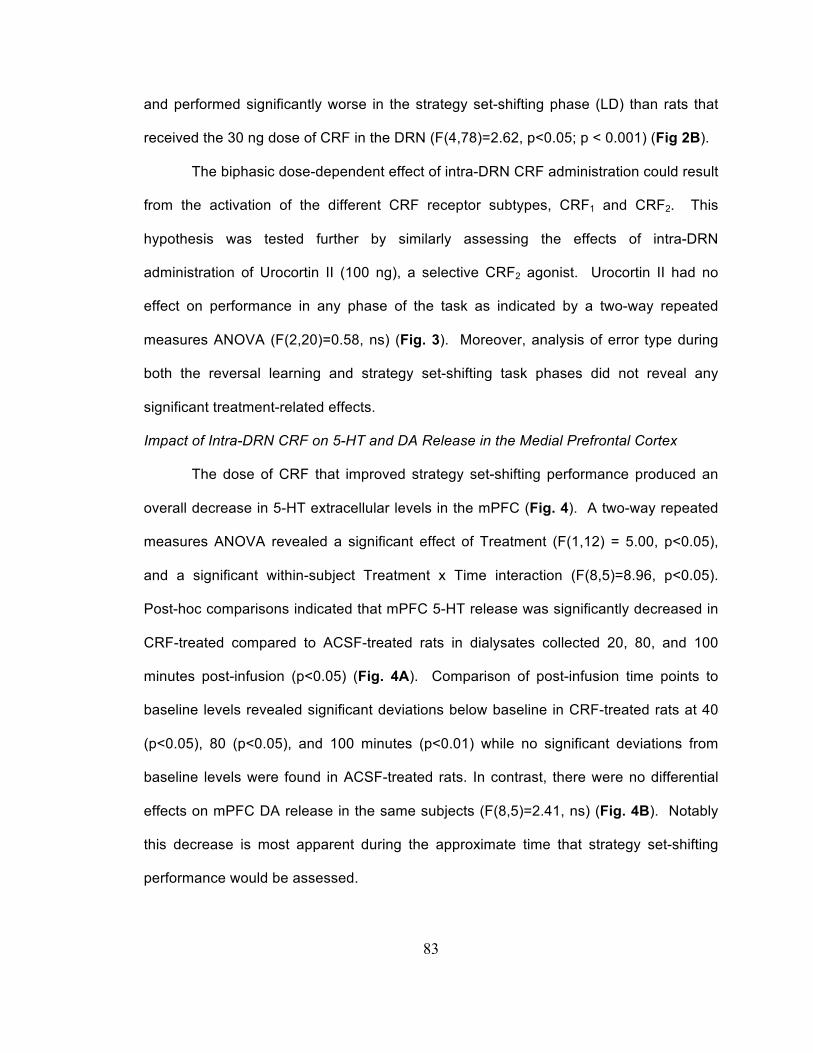
83
and performed significantly worse in the strategy set-shifting phase (LD) than rats that
received the 30 ng dose of CRF in the DRN (F(4,78)=2.62, p<0.05; p < 0.001) (Fig 2B).
The biphasic dose-dependent effect of intra-DRN CRF administration could result
from the activation of the different CRF receptor subtypes, CRF1 and CRF2. This
hypothesis was tested further by similarly assessing the effects of intra-DRN
administration of Urocortin II (100 ng), a selective CRF2 agonist. Urocortin II had no
effect on performance in any phase of the task as indicated by a two-way repeated
measures ANOVA (F(2,20)=0.58, ns) (Fig. 3). Moreover, analysis of error type during
both the reversal learning and strategy set-shifting task phases did not reveal any
significant treatment-related effects.
Impact of Intra-DRN CRF on 5-HT and DA Release in the Medial Prefrontal Cortex
The dose of CRF that improved strategy set-shifting performance produced an
overall decrease in 5-HT extracellular levels in the mPFC (Fig. 4). A two-way repeated
measures ANOVA revealed a significant effect of Treatment (F(1,12) = 5.00, p<0.05),
and a significant within-subject Treatment x Time interaction (F(8,5)=8.96, p<0.05).
Post-hoc comparisons indicated that mPFC 5-HT release was significantly decreased in
CRF-treated compared to ACSF-treated rats in dialysates collected 20, 80, and 100
minutes post-infusion (p<0.05) (Fig. 4A). Comparison of post-infusion time points to
baseline levels revealed significant deviations below baseline in CRF-treated rats at 40
(p<0.05), 80 (p<0.05), and 100 minutes (p<0.01) while no significant deviations from
baseline levels were found in ACSF-treated rats. In contrast, there were no differential
effects on mPFC DA release in the same subjects (F(8,5)=2.41, ns) (Fig. 4B). Notably
this decrease is most apparent during the approximate time that strategy set-shifting
performance would be assessed.
84
Impact of Prior Social Stress Experience on the Cognitive Effects of Intra-DRN CRF
As previously described, rats exposed to repeated social stress clustered into
two populations based on their latency to assume the defeat posture with 13 rats
classified as short latency (SL, 305 sec ± 24 sec) and 14 rats classified as long latency
(LL, 560 sec ± 20 sec) (p<0.001). Intra-DRN CRF (30 ng) produced different effects in
subpopulations of rats that were dependent upon their latency to assume the defeat
posture. CRF produced a similar facilitation of strategy set-shifting performance in SL
rats as in unstressed rats (n=6). In contrast, this effect was absent in LL rats (n=8) (Fig.
5). A two-way repeated measures ANOVA revealed a significant Treatment x Stage
interaction (F(4,40)=2.61, p<0.05), and post-hoc comparisons showed that the CRF (30
ng) SL group displayed better strategy set-shifting performance than both the ACSF SD
and the CRF (30 ng) LL treatment groups (p<0.05). Post-hoc comparisons indicated no
significant treatment effects on reversal learning. However, a one-way ANOVA of
Treatment within the SR stage revealed a trend toward significance (F(2,20)=3.22,
0.05<p<0.07) and post-hoc comparisons using Tukey’s HSD showed that CRF (30 ng)
LL rats performed significantly better in reversal learning than ACSF SD rats (p<0.05).
Analysis of error type within the SR and LD stages revealed no significant effects of
treatment. Overall, these data show that after social defeat, the 30 ng dose of CRF
retained its ability to produce CRF1-mediated facilitation of set-shifting performance
selectively in SL rats, but LL rats no longer responded in this manner to CRF.

85
Discussion
The current study identified a novel effect of CRF in the DRN to facilitate
cognitive flexibility as measured by strategy set-shifting performance. Several findings
suggested that facilitation of strategy set-shifting was mediated by CRF1 receptors. The
effect was produced by a moderate dose of CRF that interacts with CRF1 receptors but
not by a higher dose or by urocortin II, a peptide that is selective for CRF2 receptors
(Reyes et al., 2001). The behaviorally effective dose decreased extracellular 5-HT
levels in forebrain, consistent with responses mediated by CRF1 but not CRF2 receptors
(Price and Lucki, 2001; Forster et al., 2008; Lukkes et al., 2008). Importantly, in a
subpopulation of rats in which social stress has been shown to redistribute CRF1 and
CRF2 receptors so that CRF2 is more prominent on the plasma membrane (Wood et al.,
2013), the effects of CRF changed from facilitation of strategy set-shifting to facilitation
of reversal learning. This shift in CRF effects on cognitive function induced by social
stress is consistent with CRF2-mediated increases in 5-HT in the forebrain and the role
of 5-HT in reversal learning (Forster et al., 2008; Brown et al., 2012; Furr et al., 2012).
Together, the findings suggest that exposure to acute stress will differentially affect
cognitive processes depending on stress history and individual coping strategy.
Individual differences in the cellular adaptation of CRF receptor redistribution may
underlie the ability of CRF to affect different cognitive processes.
Behavioral Effects of CRF in the DRN
The DRN is densely innervated by CRF and CRF axon terminals here synapse
with both 5-HT and non-5-HT (e.g. GABA) dendrites (Valentino et al., 2001; Waselus et
al., 2005). In situ hybridization studies suggest that CRF2 is the prominent CRF receptor
subtype in the DRN (Van Pett et al., 2000). However, studies using pharmacological
manipulation of CRF receptors in the DRN to examine electrophysiological,

86
microdialysis, and behavioral endpoints provided evidence for effects mediated by both
CRF1 and CRF2 receptors (Valentino and Commons, 2005). In general, these studies
suggest that low levels of CRF, as might be released with acute mild stress, activate
CRF1 receptors on GABAergic neurons to inhibit 5-HT neuronal activity and release in
forebrain regions (Kirby et al., 2000; Price and Lucki, 2001; Waselus et al., 2005). In
contrast, this inhibition is lost as the dose of CRF is increased to doses that would
interact with CRF2 receptors (Kirby et al., 2000; Pernar et al., 2004). The behavioral
effects of engaging CRF1 and CRF2 receptors in the DRN have been best characterized
in the model of learned helplessness (Maier and Watkins, 2005). Deficits in learning
shock escape were associated with CRF2-induced activation of DRN-5-HT neurons and
5-HT forebrain release, and CRF1-mediated inhibition blocked the ability of a CRF2
agonist or uncontrollable stress to produce learned helplessness (Hammack et al.,
2003a; Hammack et al., 2003b). Although the role of CRF in the DRN on learned
helplessness has been well characterized, the DRN-5-HT system has been implicated in
other aspects of cognition and activation of CRF receptors could affect other critical
decision-making processes (Dayan and Huys, 2009; Robbins and Arnsten, 2009). The
present report is the first to examine the effects of CRF in the DRN on cognitive function
unrelated to fear.
Facilitation of Strategy Set-Shifting Performance Mediated by CRF1, not CRF2
A prominent finding of this study was that CRF facilitated strategy set-shifting
performance with an inverted U-shaped dose response relationship and this effect was
regionally specific. The effective CRF dose (30 ng) is one that produces the
characteristic CRF1-mediated inhibition of 5-HT DRN neurons (Price and Lucki, 2001;
Wood et al., 2013). The finding that raising the CRF dose to 100 ng or administering
urocortin II failed to facilitate set-shifting performance is consistent with mediation of

87
cognitive facilitation by CRF1 and not CRF2 receptors. Several studies have found that
high doses of CRF in the DRN preferentially activate CRF2 receptors, producing
opposing physiological effects to those of lower doses acting on CRF1 receptors (Price
and Lucki, 2001; Pernar et al., 2004; Forster et al., 2008; Lukkes et al., 2008).
The microdialysis results indicating that the behaviorally active 30 ng dose of
CRF decreased 5-HT extracellular levels in the mPFC at a time when behavior would be
measured, are consistent with the known inhibitory effects of CRF1 receptor activation on
5-HT release in various forebrain regions (Price et al., 1998; Price and Lucki, 2001;
Lukkes et al., 2008). Lesion studies have strongly implicated the mPFC in the
performance of strategy set-shifting tasks (Ragozzino et al., 1999; Floresco et al., 2008).
The finding that decreased mPFC 5-HT was associated with improved strategy set-
shifting performance and no effect on other task components was somewhat surprising,
given that 5-HT depletion has been reported to selectively impair reversal learning while
leaving set-shifting performance intact (Clarke et al., 2005; Lapiz-Bluhm et al., 2009).
However, the 5-HT depletion methods employed in these studies, systemic inhibition of
5-HT synthesis (Lapiz-Bluhm et al., 2009) or destruction of 5-HT neurons (Clarke et al.,
2005), produced large and chronic decreases in prefrontal 5-HT levels. In contrast, the
intra-DRN CRF treatment employed in the current study produced an acute and
moderate decrease in prefrontal 5-HT levels. Other pharmacological manipulations that
produce similar acute moderate decreases in prefrontal 5-HT also facilitate set-shifting
performance such as the 5-HT6 receptor agonist, WAY-181187 (Schechter et al., 2008;
Burnham et al., 2010). Similarly, acute systemic administration of the 5-HT1A receptor
agonist, 8-OH-DPAT, which has been shown to moderately decrease forebrain 5-HT
release (Rossi et al., 2008), improved visuospatial attentional performance in an mPFC-
dependent a five-choice serial reaction time task (Muir et al., 1996; Winstanley et al.,

88
2003). The results suggest that 5-HT inhibition by an acute stress that would engage
CRF1 receptors in the DRN can facilitate cognitive flexibility. Enhanced cognitive
flexibility may underlie the promotion of escape behavior and the ability of CRF1 receptor
activation in the DRN to inhibit learned helplessness (Hammack et al., 2003a).
Social Stress Experience and Coping Style Alter the Cognitive Impact of Intra-DRN CRF
The response of DRN-5-HT neurons to CRF (30 ng) is qualitatively changed by a
history of stress. For example, prior swim stress changed the response from a CRF1-
mediated inhibition to a CRF2-mediated excitation (Price et al., 2002). This was
associated with a redistribution of CRF receptors such that CRF1 receptors became
internalized and CRF2 receptors were recruited to the plasma membrane (Waselus et
al., 2009). The social stress used in the present study produces similar qualitative
changes in CRF responses and CRF receptor distribution (Wood et al., 2013). However,
these changes are limited to a subpopulation of rats that exhibit a coping style
characterized by a resistance to assume the defeat posture (LL rats). The present study
provides evidence that social stress-induced changes in CRF function at the cellular
level can translate to changes in cognitive performance. Accordingly, the CRF1-
mediated facilitation of strategy set-shifting performance was lost in LL rats, consistent
with CRF1 receptor internalization that is selective to rats with this coping style.
Moreover, this was replaced by an improvement in reversal learning, an effect that would
be consistent with the effects of CRF2 receptors on 5-HT transmission (Brown et al.,
2012; Furr et al., 2012). Together the results emphasize that a history of stress and
coping strategy are important determinants of how subsequent stressors will affect
cognitive function.
Clinical Implications

89
As a result of dual CRF receptor subtypes in the DRN with opposing actions,
stressors can have complex effects on the DRN-5-HT system that can be reflected in
different cognitive consequences. This study suggests that acute stress-induced
decreases in serotonergic activity facilitate strategy set-shifting whereas acute stress-
induced increases in serotonergic activity that would be observed in certain individuals
with a history of prior stress facilitate reversal learning. These individuals would not
exhibit an appropriate degree of cognitive flexibility as shown by others that have a
contrasting coping style and their lack of cognitive flexibility could render these
individuals more vulnerable to stress-related pathology. Stress-induced increases or
decreases in serotonergic activity appear to adaptively fine tune cognitive performance,
between individuals in a manner that is dependent upon their prior stress experience
and coping style. Knowledge of these individual differences in the cognitive impact of
stress as a result of coping strategy may help inform more personalized treatment of
individuals suffering from stress-related psychiatric disorders through serotonin-targeted
pharmacotherapies in combination with cognitive behavioral therapy.
Acknowledgements
This work was supported by National Institute of Health Grants MH058250 and
T32 MH14654.

90
Figures and Legends
Figure 1. Intra-DRN-administered CRF (30 ng) facilitates strategy set-shifting
performance. (A) Task performance indicated by the number of trials to reach criterion.
The abscissa indicates the task phase: side discrimination (SD), side reversal
discrimination (SR), and shift to light discrimination (LD). The ordinate indicates the
number of trials to reach criterion. Each bar is the mean of 17 ACSF, 6 CRF (10 ng), 13
CRF (30 ng), or 8 CRF (100 ng) treated rats and vertical lines indicate SEM. (B) Mean
number of different error types committed during strategy set-shifting. ** p < 0.01,
compared to ACSF; # p<0.05, ## p<0.01, compared to other CRF doses.
0
50
100
150
200
SD SR LD
ACSFCRF (10 ng)CRF (30 ng)CRF (100 ng)
Tria
ls to
Crit
erio
n
Task Phase
**
CRF (10 ng)CRF (30 ng)
##
#
A
0
10
20
30
40
50
60
70
Perseverative Regressive Random
ACSFCRF (10 ng)CRF (30 ng)CRF (100 ng)
Num
ber o
f Erro
rs
Type of Error(LD Phase)
CRF (10 ng)CRF (30 ng)##
B

91
A
Figure 2. Regional controls confirm that facilitation of strategy set-shifting by CRF (30
ng) is regionally restricted to the DRN. (A) Location of 30 ng CRF infusions in and
outside the DRN. The location of infusions was reconstructed onto plates 47, 49, and 51
(left to right) from Paxinos and Watson (1986). Black filled squares represent infusions
within the DRN. White open circles represent infusions outside of the DRN. Aq,
cerebral aqueduct; CG, central gray; CGD, central gray, dorsal; DR, dorsal raphe
nucleus; mlf, medial longitudinal fasciculus; xscp, decussation of the superior cerebellar
peduncle. (B) Task performance indicated by the number of trials to reach criterion for
rats that received ACSF or CRF inside or outside of the DRN. Each bar is the mean of
17 ACSF, 13 CRF (30 ng) IN, or 11 CRF (30 ng) OUT treated rats and vertical lines
indicated SEM. *** p<0.001 compared to ACSF, ### p<0.001 compared to other CRF
treatment group.
0
50
100
150
200
SD SR LD
ACSFCRF (30 ng) INCRF (30 ng) OUT
Tria
ls to
Crit
erio
n
Task Phase
B
***
###

92
Figure 3. Intra-DRN administration of the selective CRF2 agonist Urocortin II did not
affect task performance. Task performance indicated by the number of trials to reach
criterion. Each bar is the mean of 17 ACSF or 6 Urocortin II (100 ng) treated rats and
vertical lines indicated SEM.
0
50
100
150
200
SD SR LD
ACSFUrocortin II (100 ng)
Tria
ls to
Crit
erio
n
Task Phase

93
Figure 4. Intra-DRN CRF (30 ng) decreased mPFC 5-HT but not DA extracellular levels.
(A) Effect of ACSF or CRF (30 ng) on mPFC 5-HT. The abscissa shows time (min)
before and after the infusion which occurred at 0. The ordinate indicates the
extracellular level of 5-HT expressed as a percentage of baseline. (B) Effect of ACSF or
CRF (30 ng) on mPFC DA in same rats as shown in A. The abscissa and ordinate are
as described in A. Each point is the mean of 5 ACSF or 7 CRF (30 ng) treated rats.
Vertical lines indicate SEM. * p<0.05, ** p<0.01, compared to ACSF; # p<0.05, ##
p<0.01 compared to baseline.
50
75
100
125
150
-80 -60 -40 -20 0 20 40 60 80 100
ACSFCRF (30 ng)Baseline
Extra
cellu
lar 5
-HT
(% B
asel
ine)
Time After Infusion
A
**
**
*
# #
##
Infusion
*
50
75
100
125
150
-80 -60 -40 -20 0 20 40 60 80 100
ACSFCRF (30 ng)Baseline
Extra
cellu
lar D
A (%
Bas
elin
e)
Time After Infusion
B
Infusion

94
Figure 5. Prior social stress experience occludes the facilitatory effect of intra-DRN CRF
(30 ng) in LL but not SL rats. Task performance indicated by the number of trials to
reach criterion. Each bar is the mean of 9 ACSF SD, 6 CRF (30 ng) SL, or 8 CRF (30
ng) LL treated rats and vertical lines indicated SEM. * p<0.05, compared to ACSF SD; #
p < 0.05 compared to other CRF-treatment group.
0
25
50
75
100
125
150
175
200
225
SD SR LD
ACSF SDCRF (30 ng) SLCRF (30 ng) LL
Tria
ls to
Crit
erio
n
Task Phase
*
*
#
CRF (30 ng) SLCRF (30 ng) LL

95
References
Alexander JK, Hillier A, Smith RM, Tivarus ME, Beversdorf DQ (2007) Beta-adrenergic
modulation of cognitive flexibility during stress. J Cogn Neurosci 19:468-478.
Arnsten AF (2009) Stress signalling pathways that impair prefrontal cortex structure and
function. Nat Rev Neurosci 10:410-422.
Arnsten AF (2011) Prefrontal cortical network connections: key site of vulnerability in
stress and schizophrenia. Int J Dev Neurosci 29:215-223.
Bale TL, Vale WW (2004) CRF and CRF receptors: role in stress responsivity and other
behaviors. Annu Rev Pharmacol Toxicol 44:525-557.
Birnbaum S, Gobeske KT, Auerbach J, Taylor JR, Arnsten AF (1999) A role for
norepinephrine in stress-induced cognitive deficits: alpha-1-adrenoceptor
mediation in the prefrontal cortex. Biol Psychiatry 46:1266-1274.
Bondi CO, Jett JD, Morilak DA (2010) Beneficial effects of desipramine on cognitive
function of chronically stressed rats are mediated by alpha1-adrenergic receptors
in medial prefrontal cortex. Prog Neuropsychopharmacol Biol Psychiatry 34:913-
923.
Brown HD, Amodeo DA, Sweeney JA, Ragozzino ME (2012) The selective serotonin
reuptake inhibitor, escitalopram, enhances inhibition of prepotent responding and
spatial reversal learning. J Psychopharmacol 26:1443-1455.
Burnham KE, Baxter MG, Bainton JR, Southam E, Dawson LA, Bannerman DM, Sharp
T (2010) Activation of 5-HT(6) receptors facilitates attentional set shifting.
Psychopharmacology (Berl) 208:13-21.
Butler PD, Weiss JM, Stout JC, Nemeroff CB (1990) Corticotropin-releasing factor
produces fear-enhancing and behavioral activating effects following infusion into
the locus coeruleus. J Neurosci 10:176-183.

96
Campeau S, Liberzon I, Morilak D, Ressler K (2011) Stress modulation of cognitive and
affective processes. Stress 14:503-519.
Chen MF, Chiu TH, Lee EH (1992) Noradrenergic mediation of the memory-enhancing
effect of corticotropin-releasing factor in the locus coeruleus of rats.
Psychoneuroendocrinology 17:113-124.
Clarke HF, Walker SC, Crofts HS, Dalley JW, Robbins TW, Roberts AC (2005)
Prefrontal serotonin depletion affects reversal learning but not attentional set
shifting. J Neurosci 25:532-538.
Curtis AL, Drolet G, Valentino RJ (1993) Hemodynamic stress activates locus coeruleus
neurons of unanesthetized rats. Brain Res Bull 31:737-744.
Danet M, Lapiz-Bluhm S, Morilak DA (2010) A cognitive deficit induced in rats by chronic
intermittent cold stress is reversed by chronic antidepressant treatment. Int J
Neuropsychopharmacol 13:997-1009.
Dayan P, Huys QJ (2009) Serotonin in affective control. Annu Rev Neurosci 32:95-126.
Finlay JM, Zigmond MJ, Abercrombie ED (1995) Increased dopamine and
norepinephrine release in medial prefrontal cortex induced by acute and chronic
stress: effects of diazepam. Neuroscience 64:619-628.
Floresco SB, Block AE, Tse MT (2008) Inactivation of the medial prefrontal cortex of the
rat impairs strategy set-shifting, but not reversal learning, using a novel,
automated procedure. Behav Brain Res 190:85-96.
Forster GL, Pringle RB, Mouw NJ, Vuong SM, Watt MJ, Burke AR, Lowry CA, Summers
CH, Renner KJ (2008) Corticotropin-releasing factor in the dorsal raphe nucleus
increases medial prefrontal cortical serotonin via type 2 receptors and median
raphe nucleus activity. Eur J Neurosci 28:299-310.

97
Furr A, Lapiz-Bluhm MD, Morilak DA (2012) 5-HT2A receptors in the orbitofrontal cortex
facilitate reversal learning and contribute to the beneficial cognitive effects of
chronic citalopram treatment in rats. Int J Neuropsychopharmacol 15:1295-1305.
Hammack SE, Pepin JL, DesMarteau JS, Watkins LR, Maier SF (2003a) Low doses of
corticotropin-releasing hormone injected into the dorsal raphe nucleus block the
behavioral consequences of uncontrollable stress. Behav Brain Res 147:55-64.
Hammack SE, Richey KJ, Schmid MJ, LoPresti ML, Watkins LR, Maier SF (2002) The
role of corticotropin-releasing hormone in the dorsal raphe nucleus in mediating
the behavioral consequences of uncontrollable stress. J Neurosci 22:1020-1026.
Hammack SE, Schmid MJ, LoPresti ML, Der-Avakian A, Pellymounter MA, Foster AC,
Watkins LR, Maier SF (2003b) Corticotropin releasing hormone type 2 receptors
in the dorsal raphe nucleus mediate the behavioral consequences of
uncontrollable stress. J Neurosci 23:1019-1025.
Henderson RK, Snyder HR, Gupta T, Banich MT (2012) When does stress help or
harm? The effects of stress controllability and subjective stress response on
stroop performance. Front Psychol 3:179.
Joëls M, Baram TZ (2009) The neuro-symphony of stress. Nat Rev Neurosci 10:459-
466.
Kessler RC (1997) The effects of stressful life events on depression. Annu Rev Psychol
48:191-214.
Kirby LG, Lucki I (1997) Interaction between the forced swimming test and fluoxetine
treatment on extracellular 5-hydroxytryptamine and 5-hydroxyindoleacetic acid in
the rat. J Pharmacol Exp Ther 282:967-976.

98
Kirby LG, Rice KC, Valentino RJ (2000) Effects of corticotropin-releasing factor on
neuronal activity in the serotonergic dorsal raphe nucleus.
Neuropsychopharmacology 22:148-162.
Kreiss DS, Wieland S, Lucki I (1993) The presence of a serotonin uptake inhibitor alters
pharmacological manipulations of serotonin release. Neuroscience 52:295-301.
Lapiz-Bluhm MD, Soto-Piña AE, Hensler JG, Morilak DA (2009) Chronic intermittent cold
stress and serotonin depletion induce deficits of reversal learning in an
attentional set-shifting test in rats. Psychopharmacology (Berl) 202:329-341.
Lejeune F, Millan MJ (2003) The CRF1 receptor antagonist, DMP695, abolishes
activation of locus coeruleus noradrenergic neurones by CRF in anesthetized
rats. Eur J Pharmacol 464:127-133.
Lukkes JL, Forster GL, Renner KJ, Summers CH (2008) Corticotropin-releasing factor 1
and 2 receptors in the dorsal raphé differentially affect serotonin release in the
nucleus accumbens. Eur J Pharmacol 578:185-193.
Maier SF, Watkins LR (2005) Stressor controllability and learned helplessness: the roles
of the dorsal raphe nucleus, serotonin, and corticotropin-releasing factor.
Neurosci Biobehav Rev 29:829-841.
Marin MF, Lord C, Andrews J, Juster RP, Sindi S, Arsenault-Lapierre G, Fiocco AJ,
Lupien SJ (2011) Chronic stress, cognitive functioning and mental health.
Neurobiol Learn Mem 96:583-595.
Miczek KA (1979) A new test for aggression in rats without aversive stimulation:
differential effects of d-amphetamine and cocaine. Psychopharmacology (Berl)
60:253-259.
Millan MJ et al. (2012) Cognitive dysfunction in psychiatric disorders: characteristics,
causes and the quest for improved therapy. Nat Rev Drug Discov 11:141-168.

99
Muir JL, Everitt BJ, Robbins TW (1996) The cerebral cortex of the rat and visual
attentional function: dissociable effects of mediofrontal, cingulate, anterior
dorsolateral, and parietal cortex lesions on a five-choice serial reaction time task.
Cereb Cortex 6:470-481.
Naegeli KJ, O'Connor JA, Banerjee P, Morilak DA (2013) Effects of milnacipran on
cognitive flexibility following chronic stress in rats. Eur J Pharmacol 703:62-66.
Nuechterlein KH, Dawson ME, Gitlin M, Ventura J, Goldstein MJ, Snyder KS, Yee CM,
Mintz J (1992) Developmental Processes in Schizophrenic Disorders:
longitudinal studies of vulnerability and stress. Schizophr Bull 18:387-425.
Owens MJ, Nemeroff CB (1991) Physiology and pharmacology of corticotropin-releasing
factor. Pharmacol Rev 43:425-473.
Page ME, Valentino RJ (1994) Locus coeruleus activation by physiological challenges.
Brain Res Bull 35:557-560.
Pernar L, Curtis AL, Vale WW, Rivier JE, Valentino RJ (2004) Selective activation of
corticotropin-releasing factor-2 receptors on neurochemically identified neurons
in the rat dorsal raphe nucleus reveals dual actions. J Neurosci 24:1305-1311.
Price ML, Lucki I (2001) Regulation of serotonin release in the lateral septum and
striatum by corticotropin-releasing factor. J Neurosci 21:2833-2841.
Price ML, Kirby LG, Valentino RJ, Lucki I (2002) Evidence for corticotropin-releasing
factor regulation of serotonin in the lateral septum during acute swim stress:
adaptation produced by repeated swimming. Psychopharmacology (Berl)
162:406-414.
Price ML, Curtis AL, Kirby LG, Valentino RJ, Lucki I (1998) Effects of corticotropin-
releasing factor on brain serotonergic activity. Neuropsychopharmacology
18:492-502.

100
Ragozzino ME, Wilcox C, Raso M, Kesner RP (1999) Involvement of rodent prefrontal
cortex subregions in strategy switching. Behav Neurosci 113:32-41.
Reyes TM, Lewis K, Perrin MH, Kunitake KS, Vaughan J, Arias CA, Hogenesch JB,
Gulyas J, Rivier J, Vale WW, Sawchenko PE (2001) Urocortin II: a member of
the corticotropin-releasing factor (CRF) neuropeptide family that is selectively
bound by type 2 CRF receptors. Proc Natl Acad Sci U S A 98:2843-2848.
Robbins TW, Arnsten AF (2009) The neuropsychopharmacology of fronto-executive
function: monoaminergic modulation. Annu Rev Neurosci 32:267-287.
Rossi DV, Burke TF, McCasland M, Hensler JG (2008) Serotonin-1A receptor function in
the dorsal raphe nucleus following chronic administration of the selective
serotonin reuptake inhibitor sertraline. J Neurochem 105:1091-1099.
Salehi B, Cordero MI, Sandi C (2010) Learning under stress: the inverted-U-shape
function revisited. Learn Mem 17:522-530.
Schechter LE, Lin Q, Smith DL, Zhang G, Shan Q, Platt B, Brandt MR, Dawson LA, Cole
D, Bernotas R, Robichaud A, Rosenzweig-Lipson S, Beyer CE (2008)
Neuropharmacological profile of novel and selective 5-HT6 receptor agonists:
WAY-181187 and WAY-208466. Neuropsychopharmacology 33:1323-1335.
Smagin GN, Swiergiel AH, Dunn AJ (1995) Corticotropin-releasing factor administered
into the locus coeruleus, but not the parabrachial nucleus, stimulates
norepinephrine release in the prefrontal cortex. Brain Res Bull 36:71-76.
Snyder K, Wang WW, Han R, McFadden K, Valentino RJ (2012) Corticotropin-releasing
factor in the norepinephrine nucleus, locus coeruleus, facilitates behavioral
flexibility. Neuropsychopharmacology 37:520-530.

101
Vale W, Spiess J, Rivier C, Rivier J (1981) Characterization of a 41-residue ovine
hypothalamic peptide that stimulates secretion of corticotropin and beta-
endorphin. Science 213:1394-1397.
Vale W, Rivier C, Brown MR, Spiess J, Koob G, Swanson L, Bilezikjian L, Bloom F,
Rivier J (1983) Chemical and biological characterization of corticotropin releasing
factor. Recent Prog Horm Res 39:245-270.
Valentino RJ, Commons KG (2005) Peptides that fine-tune the serotonin system.
Neuropeptides 39:1-8.
Valentino RJ, Van Bockstaele E (2008) Convergent regulation of locus coeruleus activity
as an adaptive response to stress. Eur J Pharmacol 583:194-203.
Valentino RJ, Foote SL, Aston-Jones G (1983) Corticotropin-releasing factor activates
noradrenergic neurons of the locus coeruleus. Brain Res 270:363-367.
Valentino RJ, Liouterman L, Van Bockstaele EJ (2001) Evidence for regional
heterogeneity in corticotropin-releasing factor interactions in the dorsal raphe
nucleus. J Comp Neurol 435:450-463.
Valentino RJ, Lucki I, Van Bockstaele E (2010) Corticotropin-releasing factor in the
dorsal raphe nucleus: Linking stress coping and addiction. Brain Res 1314:29-37.
Van Pett K, Viau V, Bittencourt JC, Chan RK, Li HY, Arias C, Prins GS, Perrin M, Vale
W, Sawchenko PE (2000) Distribution of mRNAs encoding CRF receptors in
brain and pituitary of rat and mouse. J Comp Neurol 428:191-212.
Waselus M, Valentino RJ, Van Bockstaele EJ (2005) Ultrastructural evidence for a role
of gamma-aminobutyric acid in mediating the effects of corticotropin-releasing
factor on the rat dorsal raphe serotonin system. J Comp Neurol 482:155-165.
102
Waselus M, Nazzaro C, Valentino RJ, Van Bockstaele EJ (2009) Stress-induced
redistribution of corticotropin-releasing factor receptor subtypes in the dorsal
raphe nucleus. Biol Psychiatry 66:76-83.
Winstanley CA, Chudasama Y, Dalley JW, Theobald DE, Glennon JC, Robbins TW
(2003) Intra-prefrontal 8-OH-DPAT and M100907 improve visuospatial attention
and decrease impulsivity on the five-choice serial reaction time task in rats.
Psychopharmacology (Berl) 167:304-314.
Wood SK, Walker HE, Valentino RJ, Bhatnagar S (2010) Individual differences in
reactivity to social stress predict susceptibility and resilience to a depressive
phenotype: role of corticotropin-releasing factor. Endocrinology 151:1795-1805.
Wood SK, Zhang XY, Reyes BA, Lee CS, Van Bockstaele EJ, Valentino RJ (2013)
Cellular adaptations of dorsal raphe serotonin neurons associated with the
development of active coping in response to social stress. Biol Psychiatry
73:1087-1094.
Yoshitake S, Ijiri S, Kehr J, Yoshitake T (2013) Concurrent modulation of extracellular
levels of noradrenaline and cAMP during stress and by anxiogenic- or anxiolytic-
like neuropeptides in the prefrontal cortex of awake rats. Neurochem Int 62:314-
323.

103
Chapter 4
Adolescent social stress impairs behavioral flexibility during adulthood:
relationship to coping strategy
Kevin P. Snyder1, Mark Barry1, and Rita J. Valentino2
1The University of Pennsylvania
Philadelphia, PA 19104
2The Children’s Hospital of Philadelphia
Philadelphia, PA 19104
Corresponding Author: Rita J. Valentino, Ph.D.
The Children's Hospital of Philadelphia
402D Abramson Pediatric Research Center
Philadelphia, PA 19104
215-590-0650 (phone)
215-590-3364 (fax)
Number of Figures: 5
Number of Tables: 2
Number of Pages: 35

104
Abstract
Stress experience during adolescence has been strongly linked to the
development of psychiatric disorders in adulthood, many of which are associated with
deficits in prefrontal cortex function. The current study assessed the cognitive impact of
adolescent social stress on performance in a medial prefrontal cortex (mPFC)-
dependent operant strategy set-shifting task (OSST). Early adolescent (P28), mid-
adolescent (P42) and Adult (P70) rats were exposed to the resident-intruder model of
social stress for 5 days and tested in the OSST either one week after stress experience
or during adulthood. After completion of the OSST, rats were perfused with
paraformaldehyde and expression of the immediate early gene, c-fos, was
immunohistochemically quantified in the mPFC. Strategy set-shifting performance was
selectively impaired in adult rats that were stressed during adolescence. Coping
strategy in response to social stress was a determining factor in whether early
adolescents would exhibit cognitive impairments in adulthood. Stress experience had no
impact on OSST performance assessed during early or mid-adolescence. Medial
prefrontal cortical c-fos was positively correlated with strategy set-shifting performance
only in rats that were tested during adulthood. Social stress during adolescence can
produce impairments in prefrontal cortex-mediated cognition during adulthood. This
impairment may not be evident during adolescence because set-shifting performance
may involve different brain regions during adolescence than during adulthood.
Additionally, the protracted cognitive impact of adolescent social stress experience may
be dependent upon individual coping strategy.
105
Introduction
Stress has been implicated in many psychiatric disorders including depression,
schizophrenia, attentional deficit hyperactivity disorder, and obsessive-compulsive
disorder (Nuechterlein et al., 1992; Kessler, 1997; Findley et al., 2003; Marin et al.,
2011; Wigal et al., 2012). These disorders are characterized by deficits in cognitive
function, particularly executive function that is regulated by the prefrontal cortex (PFC)
(Jurado and Rosselli, 2007; Clark et al., 2009; Arnsten, 2011). Stressors are thought to
impair cognitive function as a result of structural and functional changes in the PFC
(Radley et al., 2006; Arnsten, 2009). For example, chronic self-perceived stress has
been associated with impaired set-shifting performance, a measure of executive
function, in human subjects (Orem et al., 2008). Likewise, chronic cold stress and
chronic mild stress impair attentional set-shifting behavior in rodents (Bondi et al., 2008;
Lapiz-Bluhm et al., 2009). Additionally, impaired set-shifting performance has been
observed in animal models of stress-related psychiatric disorders (Goetghebeur et al.,
2010; Chess et al., 2011; Brooks et al., 2012; Cao et al., 2012). Stress-induced
vulnerability to psychiatric diseases may derive in part from PFC dysfunctions that are
expressed as cognitive impairments.
Although stress during adulthood can influence cognitive function, the impact
may be greater during specific windows of development when defense mechanisms and
brain regions involved in cognition and emotion are still developing. The hypothalamic-
pituitary-adrenal (HPA) axis response to stress is heightened during adolescence and
does not habituate to chronic stress in the same manner as it does during adulthood
(Romeo et al., 2006; Gunnar et al., 2009). Early life stress can also produce enduring
effects that are expressed in adulthood and this has been associated with the
occurrence of psychiatric disorders in adulthood (Halligan et al., 2007; Lupien et al.,

106
2009). Consistent with this, rats with adolescent stress experience display increased
anxiety-related and depressive-like behaviors as well as impaired learning and memory
in adulthood (Isgor et al., 2004; Uys et al., 2006; McCormick et al., 2008). Both human
and animal studies demonstrate that set-shifting performance improves with
development of the prefrontal cortex (Kalkut et al., 2009; Newman and McGaughy,
2011), but the impact adolescent stress experience on set-shifting performance remains
unknown.
Social stressors are among the most prevalent and most detrimental to human
mental health and well-being (Brown and Prudo, 1981). Social stress has been
effectively modeled in rodents by the resident-intruder paradigm (Miczek, 1979). This
ethologically relevant stressor produces HPA axis dysfunctions and increased
depressive-like and substance abuse related behaviors (Rygula et al., 2008; Wood et al.,
2010; Wood et al., 2012; Bardo et al., 2013; Chaijale et al., 2013). Social stress during
adolescence acutely increases defensive behaviors and noradrenergic tone while
decreasing defensive and social interaction behaviors in adult animals that were
stressed during adolescence (Vidal et al., 2007; Bingham et al., 2011).
To better understand the impact of social stress on cognitive function the current
study evaluated the effects of social stress throughout development on performance in
an mPFC-dependent operant strategy set-shifting task (OSST), adapted from (Floresco
et al., 2008) . To determine whether stress effects on cognitive performance were
related to effects on mPFC function, mPFC activity during task performance was also
assessed by immunohistochemical quantification of the expression of the immediate
early gene, c-fos.

107
Methods
Animals
Male Sprague Dawley rats (Charles River, Wilmington, Massachusetts) served
as control or social stress “intruder” rats and were delivered to the animal facility on PND
24 (early adolescents), PND 38 (mid-adolescents), or PND 66 (adults). These animals
were given 4 days to acclimate to the colony before the onset of experimentation. Male
Long-Evans retired breeders (550-850 g) served as residents (Charles River,
Wilmington, Massachusetts). Animals were singly housed on a 12 h light/dark cycle with
lights on at 7 AM throughout experimentation. Care and use of animals was approved
by the Institutional Animal Care and Use Committee of the Children’s Hospital of
Philadelphia.
Experimental Design
Five experimental groups were studied: Early Adolescents (EA), Mid-Adolescents
(MA), Adults, Early Adolescents Tested as Adults (EA-Adults), and Mid-Adolescents
Tested as Adults (MA-Adults). After 4 days of acclimation, rats were exposed to five
consecutive days of social stress or control manipulation. EA and EA-Adult animals
began social stress on PND 28, MA and MA-Adult animals began social stress on PND
42, and Adults began social stress on PND 70. On the last day of social stress or
control manipulation EA, MA, and Adult rats began food restriction to maintain 85% free-
feeding weight. OSST training (described below) began 3 days after the last
experimental manipulation and testing occurred after 3 days of training, 6 days after the
final experimental manipulation. EA-Adult and MA-Adult animals were food restricted,
trained, and tested in the operant chamber at the same age as Adult animals (PND 74,
77, and 80, respectively).

108
Social Stress
Rats were randomly assigned to control or social stress groups. The social
stress procedure was a modification of the resident-intruder model (Miczek, 1979) and
identical to that previously described with the exception that rats were exposed to 5
consecutive days of social stress (see Supplement 1 for detailed methods) (Bingham et
al., 2011). All animals were singly housed during social stress; however, EA-Adult and
MA-Adult animals were pair housed during the time period between the end of social
stress and the beginning of food restriction and operant training/testing. EA, MA, and
Adult animals remained singly housed following social stress as they proceeded
immediately to food restriction and operant training/testing.
Operant Training and Testing
Operant training and testing was carried out in operant chambers (Med-
Associates, St. Albans, VT, USA), each within a sound-attenuating box (see
Supplement 1 for detailed methods). Animals were food restricted with the goal of
reaching and maintaining 85% of their free-feeding weight. A 4-day operant training and
testing protocol, adapted from (Floresco et al., 2008) was initiated on the fourth day of
food restriction. On the first day, animals were habituated to the chamber and shaped to
lever press on a fixed-ratio 1 schedule on one lever (randomly chosen left/right) to a
criterion of 50 presses within 30 minutes. On the second day, animals were trained to
achieve the same criterion with a fixed-ratio 1 schedule on the opposite lever. On the
third day, animals were introduced to the trial structure of the task, under conditions with
no discernable “rule” (see Supplement 1 for detailed methods). Each animal’s side bias
was determined to be toward the lever on the side that the animal pressed on the
majority of trials.
109
On the fourth day, cognitive flexibility was tested in an operant set-shifting task
(OSST), consisting of a series of three consecutive discriminations: an initial side
discrimination (SD), a side reversal discrimination (SR), and a shift to light discrimination
(LD). Animals proceeded from one stage of the task to the next after achieving a
criterion of 8 consecutive correct choices, provided 30 trials had been attempted. This
minimum of 30 trials stipulation was added to ensure that each animal experienced
enough trials in each stage of the task for the transitions from one type of discrimination
to the next to be cognitively meaningful. The trial structure and timing of light
illuminations during each stage of the task were the same as they were during the
previous day’s training session, with one exception: on each trial only one stimulus light
was illuminated. For every pair of trials, on the first trial of the pair the left or right
stimulus light was randomly selected to be illuminated, and the opposite stimulus light
was illuminated on the following trial.
During the SD stage, the lever on the side opposite the animal’s side bias was
designated to be the correct lever on every trial, regardless of the location of the
stimulus light. During the SR stage, the correct lever on each trial was designated to be
the lever opposite the correct lever during the initial side discrimination. During the LD
stage, the correct lever was designated as the lever underneath the illuminated stimulus
light on each trial. After reaching criterion in the LD stage, the task was ended, and the
animal was removed from the chamber. Trials to criterion (TTC) and number of errors
were recorded during each stage of the OSST were recorded for each rat. Omitted trials
were not included in the TTC measure.
Immunohistochemistry
Thirty minutes after completing the OSST, some rats were anesthetized with
isoflurane and transcardially perfused with heparinized saline followed by 4%

110
paraformaldehyde for processing of c-fos as previously described (Snyder et al., 2012).
See Supplement 1 for detailed methods.
Statistical Analysis
All data from animals that were stressed at the same age as they were tested in
the OSST (EA, MA, Adult) were analyzed independently from animals that were stress at
different ages but tested as adults (EA-Adult, MA-Adult, and Adult). Effects of age on
TTC were assessed by means of two-way ANOVA (Age of Stress x Stage) with repeated
measures across Stage. Effects of social stress on TTC were assessed by two-way
ANOVAs (Stress x Stage) with repeated measures across Stage performed within each
experimental group. Effects of social stress on error type during the side reversal and
shift to light stages were also assessed separately within each experimental group by
performing two-way mixed ANOVAs (Stress x Error Type) with Error Type as the within-
subject factor. Where significant main effects or interactions were found, follow-up post-
hoc comparisons were performed using the Holm-Sidak method, unless otherwise
noted.

111
Results
Effects of Social Stress During Development on Cognitive Performance
Early adolescent (EA) (P28, n=19 control, n=16 stress), mid-adolescent (MA)
(P42, n=8 control, n=8 stress), and adult (P70, n=20 control, n=28 stress) rats completed
testing in the OSST. Some rats of each group did not finish the task including 2 EA
control rats, 4 EA stressed rats, and 1 Adult control rat. There was no effect of stress
when comparing the three age groups (Fig. 1A). A three-way ANOVA (Stress x Age x
Stage) with repeated measures across Stage revealed no statistically significant (three-
way) interaction (F(4,184)=0.2, ns). Likewise there was no Stress x Stage interaction
(F(2,92)=1.4, ns). However, there was a significant Age x Stage interaction
(F(4,184)=4.2, p<0.005), and post-hoc comparisons showed that during the strategy set-
shifting stage all three age groups performed significantly differently that each other (p <
0.05) with the mid-adolescents performing the best and the adults performing the worst.
Although social stress had no effect on cognitive performance that was assessed
during the same developmental stage as the stress exposure, it had enduring effects in
rats that were stressed as adolescents and assessed in adulthood (Fig. 1B). Some rats
of these rats also not finish the task including 2 EA-Adult stressed rats and 2 MA-Adult
stressed rats. A comparison of rats that were exposed to stress or control conditions in
early adolescence (n=11 control, n=11 stress), mid-adolescence (n=13 control, n=12
stress) or adulthood (n=20 control, n=28 stress) and tested as adults revealed that early
handling or stress improved strategy set-shifting performance (Age of Stress x Stage
interaction F(4,176)=5.7, p<0.005). Post-hoc analysis showed that EA-Adult rats
performed better than MA-Adult rats (p<0.05). Additionally, a Stress x Stage Interaction
(F(2,88)=3.8, p<0.05) and post-hoc analysis indicated that social stress experience
impaired performance during strategy set-shifting in rats tested as adults.

112
For rats that were stressed as adolescents and tested as adults an effect of
stress was observed (Fig. 1B). A significant Stress x Stage interaction was found in
MA-Adult rats with social stress selectively impairing strategy set-shifting performance
(F(2,46)=3.3, p<0.05; p<0.05 post-hoc). In EA-Adult rats social stress generally
impaired OSST performance, although this effect could not be attributed to a particular
task phase (Between-Subject Stress Effect F(1,20)=5.9, p<0.05). Together these results
suggest that although some manipulation during early life may promote cognitive
flexibility in adulthood, social stress at this age reverses that benefit.
Effects of Social Stress on Strategy Set-Shifting Error Type
To better understand how adolescent social stress affected cognitive
performance, the effect of social stress on error type was analyzed by two-way mixed
ANOVAs (Stress x Error Type) performed within each experimental group (see
Supplement 1 for a detailed description of error type classification). Social stress
selectively increased perseverative errors in Adult rats during strategy set-shifting
(F(2,92)=4.6, p<0.05; p<0.01 post-hoc) (Fig. 2). Identical analysis to that described
above was also performed on error type within reversal learning; however, no significant
effects of stress were found.
Effect of Stress Coping Strategy on Cognitive Performance
Social stress was previously shown to reveal behaviorally and physiologically
distinct subpopulations of animals based on their respective passive or active stress
coping strategies, as defined by the relatively short (SL) or long (LL) latency to assume
the subordinate defeat posture, respectively (Wood et al., 2010). A cluster analysis was
performed to categorize each rat as an SL or LL animal (see Supplement 1 for details).
Table 1S shows the mean latency of each subpopulation for each experimental group.
There was an effect of coping strategy on OSST performance in EA-Adult rats such that

113
those rats with a passive coping strategy (SL) exhibited impaired performance,
particularly during the strategy set-shifting component of the task. There was a
significant between-subject effect of Latency Group in EA-Adult rats (F(2,22)=4.4,
p<0.05), and post-hoc comparisons indicated that SL rats were specifically impaired by
social stress with respect to control rats (p<0.05). A nearly significant Latency Group x
Stage interaction (F(4,44)=2.2, p<0.1) was also found in this group of animals, and
Tukey’s HSD post-hoc indicated that social stress impaired performance during strategy
set-shifting selectively in SL EA-Adult rats with respect to control EA-Adult rats (p<0.01)
(Fig. 3). No significant effects of coping strategy on strategy set-shifting performance
were found in any other experimental groups.
Effect of Stress Coping Strategy on Error Type
The SL population of EA-Adult rats made more perseverative errors compared to
controls. Although there was no Latency Group x Error Type interaction (F(4,44)=1.4,
ns) in EA-Adult rats, Tukey's HSD post-hoc comparisons revealed that SL rats
committed more perseverative errors than control rats in this group (p<0.05) (Fig. 4A).
Interestingly, a significant interaction between Latency Group and Error Type
was found for Adults during the strategy set-shift stage (F(4,86)=3.0, p<0.05) (Fig. 4B).
Social stress selectively increased perseverative errors in LL adult rats (p<0.01) as
compared to controls. Identical analysis to that described above was also performed on
error type within reversal learning; however, no significant effects of coping strategy
were found.
Effects of Social Stress and Age on Task-Associated Activation of the Medial Prefrontal
Cortex
Table 1 summarizes the mean number of c-fos profiles in the mPFC in each
group. There was no effect of Stress on the number of c-fos profiles in the mPFC

114
(F(1,38)=0.1, ns) and no Stress x Group interaction (F(4,38)=0.8, ns). However, a
significant main effect of Group (F(4,38)=6.0, p<0.001) was found, and post-hoc
comparisons indicated that MA animals had significantly less mPFC c-fos expression
than all other groups that were handled or stressed during adolescence (p<0.01). This
result was surprising given that MA rats exhibit the best strategy set-shifting
performance, a task that is though to be mPFC-mediated.
The relationship between c-fos profiles in the mPFC and trials to criterion was
then determined for rats tested in adolescence and for rats tested in adulthood.
Because this comparison across all age groups could be confounded by developmental
changes in the relationships between c-fos expression, neuronal activation, and task
performance, regression analysis was performed separately in all rats tested during
adolescence (EA and MA) and all rats tested as adults (Adults, EA-Adults, MA-Adults).
For rats tested during adolescence mPFC c-fos expression was negatively correlated to
strategy set-shifting performance (positive between c-fos and trials to criterion)
(F(1,13)=5.1, p<0.05), suggesting that mPFC activation may impair rather than facilitate
strategy set-shifting performance during adolescence (Fig. 5A). In contrast, a significant
positive correlation between mPFC c-fos expression and strategy set-shifting
performance (negative between c-fos and trials to criterion) was found for rats tested in
adulthood (F(1,28)=8.2, p<0.01 (Fig. 5B). A reciprocal transformation of the number of
mPFC c-fos profiles revealed an even stronger relationship with strategy set-shifting
performance (F(1,28)=12.4, p<0.005). This transformed relationship may be even more
appropriate because it assumes an asymptotic relationship between mPFC c-fos
expression and task performance such that even at the highest observed levels of
mPFC c-fos expression trials to criterion are reasonably still predicted to be greater than

115
the minimum criterion of 8 trials. These data suggest that strategy set-shifting
performance may not be facilitated by mPFC activation in male rats until adulthood.

116
Discussion
The current study examined the short and long-term impact of social stress
experience and coping strategy throughout development on cognitive flexibility.
Interestingly, all developmental and stress-related effects on task performance were
isolated to the mPFC-dependent strategy set-shifting phase of the OSST. The most
prominent finding was that social stress during adolescence produced a protracted
impairment of cognitive flexibility that did not manifest until adulthood, and for early
adolescent rats this was related to a coping strategy characterized by a propensity to
defeat. The lack of correlation between c-fos expression in the mPFC and task
performance in adolescents suggests that this structure is not engaged in the task in
adolescence to the same extent as it is in adults and that other brain regions that may be
less sensitive to stress regulate task performance in adolescence.
Relationship to Other Studies
Chronic restraint stress experienced during adulthood has been shown to impair
both prefrontal and hippocampal-dependent cognitive performance (Conrad et al., 1996;
Liston et al., 2006). These cognitive impairments and the neuroplastic mechanisms
underlying them were relatively transient, lasting only a few weeks (Luine et al., 1994;
Conrad et al., 1999; Radley et al., 2005; Goldwater et al., 2009; Liston et al., 2009).
Studies investigating the impact of stress throughout development suggest that it is
typically less pronounced immediately after the stress, but is expressed as behavioral or
cognitive dysfunction during adulthood, consistent with the present results using social
stress (Lupien et al., 2009; McCormick and Mathews, 2010). The cognitive impact of
adolescent chronic stress experience is typically less pronounced immediately after the
stress, but is expressed as changes in behavior or cognitive function during adulthood,
consistent with the present results using social stress. For example, chronic variable

117
stress in pre-pubertal animals impaired a hippocampal memory task and increased the
expression of anxiogenic and depressive-like behaviors in adulthood (Isgor et al., 2004;
Tsoory et al., 2007). To date, no studies have investigated the short or long-term effects
of adolescent stress on prefrontal cortex-dependent cognitive flexibility. Since the
prefrontal cortex is known to be stress-sensitive and, along with set-shifting ability,
continues to develop throughout adolescence, stress exposure during adolescence may
be much more impactful than exposure to the same stressor during adulthood (Arnsten
and Shansky, 2004; Kalkut et al., 2009; Arnsten, 2011; Cain et al., 2011; Newman and
McGaughy, 2011; Kolb et al., 2012). This study was also unique in using social stress, a
relevant stressor for humans, particularly during adolescence (Buwalda et al., 2011).
Social Stress has Minimal Immediate Effects on Cognitive Flexibility
Adolescent rats were resilient to short-term effects on cognitive performance.
This is somewhat surprising, given that the HPA axis stress response is generally
sensitized during adolescence (Romeo et al., 2006). However, the present finding is
consistent with other studies demonstrating minimal acute cognitive and behavioral
impact of stress during adolescence (Isgor et al., 2004; Hodes and Shors, 2005; Toth et
al., 2008). The finding that adolescent rats exhibited better strategy set-shifting
performance than adult rats was also unexpected as others have found that set-shifting
performance is worse during adolescence than adulthood (Kalkut et al., 2009; Newman
and McGaughy, 2011). This discrepancy may reflect procedural differences between
the rodent attentional set-shifting task (AST) used by Newman and McGaughy and the
OSST employed in this study (Newman and McGaughy, 2011). Previous studies
assaying the effects of amphetamine exposure on cognitive flexibility have also found
differential effects on strategy set-shifting compared with AST performance
(Featherstone et al., 2008; Hankosky et al., 2013).
118
The most significant immediate effect of social stress on cognitive performance
was an increase in the number of perseverative errors committed during the strategy
set-shifting in adult rats. Chronic stress in adult rats has been shown to induce atrophy
of mPFC neurons and hypertrophy of neurons in the dorsolateral striatum (DLS),
resulting in a bias toward habitual behavior and away from goal-directed performance
(Dias-Ferreira et al., 2009). Lesions of the mPFC increase perseveration during strategy
set-shifting, and DLS lesions have been associated with impaired rule acquisition
(Featherstone and McDonald, 2004; Jacquet et al., 2013). Stress-induced frontostriatal
reorganization favoring the DLS over the mPFC could account for an increase in
perseverative errors without deficits in task performance.
Adolescent Social Stress has Protracted Effects on Cognitive Flexibility that are
Expressed in Adulthood
Although social stress experience during adolescence did not alter cognitive
flexibility tested in the same developmental period, it resulted in cognitive impairments in
adulthood. This was particularly apparent when social stress was experienced during
mid-adolescence. Mid-adolescence is a period of intense synaptic pruning of mPFC
neurons (Gourley et al., 2012). These ongoing developmental changes heighten the
vulnerability of mPFC to stress (Selemon, 2013). In the present study, social stress
during early adolescence produced a general impairment in OSST performance during
adulthood that was less selective to a particular task phase. This lack of task phase
selectivity may be attributed to the greater number task relevant developing brain
regions that could be altered by social stress experience.
An unanticipated finding was that rats exposed either control or stress conditions
during adolescence displayed improved set-shifting performance in adulthood compared
to rats that experienced control or stress manipulations as adults. Previous studies have

119
shown that early life handling has enduring effects to decrease anxiogenic behaviors
and improve cognition in adulthood (Meaney et al., 1988; Caldji et al., 2000). The
current findings suggest that handling in adolescence is beneficial for cognitive flexibility
but that social stress at this time removes that benefit.
Coping Strategy in Response to Social Stress is a Determinant of Cognitive
Consequences
Exposure of rats to repeated resident-intruder stress reveals two subpopulations
that are distinguished by a relatively short (SL) or long (LL) latency to assume a
subordinate defeat posture during the resident-intruder encounter (Wood et al., 2010).
Rats in the LL group exhibit more upright postures in response to aggressive encounters
by the resident. Social stress has different behavioral and physiological consequences
in rats that exhibit these distinct coping strategies (Wood et al., 2010; Wood et al., 2012;
Bérubé et al., 2013; Wood et al., 2013). In the present study, the propensity to assume
the subordinate defeat posture was associated with impaired in set-shifting performance
and more perseverative errors. This suggests that engaging the circuits that subserve
this defensive behavior in early adolescence may affect the development of neural
substrates underlying strategy set-shifting in adulthood. The association of a specific
coping style with the consequences of social stress on cognitive function did not extend
to mid-adolescence suggesting that resistance to defeat is protective only in early
adolescent rats.
Notably, for adults exposed to social stress, the increase in perseverative errors
observed during the strategy set-shifting phase of the OSST was driven primarily by LL
rats, suggesting that resisting defeat at this point in development does not confer
protection from the cognitive effects of social stress.
Dependence of Set-Shifting Performance on mPFC Activation throughout Development
120
We previously demonstrated that the number of c-fos profiles in the mPFC was
negatively correlated with the trials to reach criterion (i.e. positively correlated to
performance) in an attentional set-shifting task (Snyder et al., 2012). In the present
study, a similar correlation was demonstrated only for rats that were tested in adulthood.
Unexpectedly, a negative correlation was found in animals that were tested during
adolescence, suggesting that increased mPFC activity was associated with impaired set-
shifting performance in these animals. This differential relationship between mPFC
activity and set-shifting performance across age groups is in line with the developmental
trajectory of the mPFC and set-shifting performance (van Eden et al., 1990; Newman
and McGaughy, 2011). Adolescent animals may be using alternative faster-developing
brain regions associated with goal directed behavior such as the basal ganglia that to
solve the task (Da Cunha et al., 2012).
Clinical Implications
Adverse experiences during adolescence have been strongly linked to the
development of psychiatric disorders in adulthood, many of which are associated with
deficits in prefrontal cortex function (Clark et al., 2009; Arnsten, 2011; Patchev et al.,
2013). The current study provides evidence that prefrontal cortex-mediated cognition in
adulthood can be altered by social stress experience during adolescence. The
dependency of this effect on coping strategy in rats that were stressed during early
adolescence suggests there may be potential therapeutic benefits to teaching children
coping strategies. Interestingly, when rats experienced stress as adults, the coping
strategy that was protective during early adolescence was associated with increased
cognitive rigidity. Thus adaptive stress coping strategies for young children may be quite
different than those that are adaptive during adulthood. While social stress experience
may be unavoidable, future research into the associations between stress coping

121
strategies throughout development and cognitive outcomes in human subjects may
reveal therapeutic strategies to effectively cope with social stress and limit its negative
consequences.
Acknowledgements
This work was supported by National Institute of Health Grants MH093981,
MH040008, and T32 MH14654 (KS). We acknowledge the assistance of Julia
Valenziano.

122
Figures and Legends
Figure 1. Social stress selectively impaired strategy set-shifting performance in MA-Adult
rats. The bars indicate the mean number of trials necessary to reach criterion for side
discrimination (SD), side reversal discrimination (SR), and shift to light discrimination
(LD) components of the task for rats tested one week after stress exposure (A) or during
adulthood (B). Vertical lines represent SEM. * p<0.05

123
Figure 2. Social stress selectively increased strategy set-shifting perseverative errors in
Adult rats. The bars indicate the mean number of perseverative, regressive, and
random errors committed during the shift to light discrimination (LD) component of the
task for rats tested one week after stress exposure (A) or during adulthood (B). Vertical
lines represent SEM. * p<0.05

124
Figure 3. The short latency (SL) coping strategy was associated with impaired strategy
set-shifting performance in EA-Adult rats. The bars indicate the mean number of trials
necessary to reach criterion for side discrimination (SD), side reversal discrimination
(SR), and shift to light discrimination (LD) components of the task. Vertical lines
represent SEM. ** p<0.01

125
Figure 4. The short latency (SL) and long latency (LL) coping strategies were associated
with increased strategy set-shifting perseverative errors in EA-Adult and Adult rats,
respectively. The bars indicate the mean number of perseverative, regressive, and
random errors committed during the shift to light discrimination (LD) component of the
task for EA-Adult (A) and Adult (B) rats. Vertical lines represent SEM. * p<0.05; **
p<0.01

126
Figure 5. Expression of c-fos in the medial prefrontal cortex (mPFC) was differentially
correlated with strategy set-shifting performance depending upon the age of testing. (A)
Each point in the scatterplot represents the number of c-fos profiles in the mPFC and
trials to criterion during the shift to light discrimination (LD) for an individual rat that was
tested during adolescence regardless of stress experience. The solid line represents the
equation describing the linear relationship. There was a significant positive relationship
between number of c-fos profiles and trials to criterion indicating a negative relationship
with performance on the task (F(1,13)=5.1, p<0.05). (B) Each point in the scatterplot

127
represents the number of c-fos profiles in the mPFC and trials to criterion during the LD
for an individual rat that was tested during adulthood regardless of stress experience.
The solid line represents the equation describing the linear relationship. There was a
significant negative relationship between number of c-fos profiles and trials to criterion
indicating a positive relationship with performance on the task (F(1,28)=8.2, p<0.01).
The dotted line represents the equation describing the relationship based on a reciprocal
transformation of the number of c-fos profiles. There was a positive reciprocal
relationship between number of c-fos profiles and trials to criterion indicating a positive
relationship with performance on the task (F(1,28)=12.4, p<0.005).

128
Table 1. Mean mPFC c-fos profile counts ± SEM in each experimental group. Experimental Group Control Stress
EA** 448.9 ± 28.3 (n = 4) 476.5 ± 55.3 (n = 4) MA 214.7 ± 45.4 (n = 3) 223.6 ± 14.7 (n = 4)
Adult 344 ± 47.5 (n = 4) 423.2 ± 49.0 (n = 8) EA-Adult*** 464.7 ± 70.0 (n = 5) 488.0 ± 61.5 (n = 8) MA-Adult*** 602.4 ± 95.6 (n = 5) 488.7 ± 121.5 (n = 3)
** p<0.01; *** p<0.005 (compared to MA)

129
Supplemental Information
Supplemental Methods
Social Stress
Intruders were placed into the cage of the resident and allowed to interact until a
defeat occurred or 15 minutes had elapsed after which animals were separated by a
wire mesh barrier for the remainder of the 30 minute session. A defeat was determined
to have occurred when an intruder assumed a supine posture for at least 3 seconds.
Because previous studies determined that adoption of the defeat posture developed
through the course of adolescence, adolescent intruders were separated from residents
after 5 attacks regardless of whether or not a defeat had occurred (Bingham et al.,
2011). The latency to defeat was recorded for each session and averaged across all 5
exposures to social stress for each intruder. Defeat latencies from sessions when
animals were separated after 5 attacks without defeat were quantitatively treated as 15-
minute no defeat sessions. Intruders were returned to their home cages after each
session. Intruders were exposed to different residents on each of the 5 consecutive
days. Control rats were placed in novel cages for 30 minutes for 5 consecutive days.
Operant Chamber
Each box was equipped with a fan to provide air ventilation and block out
potentially distracting outside noises. Each chamber contained two levers on either side
of a food receptacle where grain-based food pellet rewards (45 mg; BioServ,
Frenchtown, NJ, USA) could be delivered. A stimulus light was positioned above each
lever, and a house light was positioned top-center on the wall opposite the levers. Data
was recorded and stored onto a PC computer via an interface module.

130
Operant Training
On each trial, the house light and both stimulus lights were illuminated for 15-
seconds during which rats could press one of the two levers for food reward. The
correct lever was randomly selected to occur one, three, or five times in a row on a
particular side, such that over many trials it was equally likely to occur either side. This
was done to encourage animals to switch sides during training while not allowing them to
use spatial or light cues to reliably predict the location of the correct lever. If the correct
lever was pressed within 15 seconds of trial initiation, a single reward pellet was
delivered, and all lights remained illuminated for 3 seconds followed by darkness for a 5
second timeout before initiation of the next trial. If the incorrect lever was pressed within
15 seconds of trial initiation, no reward was delivered, and all lights were immediately
shut off for a 10 second timeout before initiation of the next trial. If neither lever was
pressed within 15 seconds of trial initiation, all lights were shut off for a 5 second timeout
before initiation of the next trial. Additionally, if either lever was pressed during a dark
timeout period, the initiation of the following trial would be reset to occur 5 seconds after
the time of this lever press. Trials continued until an animal achieved 50 correct trials.
Characterization of Error Types
Error types within both the SR and LD stages of the OSST were characterized
using logistic regression to determine whether treatments impacted perseveration on the
previous rule or the acquisition and maintenance of the new rule. For the SR stage,
every trial attempted by a particular animal was categorized as “correct” or “incorrect”
and regressed by trial number. A logistic curve of best fit, representing the probability of
a correct response with respect to trial number, was generated and the trial number after
which the value of this curve became greater than or equal to chance performance value

131
of 50% was noted. Errors that occurred on or before this trial were characterized as
perseverative errors, as they occurred while the animal was following the old rule with
greater than chance probability. Errors that occurred after this trial were characterized
as regressive errors, as these errors were made after the animal had disengaged from
following the previous rule and was in the process of acquiring the new rule.
For the LD stage, trials attempted were split into two categories: (1) trials when
the stimulus light was illuminated above the previously correct lever during the SR stage
and (2) trials when the stimulus light was illuminated above the opposite lever. Errors
from trials of the first category were classified as perseverative or regressive using the
same method described above for the side reversal stage. Errors from trials of the
second category were counted as random errors, as they were unrelated to the
previously learned rule.
Immunohistochemistry
The brains were removed and post-fixed for at least 90 minutes before being
transferred to a 20% sucrose solution containing 0.1% sodium azide for at least 48
hours. Frozen serial 30 µm coronal sections through frontal cortex were sliced on a
cryostat, collected into four wells, and stored at -20 °C in cryoprotectant. After being
rinsed to remove cryoprotectant, sections were incubated in 0.75% H2O2 in phosphate
buffer for 30 minutes. Sections were processed to visualze c-fos immunoreactivity as
previously described with the exception that the rabbit antibody directed against c-fos
was obtained from Dr. Paul Sawchenko (The Salk Institute, San Diego, CA) and used at
a concentration of 1:25,000 (Carr et al., 2010).
Sections were visualized on a Zeiss Axiovert 25 and digital images were
obtained using a Leica DFC 480 camera and imaging software by an individual blinded

132
to the treatment group. Immunoreactive profiles were sampled in the same area of
medial prefrontal cortex of each section by creating a region-of-interest shape that was
superimposed on all other sections in the same region. The c-fos profiles were counted
within these areas using Image J. At least two sections per animal were used to count
immunoreactive profiles and the number of profiles per section was averaged for each
subject.
Supplemental Statistical Analysis Methods
As previously described (Wood et al., 2013), cluster analyses (JMP 9.0; SAS,
Cary, North Carolina) were applied separately to the defeat latencies of animals within
each experimental group in order to categorize animals on the basis of their stress-
coping strategy. Two clusters were generated for each group, and animals were
classified as either short (SL) or long latency (LL) animals. In order to examine the
effect of stress coping style on task performance, identical analyses of TTC and error
type as described above were performed, except the Stress effect animals were grouped
as control, SL, or LL.
Effects of social stress and age on immunoreactive c-fos profile counts were
assessed by two-way ANOVA (Stress x Age). The relationship between mPFC neuronal
activity on strategy set-shifting performance was also assessed by performing
regression analysis.
Table 1S. Mean latency (sec) ± SEM to defeat for SL and LL rats in each experimental group.
Experimental Group SL LL EA 278.0 ± 56.5 (n = 6) 697.6 ± 102.1 (n = 13) MA 156.3 ± 12.0 (n = 3) 541.8 ± 179.6 (n = 5)
Adult 260.3 ± 26.7 (n = 9) 527 ± 22.0 (n = 22) EA-Adult 324.6 ± 28.2 (n = 7) 621.7 ± 51.3 (n = 7) MA-Adult 168.8 ± 32.3 (n = 4) 518.8 ± 62.0 (n = 10)

133
References
Arnsten AF (2009) Stress signalling pathways that impair prefrontal cortex structure and
function. Nat Rev Neurosci 10:410-422.
Arnsten AF (2011) Prefrontal cortical network connections: key site of vulnerability in
stress and schizophrenia. Int J Dev Neurosci 29:215-223.
Arnsten AF, Shansky RM (2004) Adolescence: vulnerable period for stress-induced
prefrontal cortical function? Introduction to part IV. Ann N Y Acad Sci 1021:143-
147.
Bardo MT, Neisewander JL, Kelly TH (2013) Individual differences and social influences
on the neurobehavioral pharmacology of abused drugs. Pharmacol Rev 65:255-
290.
Bingham B, McFadden K, Zhang X, Bhatnagar S, Beck S, Valentino R (2011) Early
adolescence as a critical window during which social stress distinctly alters
behavior and brain norepinephrine activity. Neuropsychopharmacology 36:896-
909.
Bondi CO, Rodriguez G, Gould GG, Frazer A, Morilak DA (2008) Chronic unpredictable
stress induces a cognitive deficit and anxiety-like behavior in rats that is
prevented by chronic antidepressant drug treatment. Neuropsychopharmacology
33:320-331.
Brooks JM, Pershing ML, Thomsen MS, Mikkelsen JD, Sarter M, Bruno JP (2012)
Transient inactivation of the neonatal ventral hippocampus impairs attentional
set-shifting behavior: reversal with an α7 nicotinic agonist.
Neuropsychopharmacology 37:2476-2486.
Brown GW, Prudo R (1981) Psychiatric disorder in a rural and an urban population: 1.
Aetiology of depression. Psychol Med 11:581-599.

134
Buwalda B, Geerdink M, Vidal J, Koolhaas JM (2011) Social behavior and social stress
in adolescence: a focus on animal models. Neurosci Biobehav Rev 35:1713-
1721.
Bérubé P, Laforest S, Bhatnagar S, Drolet G (2013) Enkephalin and dynorphin mRNA
expression are associated with resilience or vulnerability to chronic social defeat
stress. Physiol Behav.
Cain RE, Wasserman MC, Waterhouse BD, McGaughy JA (2011) Atomoxetine
facilitates attentional set shifting in adolescent rats. Dev Cogn Neurosci 1:552-
559.
Caldji C, Francis D, Sharma S, Plotsky PM, Meaney MJ (2000) The effects of early
rearing environment on the development of GABAA and central benzodiazepine
receptor levels and novelty-induced fearfulness in the rat.
Neuropsychopharmacology 22:219-229.
Cao AH, Yu L, Wang YW, Wang JM, Yang LJ, Lei GF (2012) Effects of methylphenidate
on attentional set-shifting in a genetic model of attention-deficit/hyperactivity
disorder. Behav Brain Funct 8:10.
Carr GV, Bangasser DA, Bethea T, Young M, Valentino RJ, Lucki I (2010)
Antidepressant-like effects of kappa-opioid receptor antagonists in Wistar Kyoto
rats. Neuropsychopharmacology 35:752-763.
Chaijale NN, Curtis AL, Wood SK, Zhang XY, Bhatnagar S, Reyes BA, Van Bockstaele
EJ, Valentino RJ (2013) Social Stress Engages Opioid Regulation of Locus
Coeruleus Norepinephrine Neurons and Induces a State of Cellular and Physical
Opiate Dependence. Neuropsychopharmacology.

135
Chess AC, Raymond BE, Gardner-Morse IG, Stefani MR, Green JT (2011) Set shifting
in a rodent model of attention-deficit/hyperactivity disorder. Behav Neurosci
125:372-382.
Clark L, Chamberlain SR, Sahakian BJ (2009) Neurocognitive mechanisms in
depression: implications for treatment. Annu Rev Neurosci 32:57-74.
Conrad CD, Galea LA, Kuroda Y, McEwen BS (1996) Chronic stress impairs rat spatial
memory on the Y maze, and this effect is blocked by tianeptine pretreatment.
Behav Neurosci 110:1321-1334.
Conrad CD, LeDoux JE, Magariños AM, McEwen BS (1999) Repeated restraint stress
facilitates fear conditioning independently of causing hippocampal CA3 dendritic
atrophy. Behav Neurosci 113:902-913.
Da Cunha C, Gomez-A A, Blaha CD (2012) The role of the basal ganglia in motivated
behavior. Rev Neurosci 23:747-767.
Dias-Ferreira E, Sousa JC, Melo I, Morgado P, Mesquita AR, Cerqueira JJ, Costa RM,
Sousa N (2009) Chronic stress causes frontostriatal reorganization and affects
decision-making. Science 325:621-625.
Featherstone RE, McDonald RJ (2004) Dorsal striatum and stimulus-response learning:
lesions of the dorsolateral, but not dorsomedial, striatum impair acquisition of a
simple discrimination task. Behav Brain Res 150:15-23.
Featherstone RE, Rizos Z, Kapur S, Fletcher PJ (2008) A sensitizing regimen of
amphetamine that disrupts attentional set-shifting does not disrupt working or
long-term memory. Behav Brain Res 189:170-179.
Findley DB, Leckman JF, Katsovich L, Lin H, Zhang H, Grantz H, Otka J, Lombroso PJ,
King RA (2003) Development of the Yale Children's Global Stress Index (YCGSI)

136
and its application in children and adolescents ith Tourette's syndrome and
obsessive-compulsive disorder. J Am Acad Child Adolesc Psychiatry 42:450-457.
Floresco SB, Block AE, Tse MT (2008) Inactivation of the medial prefrontal cortex of the
rat impairs strategy set-shifting, but not reversal learning, using a novel,
automated procedure. Behav Brain Res 190:85-96.
Goetghebeur PJ, Lerdrup L, Sylvest A, Dias R (2010) Erythropoietin reverses the
attentional set-shifting impairment in a rodent schizophrenia disease-like model.
Psychopharmacology (Berl) 212:635-642.
Goldwater DS, Pavlides C, Hunter RG, Bloss EB, Hof PR, McEwen BS, Morrison JH
(2009) Structural and functional alterations to rat medial prefrontal cortex
following chronic restraint stress and recovery. Neuroscience 164:798-808.
Gourley SL, Olevska A, Warren MS, Taylor JR, Koleske AJ (2012) Arg kinase regulates
prefrontal dendritic spine refinement and cocaine-induced plasticity. J Neurosci
32:2314-2323.
Gunnar MR, Wewerka S, Frenn K, Long JD, Griggs C (2009) Developmental changes in
hypothalamus-pituitary-adrenal activity over the transition to adolescence:
normative changes and associations with puberty. Dev Psychopathol 21:69-85.
Halligan SL, Herbert J, Goodyer I, Murray L (2007) Disturbances in morning cortisol
secretion in association with maternal postnatal depression predict subsequent
depressive symptomatology in adolescents. Biol Psychiatry 62:40-46.
Hankosky ER, Kofsky NM, Gulley JM (2013) Age of exposure-dependent effects of
amphetamine on behavioral flexibility. Behav Brain Res 252:117-125.
Hodes GE, Shors TJ (2005) Distinctive stress effects on learning during puberty. Horm
Behav 48:163-171.

137
Isgor C, Kabbaj M, Akil H, Watson SJ (2004) Delayed effects of chronic variable stress
during peripubertal-juvenile period on hippocampal morphology and on cognitive
and stress axis functions in rats. Hippocampus 14:636-648.
Jacquet M, Lecourtier L, Cassel R, Loureiro M, Cosquer B, Escoffier G, Migliorati M,
Cassel JC, Roman FS, Marchetti E (2013) Dorsolateral striatum and dorsal
hippocampus: a serial contribution to acquisition of cue-reward associations in
rats. Behav Brain Res 239:94-103.
Jurado MB, Rosselli M (2007) The elusive nature of executive functions: a review of our
current understanding. Neuropsychol Rev 17:213-233.
Kalkut EL, Han SD, Lansing AE, Holdnack JA, Delis DC (2009) Development of set-
shifting ability from late childhood through early adulthood. Arch Clin
Neuropsychol 24:565-574.
Kessler RC (1997) The effects of stressful life events on depression. Annu Rev Psychol
48:191-214.
Kolb B, Mychasiuk R, Muhammad A, Li Y, Frost DO, Gibb R (2012) Experience and the
developing prefrontal cortex. Proc Natl Acad Sci U S A 109 Suppl 2:17186-
17193.
Lapiz-Bluhm MD, Soto-Piña AE, Hensler JG, Morilak DA (2009) Chronic intermittent cold
stress and serotonin depletion induce deficits of reversal learning in an
attentional set-shifting test in rats. Psychopharmacology (Berl) 202:329-341.
Liston C, McEwen BS, Casey BJ (2009) Psychosocial stress reversibly disrupts
prefrontal processing and attentional control. Proc Natl Acad Sci U S A 106:912-
917.
Liston C, Miller MM, Goldwater DS, Radley JJ, Rocher AB, Hof PR, Morrison JH,
McEwen BS (2006) Stress-induced alterations in prefrontal cortical dendritic

138
morphology predict selective impairments in perceptual attentional set-shifting. J
Neurosci 26:7870-7874.
Luine V, Villegas M, Martinez C, McEwen BS (1994) Repeated stress causes reversible
impairments of spatial memory performance. Brain Res 639:167-170.
Lupien SJ, McEwen BS, Gunnar MR, Heim C (2009) Effects of stress throughout the
lifespan on the brain, behaviour and cognition. Nat Rev Neurosci 10:434-445.
Marin MF, Lord C, Andrews J, Juster RP, Sindi S, Arsenault-Lapierre G, Fiocco AJ,
Lupien SJ (2011) Chronic stress, cognitive functioning and mental health.
Neurobiol Learn Mem 96:583-595.
McCormick CM, Mathews IZ (2010) Adolescent development, hypothalamic-pituitary-
adrenal function, and programming of adult learning and memory. Prog
Neuropsychopharmacol Biol Psychiatry 34:756-765.
McCormick CM, Smith C, Mathews IZ (2008) Effects of chronic social stress in
adolescence on anxiety and neuroendocrine response to mild stress in male and
female rats. Behav Brain Res 187:228-238.
Meaney MJ, Aitken DH, van Berkel C, Bhatnagar S, Sapolsky RM (1988) Effect of
neonatal handling on age-related impairments associated with the hippocampus.
Science 239:766-768.
Miczek KA (1979) A new test for aggression in rats without aversive stimulation:
differential effects of d-amphetamine and cocaine. Psychopharmacology (Berl)
60:253-259.
Newman LA, McGaughy J (2011) Adolescent rats show cognitive rigidity in a test of
attentional set shifting. Dev Psychobiol 53:391-401.

139
Nuechterlein KH, Dawson ME, Gitlin M, Ventura J, Goldstein MJ, Snyder KS, Yee CM,
Mintz J (1992) Developmental Processes in Schizophrenic Disorders:
longitudinal studies of vulnerability and stress. Schizophr Bull 18:387-425.
Orem DM, Petrac DC, Bedwell JS (2008) Chronic self-perceived stress and set-shifting
performance in undergraduate students. Stress 11:73-78.
Patchev AV, Rodrigues AJ, Sousa N, Spengler D, Almeida OF (2013) The future is now:
early life events preset adult behaviour. Acta Physiol (Oxf).
Radley JJ, Rocher AB, Janssen WG, Hof PR, McEwen BS, Morrison JH (2005)
Reversibility of apical dendritic retraction in the rat medial prefrontal cortex
following repeated stress. Exp Neurol 196:199-203.
Radley JJ, Rocher AB, Miller M, Janssen WG, Liston C, Hof PR, McEwen BS, Morrison
JH (2006) Repeated stress induces dendritic spine loss in the rat medial
prefrontal cortex. Cereb Cortex 16:313-320.
Romeo RD, Bellani R, Karatsoreos IN, Chhua N, Vernov M, Conrad CD, McEwen BS
(2006) Stress history and pubertal development interact to shape hypothalamic-
pituitary-adrenal axis plasticity. Endocrinology 147:1664-1674.
Rygula R, Abumaria N, Havemann-Reinecke U, Rüther E, Hiemke C, Zernig G, Fuchs E,
Flügge G (2008) Pharmacological validation of a chronic social stress model of
depression in rats: effects of reboxetine, haloperidol and diazepam. Behav
Pharmacol 19:183-196.
Selemon LD (2013) A role for synaptic plasticity in the adolescent development of
executive function. Transl Psychiatry 3:e238.
Snyder K, Wang WW, Han R, McFadden K, Valentino RJ (2012) Corticotropin-releasing
factor in the norepinephrine nucleus, locus coeruleus, facilitates behavioral
flexibility. Neuropsychopharmacology 37:520-530.

140
Toth E, Gersner R, Wilf-Yarkoni A, Raizel H, Dar DE, Richter-Levin G, Levit O, Zangen
A (2008) Age-dependent effects of chronic stress on brain plasticity and
depressive behavior. J Neurochem 107:522-532.
Tsoory M, Cohen H, Richter-Levin G (2007) Juvenile stress induces a predisposition to
either anxiety or depressive-like symptoms following stress in adulthood. Eur
Neuropsychopharmacol 17:245-256.
Uys JD, Marais L, Faure J, Prevoo D, Swart P, Mohammed AH, Stein DJ, Daniels WM
(2006) Developmental trauma is associated with behavioral hyperarousal, altered
HPA axis activity, and decreased hippocampal neurotrophin expression in the
adult rat. Ann N Y Acad Sci 1071:542-546.
van Eden CG, Kros JM, Uylings HB (1990) The development of the rat prefrontal cortex.
Its size and development of connections with thalamus, spinal cord and other
cortical areas. Prog Brain Res 85:169-183.
Vidal J, Bie J, Granneman RA, Wallinga AE, Koolhaas JM, Buwalda B (2007) Social
stress during adolescence in Wistar rats induces social anxiety in adulthood
without affecting brain monoaminergic content and activity. Physiol Behav
92:824-830.
Wigal SB, Truong C, Stehli A (2012) The novel use of objective laboratory school tasks
to measure stress responses in children with ADHD. Postgrad Med 124:49-57.
Wood SK, Walker HE, Valentino RJ, Bhatnagar S (2010) Individual differences in
reactivity to social stress predict susceptibility and resilience to a depressive
phenotype: role of corticotropin-releasing factor. Endocrinology 151:1795-1805.
Wood SK, McFadden KV, Grigoriadis D, Bhatnagar S, Valentino RJ (2012) Depressive
and cardiovascular disease comorbidity in a rat model of social stress: a putative
role for corticotropin-releasing factor. Psychopharmacology (Berl) 222:325-336.

141
Wood SK, Zhang XY, Reyes BA, Lee CS, Van Bockstaele EJ, Valentino RJ (2013)
Cellular adaptations of dorsal raphe serotonin neurons associated with the
development of active coping in response to social stress. Biol Psychiatry
73:1087-1094.

142
Chapter 5: Conclusions and Future Directions
Stress-related cognitive impairments are experienced even by healthy individuals
on a daily basis, but more importantly they are a crucial yet often overlooked component
of most affective disorders (Orem et al., 2008; Clark et al., 2009; Arnsten, 2011; Marin et
al., 2011). This dissertation explored the role of the stress-response peptide,
corticotropin-releasing factor (CRF), in stress-induced modulation of the locus coeruleus
norepinephrine (LC-NE) system and the dorsal raphe nucleus serotonin (DRN-5-HT)
system as well as the developmental time course of the impact of stress on prefrontal
cortex (PFC)-dependent cognitive flexibility. In Chapter 2, intra-LC administration of
CRF was found to produce a dose-dependent inverted U-shaped effect on PFC neuronal
activation and PFC-mediated cognitive flexibility. These findings suggest that the
commonly accepted inverted U-shaped effect of stress on cognition may be mediated, at
least in part, by the actions of CRF on the LC-NE system. A similar dose-dependent
inverted U-shaped effect on cognitive flexibility was observed with respect to intra-DRN
administration of CRF in Chapter 3, implicating the DRN-5-HT system as well in
mediating the effects of stress on cognition. Additionally, this study revealed a role for
prior stress experience and coping strategy in the CRF-mediated effects of stress on
cognitive flexibility via the DRN-5-HT system. In a subpopulation of rats with prior social
stress experience that were resistant to social defeat, reversal learning performance was
facilitated by intra-DRN administration of the same dose of CRF that had facilitated set-
shifting performance in stress-naïve and submissive rats with a history of social stress
experience. In Chapter 4 the cognitive impact of social stress experience and coping
style was further investigated throughout the course of development. Deleterious
cognitive effects of chronic social stress were most prominently expressed in rats that
were stressed during adolescence but cognitively evaluated as adults. Furthermore, the
143
defeat-resistant coping style that was observed during early adolescence to be
protective against expression of stress-induced cognitive impairment during adulthood
was associated with cognitive impairment in rats that were stressed as adults.
Interestingly, PFC neuronal activation was found to be positively associated with set-
shifting performance during adulthood, yet this association was not found in rats that
were cognitively evaluated during adolescence, suggesting that cognitive flexibility may
be mediated by other brain structures during adolescence. Taken together, these
studies provide insight into the monoaminergic mechanisms underlying the beneficial
cognitive impact of acute stress as well as cognitive impact of stress coping style
throughout life.
Relationship to Previous Studies
Specific roles for monoamine neurotransmitter systems (i.e. LC-NE and DRN-5-
HT) in unique aspects of prefrontal cortex-dependent cognition have been recently
hypothesized (Doya, 2008; Robbins and Arnsten, 2009). Although a role for stress in
the modulation of these forms of cognition has been strongly implicated (Arnsten, 2009;
Campeau et al., 2011), the mechanisms by which stress impacts cognition via
modulation of monoamine neurotransmitter systems have not been clearly elucidated. A
role for the increased activity of the LC-NE system in facilitation of cognitive flexibility
has been strongly implicated (Aston-Jones et al., 2000; Lapiz and Morilak, 2006; Tait et
al., 2007). Moreover, acute stress experience or CRF infusion into the LC have both
been shown to increase LC neuronal activity and NE release in projection areas, and
this has been hypothesized to adaptively promote cognitive flexibility in stressful
environments (Curtis et al., 1993; Finlay et al., 1995; Curtis et al., 1997; Valentino and
Van Bockstaele, 2008). The findings presented in Chapter 2 were the first to show test

144
this hypothesis and showed that infusion of CRF in the LC did in fact facilitate cognitive
flexibility.
The DRN-5-HT system has also been implicated in cognitive flexibility (Homberg,
2012). Depletion of 5-HT has been associated with impaired reversal learning whereas
treatment with a selective serotonin reuptake inhibitor was associated with improved
reversal learning, indicating a positive correlation between serotonergic tone and
reversal learning ability (Clarke et al., 2005; Lapiz-Bluhm et al., 2009; Brown et al.,
2012). The DRN-5-HT system has also been implicated in set-shifting ability as
serotonin transporter knock-out rats display impaired set-shifting performance (Nonkes
et al., 2012), although it is difficult to determine whether this impairment was due to
increased or decreased serotonergic activity because developmental changes in
serotonergic innervation of the prefrontal cortex has been shown in these transgenic rats
(Witteveen et al., 2013). The relationship between stress/CRF and the DRN-5-HT
system is complicated by the opposing actions of CRF1 and CRF2 receptors, such that
low doses of CRF in the DRN decrease serotonergic activity via interaction with the
higher affinity CRF1 receptor whereas high doses increase activity via CRF2 (Valentino
and Commons, 2005). The findings in Chapter 3 were the first to suggest that intra-DRN
CRF1-mediated decreases in serotonergic tone facilitate set-shifting and do not affect
reversal learning. This was a novel and unexpected result as reversal learning and
serotonin have been linked by previous studies, but may be explained by differences
between the chronic 5-HT depletion methods used in previous studies and the acute
moderate decreases in serotonergic tone produced by CRF infusion. Notably, set-
shifting ability was also facilitated by low doses of CRF in the LC, suggesting that mild
stress may facilitate this form of cognitive flexibility via the actions of CRF on both the
LC-NE and DRN-5-HT systems.

145
A role for the DRN-5-HT system in reversal learning was, however, reinforced by
the results of intra-DRN CRF infusion in rats with a history of social stress experience.
Prior stress experience has been shown to increase the ratio of CRF2:CRF1 on the cell
surface of neurons in the DRN such that serotonergic activity is increased in response to
doses of CRF that decreased serotonergic activity in stress naïve rats (Price et al., 2002;
Waselus et al., 2009; Valentino et al., 2010). In rats with a history of social stress
experience this stress-induced redistribution of CRF receptor subtypes in the DRN only
occurs in a defeat-resistant subpopulation of rats (Wood et al., 2013). In Chapter 3, the
cognitive consequences of this stress-induced CRF receptor subtype redistribution were
investigated and facilitation of reversal learning was selectively produced in the defeat-
resistant subpopulation of rats with a history of social stress experience in response to
the same dose of CRF that facilitated set-shifting in stress naïve rats. This study
suggests that the type of cognitive flexibility (i.e. reversal learning or set-shifting) will be
affected by acute stress-induced modulation of the DRN-5-HT system depends upon the
severity of the stressor and prior stress experience and coping style.
Chronic stress during adulthood has repeatedly been shown to differentially
impair cognitive flexibility in a stressor specific manner (Liston et al., 2006; Bondi et al.,
2008; Lapiz-Bluhm et al., 2009), yet the impact of social stress on cognitive flexibility has
not been previously studied. Additionally, chronic stress experience has been shown to
produce different physiological and cognitive consequences when experienced during
adolescence that often persist into adulthood (Isgor et al., 2004; Uys et al., 2006;
McCormick and Mathews, 2010; Bingham et al., 2011). Chapter 4 found that although
social stress during adulthood only produced relatively subtle impairments in cognitive
flexibility, social stress during adolescence significantly impaired cognitive flexibility in
adulthood. The impact of stress coping strategy has not been previously evaluated in

146
the context of cognitive flexibility so it was interesting that social stress induced
impairment of cognitive flexibility were associated with different coping styles when the
stress was experienced during adulthood vs. adolescence. Previous studies in humans
and rats have examined cognitive flexibility during adolescence and found impaired
performance during adolescence compared to adulthood (Kalkut et al., 2009; Newman
and McGaughy, 2011). In contrast, the study in Chapter 4 found that cognitive flexibility
was enhanced during adolescence. This may be explained by the additional finding that
expression of the immediate early gene c-fos, a biomarker for neuronal activity, in the
PFC did not positively correlate with set-shifting performance in adolescent rats as it did
in adult rats. In fact higher PFC c-fos expression was associated with worse set-shifting
performance, suggesting that the particular task used in this study may not be
appropriate for assessment of PFC-dependent cognitive flexibility during adolescence.
This work has confirmed the hypothesized role of CRF in stress-induced facilitation of
cognitive flexibility via the LC-NE system, discovered a novel stress experience-
dependent role of CRF in stress-induced facilitation of cognitive flexibility via the DRN-5-
HT system, and extended the current understanding of the cognitive impact of chronic
stress experience throughout development to include the effects of social stress and
coping style in the context of cognitive flexibility.
Monoaminergic Mechanisms Underlying the Cognitive Impact of Stress: Implications for
the Development of Novel Psychiatric Therapies
The effectiveness of antidepressant treatments that target the 5-HT and/or NE
system in the treatment of several stress-related affective disorders suggests a role for
these monoamine systems in the etiology of these disorders (Goddard et al., 2008;
López-Muñoz and Alamo, 2009; Bespalov et al., 2010). Cognitive therapy has also
been found to be effective in the treatment of these disorders, but the current
147
understanding of the relationship between cognitive and pharmacological treatments
remains incomplete (Beck, 2008; Pringle et al., 2011; Hanrahan et al., 2013). The
findings of Chapter 2 and Chapter 3 provide mechanistic evidence for the role of these
monoamine systems in cognitive limb of the stress response, which may be disrupted in
psychiatric patients. The consistently observed CRF1-mediated facilitation of set-shifting
performance by local administration of low dose CRF into both the LC and the DRN
suggests that administration of low doses of a CRF1 agonist may be useful in treating
psychiatric disorders associated with poor set-shifting ability. As this CRF1-mediated
facilitation of set-shifting was produced by increased noradrenergic and decreased
serotonergic tone, co-administration of both a low dose of a norepinephrine reuptake
inhibitor (e.g. reboxetine) along with a serotonin reuptake enhancer (e.g. tianeptine) may
also prove useful in the treatment of stress-related cognitive impairments. Furthermore,
co-administration of these pharmacological treatments alongside cognitive therapy may
particularly augment the efficacy of therapies that are specifically directed at improving
cognitive flexibility, such as cognitive remediation therapy (Tchanturia and Hambrook,
2009).
Importance of Stress Coping Style in the Determination of the Cognitive Outcomes
Differences of coping style in response to social stress experience have been
reliably shown to result in differential physiological outcomes (Salvador, 2005; Wood et
al., 2010; Wood et al., 2012; Bérubé et al., 2013; Wood et al., 2013). The findings
presented in Chapter 3 and Chapter 4 suggest that coping style also impacts the
cognitive consequences of stress experience. In adult rats, a defeat-resistant coping
style was associated with an increase in perseverative errors committed during set-
shifting performance and a shift from intra-DRN CRF1-mediated facilitation of set-shifting
to CRF2-mediated facilitation of reversal learning. In contrast, adult rats that exhibited a

148
submissive coping style were completely unaffected by social stress experience, with
respect to the cognitive endpoints evaluated in these studies. Given that similar CRF2-
mediated increases in 5-HT release has been associated with learned helplessness in
response to inescapable shock, it is tempting to speculate that the defeat-resistant
subpopulation of rats may perceive social stress as an uncontrollable stressor, and that
the observed cognitive deficit in set-shifting performance may be related to the
behavioral inflexibility that underlies learned helplessness behavior (Minor et al., 1984;
Bland et al., 2003; Hammack et al., 2003). This hypothesis could be tested by
evaluating the impact of social stress coping style on shock-induced escape behavior.
Interestingly, the cognitive impact of social stress coping style was found to be
dependent upon the age at which social stress was experienced. Rats that were
stressed during early adolescence but cognitively evaluated as adults displayed the
opposite pattern (i.e. submissive coping, not defeat-resistant coping, was associated
with impaired set-shifting performance). This implies that submissive coping styles that
are cognitively benign during adulthood may be maladaptive during adolescence. This
finding in combination with the delayed onset of cognitive impairment observed in rats
that were stressed during adolescence suggests that the cognitive impact of social
stress experience during adolescence may be highly nuanced yet profoundly significant.
Longitudinal studies of the cognitive impact of adolescent stress coping style may reveal
ways to teach children appropriate and adaptive coping strategies during this critical
period of life.
Translational Validity of Cognitive Models of Stress-Related Psychiatric Disorders
Animal models allow researchers to probe the neurobiological mechanisms
underlying psychiatric disorders via invasive experimentation that could never be
performed in human subjects. Unfortunately, stress-related affective disorders are

149
characterized by highly salient alterations of mood that are inherently difficult to study
effectively using animal models (Frazer and Morilak, 2005). For example, the forced
swim test has been validated as a predictive model for the screening of antidepressant
compounds (Porsolt et al., 1978; Detke et al., 1995), yet it provides little insight into the
mechanisms by which these compounds alleviate actual symptoms of depression in
human patients. Studies of human patients suffering from stress-related affective
disorders have identified specific cognitive deficits (e.g. impaired working memory,
cognitive rigidity) associated with these illnesses and in some cases have even isolated
candidate brain regions (e.g. the prefrontal cortex) whose dysregulation may underlie
these cognitive symptoms (Weinberger et al., 1986; Elliott et al., 1997; Barch, 2005;
Clark et al., 2009). These cognitive deficits can be explicitly modeled in animal subjects
via performance evaluation in analogous cognitive tasks. For example, poor
performance in the Wisconsin Card Sorting Task (WCST), which has been used for
decades to detect prefrontal lobe damage in human subjects, has been found in patients
suffering from stress-related affective disorders (Banno et al., 2012; Oral et al., 2012).
The attentional set-shifting task (AST), a rodent analog of the WCST that is also
prefrontal cortex-dependent, has been used to study the mechanisms underlying stress-
induced cognitive impairments and how they can be resolved by chronic antidepressant
treatment (Birrell and Brown, 2000; Lapiz and Morilak, 2006; Liston et al., 2006; Naegeli
et al., 2013). The recent development of several well-designed, operant chamber-based
cognitive tasks for rodent subjects (e.g. strategy set-shifting, probabilistic reversal, and
gambling tasks) that are directly analogous to human tasks that detect psychiatric
disorder-associated cognitive deficits may streamline translational research and
revolutionize the current understanding and treatment of psychiatric illness (Floresco et
al., 2008; Zeeb et al., 2009; Bari et al., 2010; Millan et al., 2012; Homberg, 2013).

150
Future Directions
Several follow-up experiments could be performed to further support or extend
the conclusions drawn from this dissertation. Some of the results described were
interpreted on the basis of reasonable assumptions that could be validated by further
experimentation. For example, the experiments described in Chapter 2 and Chapter 3
imply that CRF-mediated modulation of the LC-NE or DRN-5-HT system, respectively,
impacted cognitive flexibility via alterations of NE or 5-HT release in the PFC. This
assumption could be validated by using optogenetic tools to selectively alter NE or 5-HT
release in specific regions of the PFC. Another assumption was made in the design of
the experiments described in Chapter 3 and Chapter 4, namely that performance during
the light discrimination phase of the operant set-shifting task was indicative of set-
shifting performance and not simply cue-based discrimination learning. This could be
validated by switching the task order to start with the cue-based light discrimination so
that the set-shift would be the subsequent side discrimination. This counterbalancing
was deemed unnecessary as it was performed during the initial study the validated the
task to be PFC-dependent (Floresco et al., 2008), but it would provide further validation
of the operant set-shifting task in the context of these studies.
Other experiments could be performed to answer questions that arose from the
results this dissertation. For example, the finding that c-fos expression in the PFC of
adolescent rats was anti-correlated with set-shifting performance suggests that other
brain regions may be utilized in the performance of this task during adolescence.
Evaluation of c-fos expression in other brain regions that develop earlier in life and have
been implicated in learning such as the striatum and hippocampus could be assessed in
animals performing the task during adolescence. Another question that arose from
these studies was whether the differential cognitive performance observed in the two

151
subpopulations of rats with different coping strategies was produced by social stress
experience or whether it was due to inherent differences in these two subpopulations of
rats and was unrelated to the actual stress experience. This question could be
addressed by evaluating rats in the cognitive task prior to social stress experience, and
then after determining the coping style of each rats by exposing them to social stress,
comparing the cognitive performance between rats from both coping styles.
Conclusion
These studies have provided concrete evidence for the hypothesized role of the
LC-NE system in the cognitively adaptive response to acute stress, suggested a novel
role for the DR-5-HT system in stress-induced facilitation of cognitive flexibility, and
revealed the cognitive impact of coping style in response to social stress throughout
development. This work will help inform future research on the cognitive impact of
stress and may lead to improved treatment of patients suffering from stress-related
psychiatric disorders.

152
References
Arnsten AF (2009) Stress signalling pathways that impair prefrontal cortex structure and
function. Nat Rev Neurosci 10:410-422.
Arnsten AF (2011) Prefrontal cortical network connections: key site of vulnerability in
stress and schizophrenia. Int J Dev Neurosci 29:215-223.
Aston-Jones G, Rajkowski J, Cohen J (2000) Locus coeruleus and regulation of
behavioral flexibility and attention. Prog Brain Res 126:165-182.
Banno M, Koide T, Aleksic B, Okada T, Kikuchi T, Kohmura K, Adachi Y, Kawano N,
Iidaka T, Ozaki N (2012) Wisconsin Card Sorting Test scores and clinical and
sociodemographic correlates in Schizophrenia: multiple logistic regression
analysis. BMJ Open 2.
Barch DM (2005) The cognitive neuroscience of schizophrenia. Annu Rev Clin Psychol
1:321-353.
Bari A, Theobald DE, Caprioli D, Mar AC, Aidoo-Micah A, Dalley JW, Robbins TW
(2010) Serotonin modulates sensitivity to reward and negative feedback in a
probabilistic reversal learning task in rats. Neuropsychopharmacology 35:1290-
1301.
Beck AT (2008) The evolution of the cognitive model of depression and its
neurobiological correlates. Am J Psychiatry 165:969-977.
Bespalov AY, van Gaalen MM, Gross G (2010) Antidepressant treatment in anxiety
disorders. Curr Top Behav Neurosci 2:361-390.
Bingham B, McFadden K, Zhang X, Bhatnagar S, Beck S, Valentino R (2011) Early
adolescence as a critical window during which social stress distinctly alters
behavior and brain norepinephrine activity. Neuropsychopharmacology 36:896-
909.

153
Birrell JM, Brown VJ (2000) Medial frontal cortex mediates perceptual attentional set
shifting in the rat. J Neurosci 20:4320-4324.
Bland ST, Hargrave D, Pepin JL, Amat J, Watkins LR, Maier SF (2003) Stressor
controllability modulates stress-induced dopamine and serotonin efflux and
morphine-induced serotonin efflux in the medial prefrontal cortex.
Neuropsychopharmacology 28:1589-1596.
Bondi CO, Rodriguez G, Gould GG, Frazer A, Morilak DA (2008) Chronic unpredictable
stress induces a cognitive deficit and anxiety-like behavior in rats that is
prevented by chronic antidepressant drug treatment. Neuropsychopharmacology
33:320-331.
Brown HD, Amodeo DA, Sweeney JA, Ragozzino ME (2012) The selective serotonin
reuptake inhibitor, escitalopram, enhances inhibition of prepotent responding and
spatial reversal learning. J Psychopharmacol 26:1443-1455.
Bérubé P, Laforest S, Bhatnagar S, Drolet G (2013) Enkephalin and dynorphin mRNA
expression are associated with resilience or vulnerability to chronic social defeat
stress. Physiol Behav.
Campeau S, Liberzon I, Morilak D, Ressler K (2011) Stress modulation of cognitive and
affective processes. Stress 14:503-519.
Clark L, Chamberlain SR, Sahakian BJ (2009) Neurocognitive mechanisms in
depression: implications for treatment. Annu Rev Neurosci 32:57-74.
Clarke HF, Walker SC, Crofts HS, Dalley JW, Robbins TW, Roberts AC (2005)
Prefrontal serotonin depletion affects reversal learning but not attentional set
shifting. J Neurosci 25:532-538.
Curtis AL, Drolet G, Valentino RJ (1993) Hemodynamic stress activates locus coeruleus
neurons of unanesthetized rats. Brain Res Bull 31:737-744.

154
Curtis AL, Lechner SM, Pavcovich LA, Valentino RJ (1997) Activation of the locus
coeruleus noradrenergic system by intracoerulear microinfusion of corticotropin-
releasing factor: effects on discharge rate, cortical norepinephrine levels and
cortical electroencephalographic activity. J Pharmacol Exp Ther 281:163-172.
Detke MJ, Rickels M, Lucki I (1995) Active behaviors in the rat forced swimming test
differentially produced by serotonergic and noradrenergic antidepressants.
Psychopharmacology (Berl) 121:66-72.
Doya K (2008) Modulators of decision making. Nat Neurosci 11:410-416.
Elliott R, Baker SC, Rogers RD, O'Leary DA, Paykel ES, Frith CD, Dolan RJ, Sahakian
BJ (1997) Prefrontal dysfunction in depressed patients performing a complex
planning task: a study using positron emission tomography. Psychol Med 27:931-
942.
Finlay JM, Zigmond MJ, Abercrombie ED (1995) Increased dopamine and
norepinephrine release in medial prefrontal cortex induced by acute and chronic
stress: effects of diazepam. Neuroscience 64:619-628.
Floresco SB, Block AE, Tse MT (2008) Inactivation of the medial prefrontal cortex of the
rat impairs strategy set-shifting, but not reversal learning, using a novel,
automated procedure. Behav Brain Res 190:85-96.
Frazer A, Morilak DA (2005) What should animal models of depression model? Neurosci
Biobehav Rev 29:515-523.
Goddard AW, Shekhar A, Whiteman AF, McDougle CJ (2008) Serotoninergic
mechanisms in the treatment of obsessive-compulsive disorder. Drug Discov
Today 13:325-332.
Hammack SE, Schmid MJ, LoPresti ML, Der-Avakian A, Pellymounter MA, Foster AC,
Watkins LR, Maier SF (2003) Corticotropin releasing hormone type 2 receptors in

155
the dorsal raphe nucleus mediate the behavioral consequences of uncontrollable
stress. J Neurosci 23:1019-1025.
Hanrahan F, Field AP, Jones FW, Davey GC (2013) A meta-analysis of cognitive
therapy for worry in generalized anxiety disorder. Clin Psychol Rev 33:120-132.
Homberg JR (2012) Serotonin and decision making processes. Neuroscience &
Biobehavioral Reviews 36:218-236.
Homberg JR (2013) Measuring behaviour in rodents: towards translational
neuropsychiatric research. Behav Brain Res 236:295-306.
Isgor C, Kabbaj M, Akil H, Watson SJ (2004) Delayed effects of chronic variable stress
during peripubertal-juvenile period on hippocampal morphology and on cognitive
and stress axis functions in rats. Hippocampus 14:636-648.
Kalkut EL, Han SD, Lansing AE, Holdnack JA, Delis DC (2009) Development of set-
shifting ability from late childhood through early adulthood. Arch Clin
Neuropsychol 24:565-574.
Lapiz MD, Morilak DA (2006) Noradrenergic modulation of cognitive function in rat
medial prefrontal cortex as measured by attentional set shifting capability.
Neuroscience 137:1039-1049.
Lapiz-Bluhm MD, Soto-Piña AE, Hensler JG, Morilak DA (2009) Chronic intermittent cold
stress and serotonin depletion induce deficits of reversal learning in an
attentional set-shifting test in rats. Psychopharmacology (Berl) 202:329-341.
Liston C, Miller MM, Goldwater DS, Radley JJ, Rocher AB, Hof PR, Morrison JH,
McEwen BS (2006) Stress-induced alterations in prefrontal cortical dendritic
morphology predict selective impairments in perceptual attentional set-shifting. J
Neurosci 26:7870-7874.

156
López-Muñoz F, Alamo C (2009) Monoaminergic neurotransmission: the history of the
discovery of antidepressants from 1950s until today. Curr Pharm Des 15:1563-
1586.
Marin MF, Lord C, Andrews J, Juster RP, Sindi S, Arsenault-Lapierre G, Fiocco AJ,
Lupien SJ (2011) Chronic stress, cognitive functioning and mental health.
Neurobiol Learn Mem 96:583-595.
McCormick CM, Mathews IZ (2010) Adolescent development, hypothalamic-pituitary-
adrenal function, and programming of adult learning and memory. Prog
Neuropsychopharmacol Biol Psychiatry 34:756-765.
Millan MJ et al. (2012) Cognitive dysfunction in psychiatric disorders: characteristics,
causes and the quest for improved therapy. Nat Rev Drug Discov 11:141-168.
Minor TR, Jackson RL, Maier SF (1984) Effects of task-irrelevant cues and
reinforcement delay on choice-escape learning following inescapable shock:
evidence for a deficit in selective attention. J Exp Psychol Anim Behav Process
10:543-556.
Naegeli KJ, O'Connor JA, Banerjee P, Morilak DA (2013) Effects of milnacipran on
cognitive flexibility following chronic stress in rats. Eur J Pharmacol 703:62-66.
Newman LA, McGaughy J (2011) Adolescent rats show cognitive rigidity in a test of
attentional set shifting. Dev Psychobiol 53:391-401.
Nonkes LJ, van de Vondervoort II, de Leeuw MJ, Wijlaars LP, Maes JH, Homberg JR
(2012) Serotonin transporter knockout rats show improved strategy set-shifting
and reduced latent inhibition. Learning & Memory 19:190-193.
Oral E, Canpolat S, Yildirim S, Gulec M, Aliyev E, Aydin N (2012) Cognitive functions
and serum levels of brain-derived neurotrophic factor in patients with major
depressive disorder. Brain Res Bull 88:454-459.

157
Orem DM, Petrac DC, Bedwell JS (2008) Chronic self-perceived stress and set-shifting
performance in undergraduate students. Stress 11:73-78.
Porsolt RD, Anton G, Blavet N, Jalfre M (1978) Behavioural despair in rats: a new model
sensitive to antidepressant treatments. Eur J Pharmacol 47:379-391.
Price ML, Kirby LG, Valentino RJ, Lucki I (2002) Evidence for corticotropin-releasing
factor regulation of serotonin in the lateral septum during acute swim stress:
adaptation produced by repeated swimming. Psychopharmacology (Berl)
162:406-414.
Pringle A, Browning M, Cowen PJ, Harmer CJ (2011) A cognitive neuropsychological
model of antidepressant drug action. Prog Neuropsychopharmacol Biol
Psychiatry 35:1586-1592.
Robbins TW, Arnsten AF (2009) The neuropsychopharmacology of fronto-executive
function: monoaminergic modulation. Annu Rev Neurosci 32:267-287.
Salvador A (2005) Coping with competitive situations in humans. Neurosci Biobehav
Rev 29:195-205.
Tait DS, Brown VJ, Farovik A, Theobald DE, Dalley JW, Robbins TW (2007) Lesions of
the dorsal noradrenergic bundle impair attentional set-shifting in the rat. Eur J
Neurosci 25:3719-3724.
Tchanturia K, Hambrook D (2009) Cognitive remediation therapy for anorexia nervosa.
Treatment of Eating Disorders:130-150.
Uys JD, Marais L, Faure J, Prevoo D, Swart P, Mohammed AH, Stein DJ, Daniels WM
(2006) Developmental trauma is associated with behavioral hyperarousal, altered
HPA axis activity, and decreased hippocampal neurotrophin expression in the
adult rat. Ann N Y Acad Sci 1071:542-546.

158
Valentino RJ, Commons KG (2005) Peptides that fine-tune the serotonin system.
Neuropeptides 39:1-8.
Valentino RJ, Van Bockstaele E (2008) Convergent regulation of locus coeruleus activity
as an adaptive response to stress. Eur J Pharmacol 583:194-203.
Valentino RJ, Lucki I, Van Bockstaele E (2010) Corticotropin-releasing factor in the
dorsal raphe nucleus: Linking stress coping and addiction. Brain Res 1314:29-37.
Waselus M, Nazzaro C, Valentino RJ, Van Bockstaele EJ (2009) Stress-induced
redistribution of corticotropin-releasing factor receptor subtypes in the dorsal
raphe nucleus. Biol Psychiatry 66:76-83.
Weinberger DR, Berman KF, Zec RF (1986) Physiologic dysfunction of dorsolateral
prefrontal cortex in schizophrenia. I. Regional cerebral blood flow evidence. Arch
Gen Psychiatry 43:114-124.
Witteveen JS, Middelman A, van Hulten JA, Martens GJ, Homberg JR, Kolk SM (2013)
Lack of serotonin reuptake during brain development alters rostral raphe-
prefrontal network formation. Frontiers in cellular neuroscience 7.
Wood SK, Walker HE, Valentino RJ, Bhatnagar S (2010) Individual differences in
reactivity to social stress predict susceptibility and resilience to a depressive
phenotype: role of corticotropin-releasing factor. Endocrinology 151:1795-1805.
Wood SK, McFadden KV, Grigoriadis D, Bhatnagar S, Valentino RJ (2012) Depressive
and cardiovascular disease comorbidity in a rat model of social stress: a putative
role for corticotropin-releasing factor. Psychopharmacology (Berl) 222:325-336.
Wood SK, Zhang XY, Reyes BA, Lee CS, Van Bockstaele EJ, Valentino RJ (2013)
Cellular adaptations of dorsal raphe serotonin neurons associated with the
development of active coping in response to social stress. Biol Psychiatry
73:1087-1094.

159
Zeeb FD, Robbins TW, Winstanley CA (2009) Serotonergic and dopaminergic
modulation of gambling behavior as assessed using a novel rat gambling task.
Neuropsychopharmacology 34:2329-2343.