Embed Size (px)
Citation preview
For personal use. Only reproduce with permission from Elsevier Ltd.528
Review Strategies for stroke rehabilitation
Neurology Vol 3 September 2004 http://neurology.thelancet.com
Rehabilitation after hemiplegic stroke has typically relied onthe training of patients in compensatory strategies. Thetranslation of neuroscientific research into care has led tonew approaches and renewed promise for better outcomes.Improved motor control can progress with task-specifictraining incorporating increased use of proximal and distalmovements during intensive practice of real-worldactivities. Functional gains are incorrectly said to plateau by3–6 months. Many patients retain latent sensorimotorfunction that can be realised any time after stroke with apulse of goal-directed therapy. The amount of practiceprobably best determines gains for a given level of residualmovement ability. Clinicians should encourage patients tobuild greater strength, speed, endurance, and precision ofmultijoint movements on tasks that increase independenceand enrich daily activity. Imaging tools may help cliniciansdetermine the capacity of residual networks to respond to atherapeutic approach and help establish optimal dose-response curves for training. Promising adjunct approachesinclude practice with robotic devices or in a virtualenvironment, electrical stimulation to increase corticalexcitability during training, and drugs to optimise molecularmechanisms for learning. Biological strategies for neuralrepair may augment rehabilitation in the next decade.
Lancet Neurol 2004; 3: 528–36
Rehabilitation of patients with hemiplegia after stroke hasbeen limited for decades by a lack of theory-driven strategiesleading to successful clinical trials with improvement inmotor skills for daily activities. Recent therapeutic approacheshave begun to build on methods to manipulate theremarkable adaptability or plasticity of the brain in responseto task-specific practice, drugs, robotic trainers, and otherways to augment motor learning.1 In this review, I offerconceptual bases for a clinical science of neurorehabilitationand emphasise mechanisms and procedures to improvewalking and arm movement to lessen disability andlimitations in daily life. One goal is to diminish the pessimismof physicians regarding the effect of additional rehabilitationand to provide them with sensible advice to offer patients whoseek to improve motor skills at any time after stroke.
The art of rehabilitation Stroke is among the most common causes of adult-onsetdisability. 70–85% of first strokes are accompanied byhemiplegia.1 6 months after stroke, only 60% of people withhemiparesis who need inpatient rehabilitation have achievedfunctional independence in simple activities of daily living(ADL) such as toileting and walking short distances.2,3 Patientswith sensorimotor and visual-field loss are much moredependent on carers than those with pure motor
impairments, but even the latter may walk too slowly toparticipate in out-of-home activities or may be unable tointegrate the use of an affected arm into personal care.
Rehabilitation for hemiplegic stroke includes organisedmultidisciplinary, supportive services that begin 48 h afteronset in stable patients. Inpatient and outpatientrehabilitation works to the advantage of patients and familiesin a general sense, but the effectiveness of each component ofcare falls short of evidence-based practice standards. Thetraining of patients to compensate with the unaffected arm orleg has been a mainstay of rehabilitation. Physical,occupational, and speech therapists primarily build skills andchange the environment to maintain patients at home with aslittle carer support as possible. Problem-solving, supportivesocial and psychological services, removal of architecturalbarriers to mobility, braces and other orthotics, and devicessuch as wheelchairs and walkers continue to play animportant part in helping patients adapt to disability.
Well-designed clinical trials that deal with criticalconceptual and therapeutic issues have developedsubstantially only in the past 5–10 years.4 As of 2002, about125 clinical trials of therapies for stroke rehabilitation hadbeen designed as randomised trials with blinded proceduresfor the measurement of outcomes.1,5,6 Most trials examinedthe organisation, location, or intensity of generalrehabilitation services, the prevention and management ofmedical complications,1,7,8 and support for communityreintegration; for example, organised care in a stroke unitimproves outcomes,9 and home services may help preventdeterioration.10 Small pilot trials have also examined specificdrug, cognitive, or physical approaches to therapy, but theirdesign prevents generalisation of results. Very few trials havelooked into the optimum intensity and duration of a specificintervention. Without studies of dose-response interactions,interventions may be stopped before rehabilitations gainspeak.
Neuroscientific bases for rehabilitationWithin the past 12 years, empirical bases for rehabilitativepractices have developed in parallel to the growth ofneuroscientific knowledge about basic mechanisms for motorcontrol, cognition, learning, and memory. This neurobiologyof rehabilitation (panel 1) may lead to improved methods forearly and late therapeutic interventions to lessen physical and
BHD is Professor of Neurology, at the Neurologic Rehabilitation andResearch Program, Department of Neurology, David Geffen Schoolof Medicine, University of California Los Angeles, USA.
Correspondence: Prof Bruce H Dobkin, Department of Neurology,University of California Los Angeles, Reed Neurologic ResearchCenter, 710 Westwood Plaza, Los Angeles, CA, USA. Tel +1 310 206 6500; fax +1 310 794 9486; email [email protected]
Strategies for stroke rehabilitation
Bruce H Dobkin
For personal use. Only reproduce with permission from Elsevier Ltd.529
ReviewStrategies for stroke rehabilitation
Neurology Vol 3 September 2004 http://neurology.thelancet.com
cognitive impairments and functional disabilities.11 A briefintroduction to this rapidly growing subject will helpclinicians appreciate some of the elements that may increaseor limit gains.
The bases for the acquisition, retention, and retrieval ofinformation in the healthy brain are no different from those inpatients with stroke, who have a lesser complement of intactneural pathways. For example, long-term potentiation is oneof the most likely molecular mechanisms by which synapsesand groups of neurons encode new information to represent amovement skill (procedural memory) or retain informationabout facts and events (declarative memory). Long-termpotentiantion develops from repeated associated inputs,called Hebbian synaptic learning, onto neurons in the motorcortex as they participate, for example, in the coding of a newsequence of finger movements for playing the piano. Thoseneurons, and others linked to them in related sensorimotorregions of the brain and spinal cord, come to represent theacquired skilled movements. The same neurons participate in
other movements as well. Various neurotransmitters mayexcite or inhibit this learning. For example, lorazepam mayimpair the acquisition of a new motor skill by activating theGABA receptor, without interfering with the ability to do apreviously learned task.12
The neurobiology of rehabilitation-induced neuraladaptations has developed from experiments in animalmodels13–16 and from neurophysiological and neuroimagingstudies in patients after focal brain lesions.17 Early gains afterstroke occur with the resolution of oedema, ionic fluxes,inflammatory and oxidative processes, and impairedneurotransmitter actions. In addition, residual tissue andgenes like those expressed during brain development serve asa potential substrate for reorganisation that may bemodulated by rehabilitation therapies.18–21
Experience and training induce physiological andmorphological plasticity after stroke (panel 2).1 Strongersynaptic connections arise between groups of neurons thatrepresent more skilled movement.22 Cortical neuronsdischarge at different rates depending on the direction,acceleration, and force of movement into space for reachingor stepping.23 With new demands and training, these neuronscan represent different movements. Sensory feedback, such asproprioception, has a crucial effect on motor ability at corticaland spinal levels as it reshapes sensorimotor integration.Central controllers for movements interact with theperipheral motor system and environment and use inputfrom these interactions to plan and modify a movement.24,25
The capacity of spared networks and assemblies ofneurons to contribute to partial restoration of skilledbehaviours is critical to improving the use of the affected armor leg. Functional neuroimaging studies show that the bestgains after stroke generally coincide with greater engagementof spared primary regions that ordinarily contribute to thecontrol of a behaviour.26 Reorganisation also occurs withinbilaterally distributed networks that are normally only neededfor complex activities. Homologous sensorimotor structuresof the uninjured hemisphere, for example, commonly developa larger role. The brain’s reward systems within the basalganglia,27 tweaked by motivation and feedback, and itsworking memory regions in the frontal lobe, becomeimportant contributors to the use of informational cuesprovided by therapists to improve motor skills. Gains mayalso arise from within subcomponents of hierarchical systems,such as the cortical and spinal organisations for certainsemiautomatic movements within the usual workspace ofeach limb—such as stepping, reaching for a nearby object, orbringing the hand to the mouth for feeding.28–30
Clinicians, therefore, can take advantage of the injuredbrain’s residual distributed system. This resource forneurorestoration primarily deploys changes in synapticplasticity in response to motor learning. Well-defined trainingmethods are essential for optimum manipulation of rehab-ilitation-induced neural adaptations that produce behaviouralgains.
Arm trainingForms of practice characterise all rehabilitation efforts toregain motor control. To some degree, most approaches
Panel 1. Neurobiological rehabilitation strategies(approaches that manipulate brain processes andbehaviours)
1. Restitution of activity in neurons and networks associated with internalbiological events—eg, gene expression and resolution of detrimentalconditions such as oedema, heme toxicity, calcium and sodium ionicfluxes, diminished neurotransmitter release, and transynaptic failure inregions remote but connected to damaged neurons (diaschisis)
2. Extrinsic activity-induced adaptations in partly spared networks within adistributed system for motor and cognitive function, elicited by practicethat is progressive and motivating and drives synaptic activity for learning
3. Extrinsic pharmacological replacement of neurotransmitters frominterrupted cortical projections by use of neuromodulators and moleculesthat improve synaptic efficacy and long-term potentiation or activate geneexpression for growth of dendritic sprouts and spines
4. Endogenous and exogenous biological manipulations of geneexpression, axon regeneration, axon guidance, dendritic sprouting,synaptogenesis, and cell replacement
Panel 2. Steps for motor learning after stroke
1. Progressively train components of goal-oriented, skilled movementtasks
2. Reinforce behaviour by a specified learning framework
3. Optimise kinematic, force, acceleration, directional, and temporalsensorimotor components of movement for feedback into thesensorimotor network
4. Repetitive practice under varying conditions for procedural or episodiclearning
5. Evolution of neuronal representational maps for skilled movement byunmasking of latent synapses and recruitment of areas needed when taskdifficulty increases
6. Greater neuronal membrane excitability and synaptic efficacy ofconnections within related primary and secondary sensorimotor corticesand spinal motor pools
7. Morphological changes in dendritic branches and spines associatedwith long-term potentiation and long-term depression
8. Adaptation of the remaining cortical, subcortical, and spinal network forskilled movement that takes into account the mechanical properties of thelimb and estimates motor commands from signals associated withintended limb motion, spatial targets, and goals
For personal use. Only reproduce with permission from Elsevier Ltd.530
Review Strategies for stroke rehabilitation
Neurology Vol 3 September 2004 http://neurology.thelancet.com
encourage the practice of feasible submovements and largersequences of action by varied means to target better multijointmovement patterns. Constraint-induced movement therapy(CIMT) progressively shapes more functionally usefulmovements with an operant approach, coupled tosimultaneous restraint of the unaffected arm.31 A standard setof tasks for reaching, grasping, and pinching are used. Theconceptual basis for CIMT is the idea that learned nonuse ofthe affected hand is common after the completion of formalrehabilitation because of pain, slow and high-effort attemptsto use the arm, and ease of use of the unaffected arm. Forceduse of the arm throughout the day and formal training for 6 ha day for 2 weeks during CIMT are recommended, but lessintensity may work as well. Many patients make up to halftheir improvement in the first several days of practice becausethey have substantial latent motor control. Treatment withCIMT so far only seems beneficial if patients have a minimumof 10� of voluntary wrist and finger extension; perhaps fewerthan 10% of outpatients can benefit from this massed-practiceapproach.
Forced use tends to negate other approaches for training,such as bimanual arm practice.32 Also, published informationon motor learning suggests that blocks of massed practice mayresult in less need for memory retrieval over the course oftraining, which could lead to less retention.33 Spaced practicewith some interval between sessions may lead to betterretention of a skill. A well-designed, multicentre trial of CIMTin patients with moderate and more severe wrist and fingerimpairment 3–9 months after stroke will be reported thisyear.34
Some concern about massed practice in the first few daysafter stroke has been raised after the experimental finding inrats that more neurons may be damaged or the size of theinfarct increased by early overuse of a paretic limb.35,36
However, the level of exercise of a rat running on a rotating wheel is much greater than that a patient couldpossibly experience. In general, exercise by rodents startingseveral days after an induced stroke and in healthy rodents has positive effects on mechanisms of plasticity, such as production of brain-derived neurotrophicfactor.37 However, these results in caged inbred rats do notprove that this exercise phenomenon occurs in human beings.
Task-oriented functional training customises therapy forrepetitive practice of tasks by use of whatever proximal anddistal function is present. This approach uses motor learningprinciples of spaced practice and intermittent feedback tofacilitate real-world activities. Tasks that are relevant to apatient’s daily life, and hence are motivating, are done inrandom order to optimise learning.38 A pilot trial thatcompared conventional therapy for the arm with functional
task practice and strengthening for an additional 20 h during4–6 weeks of inpatient rehabilitation found both short andlong-term gains in motor control for those who received themore focused interventions.39
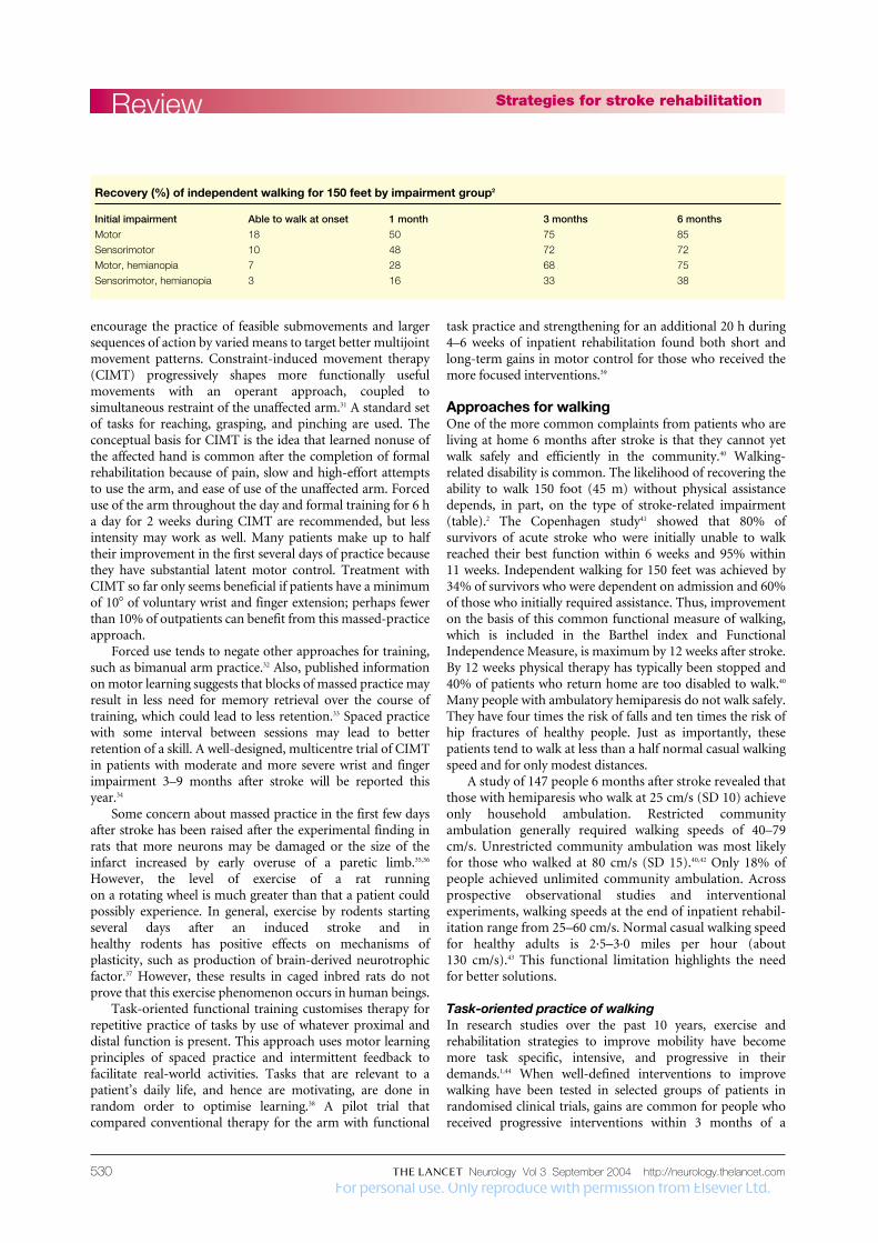
Approaches for walkingOne of the more common complaints from patients who areliving at home 6 months after stroke is that they cannot yetwalk safely and efficiently in the community.40 Walking-related disability is common. The likelihood of recovering theability to walk 150 foot (45 m) without physical assistancedepends, in part, on the type of stroke-related impairment(table).2 The Copenhagen study41 showed that 80% ofsurvivors of acute stroke who were initially unable to walkreached their best function within 6 weeks and 95% within11 weeks. Independent walking for 150 feet was achieved by34% of survivors who were dependent on admission and 60%of those who initially required assistance. Thus, improvementon the basis of this common functional measure of walking,which is included in the Barthel index and FunctionalIndependence Measure, is maximum by 12 weeks after stroke.By 12 weeks physical therapy has typically been stopped and40% of patients who return home are too disabled to walk.40
Many people with ambulatory hemiparesis do not walk safely.They have four times the risk of falls and ten times the risk ofhip fractures of healthy people. Just as importantly, thesepatients tend to walk at less than a half normal casual walkingspeed and for only modest distances.
A study of 147 people 6 months after stroke revealed thatthose with hemiparesis who walk at 25 cm/s (SD 10) achieveonly household ambulation. Restricted communityambulation generally required walking speeds of 40–79cm/s. Unrestricted community ambulation was most likelyfor those who walked at 80 cm/s (SD 15).40,42 Only 18% ofpeople achieved unlimited community ambulation. Acrossprospective observational studies and interventionalexperiments, walking speeds at the end of inpatient rehabil-itation range from 25–60 cm/s. Normal casual walking speedfor healthy adults is 2·5–3·0 miles per hour (about130 cm/s).43 This functional limitation highlights the needfor better solutions.
Task-oriented practice of walkingIn research studies over the past 10 years, exercise andrehabilitation strategies to improve mobility have becomemore task specific, intensive, and progressive in theirdemands.1,44 When well-defined interventions to improvewalking have been tested in selected groups of patients inrandomised clinical trials, gains are common for people whoreceived progressive interventions within 3 months of a
Recovery (%) of independent walking for 150 feet by impairment group2
Initial impairment Able to walk at onset 1 month 3 months 6 months
Motor 18 50 75 85
Sensorimotor 10 48 72 72
Motor, hemianopia 7 28 68 75
Sensorimotor, hemianopia 3 16 33 38
For personal use. Only reproduce with permission from Elsevier Ltd.531
ReviewStrategies for stroke rehabilitation
Neurology Vol 3 September 2004 http://neurology.thelancet.com
stroke.44,45 Conventional physiotherapy—with or withouttreadmill activities and of modest intensity—have also led toimproved walking skills, including level of independence andwalking speed, when started 6–18 months after stroke.46–53
Although patients who make large gains tend to be amongthose with more modest impairments, these studies confirmthe potential to improve function by trying a pulse of goal-directed physical therapy at any time after stroke.
One approach for massed practice is treadmill trainingwith partial support of body weight. This strategy is developedfrom studies of cats with transected spinal cords,54 but thistheoretical basis seems less translatable to humans than mostreports suggest. Many postural and automatic features of theneural control for quadrupedal walking are managed at thelevel of the lumbar motor pools, which probably includes theconservation of a subcomponent of the locomotor systemcalled central pattern generators. This system facilitatesautomatic reciprocal flexor and extensor stepping movementsof the hindlimbs.55,56 Remarkably, cats have been trained towalk on a moving treadmill belt with trunk support after a lowthoracic cord transection with their dorsal and ventral rootsintact, but they cannot walk over ground.57 Human beings,however, probably depend less than other mammals onpattern generation. Bipedal walking required a number ofevolutionary musculoskeletal adaptations and accompanyingneural computations to manage human locomotion.58 Forexample, human beings must land on the heel and rollforward to the ball of the foot to push off for greater energyefficiency; quadrupeds do not. Supraspinal motor regions arequite active in human beings during over ground or treadmillwalking as revealed by functional imaging techniques.59,60
Despite its theoretical basis, body-weight-supportedtreadmill training (BWSTT) has had a disappointing effect onwalking outcomes when compared with conventional trainingof the same duration, at least in randomised clinical trialsbegun within 2 months of onset of hemiplegia. In these trialsduring inpatient rehabilitation, patients were trained at slowtreadmill speeds (from 0·2–1·0 mph) and body-weightsupport was used only until patients could step at slow speedswithout assistance, rather than using support to enablepractice at faster speeds.45,61–64 Training at fast treadmill speedsleads to fast walking over ground.51,65 Patients withhemiparesis can safely exercise at a level of effort that providesa conditioning response.47 The energy cost of walking maydecrease by 50% as walking speeds increase from 40 cm/s to150 cm/s.66
BWSTT may have a greater effect in patients with morechronic hemiparesis who walk with a poor gait pattern andslowly (less than 80 cm/s) beyond the time usually allotted forinpatient and outpatient rehabilitation.51,61,67 Importantfeatures of training include practice at treadmill speedsconsistently faster than the subject can walk over ground andphysical and verbal cues from therapists who help to optimisethe symmetry of stepping, joint angles, and timing of loadingand unloading each leg by manipulating the legs and pelvis.No randomised clinical trial has been done. A recentCochrane review of 11 trials with a total of 458 participantsconcluded that a well-designed large-scale study to assessBWSTT after stroke is urgently needed.68
An approach such as BWSTT is not a stand-alone therapy.It must be complemented by training on various surfaces thatfurther help patients recapture motor control for a better gaitpattern, as well as by working on strengthening, conditioning,and balance. Physicians can encourage patients to work onthese features of walking. One simple technique to rapidlyimprove the symmetry and speed of walking is to have thepatient more consciously try to push the leg that is in mid-stance into the floor and, at that moment, pull the other legfrom the hip to initiate its swing forward. This load and swingcue improves attention to the timing of loading the stance leg(particularly the unaffected one) and swinging the other(especially the affected leg). The strategy is supported by datafrom studies of sensory triggers for rhythmic stepping inhuman beings and other mammals.69,70
SpasticityClinicians may spend needless effort in trying to treatincreases in muscle tone, despite the fact that spasticity is notcausing any fuctional disability. Clinically significant spasticityis rather uncommon after stroke.71 The loss of independentmovements, such as hip flexion and knee extension forwalking or elbow extension for reaching with simultaneouswrist extension (movement out of synergy), is commonlythought to be treatable by interventions that decrease muscletone. Impairment in making a reaching movement, such asraising the hemiparetic arm to reach while extending theelbow, seems to reflect a problem in motor control forisolation of individualised movements, not due to spasticity.72
Muscle tone is poorly related with functional disability.Indeed, bad motor control—as assessed by paresis, impaireddexterity, and fatigability, along with tissue changes inmuscle73—is usually more limiting than increased tone.1
Hypertonicity that leads to dystonic flexor postures of thehand and inversion of the foot is perhaps most commonlycaused by pain, overuse, and impaired postural control thatdrives flexor or extensor activity. Immediate medical andrehabilitation management will prevent dystonic postures. Abrief course of antispasticity drugs may also help, butcontinued drug use should be reassessed at least quarterly.Selective serotonin reuptake inhibitors and dopamineblockers can sometimes increase hypertonicity, so they shouldbe stopped. Antispasticity drugs and locally injectedbotulinum toxin rarely improve function of the arms. Studiesthat report efficacy for the hand and arm rely on scales that donot address functional limitations.74 Also, trials do notcombine the intervention with range of motion, weightbearing, and other rehabilitation interventions that mayeliminate the need to denervate a muscle every 3–4 months.Botulinum toxin injected into the tibialis posterior and toeplantar flexors may improve the gait of patients with plantarflexion and inversion that limits heel strike and stance, butmust be followed by stretching and gait training.
How much training?Many small trials reveal motor and behaviouralimprovements in patients with chronic disability after a pulseof therapy aimed at a specific impairment or functional goal atany time after a stroke (panel 3).51,61,65,75–82 An ethical challenge
For personal use. Only reproduce with permission from Elsevier Ltd.532
Review Strategies for stroke rehabilitation
Neurology Vol 3 September 2004 http://neurology.thelancet.com
for the providers of therapies is to find common agreementwith patients about realistic functional and quality-of-lifegoals. Clinicians should test adjunct strategies on patients whohave at least modest motor control before deciding that nofurther gains can be made.
The intensity and duration of therapy varies widely instudies. The decision about frequency and duration of therapyis, in many cases, made entirely on the basis of what ispractical for patients and investigators in a research study oron the number of treatment sessions covered by paymentsfrom a third party. Some general guidelines can be drawn.Most studies that led to a very significant 10 point gain on theFugl–Meyer motor assessment of selective movements orgreater strength provided specific treatment by a therapist for12–60 h, running from 1–6 h a day, 3–5 days a week for2–12 weeks. Most likely, the amount of goal-directed therapy,rather than the daily intensity, is the most important factor.83
Many outpatient clinical trials offer the experimentalintervention for 1 h/day, 3 days a week for 2–3 months.44 Wehave found that task-specific functional therapy for the armleads to significant gains in motor control and strength if
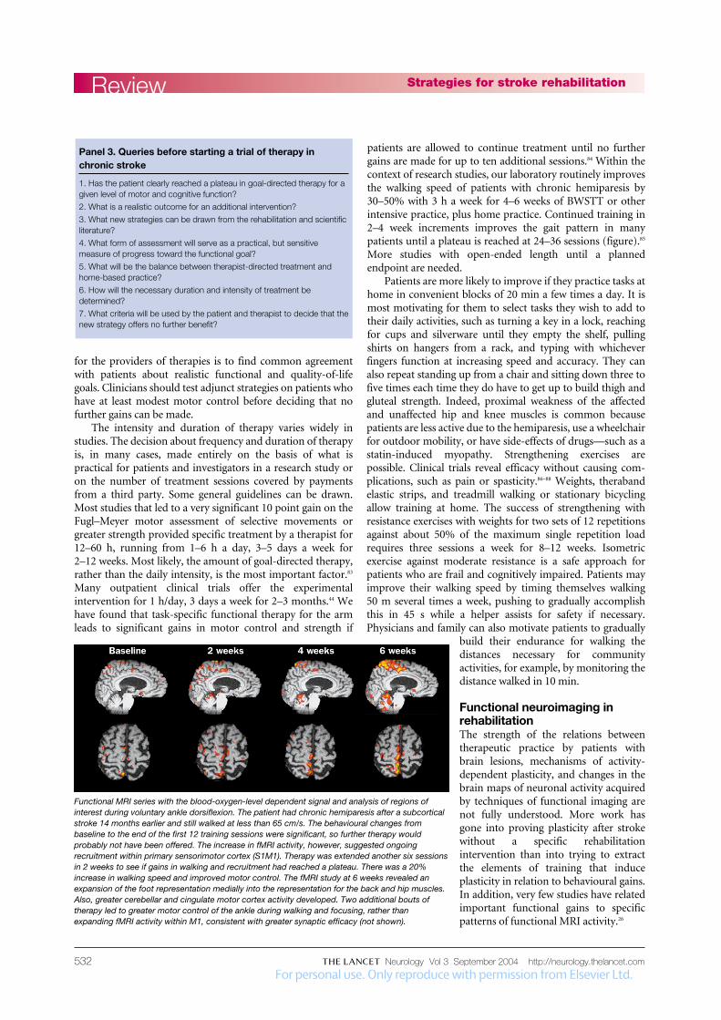
patients are allowed to continue treatment until no furthergains are made for up to ten additional sessions.84 Within thecontext of research studies, our laboratory routinely improvesthe walking speed of patients with chronic hemiparesis by30–50% with 3 h a week for 4–6 weeks of BWSTT or otherintensive practice, plus home practice. Continued training in2–4 week increments improves the gait pattern in manypatients until a plateau is reached at 24–36 sessions (figure).85
More studies with open-ended length until a plannedendpoint are needed.
Patients are more likely to improve if they practice tasks athome in convenient blocks of 20 min a few times a day. It ismost motivating for them to select tasks they wish to add totheir daily activities, such as turning a key in a lock, reachingfor cups and silverware until they empty the shelf, pullingshirts on hangers from a rack, and typing with whicheverfingers function at increasing speed and accuracy. They canalso repeat standing up from a chair and sitting down three tofive times each time they do have to get up to build thigh andgluteal strength. Indeed, proximal weakness of the affectedand unaffected hip and knee muscles is common becausepatients are less active due to the hemiparesis, use a wheelchairfor outdoor mobility, or have side-effects of drugs—such as astatin-induced myopathy. Strengthening exercises arepossible. Clinical trials reveal efficacy without causing com-plications, such as pain or spasticity.86–88 Weights, therabandelastic strips, and treadmill walking or stationary bicyclingallow training at home. The success of strengthening withresistance exercises with weights for two sets of 12 repetitionsagainst about 50% of the maximum single repetition loadrequires three sessions a week for 8–12 weeks. Isometricexercise against moderate resistance is a safe approach forpatients who are frail and cognitively impaired. Patients mayimprove their walking speed by timing themselves walking50 m several times a week, pushing to gradually accomplishthis in 45 s while a helper assists for safety if necessary.Physicians and family can also motivate patients to gradually
build their endurance for walking thedistances necessary for communityactivities, for example, by monitoring thedistance walked in 10 min.
Functional neuroimaging inrehabilitationThe strength of the relations betweentherapeutic practice by patients withbrain lesions, mechanisms of activity-dependent plasticity, and changes in thebrain maps of neuronal activity acquiredby techniques of functional imaging arenot fully understood. More work hasgone into proving plasticity after strokewithout a specific rehabilitationintervention than into trying to extractthe elements of training that induceplasticity in relation to behavioural gains.In addition, very few studies have relatedimportant functional gains to specificpatterns of functional MRI activity.26
Functional MRI series with the blood-oxygen-level dependent signal and analysis of regions ofinterest during voluntary ankle dorsiflexion. The patient had chronic hemiparesis after a subcorticalstroke 14 months earlier and still walked at less than 65 cm/s. The behavioural changes frombaseline to the end of the first 12 training sessions were significant, so further therapy wouldprobably not have been offered. The increase in fMRI activity, however, suggested ongoingrecruitment within primary sensorimotor cortex (S1M1). Therapy was extended another six sessionsin 2 weeks to see if gains in walking and recruitment had reached a plateau. There was a 20%increase in walking speed and improved motor control. The fMRI study at 6 weeks revealed anexpansion of the foot representation medially into the representation for the back and hip muscles.Also, greater cerebellar and cingulate motor cortex activity developed. Two additional bouts oftherapy led to greater motor control of the ankle during walking and focusing, rather thanexpanding fMRI activity within M1, consistent with greater synaptic efficacy (not shown).
Panel 3. Queries before starting a trial of therapy inchronic stroke
1. Has the patient clearly reached a plateau in goal-directed therapy for agiven level of motor and cognitive function?
2. What is a realistic outcome for an additional intervention?
3. What new strategies can be drawn from the rehabilitation and scientificliterature?
4. What form of assessment will serve as a practical, but sensitivemeasure of progress toward the functional goal?
5. What will be the balance between therapist-directed treatment andhome-based practice?
6. How will the necessary duration and intensity of treatment bedetermined?
7. What criteria will be used by the patient and therapist to decide that thenew strategy offers no further benefit?
For personal use. Only reproduce with permission from Elsevier Ltd.533
ReviewStrategies for stroke rehabilitation
Neurology Vol 3 September 2004 http://neurology.thelancet.com
Functional imaging, however, may offer clinicians insight into how to better engage a network in the hope ofimproving rehabilitation training techniques. For example,the magnitude of the blood-oxygen-level-dependent signal in functional MRI studies is greater within M1 and thesupplementary motor cortex when the eyes gaze in thedirection of the hand doing a sequential motor task than whenlooking at the inactive hand. Activity within the dorsal premotor cortex for the affected hand after strokereveals its contribution as a substrate of functional gains for use of the hand, bolstered by connections to visuomotor regions.89 Thus, focused visual input may be an important feature of training. Transcranial magneticstimulation (TMS) and functional MRI studies over the time of a treatment strategy could take snapshots of anetwork, to ensure it was engaged by the intervention.Imaging could also be done during a verbal or visuospatialworking memory task to help estimate a patient’s residualneural resources for learning and potential to employ mentalimagery and observation of arm actions as forms ofpractice.90,91
PET, functional MRI, and TMS92 has detected functionalreorganisation as a patient’s sensorimotor and cognitiveexperience was manipulated, primarily in studies of armrecovery.26,93 For walking, near-infrared spectroscopy94 duringstepping on a treadmill and functional MRI during ankledorsiflexion (figure)85,95,96 serve as markers of training-inducedplasticity. Improved motor control of the leg and faster walking speed were accompanied by recruitment ofsensorimotor system neurons and representational changes.Longitudinal functional MRI mapping studies may offerinsight into the optimal intensity or duration of a task-specific therapy.85 Mapping of brain-activity patterns mayalso help to predict the capacity of a movement representationto reorganise in response to a particular therapy soon after thestart of the intervention. For example, TMS of the handrepresentation in M1 of the affected hemisphere reveals lessexcitability after stroke than stimulation of the unaffectedside. Increased excitability of affected M1 with greatersymmetry between the two sides in the first several sessions oftask-oriented training of the hemiparetic arm was associatedwith long-term gains in the Fugl–Meyer score for motorcontrol.84
NeuroaugmentationEven with optimised approaches and intensities of physical,occupational, language, and cognitive rehabilitation, othermanipulations may be needed to increase gains. Theseinterventions require further study but build on growingresearch rationales.
Neuropharmacological adjunctsCascades of molecular interactions underlie activity-dependent plasticity and skills learning. Many of theseprocesses involve the major neurotransmitters.97 After stroke,dopamine, acetylcholine, serotonin, and norepinephrine maybe interrupted or downregulated in their projections to cortexfrom sites of origin in the brainstem. This may contribute todiaschisis, the failure of transynaptic neurotransmission.
Clinicians may soon identify individuals with genotypes thatalso reduce central aminergic or cholinergic tone. These “low-aminergic” patients may benefit, for example, fromamphetamine after a brain injury.98 Pertinent to clinicalpractice is the possibility that commonly used drugs maychange neurotransmitter concentrations or block theirreceptors, leading to iatrogenic loss of tone. Stroke trialssuggest at least a transient decline in function fromantiepileptic drugs (which affect cognitive processing),dopamine blockers such as antipsychotics, and �2 adrenergicdrugs.99
Augmentation of one of these chemical transmitters,especially activation of NMDA receptors,100 may drive long-term potentiation and optimise activity-dependent relearningof skills after stroke. Available acetylcholinesterase inhibitorsmay aid both declarative and procedural learning.16,101 Amongtheir functions, acetylcholine projections from the nucleusbasalis transmit behaviourally relevant sensory information.Dopaminergic drugs such as levodopa,102 as well as drugs thatincrease the availability of norepinephrine, such asmethylphenidate103 and amphetamine,104,105 have shown someefficacy in stroke-rehabilitation trials when combined withphysical or language therapy. Dopaminergic projections fromthe ventral tegmental tract relate a reward to the cognitiveeffort of the task, which reinforces associative learning.Norepinephrine projections from the locus coeruleus alsomodulate the saliency of sensory inputs, attention, andfeatures of memory.106 Drugs that act on theseneurotransmitter receptors improve task-specific signalling.107
Selective serotonin reuptake inhibitors have modestlyimproved motor learning in healthy people and patients withstroke for arm tasks, as the drugs increased regional brainactivity.108,109 Studies with TMS suggest that selective serotoninreuptake inhibitors increase the excitability of corticospinalneurons.110
Learning and synaptic plasticity are also modulated byother molecules, such as cAMP response element-bindingprotein (CREB), which can be activated by drugs.111
Pharmaceutical research is very active in the development of“memory” drugs for Alzheimer’s disease that could alsoenable task-specific learning. The propensity forresponsiveness to a drug may be predicted by fMRI and PETactivation studies112 and by TMS.113,114 These tools, then, mayhelp clinicians choose a drug for an individualised approachto augment rehabilitation.115
Electrical neurostimulationMotor gains and limited functional gains have been elicited byelectrical neuromuscular stimulation triggered by feedback.One approach stimulates a weak muscle, such as the wristextensors, on the basis of the amplitude of theelectromyographic signal during attempted voluntarymovement of a joint.32,116 Other biofeedback techniques mayhelp patients activate a muscle or joint movement, althoughthere are few data. Direct neuromuscular electricalstimulation over the surface of key muscles for a grasp andpinch movement may improve motor skills when combinedwith task-oriented practice.117 Functional electricalstimulation is also used to activate paretic muscles timed
For personal use. Only reproduce with permission from Elsevier Ltd.534
Review Strategies for stroke rehabilitation
Neurology Vol 3 September 2004 http://neurology.thelancet.com
to a movement, such as contraction of the tibialis anterior toclear the foot during the swing phase of walking. InjectableBION electrodes triggered by an external transmitter haveshown potential as a muscle-stimulating neuroprosthesis.118
Cortical plasticity has been modulated for up to severalhours in animals and human beings by peripheral-nerve anddirect cortical stimulation, independently and incombination.119 These methods include cortical motor stim-ulation by repetitive TMS,120 direct electrode array stimulationover the dura of M1,121 repetitive peripheral nervestimulation,122 and paired associative stimulation with a TMSpulse immediately after a peripheral nerve stimulus.123,124
Mechanisms of action include an increase in NMDAexcitation, a decrease in GABA inhibition, an increase inneuronal excitability, and Hebbian associative synapticlearning. In a similar vein, local anaesthesia of a portion of theaffected hand rapidly changed representational plasticity andimproved function.125 The inducing of anaesthesia in theunaffected hand seemed to reverse inhibition of theunaffected hemisphere on the affected side,126 leading totransient gains in hand strength.127 Practice combined withcarefully chosen stimulation may come into clinical use toaugment short-term learning and function.
Robotic trainers and virtual-reality practiceThese adjuncts may maximise the intensity and convenienceof task-oriented practice. Computerised virtual reality-basedactivities have a reasonable conceptual basis forrehabilitation.128–130 A virtual-reality system may include acomputer or television screen that shows the virtualenvironment, a device on the patient’s hand that recordsmovement direction and acceleration as he or she reaches intocyberspace, and motivational games that encourage reaching,grasping, and manipulation. People can watch and attempt tomimic a movement that may activate a class of mirrorneurons to drive visuomotor neuroplasticity.131 Theinvestigator can give quantitative feedback about the accuracyand speed of movement. A few small trials suggest possibleeffectiveness for these forms of practice.132
For reaching or walking, a range of clever automatedassistive devices are in development.133 They may offer moreintensive practice opportunities without increasing time spenton supervision by the treating therapist. Robotic devices canalso measure motor skills, especially during movements of thewhole limb. For the arm, devices have been limited toshoulder and elbow movements. Results from small trialspoint more to improved strength79 than to improvedfunction.134,135 For walking, a stepper device produced equalresults to conventional gait training in terms of walkingspeed.136 Future trials of the devices should seek betterdefinition of the optimal intensity and duration of treatment.
Neural Repair Animal models of stroke and spinal-cord injury reveal thepotential for clinicians to modulate similar acute and chronicresponses for repair.137 Biological interventions may promoteregeneration of cells, axons, and neural circuitry. Endogenousneurons and glia from the subventricular zone near an infarctmay proliferate and migrate toward the stroke in response tolocal signals. Exogenous stem cells, neural progenitors, bone-marrow stromal cells,138 and other types139 offer hope forrestoration of cells and their proteins for repair.140 These cellsmay bridge an injury site to enable greater connectivity. Stemcells and engineered fibroblasts are readily engineered to makebrain-derived neurotrophic factor and acetylcholine, forexample, which could allow placement of the growth factor orneurotransmitter where needed. Pharmacological andimmunological approaches may target growth-cone receptorsto provide signals for regeneration or to block inhibitoryfactors in the environment.141 However, incorporation of newneurons and axons that augment functional recovery will also depend on rehabilitation therapies, especially those that drive activity-dependent plasticity. Without such drives, thesenew elements and cortical and subcortical projections maynot become incorporated into learning networks.36,142,143
ConclusionsRehabilitation after stroke must continue to address seriousfunctional limitations, such as walking speed and distance thatpermit community activities and better use of a hemipareticarm and hand. A growing understanding of the molecules andphysiology of neuroplasticity during motor-skills learning hasmade an exciting contribution to new strategies for strokerehabilitation. Clinicians and scientists can now design andtest therapies that manipulate cerebral adaptations to lessenimpairments, disabilities, and functional limitations. Funct-ional MRI, TMS, and other physiological windows on brainfunction may offer guidance about whether relevant networksare engaged and manipulated over the time that a training,pharmacological, or biological intervention proceeds. Insightsmay be gained about the optimal intensity and duration oftherapies and about how different therapies interact. This information may one day help individualiserehabilitation approaches and lessen the need for large clinicaltrials.
The recent emphasis of neurological rehabilitation onbasic and clinical scientific research, theory-basedinterventions, better clinical-trial designs and outcomemeasures, systematic testing of interventions, and task-oriented therapies at any time after stroke will better realisethe potential of millions of disabled people.
Conflict of interestI have no conflicts of interest.
Role of the funding sourceResearch in my laboratory contributing to this review is supported by theNational Institutes of Health at the National Institute of Child Healthand Human Development (HD39629) and the National Institute ofNeurological Disorders and Stroke (HD0741), the Larry L HillblomFoundation, and the Nathan Shapell Foundation. None of these fundingsources had a role in the preparation of this review or in the decision tosubmit it for publication.
Search strategy and selection criteriaArticles were identified by searches of MEDLINE with thewords “rehabilitation”, “stroke”, “motor control”, and “recoveryof function”, from 1990–2004, from the author’s files andtextbooks. Only references published in English were used.
For personal use. Only reproduce with permission from Elsevier Ltd.535
ReviewStrategies for stroke rehabilitation
Neurology Vol 3 September 2004 http://neurology.thelancet.com
References1 Dobkin B. The clinical science of neurologic
rehabilitation. New York: Oxford University Press;2003.
2 Patel A, Duncan P, Lai S, Studenski S. The relationbetween impairments and functional outcomespoststroke. Arch Phys Med Rehabil 2000;81: 1357–63.
3 Jorgensen H, Nakayama H, Raaschou H, et al.Outcome and time course of recovery in stroke, part I: time course. Arch Phys Med Rehabil 1995;76: 406–12.
4 Dobkin B. Focused stroke rehabilitation programs donot improve outcome. Arch Neurol 1989;46: 701–03.
5 Gresham C, Duncan P, Stason W, et al. Post-strokerehabilitation: assessment, referral, and patientmanagement: clinical practice guideline No 16, reportno 95-0663. Rockville, MD: U.S. Department ofHealth and Human Services, Public Health Service,Agency for Health Care Policy and Research, 1995.
6 Teasell R, Foley N, Bhogal S, M S. An evidence-basedreview of stroke rehabilitation. Top Stroke Rehabil2003; 10: 29-58.
7 Roth E, Lovell L, Harvey R, Heinemann A, Semik P,Diaz S. Incidence of and risk factors for medicalcomplications during stroke rehabilitation. Stroke2001; 32: 523-9.
8 Stineman M, Ross R, Maislin G, Fiedler R, Granger C.Risks of acute hospital transfer and mortality duringstroke rehabilitation. Arch Phys Med Rehabil 2003;84: 712–18.
9 Stroke Unit Trialists’ Collaboration. Organisedinpatient (stroke unit) care for stroke (CochraneReview). In: The Cochrane Library, Issue 2, 2004.Chichester, UK: John Wiley & Sons, Ltd.
10 Legg L. Outpatient Service Trialists: Rehabilitationtherapy services for stroke patients living at home:systematic review of randomised trials. Lancet 2004;363: 352–56.
11 Dobkin B. The neurobiology of rehabilitation. In:Kaler SNN, ed. Understanding and optimizing humandevelopment from cells to patients to populations.New York: New York Acad Sci, (in press).
12 Donchin O, Sawaki L, Madupu G, Cohen LG,Shadmehr R. Mechanisms influencing acquisition andrecall of motor memories. J Neurophysiol 2002;88: 2114–23.
13 Frost S, Barbay S, Friel K, Plautz E, Nudo R.Reorganization of remote cortical regions afterischemic brain injury: a potential substrate for strokerecovery. J Neurophysiol 2003; 89: 3205–14.
14 Bury S, Jones T. Unilateral sensorimotor cortexlesions in adult rats facilitate motor skill learning withthe “unaffected” forelimb and training-induceddendritic structural plasticity in the motor cortex. J Neurosci 2002; 22: 8597–606.
15 Carmichael S. Plasticity of cortical projections afterstroke. Neuroscientist 2003; 9: 64–73.
16 Conner J, Culberson A, Packowski C, Chiba A,Tuszynski M. Lesions of the basal forebraincholinergic system impair task acquisition and abolishcortical plasticity associated with motor skill learning.Neuron 2003; 38: 819–29.
17 Rossini P, Calautti C, Pauri F, Baron J-C. Post-strokeplastic reorganization in the adult brain. LancetNeurol 2003; 2: 493–502.
18 Dobkin B. Activity-dependent learning contributes tomotor recovery. Ann Neurol 1998; 44: 158–60.
19 Wei L, Erinjeri J, Rovainen C, Woolsey T. Collateralgrowth and angiogenesis around cortical stroke.Stroke 2001; 32: 2179–84.
20 Katsman D, Zheng J, Spinelli K, Carmichael S. Tissue microenvironments within functional corticalsubdivisions adjacent to focal stroke. J Cereb Blood Flow Metab 2003; 23: 997–1009.
21 Croquelois A, Wintermark M, Reichhart M, Meuli R,Bogousslavsky J. Aphasia in hyperacute stroke:language follows brain penumbra dynamics. Ann Neurol 2003; 54: 321–29.
22 Nudo R, Wise B, SiFuentes F, Milliken G. Neuralsubstrates for the effects of rehabilitative training onmotor recovery after ischemic infarct. Science 1996;272: 1791–94.
23 Sergio L, Kalaska J. Systematic changes in motorcortex cell activity with arm posture duringdirectional isometric force generation. J Neurophysiol2003; 89: 212–28.
24 Todorov E, Jordan M. Optimal feedback control as atheory of motor coordination. Nat Neurosci 2002;5: 1226–35.
25 Scott S, Norman K. Computational approaches tomotor control and their potential for interpretingmotor dysfunction. Curr Opin Neurol 2003;16: 693–98.
26 Calautti C, Baron J-C. Functional neuroimagingstudies of motor recovery after stroke in adults: areview. Stroke 2003; 34: 1553–66.
27 Graybiel A, Aosaki T, Flaherty A, Kimura M. Thebasal ganglia and adaptive motor control. Science1994; 265: 1826–31.
28 Dietz V. Spinal cord pattern generators forlocomotion. Clin Neurophysiol 2003; 114: 1379–89.
29 Bizzi E, Tresch M, Saltiel P, d’Avella A. Newperspectives on spinal motor systems. Nat Rev Neurosci 2000; 1: 101–08.
30 Graziano M, Taylor C, Moore T. Complexmovements evoked by microstimulation of precentralcortex. Neuron 2002; 34: 841–51.
31 Taub E, Uswatte G, Elbert T. New treatments inneurorehabilitation founded on basic research. Nat Rev Neurosci 2002; 3: 228–36.
32 Cauraugh JH, Kim SB. Stroke motor recovery: activeneuromuscular stimulation and repetitive practiceschedules. J Neurol Neurosurg Psychiatry 2003;74: 1562–66.
33 Schmidt R, Wrisbert C. Motor learning andperformance: a problem-based learning approach.Champaign, IL: Human Kinetics; 2000.
34 Winstein C, Miller J, Blanton S, et al. Methods for amulti-site randomized trial to investigate the effect ofconstraint-induced movement therapy in improvingupper extremity function among adults recoveringfrom a cerebrovascular stroke. Neurorehabil NeuralRepair 2003; 17: 137–52.
35 Bland S, Pillai R, Aronowski J, Grotta J, Schallert T.Early overuse and disuse of the affected forelimb aftermoderately severe intraluminal suture occlusion ofthe middle cerebral artery in rats. Behav Brain Res 2001; 126: 33–41.
36 Biernaskie J, Chernenko G, Corbett D. Efficacy ofrehabiliitative experience declines with time after focalischemic brain injury. Neurobiol Dis 2004;24: 1245–54.
37 van Praag H, Kempermann G, Gage F. Neuralconsequences of environmental enrichment. Nat Rev Neurosci 2000; 1: 191–98.
38 Hanlon R. Motor learning following unilateral stroke.Arch Phys Med Rehabil 1996; 77: 811–15.
39 Winstein C, Rose D, Tan S, Lewthwaite R, Chui H,Azen S. A randomized controlled comparison ofupper-extremity rehabilitation strategies in acutestroke: a pilot study of immediate and long-termoutcomes. Arch Phys Med Rehabil 2004; 85: 620–28.
40 Lord S, McPherson K, McNaughton H, Rochester L,Weatherall M. Community ambulation after stroke:how important and obtainable is it and whatmeasures appear predictive? Arch Phys Med Rehabil2004; 85: 234–39.
41 Jorgensen H, Nakayama H, Raaschou H, Olsen T.Recovery of walking function in stroke patients: TheCopenhagen Stroke Study. Arch Phys Med Rehabil1995; 76: 27–32.
42 Perry J, Garrett M, Gromley J, Mulroy S.Classification of walking handicap in the strokepopulation. Stroke 1995; 26: 982–89.
43 Bohannon R. Comfortable and maximum walkingspeed of adults aged 20 to 79 years: reference valuesand determinants. Age Ageing 1997; 26: 15–19.
44 Duncan P, Studenski S, Richards L, et al. Randomizedclinical trial of therapeutic exercise in subacute stroke.Stroke 2003; 34: 2173–80.
45 Visintin M, Barbeau H, Korner-Bitensky N, Mayo N.A new approach to retrain gait in stroke patientsthrough body weight support and treadmillstimulation. Stroke 1998; 29: 1122–28.
46 Green J, Forster A, Bogle S, Young J. Physiotherapyfor patients with mobility problems more than 1 yearafter a stroke: a randomised controlled trial. Lancet2002; 359: 199–203.
47 Macko R, Smith G, Dobrovolny C, Sorkin J, Goldberg A, Silver K. Treadmill training improvesfitness reserve in chronic stroke patients. Arch Phys Med Rehabil 2001; 82: 879–84.
48 Dean C, Richards C, Malouin F. Task-related circuittraining improves performance of locomotor tasks inchronic stroke: a randomized, controlled pilot trial.Arch Phys Med Rehabil 2000; 81: 409–17.
49 Dam M, Tonin P, Casson S, et al. The effects of long-term rehabilitation therapy on poststroke hemiplegicpatients. Stroke 1993; 24: 1886–91.
50 Werner C, Bardeleben A, Mauritz KH, Kirker S, Hesse S. Treadmill training with partial body weightsupport and physiotherapy in stroke patients: apreliminary comparison. Eur J Neurol 2002; 9:639–44.
51 Sullivan K, Knowlton B, Dobkin B. Step training withbody weight support: Effect of treadmill speed andpractice paradigms on post-stroke locomotorrecovery. Arch Phys Med Rehabil 2002; 83: 683–91.
52 Ada L, Dean CM, Hall JM, Bampton J, Crompton S.A treadmill and overground walking programimproves walking in persons residing in thecommunity after stroke: a placebo-controlled,randomized trial. Arch Phys Med Rehabil 2003; 84:1486–91.
53 Yagura H, Miyai I, Seike Y, Suzuki T, Yanagihara T.Benefit of inpatient multidisciplinary rehabilitationup to 1 year after stroke. Arch Phys Med Rehabil 2003;84: 1687–91.
54 Barbeau H. Locomotor training inneurorehabilitation: emerging rehabilitation concepts.Neurorehabil Neural Repair 2003; 17: 3–11.
55 Edgerton V, Roy R. Paralysis recovery in human andmodel systems. Curr Opin Neurobiol 2002; 12: 658–67.
56 Grillner S. The motor infrastructure: from ionchannels to neuronal networks. Nat Rev Neurosci2003; 4: 573–86.
57 Rossignol S, Bouyer L, Barthelemy D, Langlet C,Leblond H. Recovery of locomotion in the catfollowing spinal cord lesions. Brain Res Rev 2002;40: 257–66.
58 Nielsen J. How we walk: Central control of muscleactivity during human walking. Neuroscientist 2003;9: 195–204.
59 Dobkin B. Recovery of locomotor control. Neurologist1996; 2: 239–49.
60 Miyai I, Tanabe H, Sase I, et al. Cortical mapping ofgait in humans: a near-infrared spectroscopictopography study. NeuroImage 2001; 14: 1186–92.
61 Hesse S, Bertelt C, Jahnke M, Baake P, Mauritz K.Treadmill training with partial body weight supportcompared with physiotherapy in nonambulatoryhemiparetic patients. Stroke 1995; 26: 976–81.
62 Nilsson L, Carlsson J, Danielsson A, et al. Walkingtraining of patients with hemiparesis at an early stageafter stroke: a comparison of walking training on atreadmill with body weight support and walkingtraining on the ground. Clin Rehabil 2001; 15: 515–27.
63 Kosak M, Reding M. Comparison of partial bodyweight-supported treadmill gait training versusaggressive bracing assisted walking post stroke.Neurorehabil Neural Repair 2000; 14: 13–19.
64 Lennihan L, Wootten M, Wainwright M,Tenteromano L, McMahon D, Cotier J. Treadmillwith partial body-weight support versus conventionalgait training after stroke. Arch Phys Med Rehabil 2003;84: E5.
65 Pohl M, Mehrholz J, Ritschel C, Ruckriem S. Speed-dependent treadmill training in ambulatoryhemiparetic stroke patients. Stroke 2002; 33: 553–58.
66 Hesse S, Weerner C, Paul T, Bardeleben A, Chaler J.Influence of walking speed on lower limb muscleactivity and energy consumption during treadmillwalking of hemiparetic patients. Arch Phys Med Rehabil 2001; 82: 1547–50.
67 Hesse S, Bertelt C, Schaffrin A, Malezic M, Mauritz K.Restoration of gait in nonambulatory hemipareticpatients by treadmill training with partial body-weightsupport. Arch Phys Med Rehabil 1994; 75: 1087–93.
68 Moseley AM, Stark A, Cameron ID, Pollock A.Treadmill training and body weight support forwalking after stroke (Cochrane Review). In: The Cochrane Library, Issue 2, 2004. Chichester,UK: John Wiley & Sons, Ltd.
69 Dietz V, Duysens J. Significance of load receptor inputduring locomotion: a review. Gait Posture 2000; 11:102–10.
70 Harkema S, Hurley S, Patel U, Dobkin B, Edgerton V.Human lumbosacral spinal cord interprets loadingduring stepping. J Neurophysiol 1997; 77: 797–811.
71 Sommerfeld D, Eek E, Svensson A-K, Holmqvist L,von Arbin M. Spasticity after stroke. Stroke 2004;35: 134–39.
72 Zackowski K, Dromerick A, Sahrmann S, Thach W,Bastian A. How do strength, sensation, spasticity andjoint individuation relate to the reaching deficits ofpeople with chronic hemiparesis? Brain 2004;127: 1035–46.
73 Lieber R, Einarsson F, Friden J. Inferior mechanicalproperties of spastic muscle bundles due tohypertrophic but compromised extracellular matrixmaterial. Muscle Nerve 2003; 28: 464–71.
74 Brashear A, Gordon M, Elovic E, et al. Intramuscularinjection of botulinum toxin for the treatment ofwrist and finger spasticity after a stroke. N Engl J Med2002; 347: 395–400.
75 Bonan I, Yelnik A, Colle F, et al. Reliance on visualinformation after stroke, part II: effectiveness of abalance rehabilitation program with visual cuedeprivation after stroke, a randomized controlledtrial. Arch Phys Med Rehabil 2004; 85: 274–78.
76 Pulvermuller F, Neininger B, Elbert T, et al.Constraint-induced therapy of chronic aphasia afterstroke. Stroke 2001; 32: 1621–26.
For personal use. Only reproduce with permission from Elsevier Ltd.536
Review Strategies for stroke rehabilitation
Neurology Vol 3 September 2004 http://neurology.thelancet.com
77 Carlomagno S, Pandolfi M, Labruna L, Colombo A,Razzano C. Recovery from moderate aphasia in thefirst year poststroke: effect of type of therapy. Arch Phys Med Rehabil 2001; 82: 1073–80.
78 Musso M, Weiller C, Kiebel S. Training-inducedbrain plasticity in aphasia. Brain 1999; 122:1781–90.
79 Fasoli S, Krebs I, Stein J, et al. Effects of robotictherapy on motor impairment and recovery inchronic stroke. Arch Phys Med Rehabil 2003;84: 477–82.
80 Wolf S, Blanton S, Baer H, Breshears J, Butler A.Repetitive task practice: a critical review ofconstraint-induced movement therapy in stroke.Neurologist 2002; 8: 325–38.
81 Pizzamiglio L, Perani D, Cappa S, Vallar G,Paolucci S, Fazio F. Recovery of neglect after righthemisphere damage. Arch Neurol 1998; 55: 561–68.
82 Frassinetti F, Angeli V, Meneghello F, Avanzi S,Ladavas E. Long-lasting amelioration of visuospatialneglect by prism adaptation. Brain 2002;125: 608–23.
83 Page SJ, Sisto S, Levine P, McGrath RE. Efficacy ofmodified constraint-induced movement therapy inchronic stroke: a single-blinded randomizedcontrolled trial. Arch Phys Med Rehabil 2004;85: 14–18.
84 Koski L, Mernar T, Dobkin B. Immediate and long-term changes in corticomotor output response torehabilitation: correlation with functionalimprovements in chronic stroke. Neurorehabil Neural Repair (in press).
85 Dobkin B, Firestine A, West M, Saremi K, Woods R.Ankle dorsiflexion as an fMRI paradigm to assaymotor control for walking during rehabilitation.NeuroImage (in press).
86 Smith G, Silver K, Goldberg A, Macko R. “Task-oriented” exercise improves hamstring strength andspastic reflexes in chronic stroke patients. Stroke1999; 30: 2112–18.
87 Teixeira-Salmela L, Olney S, Nadeau S. Musclestrengthening and physical conditioning to reduceimpairment and disability in chronic strokesurvivors. Arch Phys Med Rehabil 1999; 80: 1211–18.
88 Weiss A, Suzuki T, Bean J, Fielding RA. Highintensity strength training improves strength andfunctional performance after stroke. Am J Phys Med Rehabil 2000; 79: 369–76.
89 Fridman E, Hanakawa T, Chung M, Hummel F,Leiguarda R, Cohen L. Reorganization of the humanipsilesional premotor cortex after stroke. Brain 2004;127: 747–58.
90 Daselaar SM, Veltman DJ, Rombouts SA, Raaijmakers JG, Jonker C. Neuroanatomicalcorrelates of episodic encoding and retrieval inyoung and elderly subjects. Brain 2003; 126: 43–56.
91 Malouin F, Belleville S, Richards C, Desrosiers J,Doyon J. Working memory and mental practiceoutcomes after stroke. Arch Phys Med Rehabil 2004;85: 177–83.
92 Liepert J, Bauder H, Miltner W, Taub E, Weiller C.Treatment-induced cortical reorganization afterstroke in humans. Stroke 2000; 31: 1210–16.
93 Ward N, Brown M, Thompson A, Frackowiak R.Neural correlates of motor recovery after stroke: alongitudinal fMRI study. Brain 2003; 126: 1–21.
94 Miyai I, Yagura H, Hatakenaka M, Oda I, Konishi I,Kubota K. Longitudinal optical imaging study forlocomotor recovery after stroke. Stroke 2003;34: 2866–70.
95 Dobkin B, Davis B, Bookheimer S. Functionalmagnetic resonance imaging assesses plasticity in locomotor networks. Neurology 2000;54 (suppl 3): A8.
96 Sahyoun C, Floyer-Lea A, Johansen-Bereg H,Matthews P. Towards an understanding of gaitcontrol: brain activation during the anticipation,preparation and execution of foot movements.NeuroImage 2004; 21: 568–75.
97 Gu Q. Neuromodulatory transmitter systems in thecortex and their role in cortical plasticity.Neuroscience 2002; 111: 815–35.
98 Mattay V, Goldberg T, Fera F, et al. Catechol 0-methyltransferace val 158-met genotype andindividual variation in the brain response toamphetamine. Proc Natl Acad Sci USA 2003;100: 6186–91.
99 Goldstein L, Dromerick A, Good D, et al. Possibletime window for the detrimental effects of drugs onpoststroke recovery. Neurology (suppl 3) 2002; 58: A5–A6.
100 Dinse H, Ragert P, Pleger B, Schwenkreis P,Tegenthoff M. Pharmacological modulation ofperceptual learning and associated corticalreorganization. Science 2003; 301: 91–94.
101 Sawaki L, Boroojerdi B, Kaelin-Lang A, et al.Cholinergic influences on use-dependent plasticity. J Neurophysiol 2002; 87: 166–71.
102 Scheidtmann K, Fries W, Muller F, Koenig E. Effect oflevodopa in combination with physiotherapy onfunctional motor recovery after stroke: a prospective,randomised, double-blinded study. Lancet 2001;358: 787–90.
103 Grade C, Redford B, Chrostowski J, Toussaint L,Blackwell B. Methylphenidate in early poststrokereovery: a double-blind, placebo-controlled study.Arch Phys Med Rehabil 1998; 79: 1047–50.
104 Walker-Batson D, Smith P, Curtis S, Unwin H,Greenlee R. Amphetamine paired with physicaltherapy accelerates motor recovery after stroke. Stroke1995; 26: 2254–59.
105 Sawaki L, Cohen L, Classen J, Davis B, Butefisch C.Enhancement of use-dependent plasticity by d-amphetamine. Neurology 2002; 59: 1262–64.
106 Berridge CW, Waterhouse BD. The locus coeruleus-noradrenergic system: modulation of behavioral stateand state-dependent cognitive processes. Brain Res Brain Res Rev 2003; 42: 33–84.
107 Bao S, Chan V, Merzenich M. Cortical remodellinginduced by activity in ventral tegmental dopamineneurons. Nature 2001; 412: 79–83.
108 Loubinoux I, Pariente J, Boulanouar K, et al. A singledose of the serotonin neurotransmission agonistparoxetine enhances motor output: double-blind,placebo-controlled, fMRI study in healthy subjects.NeuroImage 2002; 15: 26–36.
109 Pariente J, Loubinoux I, Carel C, et al. Fluoxetinemodulates motor performance and cerebral activationof patients recovering from stroke. Ann Neurol 2001;50: 718–29.
110 Ilic T, Korchounov A, Ziemann U. Complexmodulation of human motor cortex excitability by thespecific serotonin re-uptake inhibitor sertraline.Neurosci Lett 2002; 319: 116–20.
111 Lonze BE, Ginty DD. Function and regulation ofCREB family transcription factors in the nervoussystem. Neuron 2002; 35: 605–23.
112 Mattay V, Tessitore A, Callicott J, et al. Dopaminergicmodulation of cortical function in patients withParkinson’s disease. Ann Neurol 2002; 51: 156–64.
113 Boroojerdi B, Ziemann U, Chen R, Butefisch C,Cohen L. Mechanisms underlying human motor system plasticity. Muscle Nerve 2001; 24:602–13.
114 Butefisch C, Davis B, Sawaki L, et al. Modulation ofuse-dependent plasticity by d-amphetamine. Ann Neurol 2002; 51: 59–68.
115 Thiel C. Cholinergic modulation of learning andmemory in the human brain as detected withfunctional neuroimaging. Neurobiol Learn Mem 2003;80: 234–44.
116 Cauraugh J, Light K, Kim S, Thigpen M, Behrman A.Chronic motor dysfunction after stroke: recoveringwrist and finger extension by electromyography-triggered neuromuscular stimulation. Stroke 2000;31: 1360–64.
117 Alon G, McBride S, Ring H. Improving selected handfunctions using a noninvasive neuroprosthesis inpersons with chronic stroke. J Stroke Cerebrovasc Dis2002; 11: 99–106.
118 Salter A-C, Bagg S, Creasy J, et al. First clinicalexperience with BION implants for therapeuticelectrical stimulation. Neuromodulation 2004;7: 38–47.
119 Dobkin B. Do electrically stimulated sensory inputsand movements lead to long-term plasticity andrehabilitation gains? Curr Opin Neurol 2003;16: 685–92.
120 Fraser C, Power M, Hamdy S, et al. Driving plasticityin human adult motor cortex is associated withimproved motor function after brain injury. Neuron2002; 34: 831–40.
121 Plautz E, Barbay S, Frost S, et al. Post-infarct corticalplasticity and behavioral recovery using concurrent
cortical stimulation and rehabilitative training: afeasibility study in primates. Neurol Res 2003;25: 801–10.
122 Conforto A, Kaelin-Lang A, Cohen L. Increase inhand muscle strength of stroke patients aftersomatosensory stimulation. Ann Neurol 2002;51: 122–25.
123 Stefan K, Kunesch E, Cohen L, Benecke R, Classen J.Induction of plasticity in the human motor cortex bypaired associative stimulation. Brain 2000; 123:572–84.
124 Uy J, Ridding MC, Hillier S, Thompson P, Miles T.Does induction of plastic change in motor corteximprove leg function after stroke? Neurology 2003;61: 982–84.
125 Muellbacher W, Richards C, Ziemann U, et al.Improving hand function in chronic stroke.Arch Neurol 2002; 59: 1278–82.
126 Murase N, Duque J, Mazzocchio R, Cohen L.Influence of interhemispheric interactions on motorfunction in chronic stroke. Ann Neurol 2004;55: 400–09.
127 Werhahn KJ, Mortensen J, Van Boven RW,Zeuner KE, Cohen LG. Enhanced tactile spatial acuityand cortical processing during acute handdeafferentation. Nat Neurosci 2002; 5: 936–38.
128 Nair DG, Purcott KL, Fuchs A, Steinberg F, Kelso JA.Cortical and cerebellar activity of the human brainduring imagined and executed unimanual andbimanual action sequences: a functional MRI study.Cogn Brain Res 2003; 15: 250–60.
129 Decety J. Can motor imagery be used as a form oftherapy? J NIH Res 1995; 7: 47–48.
130 Jack D, Boian R, Merians A, et al. Virtual reality-enhanced stroke rehabilitation. IEEE Trans Neural Syst Rehabil Eng 2001; 9: 308–18.
131 Iacoboni M, Woods R, Brass M, Bekkering H,Mazziottal J, Rizzolatti G. Cortical mechanisms ofhuman imitation. Science 1999; 286: 2526–28.
132 Holden M, Dyar T. Virtual environment training: anew tool for neurorehabilitation. Neurol Report 2002;26: 62–74.
133 Hesse S, Schmidt H, Werner C, Bardeleben A. Upperand lower extremity robotic devices for rehabilitationand for studying motor control. Curr Opin Neurol2003; 16: 705–10.
134 Lum P, Burgar C, Majimundar M, Van der Loos M.Robot-assisted movement training compared toconventional therapy techniques for the rehabilitationof upper limb motor function following stroke. Arch Phys Med Rehabil 2002; 83: 952–59.
135 Hesse S, Schulte-Tigges G, Konrad M, Bardelen A,Werner C. Robot-assisted arm trainer for the passiveand active practice of bilateral forearm and wristmovements in hemiparetic subjects. Arch Phys Med Rehabil 2003; 84: 915–20.
136 Werner C, von Frankenberg S, Treig T, Konrad M,Hesse S. Treadmill training with partial body weightsupport and electromechanical gait trainer forrestoration of gait in subacute stroke patients. Stroke2002; 33: 2895–901.
137 Dobkin B, Havton L. Basic advances and new avenuesin therapy of spinal cord injury. Annu Rev Med 2004;55: 255–82.
138 Chen J, Li Y, Wang L, et al. Therapeutic benefit ofintravenous administration of bone marrow stromalcells after cerebral ischemia in rats. Stroke 2001;32: 1005–11.
139 Kondziolka D, Wechsler L, Achim C. Neuraltransplantation for stroke. J Clin Neurosci 2002;9: 225–30.
140 Rothstein J, Snyder E. Reality and immortality—neural stem cells for therapies. Nat Biotech 2004;22: 283–85.
141 Emerick A, Neafsey E, Schwab M, Kartje G.Functional reorganization of the motor cortex inadult rats after cortical lesion and treatment withmonoclonal antibody IN-1. J Neurosci 2003;23: 4826–30.
142 Schabitz W-R, Berger C, Kollmaar R, et al. Effect ofbrain-derived neurotrophic factor treatment andforced use on functional recovery after small corticalischemia. Stroke 2004; 35: 992–97.
143 Johansson B, Belichenko P. Neuronal plasticity anddendritic spines: effect of environmental enrichmenton intact and postischemic rat brain. J Cereb Blood Flow Metab 2002; 22: 89–96.


![The Elements of Stroke Rehabilitation - EBRSR 6... · The Elements of Stroke Rehabilitation pg. 1 of 44 EBRSR [Evidence-Based Review of Stroke Rehabilitation] 6 ... rehabilitation](https://img.dokumen.tips/doc/110x75/5f09ef647e708231d429361a/the-elements-of-stroke-rehabilitation-6-the-elements-of-stroke-rehabilitation.jpg)