Embed Size (px)
Citation preview
Biological Imaging Notes Andres Collazo
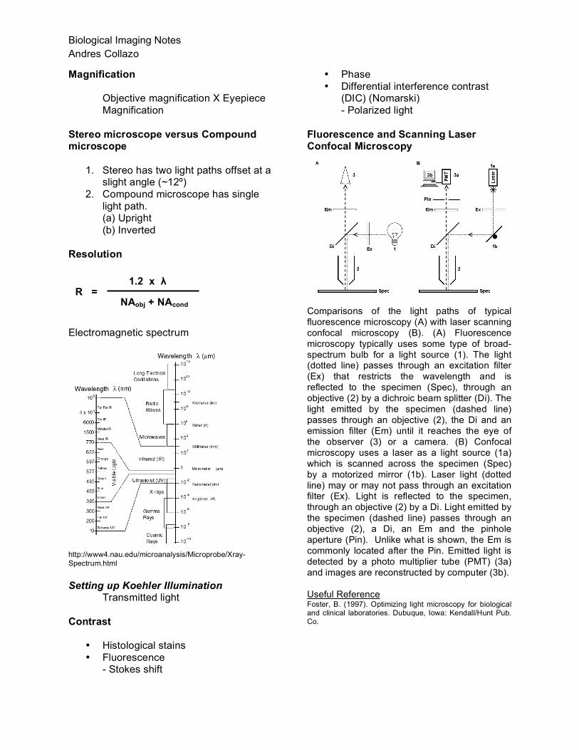
Magnification
Objective magnification X Eyepiece Magnification
Stereo microscope versus Compound microscope
1. Stereo has two light paths offset at a slight angle (~12º)
2. Compound microscope has single light path. (a) Upright (b) Inverted
Resolution
Electromagnetic spectrum
http://www4.nau.edu/microanalysis/Microprobe/Xray-Spectrum.html Setting up Koehler Illumination
Transmitted light
Contrast
• Histological stains • Fluorescence
- Stokes shift
• Phase • Differential interference contrast
(DIC) (Nomarski) - Polarized light
Fluorescence and Scanning Laser Confocal Microscopy
Comparisons of the light paths of typical fluorescence microscopy (A) with laser scanning confocal microscopy (B). (A) Fluorescence microscopy typically uses some type of broad-spectrum bulb for a light source (1). The light (dotted line) passes through an excitation filter (Ex) that restricts the wavelength and is reflected to the specimen (Spec), through an objective (2) by a dichroic beam splitter (Di). The light emitted by the specimen (dashed line) passes through an objective (2), the Di and an emission filter (Em) until it reaches the eye of the observer (3) or a camera. (B) Confocal microscopy uses a laser as a light source (1a) which is scanned across the specimen (Spec) by a motorized mirror (1b). Laser light (dotted line) may or may not pass through an excitation filter (Ex). Light is reflected to the specimen, through an objective (2) by a Di. Light emitted by the specimen (dashed line) passes through an objective (2), a Di, an Em and the pinhole aperture (Pin). Unlike what is shown, the Em is commonly located after the Pin. Emitted light is detected by a photo multiplier tube (PMT) (3a) and images are reconstructed by computer (3b). Useful Reference Foster, B. (1997). Optimizing light microscopy for biological and clinical laboratories. Dubuque, Iowa: Kendall/Hunt Pub. Co.
R = 1.2 x λ
NAobj + NAcond
Biological Imaging Notes Andres Collazo ([email protected]) Resolution (the minimum distance between two objects that allows them to be distinguished as separate) and contrast are the two often conflicting goals of any imaging experiment. Different imaging techniques provide different advantages and disadvantages. Magnetic resonance imaging (MRI) can be used in vivo but the resolution is relatively poor, 1 mm for clinical machines and 10 microns for the best research instruments. Electron microscopy (transmission, TEM, more than scanning, SEM) provides the best resolution (Angstroms, 0.1 nanometers) but it can only be used with fixed tissues. Most of what we will talk about is light microscopy. The most common wavelengths are the visible spectrum from 400 to 700 nanometers. We will be concentrating on imaging with these visible wavelengths. Brightfield images are looking at all these wavelengths while fluorescence images look at a limited range of wavelengths (i.e. blue, green or red). Fluorescence microscopy is usually monochromatic so black and white cameras are perfect for such applications. Cameras: Analog vs. Digital. Most cameras used for image acquisition today are digital. Even many analog cameras (such as camcorders) acquire the image digitally then convert it to analog for viewing on video monitors. Most digital cameras and camcorders acquire images with CCD’s (charge-coupled devices) which are rectangular silicon chips with their point light detectors arranged in columns and rows (i.e. 1200 x 1000). Imaging Software: There are many imaging software packages. These software packages are used mainly for morphometric analysis, image acquisition with a camera, and control of microscopes, X-Y stages and shutters. Almost all are PC based though there are some good Mac ones as well (for example Openlab from Improvision, http://www.improvision.com). We will be mainly showing you two packages that are restricted to the PC. Metamorph from Molecular Devices (http://www.moleculardevices.com/pages/software/metamorph.html) and the Axiovision software from Zeiss (http://www.zeiss.com/us/). Labeling techniques for following cells during development: Ease of
Labeling Initial position and time of labeling
Dilution Single cell lineage
Retrovirus ++++ - ++++ +++ Lipophilic dye (i.e. DiI)
++++ ++++ ++ +
Dextrans +++ ++++ ++ ++++ GFP and ilk Transgenics
++++ ++++ +++ ++
GFP and ilk Electroporation
+++ + +++ ++
Homotypic grafting
+ ++++ ++ ++
Caged Dextrans
++++ ++++ ++ ++

How much does it all cost? (Approximate estimates in USD) Good digital Camera $12,000.00 Computer Software (upper end) 12,000.00 Computer 3,000.00 X-Y Stage 12,000.00 (sometimes included in the price of the scope) Compound Microscope With good objectives, fluorescence and D.I.C. 45,000.00 Grand total $84,000.00 (can bargain and get a really great system for ~$50K)

Andres Collazo Zebrafish 2007
Notes on Biological Imaging Zebrafish Course 2007 Andres Collazo ([email protected]) Scott E. Fraser ([email protected]) Resolution (the minimum distance between two objects that allows them to be distinguished as separate) and contrast are the two often conflicting goals of any imaging experiment. Different imaging techniques provide different advantages and disadvantages. Magnetic resonance imaging (MRI) can be used in vivo but the resolution is relatively poor, 1 mm for clinical machines and 10 microns for the best research instruments. Electron microscopy (transmission, TEM, more than scanning, SEM) provides the best resolution (Angstroms, 0.1 nanometers) but it can only be used with fixed tissues. Most of what we will talk about is light microscopy. The most common wavelengths are the visible spectrum from 400 to 700 nanometers. We will be concentrating on imaging with these visible wavelengths. Brightfield images are looking at all these wavelengths while fluorescence images look at a limited range of wavelengths (i.e. blue, green or red). Fluorescence microscopy is usually monochromatic so black and white cameras are perfect for such applications. Cameras: Analog vs. Digital. Most cameras used for image acquisition today are digital. Even many analog cameras (such as camcorders) acquire the image digitally then convert it to analog for viewing on video monitors. Most digital cameras and camcorders acquire images with CCD’s (charge-coupled devices) which are rectangular silicon chips with their point light detectors arranged in columns and rows (i.e. 1200 x 1000). Imaging Software: There are many imaging software packages. These software packages are used mainly for morphometric analysis, image acquisition with a camera, and control of microscopes, X-Y stages and shutters. Almost all are PC based though there are some good Mac ones as well (for example Openlab from Improvision, http://www.improvision.com). We will be mainly showing you two packages that are restricted to the PC. Metamorph from Molecular Devices (http://www.moleculardevices.com/pages/software/metamorph.html) and the Axiovision software from Zeiss (http://www.zeiss.com/us/). Labeling techniques for following cells during development: Ease of
Labeling Initial position and time of labeling
Dilution Single cell lineage
Retrovirus ++++ - ++++ +++ Lipophilic dye (i.e. DiI)
++++ ++++ ++ +
Dextrans +++ ++++ ++ ++++ GFP and ilk Transgenics
++++ ++++ +++ ++
GFP and ilk Electroporation
+++ + +++ ++
Homotypic grafting
+ ++++ ++ ++
Caged Dextrans
++++ ++++ ++ ++

DiI or DiD labeling. Both stocks are at a concentration of 0.1 or 1% in Ethanol which are then diluted 1 to 10 in 0.3 M Sucrose, both heated to about 37 degrees C°. Back fill needles with 5 microliters of solution which is more than you need but it keeps the dye from evaporating and precipitating. How much does it all cost? Good digital Camera $12,000.00 Computer Software (upper end) 12,000.00 Computer 3,000.00 X-Y Stage 12,000.00 Compound Microscope With good objectives, fluorescence and D.I.C. 75,000.00 Grand total $114,000.00

Start Metamorph software from Desktop shortcut labeled Metamorph
What Metamorph looks like after it starts up.
Illumination controls drop down from this menu.
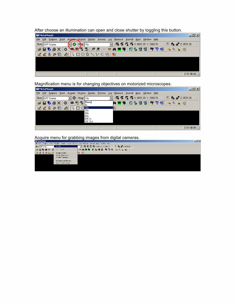
After choose an illumination can open and close shutter by toggling this button.
Magnification menu is for changing objectives on motorized microscopes.
Acquire menu for grabbing images from digital cameras.

Acquire menu for snapping single images. Adjust Exposure time by adjusting Exposure Time while looking at Live image. DON’T USE AutoExpose!
Apps menu is the one you need to use for Time-Lapse imaging. Use the Multi Dimensional Acquisition feature.

Depending on the microscope you will have control of the X-Y stage, Fluorescent turret (Wavelengths) and the focus (Z Series).