Embed Size (px)
Citation preview
STANDARDIZATION OF CEPHALOMETRIC MEASUREMENTS TO CREATE A DATA REPOSITORY OF ORTHODONTICS PATIENT CASES
Shamsi Daneshvari, PhDAMIA Dental Informatics Working Group WebinarSeptember 4, 2014
Outline2
Cephalometric history Orthodontic Case File System Cephalometric standardization Current research The future

Cephalometrics
Technique using lateral radiographs to make measurements of the head Planes, Angles and Linear measurements Used primarily in: DentistryOrthodontics Anthropology
3
History of Cephalometrics
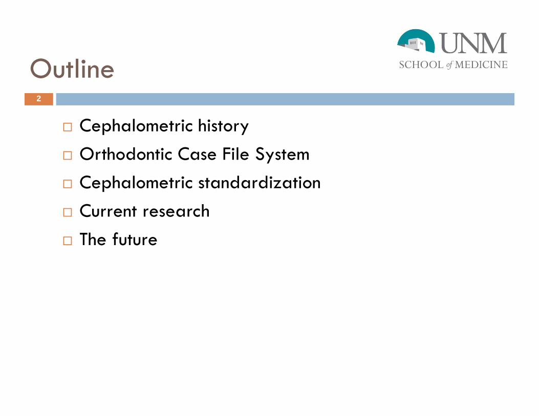
Images from: Essentials of Facial Growth and Radiographic Cephalometry
4
Began with Craniometry in Anthropology Standard landmarks
Cephalostat and methods Developed in 1931 by:Broadbent (USA) Hofrath (Germany)
History of Cephalometrics5
Multiple standards developed over timeMichigan Atlas- Riolo et al. (1974)
London Atlas- Bhaita and Leighton (1993)
Downs Analysis- Downs (1948)
Ricketts Analysis- Rickets et al. (1982)
History of Cephalometrics
Reference samples for “Normal Ranges” Usually small, white samples Downs Analysis: 1948
20 White subjects
Riolo “Michigan Atlas”: 1974 83 White subjects
Are there other racial/ethnic norms?
6

History of Cephalometrics
Tracings of Radiograph
Computerized cephalometrics Dolphin
Imaging CephX
7

Orthodontic Case File System
James K. Economides Donated in 2005 In practice since 1972
Unique collection New Mexicans Hispanic Native American
5,910 Unique patients 600 sibling pairsMulti-generational families
Available online and in person
8

Orthodontic Case File System
Online records can contain: Lateral X-rays Cephalometric measuements Panoramic X-rays Intra-oral photographs Diagnoses
9

Orthodontic Case File System
Available in Physical collection only: Dental casts Full facial and profile photographs Cephalometric tracings Treatment records
What wasn’t included: Race/ethnicity
10
Orthodontic Case File System
Cephalometrics and Dr. Economides Relatively static Changed measurement group 3 times over 20+ years 88 used most commonly
Used multiple standards Recorded for large number of patients
Cephalometrics and the Case File System Wanted to be able to add additional cases with
cephalometrics
11

Cephalometric Standardization
Reviewed and consulted: 2 atlases 8 analyses
Compared and contrasted Dr. Economides’s measurements and landmarks with the existing atlases and analyses
12

Cephalometric Standardization
“Downs Analysis” Downs WB (1948). Variations in Facial Relationships: Their Significance in Treatment
and Prognosis. American Journal of Orthodontics, 34:812-840.
“Frontal Analysis” Grummons DC, Kappeneye van de Coppelo MA (1987). A Frontal Asymmetrical
Analysis. The Journal of Clinical Orthodontics, 21: 448-465.
Grummons DC, Ricketts RM (2004). Frontal Cephalometrics: Practical Application. World Journal of Orthodontics, 5: 99-119.
“Jarabak-Bjork Analysis” Jarabak JR, Fizzell JA (1972). Technique and treatment with light-wire edgewise
appliances. Saint Louis: C. V. Mosby Co.
13

Cephalometric Standardization
“London Atlas” Bhatia SN, Leighton BC (1993). A Manual of Facial Growth: A Computer Analysis of
Longitudinal Cephalometric Growth Data. Oxford: Oxford university Press.
“McNamara Analysis” McNamara JA Jr, Brundon WL (2001). Orhtodontic and Dentofacial Orthopedics.
Ann Arbor: Needham Press.
“Michigan Atlas” Riolo ML, Moyers RE, McNamara JA, Hunter WS (1974). An Atlas of Craniofacial
Growth: Cephalometric Standards from the University School Growth Study, The University of Michigan. Ann Arbor: The University of Michigan.
14

Cephalometric Standardization
“Ricketts Analysis” Ricketts RM, Roth RH, Chaconas SJ, Schnlhof RJ, Engel A (1982). Orthodontic
Diagnosis and Planning, Volumes I and II. Denver: Rocky Mountain Orthodontics
“Soft Tissue Analysis” Holdaway RA (1983). A Soft Tissue Cephalometric Analysis and its Use in
Orthodontic Planning, Part I. American Journal of Orthodontics, 84: 1-28.
Holdaway RA (1984). A Soft Tissue Cephalometric Analysis and its Use in Orthodontic Planning, Part II. American Journal of Orthodontics, 85: 279-293.
“Tweed Analysis” Tweed CH (1954). The Frankfort Mandibular Incisal Angle (FMIA) in Orthodontic
Diagnosis, Treatment Planning, and Prognosis. The Angle Orthodontics, 24: 121-169.
“Steiner Analysis” Steiner CC (1953). Cephalometrics for You and Me. American Journal of
Orthodontics, 39: 729-755.
15
Cephalometric Standardization
Method for cross-referencing: Identify anatomical landmarks Verify variable name is consistent with existing point
naming convention Define cephalometric measurements used in our case
file systemManually examine the atlases and analyses and cross
reference
16

Cephalometric Standardization
Discovered: Not all atlases and analyses use the same vocabulary Identical names
Different landmark
Different names Same landmark
17

Cephalometric Standardization
Gonion- Defined differently
18
Gonion-GO the midpoint at the angle of the mandible Bhatia and Leighton 1993
Gonion-GO intersection of the line connecting the most distal aspect of the condyle to the distal border of the ramus, and the line at the base of the mandible
Ricketts et al. 1982
Gonial Intersection-GOI the intersection of the mandibular plane with a plane through Articulare, Posterior and along the portion of the mandibular ramus inferior to it
Riolo et al. 1974
Gonion-GO the midpoint of the angle of the mandible, found by bisecting the angle formed by the mandibular plane and a plane through Articulare, Posterior and along the portion of the mandibular ramus inferior to it
Riolo et al. 1974
Cephalometric Standardization19
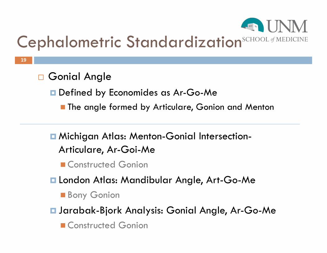
Gonial Angle Defined by Economides as Ar-Go-Me The angle formed by Articulare, Gonion and Menton
Michigan Atlas: Menton-Gonial Intersection-Articulare, Ar-Goi-MeConstructed Gonion
London Atlas: Mandibular Angle, Art-Go-Me Bony Gonion
Jarabak-Bjork Analysis: Gonial Angle, Ar-Go-MeConstructed Gonion

Cephalometric Standardization
LOINC Free to access Internationally recognized Maintained and allows for expansion Allows mapping to each atlas and analysis
measurement
http://search.loinc.org/search.zul?query=ar-go-me
20

Cephalometric Standardization
Created: 88 LOINC terms 11 LOINC parts
LOINC added a graphical portion
21

Case File System
http://hsc.unm.edu/programs/ocfs/
22

Research
Estimating race Cephalometric standardization Dental crowding and cephalometrics Dental development Secular change Tooth size and ethnic variation Eruption Juvenile sexing
23

The Future
Additional cases?? “Hard” cases
Research?? Informatics Anthropology Orthodontics
Cephalometric Manual
24

Acknowledgements
Supported by Award Number NIH 1 G08 LM009381-01A1 from the National Library of Medicine
Philip J. Kroth, MD Heather J. H. Edgar, PhD
25
References
Kroth PJ, Daneshvari S, Harris EF, Vreeman DJ, Edgar HJ. Using LOINC to link 10 terminology standards to one unified standard in a specialized domain. J Biomed Inform. 2012; 45(4):674-82. PMCID: PMC3288380.
Edgar HJH, Daneshvari S, Harris EF, Kroth P. Inter-observer agreement on subjects' race and race-informative characteristics. PloS One. 2011; 6(8): e23986. PMCID: MC3163683.
26