Embed Size (px)
Citation preview
OCT Angiography
SriniVas Sadda, MDProfessor of OphthalmologyProfessor of OphthalmologyDirector,
Medical Retina UnitOphthalmic Imaging Unit
University of Southern California Los Angeles, California, USA

Disclosure
Consulting Fee: Allergan; Carl Zeiss Meditec; Genentech; Optos; RegeneronGenentech; Optos; Regeneron
OCT AngiographyOCT Angiography
Phase Variance OCT
Phase Variance OCTase a a ce OC
• Using the complex data encoded withinUsing the complex data encoded within the OCT images (complex data is generally discarded by most commercialgenerally discarded by most commercial devices), structures with motion may be selectively isolated.selectively isolated.
• After eliminating Brownian motion and• After eliminating Brownian motion and fixation artifact, most of the residual motion in the eye is blood flowmotion in the eye is blood flow.
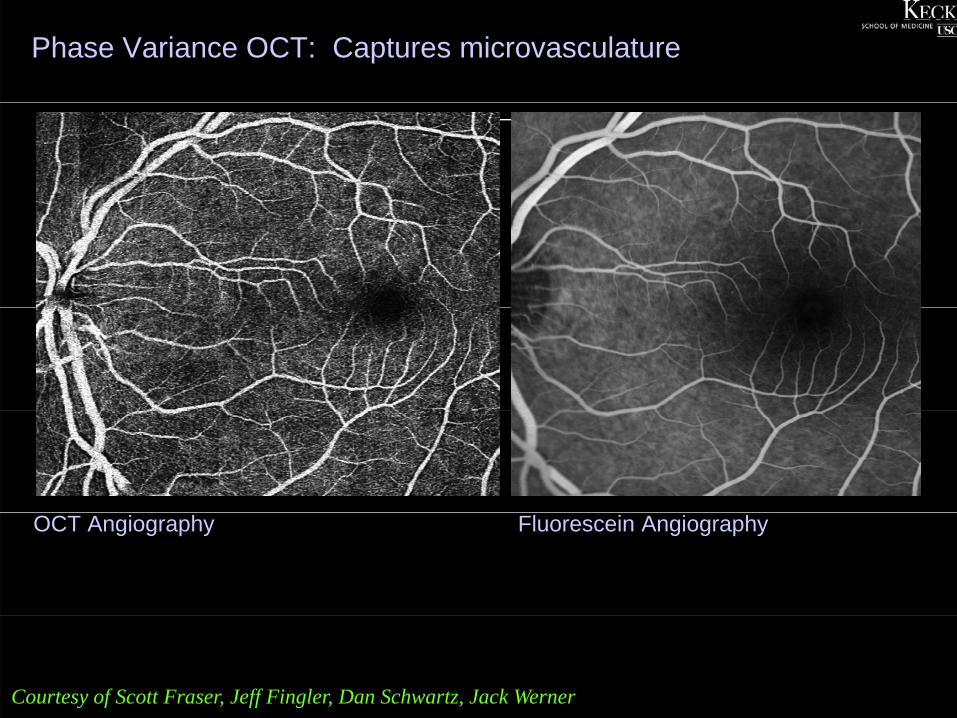
Phase Variance OCT: Captures microvasculature
OCT Angiography Fluorescein Angiography
Courtesy of Scott Fraser, Jeff Fingler, Dan Schwartz, Jack Werner
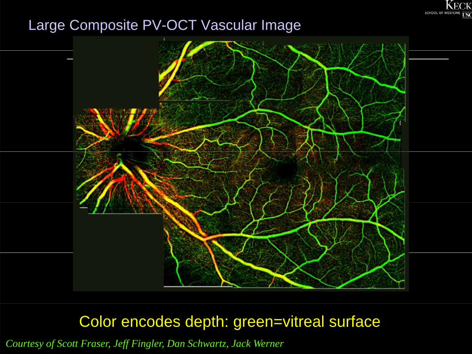
Large Composite PV-OCT Vascular Image
Color encodes depth: green=vitreal surfaceCourtesy of Scott Fraser, Jeff Fingler, Dan Schwartz, Jack Werner
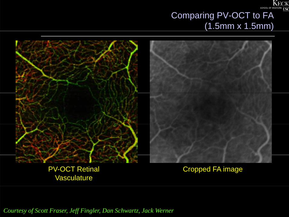
Comparing PV-OCT to FA(1.5mm x 1.5mm)
PV-OCT Retinal Vasculature
Cropped FA image
Courtesy of Scott Fraser, Jeff Fingler, Dan Schwartz, Jack Werner
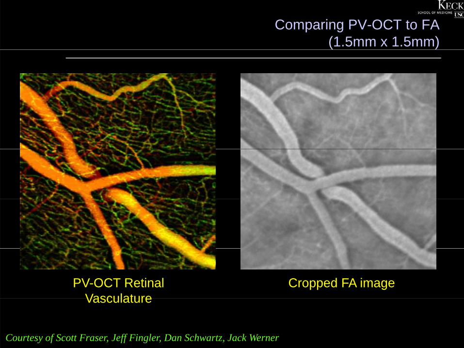
Comparing PV-OCT to FA(1.5mm x 1.5mm)
PV-OCT Retinal Vasculature
Cropped FA imageVasculature
Courtesy of Scott Fraser, Jeff Fingler, Dan Schwartz, Jack Werner
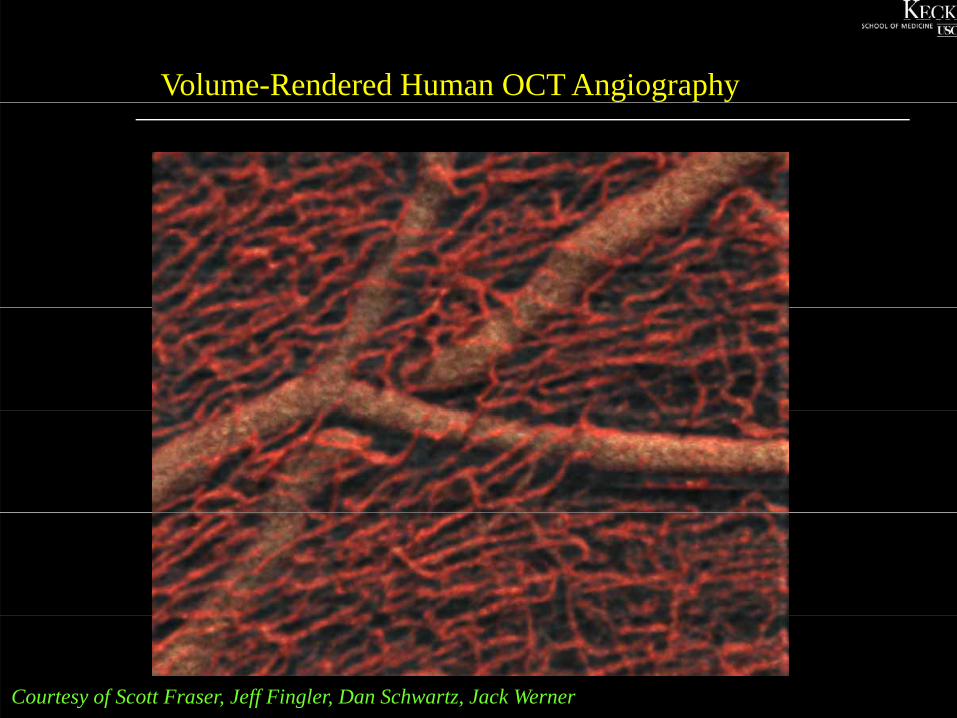
Volume-Rendered Human OCT Angiography
Courtesy of Scott Fraser, Jeff Fingler, Dan Schwartz, Jack Werner

Comparing FA to PV-OCT
Diabetic Retinopathy imaged with 125kHz PV-OCTDiabetic Retinopathy imaged with 125kHz PV OCT
3mm x 3mm
Courtesy of Scott Fraser, Jeff Fingler, Dan Schwartz, Jack Werner
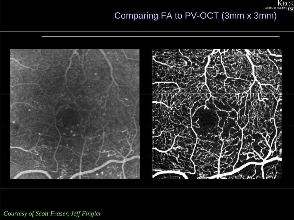
Comparing FA to PV-OCT (3mm x 3mm)
Courtesy of Scott Fraser, Jeff Fingler
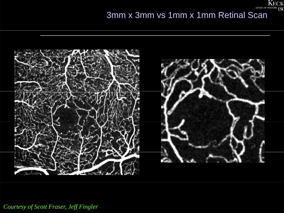
3mm x 3mm vs 1mm x 1mm Retinal Scan
Courtesy of Scott Fraser, Jeff Fingler
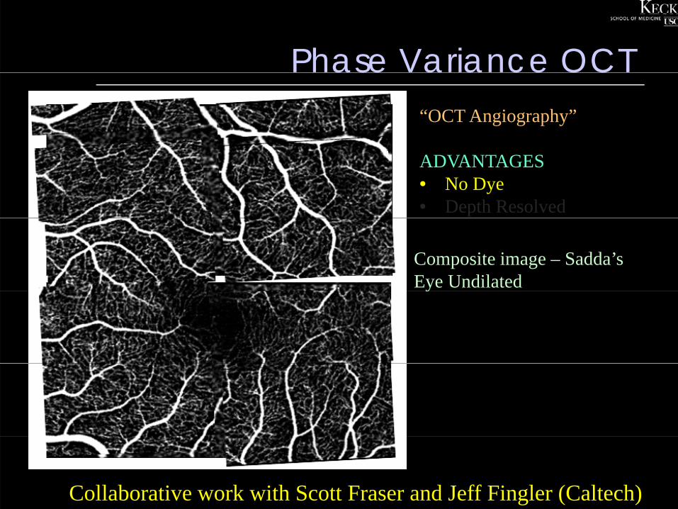
Phase Variance OCTase a a ce OC“OCT Angiography”
ADVANTAGES• No Dye• Depth Resolved
Composite image – Sadda’s Eye Undilatedy
Collaborative work with Scott Fraser and Jeff Fingler (Caltech)
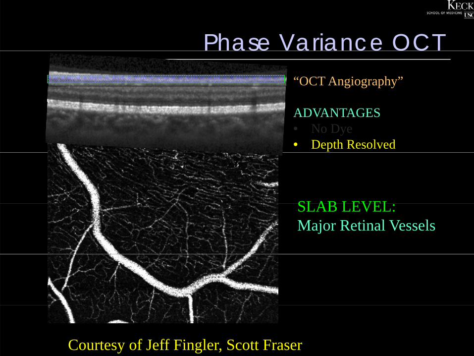
Phase Variance OCTase a a ce OC“OCT Angiography”
ADVANTAGES• No Dye• Depth Resolved
SLAB LEVELSLAB LEVEL:Major Retinal Vessels
Courtesy of Jeff Fingler, Scott Fraser
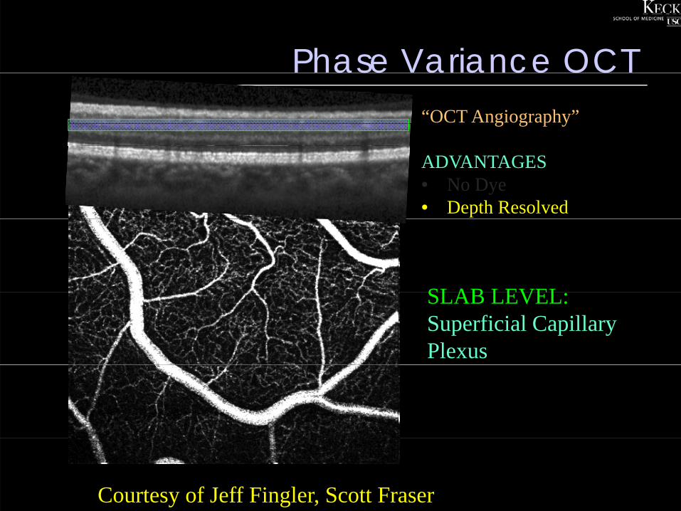
Phase Variance OCTase a a ce OC“OCT Angiography”
ADVANTAGES• No Dye• Depth Resolved
SLAB LEVELSLAB LEVEL:Superficial Capillary Plexus
Courtesy of Jeff Fingler, Scott Fraser
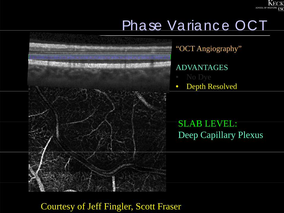
Phase Variance OCTase a a ce OC“OCT Angiography”
ADVANTAGES• No Dye• Depth Resolved
SLAB LEVELSLAB LEVEL:Deep Capillary Plexus
Courtesy of Jeff Fingler, Scott Fraser
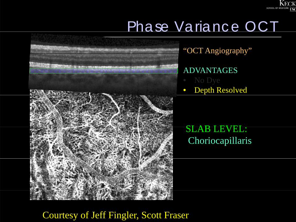
Phase Variance OCTase a a ce OC“OCT Angiography”
ADVANTAGES• No Dye• Depth Resolved
SLAB LEVELSLAB LEVEL:Choriocapillaris
Courtesy of Jeff Fingler, Scott Fraser
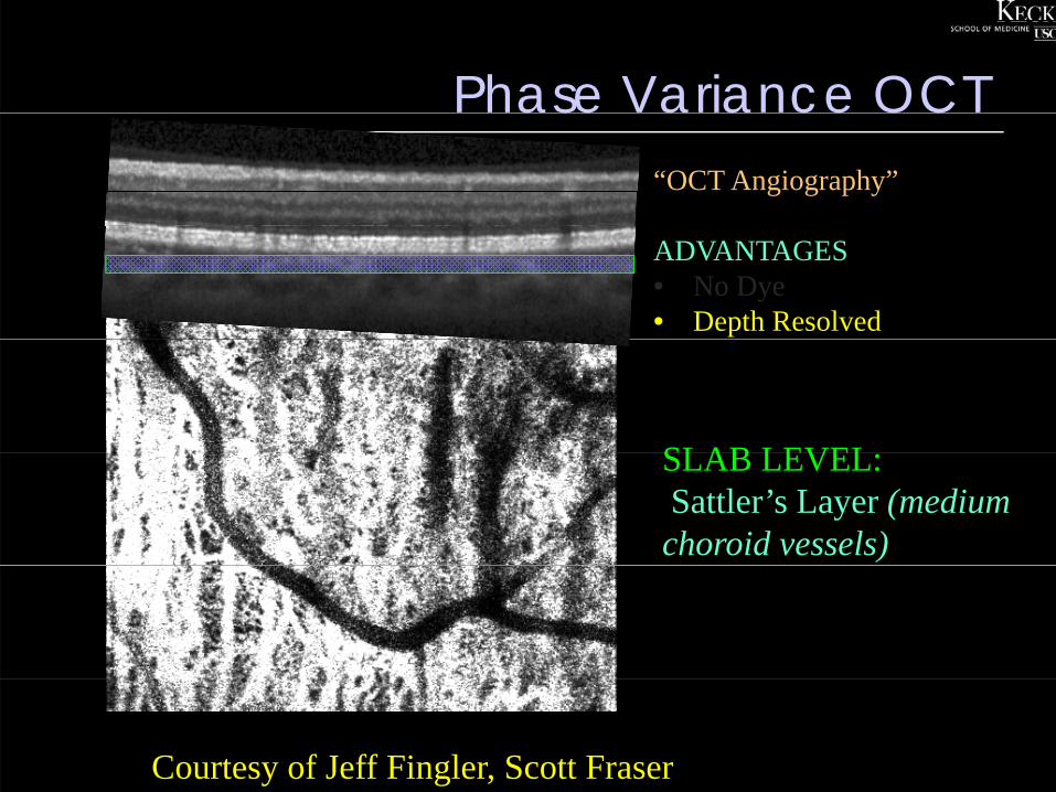
Phase Variance OCTase a a ce OC“OCT Angiography”
ADVANTAGES• No Dye• Depth Resolved
SLAB LEVELSLAB LEVEL:Sattler’s Layer (medium choroid vessels)
Courtesy of Jeff Fingler, Scott Fraser
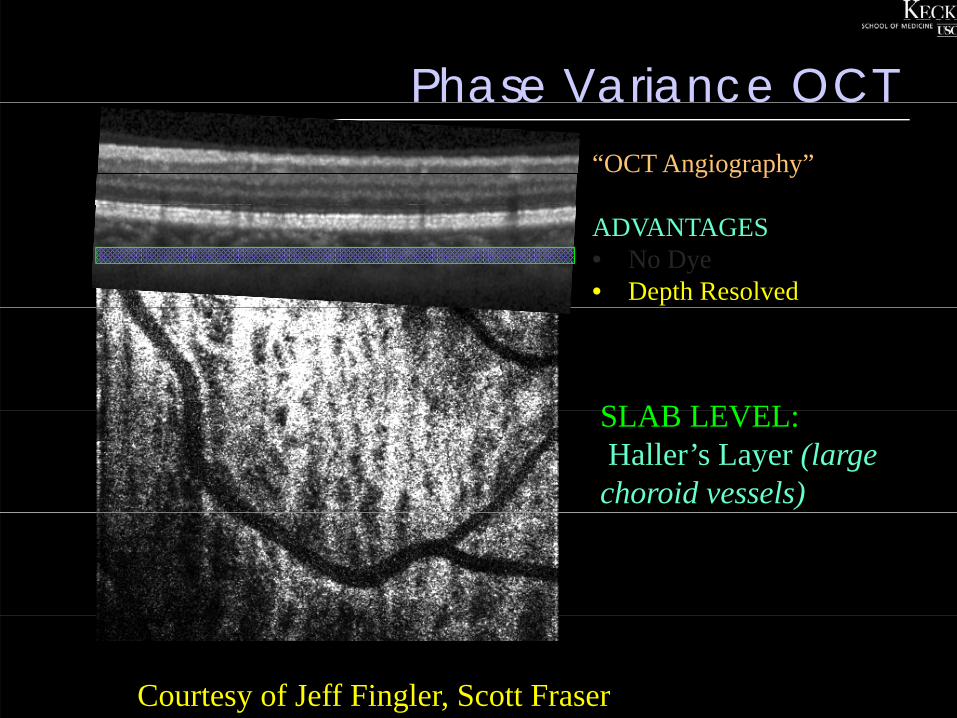
Phase Variance OCTase a a ce OC“OCT Angiography”
ADVANTAGES• No Dye• Depth Resolved
SLAB LEVELSLAB LEVEL:Haller’s Layer (large choroid vessels)
Courtesy of Jeff Fingler, Scott Fraser
PV OCT pitfallsOC p a s
• Motion artifact can be a problem forMotion artifact can be a problem for obtaining high-quality images in some patients.some patients.
Fixation tracking may be a key• Fixation tracking may be a key requirement for optimal imaging
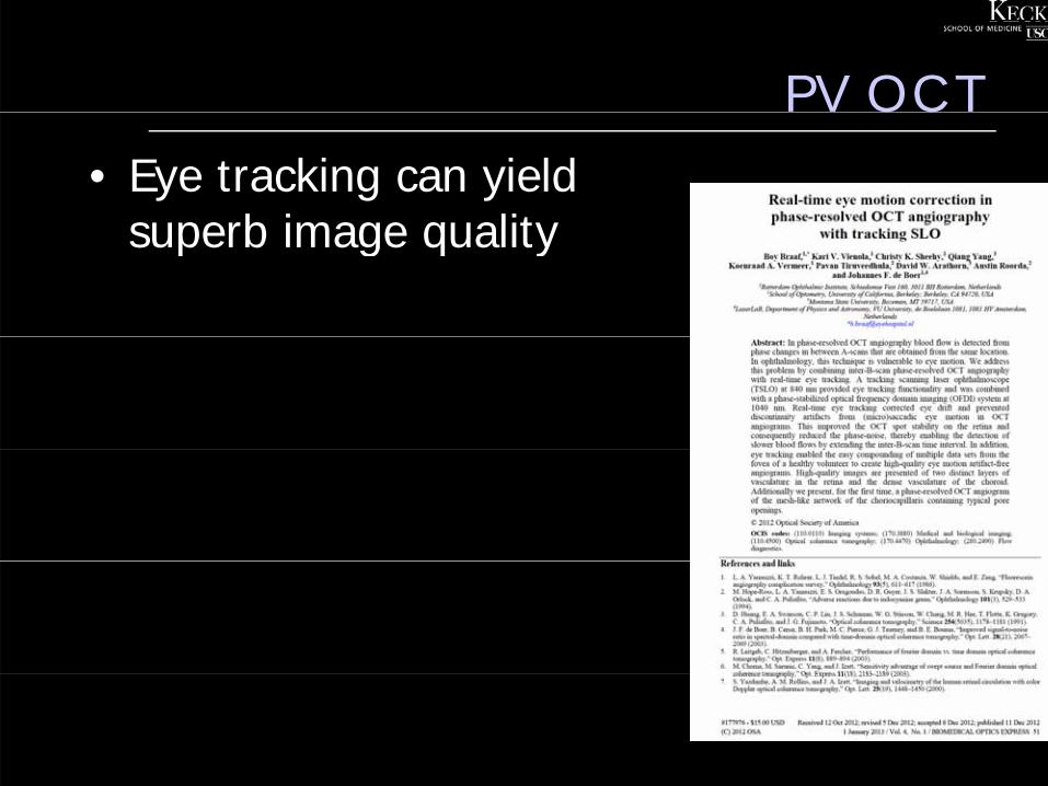
PV OCTOC• Eye tracking can yield
superb image qualitysuperb image quality
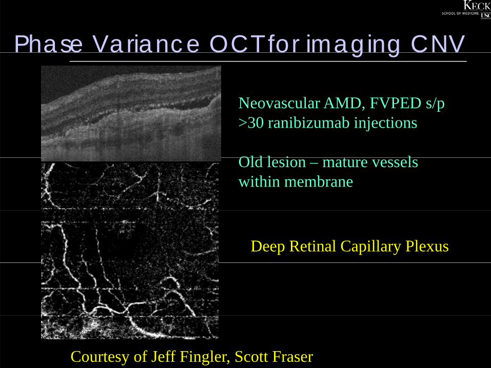
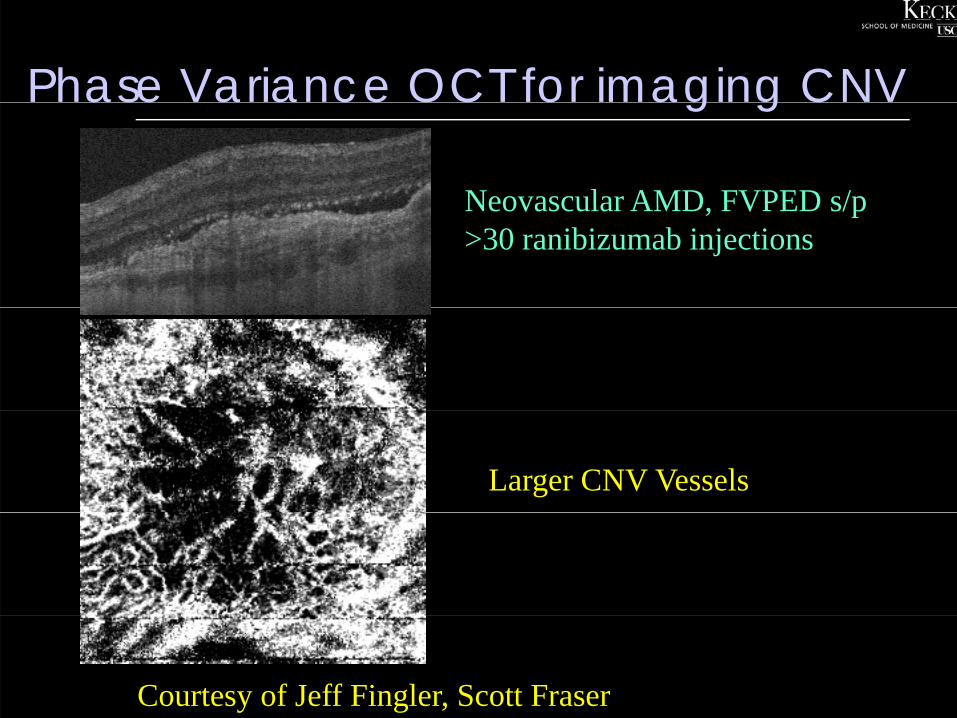
Phase Variance OCT for imaging CNVase a a ce OC o ag g C
Neovascular AMD FVPED s/pNeovascular AMD, FVPED s/p >30 ranibizumab injections
Old l i t lOld lesion – mature vessels within membrane
Deep Retinal Capillary Plexus
Courtesy of Jeff Fingler, Scott Fraser
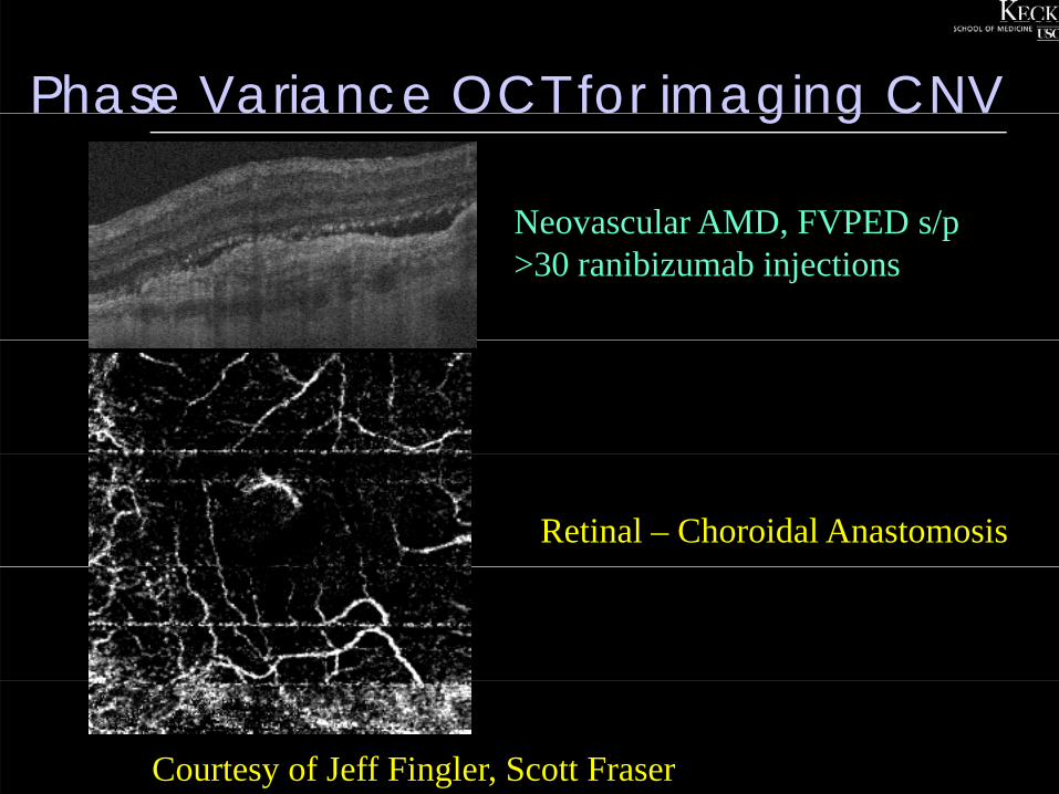
Phase Variance OCT for imaging CNVase a a ce OC o ag g C
Neovascular AMD FVPED s/pNeovascular AMD, FVPED s/p >30 ranibizumab injections
Retinal – Choroidal Anastomosis
Courtesy of Jeff Fingler, Scott Fraser
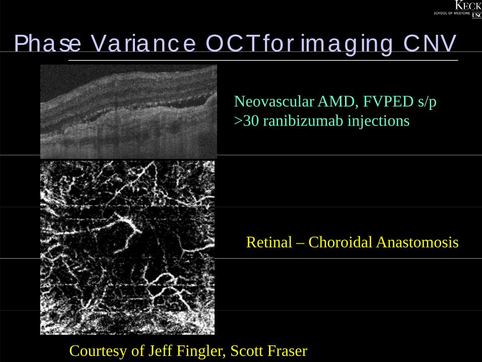
Phase Variance OCT for imaging CNVase a a ce OC o ag g C
Neovascular AMD FVPED s/pNeovascular AMD, FVPED s/p >30 ranibizumab injections
Retinal – Choroidal Anastomosis
Courtesy of Jeff Fingler, Scott Fraser
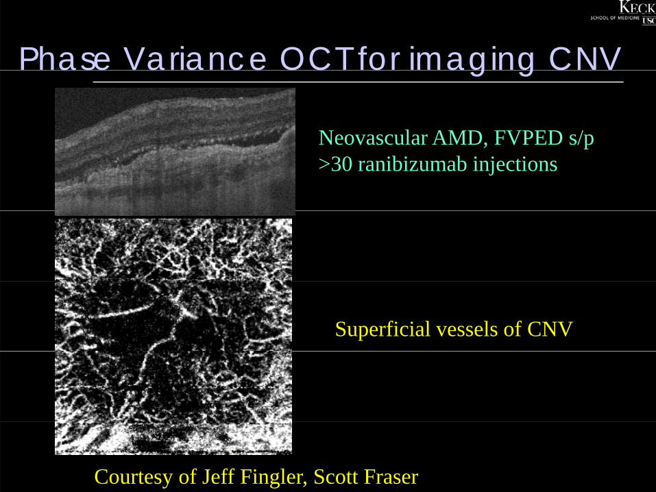
Phase Variance OCT for imaging CNVase a a ce OC o ag g C
Neovascular AMD FVPED s/pNeovascular AMD, FVPED s/p >30 ranibizumab injections
Superficial vessels of CNV
Courtesy of Jeff Fingler, Scott Fraser
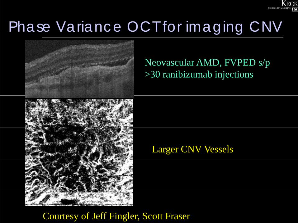
Phase Variance OCT for imaging CNVase a a ce OC o ag g C
Neovascular AMD FVPED s/pNeovascular AMD, FVPED s/p >30 ranibizumab injections
Larger CNV Vessels
Courtesy of Jeff Fingler, Scott Fraser
Phase Variance OCT for imaging CNVase a a ce OC o ag g C
Neovascular AMD FVPED s/pNeovascular AMD, FVPED s/p >30 ranibizumab injections
Larger CNV Vessels
Courtesy of Jeff Fingler, Scott Fraser

Increases confidence in our detection of CNV with OCT
Spectrum of Pigment Epithelial Detachments
D id PEDDrusenoid PED(medium homogenous)
Serous PED(low homogenous)(low homogenous)
Fibrovascular PED(low heterogenous)(low heterogenous)
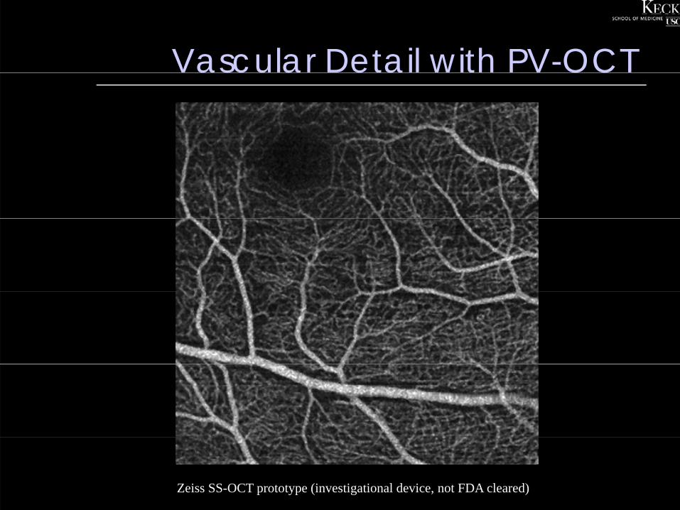
Vascular Detail with PV-OCTascu a e a OC
Zeiss SS-OCT prototype (investigational device, not FDA cleared)
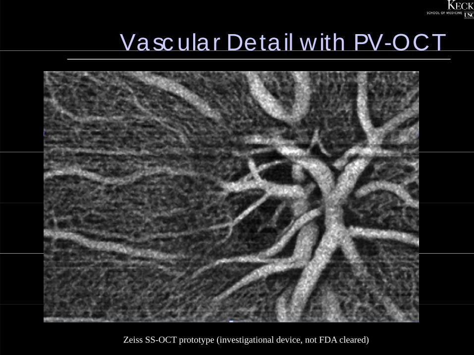
Vascular Detail with PV-OCTascu a e a OC
Zeiss SS-OCT prototype (investigational device, not FDA cleared)
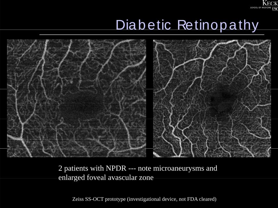
Diabetic Retinopathyabe c e opa y
2 patients with NPDR --- note microaneurysms and enlarged foveal avascular zoneenlarged foveal avascular zone
Zeiss SS-OCT prototype (investigational device, not FDA cleared)
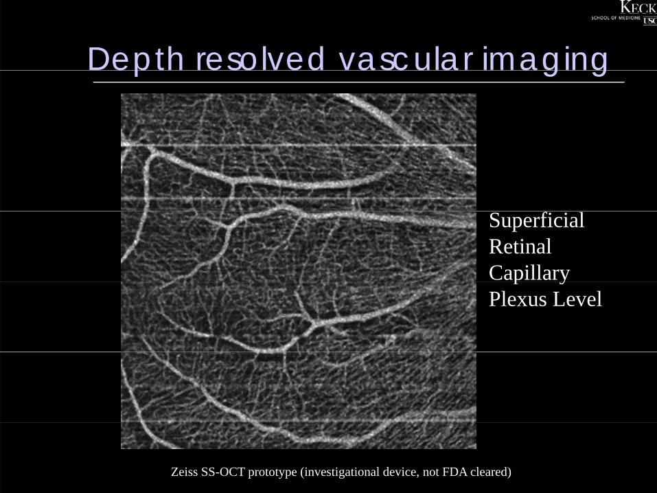
Depth resolved vascular imagingep eso ed ascu a ag g
Superficial Retinal Capillary p yPlexus Level
Zeiss SS-OCT prototype (investigational device, not FDA cleared)
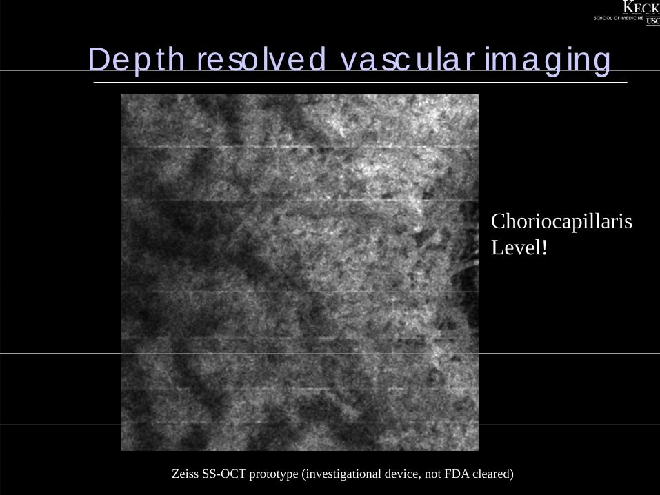
Depth resolved vascular imagingep eso ed ascu a ag g
ChoriocapillarisLevel!
Zeiss SS-OCT prototype (investigational device, not FDA cleared)
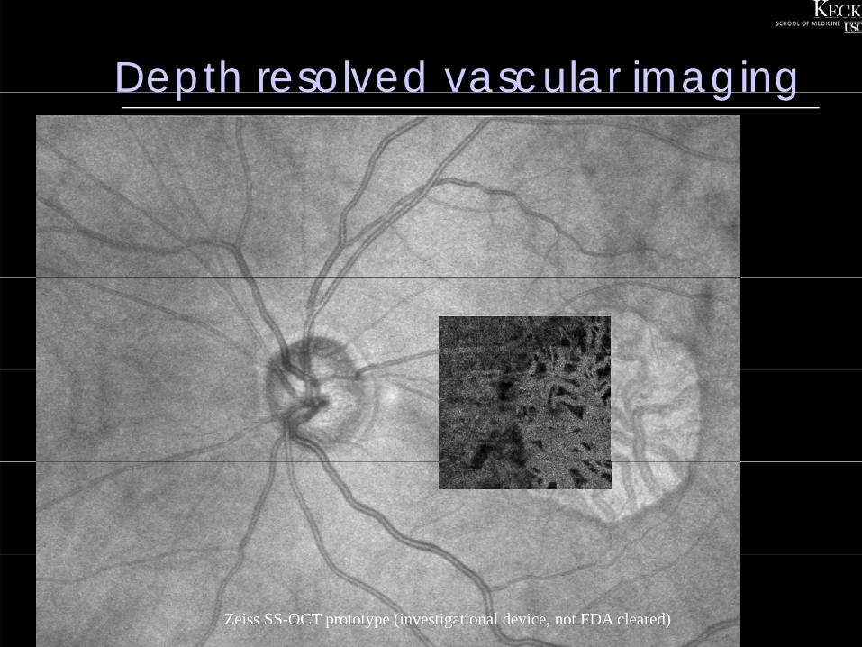
Depth resolved vascular imagingep eso ed ascu a ag g
Zeiss SS-OCT prototype (investigational device, not FDA cleared)

Depth resolved vascular imagingep eso ed ascu a ag g
Zeiss SS-OCT prototype (investigational device, not FDA cleared)
OCT AngiographyOCT Angiography
Split-Spectrum Amplitude Decorrelation Angiographyg g p y
SSADASS
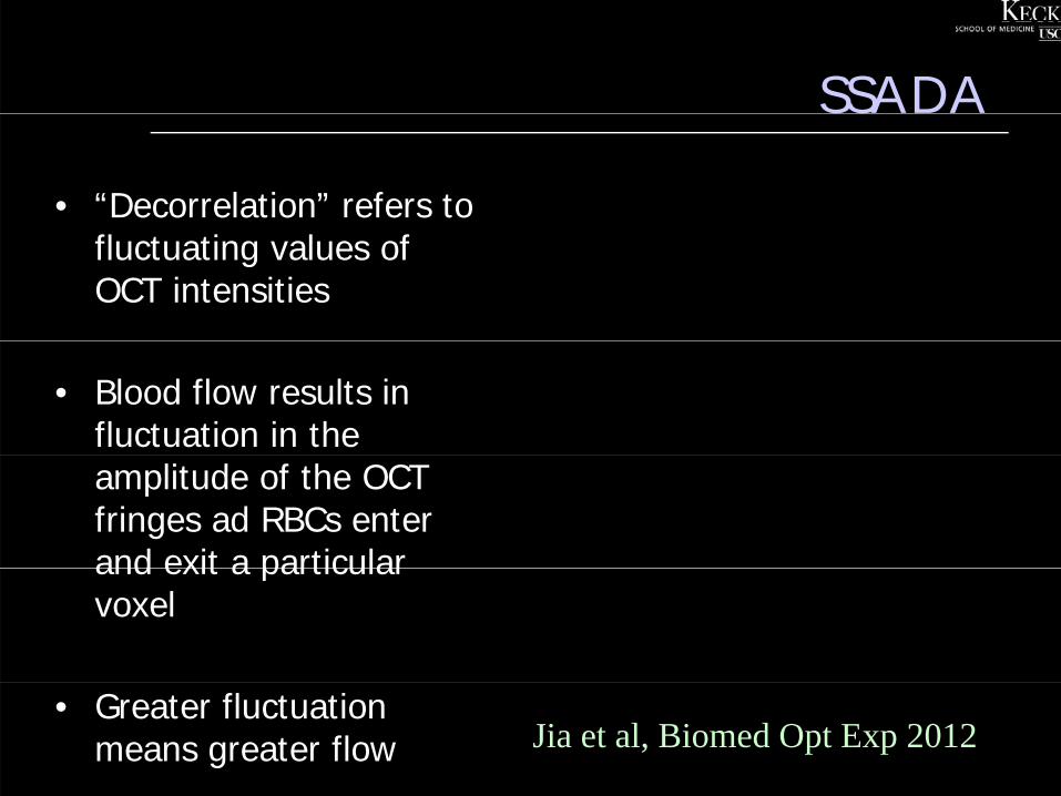
• “Decorrelation” refers to fluctuating values of OCT intensities
• Blood flow results in fluctuation in the amplitude of the OCT fringes ad RBCs enter and exit a particularand exit a particular voxel
• Greater fluctuation means greater flow Jia et al, Biomed Opt Exp 2012
En face retinal and choroidal angiograms at different Z coordinates at macula
Yali Jia, PhD; David Huang, MD, PhD. www.COOLLab.net
En face retinal and choroidal angiograms at different Z coordinates at ONH
Yali Jia, PhD; David Huang, MD, PhD. www.COOLLab.net
En face ONH angiograms separately showing the microcirculation within retina choroid and lamina cribrosamicrocirculation within retina , choroid and lamina cribrosa
Yali Jia, PhD; David Huang, MD, PhD. www.COOLLab.net
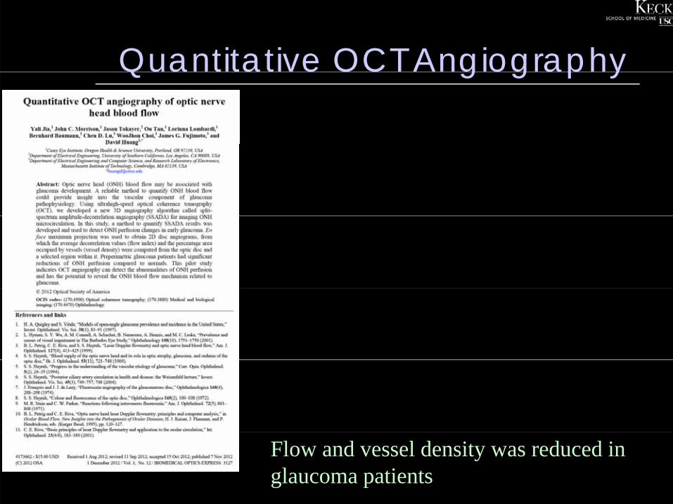
Quantitative OCT AngiographyQua a e OC g og ap y
Flow and vessel density was reduced in glaucoma patients
SummarySu a y• OCT angiography is an exciting new development in non-
invasive imaging
• The ability to acquire detailed imaging of the retinal and choroidal microvasculature in a depth-resolved fashion, without dye injection, represents a significant advance– The prospect of quantitative flow data is an additional major
benefit
• Further refinement of the technology is required to allow ascertainment of leakage
• The scope/purview of conventional angiography will likely continue to narrow

Thank you!Thank you!


![DTIC · 2011-05-14 · [Srinivas b] SRINIVAS, Y. V. Deriving parsing algorithms using sheaves. In 0 Preparation. [Srinivas 92] SRINIVAS, Y. V. Derivation of a parallel matching algorithm](https://img.dokumen.tips/doc/110x75/5f8689e2fffa8812255e2550/dtic-2011-05-14-srinivas-b-srinivas-y-v-deriving-parsing-algorithms-using.jpg)