Embed Size (px)
Citation preview
Acta Orthop Scand 1988;59(2):117-121 117
Spinal cord compression by epidural metastases Fibrosarcoma experiments in rats
Shohei Manabe, Tohgo Ohno, Hiromitsu Tanaka and Pyoyun Park
Morphologic changeswere studied in 33 rats that were paralyzed by an epidural fibrosarcoma inoculated through the spinous process. By this technique, of 29 rats examined radiographically, the tumor destroyed vertebral bone in 22. Changes of the cord in the early stage of paralysis were specially analyzed by Marchi's stain and microangiograms. In compromised cord segments in the early stage, extravasation of contrast medium was observed in the gray matter and the dorsal funiculus; and in the dorsal funiculus just proximal or distal to the compressed portion, hemorrhagic areas were present. Ascending dege- nerated fibers in the dorsal funiculus, 4 hich were derived from the degenerated posterior nerve root or the degeneration of the dorsal funiculus in the compressed segments, were characteristically detected in rats in the early stages. In the advanced stages a transverse cord lesion was observed a1 the involved level. Based on the present analyses, to prevent more advanced damage to the spinal cord, the tumor should be removed at an early stage that clinically coincides with the period when radicular signs appear.
In spite of many clinical reports on cord com- pression caused by metastatic epidural tumors, only a few systematic analyses of cord changes caused by the tumor have been performed by experimental or clinocopathologic research (Bar- ron et al. 1959, Boland et al. 1952, Ikeda et al. 1980, Ushio et al. 1977a, 1977b). Little is known of the changes in the fasciculus of the cord, which degenerates in the early stage of the compression by metastatic tumor. We have studied epidural metastases in rats in order to analyze how the spinal cord is compromised in the early stage of the compression and the timetable for cord dam- age.
Materials and methods
A cell suspension, containing 5 x lo5 tumor cells per 0.1 mL in TC-Medium 199, from a serially subcutaneously transplanted fibrosarcoma in-
Department of Orthopedics, Teikyo University School of Medicine, Tokyo, Japan
Correspondence: Dr. Shohei Manabe. Department of Orthopedics, Teikyo University School of Medicine. 11-1, Kaga, 2 Chome, Itabashi-Ku. Tokyo 173, Japan
duced by methylcholanthrene in our laboratory in a Lewis rat was inoculated into the spinous process of rats at the following levels: T7 in 15 rats, T11 in 16 rats, and L2 in 12 rats.
After the inoculation, the rats were checked daily for neurologic symptoms. Paralysis of the hind limbs was graded according to the following modified scale (stage) (Ikeda et al. 1980): Stage 0 = normal; Stage 1 = weak, but able to walk; Stage 2 = attempts to walk; Stage 3 = only moves hind limbs when pricked with a pin; Stage 4 = paraplegia.
The rats were killed at various stages of paral- ysis (Table 1). The spinal cord was removed carefully under the microscope, followed by his- tologic and vascular observations.
The spinal cord was classified depending on the site of compression:
Type 1. Anterior compression with or without anterolateral involvement.
Type 2. Posterior compression with or without posterolateral involvement.
Type 3. Lateral compression. Type 4. Cufflike encirclement of the cord by
The morphologic and vascular changes were the tumor.
analyzed according to the stage of paralysis.
Act
a O
rtho
p D
ownl
oade
d fr
om in
form
ahea
lthca
re.c
om b
y C
DL
-UC
San
ta C
ruz
on 1
0/26
/14
For
pers
onal
use
onl
y.
118 Acta Orthop Scand 1988;59(2):117-121
Table 1. Paralysis stages and the time of killing of the animals
Stage Numbeiof rats Time (days)
1 2 14 2 4 15 3 6 17 4 31 20
Marchi’s investigation was performed on 25 spinal cords. Each cord was fixed and stained in Marchi’s solution, after which serial transverse sections of the cord were analyzed. Nine other cords were stained with hematoxylin-eosin and lux01 fast blue. Marchi’s investigation: The pro- duct from degenerated myelin combines with osmium oxide, resulting in black granules. This reaction is observed in secondary degeneration of myelinated nerve fibers within 3 months after the damage of nerve cells and/or nerve fibers in the spinal cord.
Before death, the 9 remaining rats paralyzed in various stages were perfused with Indian ink. Transverse serial sections and longitudinal trans- verse serial sections of the perfused cord were prepared for microscopic histologic observation of spinal-cord blood vessels.
Of the 43 rats, 29 rats were examined radiogra- phically after death.
Results
Neurologic deficits. When the rats were killed, they had all developed neurologic deficits; the majority were Stage 4 (Table 2). The average duration from inoculation was 17 days for those in Stages 1 and 2, 18 days for those in Stage 3. and 24 days for the rats in Stage 4. In all the rats. the neurologic symptoms were progressive. and 3 1 rats that were not killed until Stage 3 became paraplegic and had complete sphincter involve- ment by the third day after they had reached Stage 3.
Table 2. Paralysis stages and tumor types (see text)
Type Paralysls stage
0 1 2 3 4 Total
1 0 1 1 0 4 8 2 0 1 0 0 5 6 3 0 0 1 4 8 13 4 . 0 0 2 2 14 18
Total 0 2 4 6 31 43
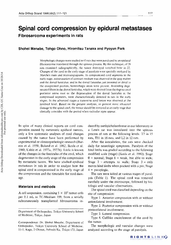
Figure 1 . Twenty-three days after inoculation. Osteolytic tumor was observed in a vertebral body and a transverse process (arrow).
Radiographic and macroscopic findings. Of the 29 rats that were checked radiographically. an osteolytic tumor of the spine (Figure 1 ) was observed in 22 rats: the lamina was involved in I0 rats, whereas the lamina and vertebral body were osteolytic in 8 rats, and the vertebral body with a pedicle and a transverse process was destroyed in 4 rats. In 7 rats that had a paravertebral tumor, but no bone destruction, the tumor was revealed to infiltrate the intervertebral foramina into the epidural space.
After the laminectomy. tumors were found to invade epidurally from the destroyed pedicle. lamina, or vertebral body, and appeared to extend over about two spinal segments, involving nerve roots. Among the 43 rats, the tumor formed a cufflike encirclement around the cord in 18 of them, and 13 had a tumor lying laterally to the cord, whereas in the remaining 12 the tumor caused anterior or posterior cord compression.
The cords of 14 rats, or half of those with complete paraplegia, had T y p e 4 cufflike c o n - pression of the cord (Table 2).
Vascular changes. In the less advanced coni- pression (Stages 1 and 2). a moderate decrease of
Act
a O
rtho
p D
ownl
oade
d fr
om in
form
ahea
lthca
re.c
om b
y C
DL
-UC
San
ta C
ruz
on 1
0/26
/14
For
pers
onal
use
onl
y.
Acta Orthop Scand 1988;59(2):117-121 119
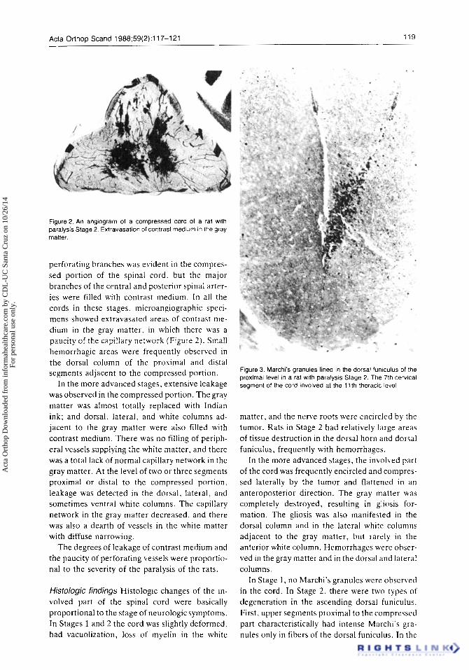
Figure 2. An angiogram of a compressed cord of a rat with paralysis Stage 2. Extravasation of contrast medium in the gray matter.
perforating branches was evident in the compres- sed portion of the spinal cord. but the major branches of the central and posterior spinal arter- ies were filled with contrast medium. In all the cords in these stages, microangiographic speci- mens showed extravasated areas of contrast me- dium in the gray matter, in which there was a paucity of the capillary network (Figure 2). Small hemorrhagic areas were frequently observed in the dorsal column of the proximal and distal segments adjacent to the compressed portion.
In the more advanced stages, extensive leakage was observed in the compressed portion. The gray matter was almost totally replaced with Indian ink; and dorsal, lateral, and white columns ad- jacent to the gray matter were also filled with contrast medium. There was no filling of periph- eral vessels supplying the white matter, and there was a total lack of normal capillary network in the gray matter. At the level of two or three segments proximal or distal to the compressed portion, leakage was detected in the dorsal, lateral, and sometimes ventral white columns. The capillary network in the gray matter decreased. and there was also a dearth of vessels in the white matter with diffuse narrowing.
The degrees of leakage of contrast medium and the paucity of perforating vessels were proportio- nal to the severity of the paralysis of the rats.
Histologic findings Histologic changes of the in- volved part of the spinal cord were basically proportional to the stage of neurologic symptoms. In Stages 1 and 2 the cord was slightly deformed, had vacuolization, loss of myelin in the white
. . . . .
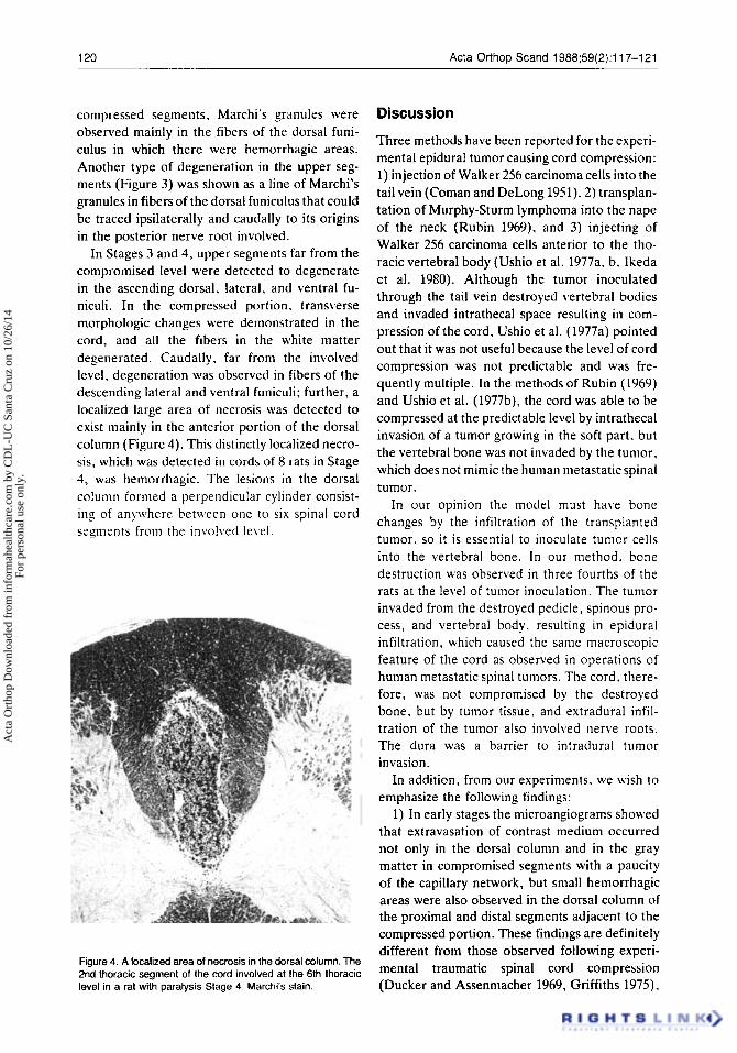
Figure 3 Marchi's granules lined in the dorsal funiculus of the proximal level in a rat with paralysis Stage 2 The 7th cerwal segment of the cord involved at the 1 l t h thoracic level
matter, and the nerve roots were encircled by the tumor. Rats in Stage 2 had relatively large areas of tissue destruction in the dorsal horn and dorsal funiculus, frequently with hemorrhages.
In the more advanced stages, the involved part of the cord was frequently encircled and compres- sed laterally by the tumor and flattened in an anteroposterior direction. The gray matter was completely destroyed, resulting in gliosis for- mation. The gliosis was also manifested in the dorsal column and in the lateral white columns adjacent to the gray matter, but rarely in the anterior white column. Hemorrhages were ohser- ved in the gray matter and in the dorsal and lateral columns.
In Stage 1, no Marchi's granules were observed in the cord. In Stage 2 , there were two types of degeneration in the ascending dorsal funiculus. First, upper segments proximal to the compressed part characteristically had intense Marchi's gi-a- nules only in fibers of the dorsal funiculus. In the
Act
a O
rtho
p D
ownl
oade
d fr
om in
form
ahea
lthca
re.c
om b
y C
DL
-UC
San
ta C
ruz
on 1
0/26
/14
For
pers
onal
use
onl
y.
120 Acta Orthop Scand 1988;59(2):117-121
compressed segments, Marchi‘s granules were observed mainly in the fibers of the dorsal funi- culus in which there were hemorrhagic areas. Another type of degeneration in the upper seg- ments (Figure 3) was shown as a line of Marchi’s granules in fibers of the dorsal funiculus that could be traced ipsilaterally and caudally to its origins in the posterior nerve root involved.
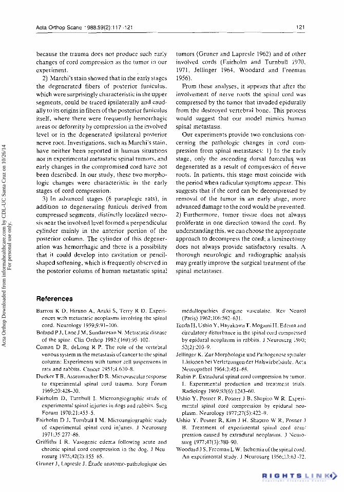
In Stages 3 and 4, upper segments far from the compromised level were detected to degenerate in the ascending dorsal. lateral, and ventral fu- niculi. In the compressed portion. transverse morphologic changes were demonstrated in the cord, and all the fibers in the white matter degenerated. Caudally. far from the involved level, degeneration was observed in fibers of the descending lateral and ventral funiculi; further, a localized large area of necrosis was detected t o exist mainly in the anterior portion of the dorsal column (Figure 4). This distinctly localized necro- sis, which was detected in cords of 8 rats in Stage 4, was hemorrhagic. The lesions in the dorsal column formed a perpendicular cylinder consist- ing of anywhere between one to six spinal cord segments from the involved level.
Figure 4. A localized area of necrosis in the dorsal column. The 2nd thoracic segment of the cord involved at the 6th thoracic level in a rat with paralysis Stage 4. Marchi’s stain.
Discussion
Three methods have been reported for the experi- mental epidural tumor causing cord compression: 1) injection of Walker 256 carcinoma cells into the tail vein (Coman and DeLong 1951). 2 ) transplan- tation of Murphy-Sturni lymphoma into the nape of the neck (Rubin 1969). and 3) injecting of Walker 256 carcinoma cells anterior t o the tho- racic vertebral body (Ushio et al. 1977a, b. Ikeda e t at. 1980). Although the tumor inoculated through the tail vein destroyed vertebral bodies and invaded intrathecal space resulting in com- pression of the cord, Ushio et al. (1977a) pointed out that it was not useful because the level of cord compression was not predictable and was fre- quently multiple. In the methods of Rubin (1969) and Ushio et al. (1977b), the cord was able to be compressed at the predictable level by intrathecal invasion of a tumor growing in the soft part. but the vertebral bone was not invaded by the tumor. which does not mimic the human metastatic spinal tumor.
In our opinion the model must have bone changes by the infiltration of the transplanted tumor, so it is essential to inoculate tumor cells into the vertebral bone. I n our method. bone destruction was observed in three fourths of the rats at the level of tumor inoculation. The tumor invaded from the destroyed pedicle, spinous pro- cess, and vertebral body. resulting in epidural infiltration, which caused the same macroscopic feature of the cord as observed in operations of human metastatic spinal tumors. The cord, there- fore, was not compromised by the destroyed bone, but by tumor tissue, and extradural infil- tration of the tumor also involved nerve roots. The dura was a barrier to intradural tumor invasion.
In addition, from our experiments. we wish to emphasize the following findings:
1) In early stages the niicroangiograms showed that extravasation of contrast medium occurred not only in the dorsal column and in the gray matter in compromised segments with a paucity of the capillary network, but small hemorrhagic areas were also observed in the dorsal column of the proximal and distal segments adjacent t o the compressed portion. These findings are definitely different from those observed following experi- mental traumatic spinal cord compression (Ducker and Assenmacher 1969, Griffiths 1975).
Act
a O
rtho
p D
ownl
oade
d fr
om in
form
ahea
lthca
re.c
om b
y C
DL
-UC
San
ta C
ruz
on 1
0/26
/14
For
pers
onal
use
onl
y.
Acta Orthop Scand 1988;59(2):117-121 121
because the trauma does not produce such early changes of cord compression as the tumor in our experiment.
2) Marchi’s stain showed that in the early stages the degenerated fibers of posterior funiculus, which were surprisingly characteristic in the upper segments, could be traced ipsilaterally and caud- ally to its origins in fibers of the posterior funiculus itself, where there were frequently hemorrhagic areas or deformity by compression in the involved level or in the degenerated ipsilateral posterior nerve root. Investigations, such as Marchi’s stain. have neither been reported in human situations nor in experimental metastatic spinal tumors. and early changes in the compromised cord have not been described. In our study, these two morpho- logic changes were characteristic in the early stages of cord compression.
3 ) In advanced stages (8 paraplegic rats), in addition to degenerating funiculi derived from compressed segments, distinctly localized necro- sis near the involved level formed a perpendicular cylinder mainly in the anterior portion of the posterior column. The cylinder of this degener- ation was hemorrhagic and there is a possibility that it could develop into cavitation or pencil- shaped softening, which is frequently observed in the posterior column of human metastatic spinal
References Barron K D, Hirano A , Araki S, Terry R D . Experi-
ences with metastatic neoplasms involving the spinal cord. Neurology 1959;9:9 1-1 Oh.
Boland P J , Lane J M. Sundaresan N. Metastatic disease of the spine. Clin Orthop 19X2:(IhY):Y5-102.
Coman D R, deLong R P. The role of the vertebral venous system in the metastasis of cancer to the spinal column: Experiments with tunlor cell suspensions in rats and rabbits. Cancer 1951 :4:61(M.
Ducker T B, Assenmacher D R . Microvascular response t o experimental spinal cord trauma. Surg Forum 1969 ;?O: 428-30.
Fairholm D. Turnbull I . Microangiographic study of experimental spinal injuries in dogs and rabbits. Surg Forum 1970:21:453-5.
Fairholm D J, Turnbull I M. Microangiograpliic study of experimental spinal cord injuries. J Neurosurg
Griffiths I R. Vasogenic edema following acute and chronic spinal cord compression in the dog. J Neu- rosurg 1975;42(2):155-65.
Gruner J , Lapresle J . Etude anatomo-pathologique des
1971 ;35:277-86.
tumors (Gruner and Lapresle 1962) and of other involved cords (Fairholm and Turnbull 1970, 1971, Jellinger 1964. Woodard and Freeman 1956).
From these analyses, it appears that after the involvement of nerve roots the spinal cord was compressed by the tumor that invaded epidurally from the destroyed vertebral bone. This process would suggest that our model mimics human spinal metastasis.
Our experiments provide two conclusions con- cerning the pathologic changes i n cord com- pression from spinal metastases: 1) I n the Early stage, only the ascending dorsal funiculu{ was degenerated as a result of compression of Aerve roots. In patients, this stage must coincide with the period when radicular symptoms appear. This suggests that if the cord can be decompressed by removal of the tumor in an early stage, more advanced damage to the cord would be prevented. 2) Furthermore, tumor tissue does not always proliferate in one direction toward the cord. By understanding this, we can choose the appropriate approach to decompress the cord; a laminectomy does not always provide satisfactory results. A thorough neurologic and radiographic analysis may greatly improve the surgical treatment of the spinal metastases.
medullopathies d’origine vasculaire. Rev Neurol (Paris) 1962: 106: 592-63 1.
Ikeda H , Ushio Y. Hayakawa T. Mogami H . Edema and circulatory disturbance in the spinal cord compressed by epidural neoplasms in rabbits. J Neurosurg 1YSU; j2 ( 2) : 203-9.
Jellinger K. Zur Morphologie und Pathogenese spinaler Lasionen bei Verletzungen der Halswirbelsiule. Acta Neuropathol 1964 :3:45 1-68.
Rubin P. Extradural spinal cord compression by tumor. I . Experimental production and treatment trials. Radiology 1969;93(6): 124340.
Ushio Y, Posner R. Posner J B. Shapiro W R . Experi- mental spinal cord compression by epidural neo- plasm. Neurology 1977;27(5):422-Y.
Ushio Y, Posner R , Kim J H. Shapiro W R. Posner J B . Treatment of experimental spinal cord com- pression caused by extradural neoplasms. J Neuro- surg 1977;47(3): 38&90.
Woodard J S , Freeman L W. Ischemia of the spinal cord. A n experimental study. J Neurosurg 1956;13:63-72.
Act
a O
rtho
p D
ownl
oade
d fr
om in
form
ahea
lthca
re.c
om b
y C
DL
-UC
San
ta C
ruz
on 1
0/26
/14
For
pers
onal
use
onl
y.