Embed Size (px)
Citation preview

Specialists in Endocrinology. Experts in Immunoassay.
SPECIALISTLABORATORYSERVICESINFORMATIONMANUAL18th EDITION, MARCH 2016
Specialists in Endocrinology. Experts in Immunoassay.
SPECIALISTLABORATORYSERVICESINFORMATIONMANUAL18th EDITION, MARCH 2016

Laboratory Services Manual 18th edition, March 2016 Page 1
Table of Contents
Page
TABLE OF CONTENTS 1
NATIONWIDE SPECIALIST LABORATORY 5
The Laboratory 5 Methodology 5 Quality Assurance 5 Personnel 6 Research and Clinical Trial Assays 6
Using our Services 6 Request Forms 6 Sample Collection Procedures 6 Sample Dispatch 6 Sample Results 6 Turnaround Time 7 SI units 7 Reference Ranges 7
Web Site 7
ClinPath Club Meetings – free CPD 7
Specialist Clinical and Laboratory Endocrinology Advisors 8
About this manual 9
THYROID FUNCTION 10
Canine Hypothyroidism 10 Individual tests 11 Thyroid Panels and Dynamic Tests 12 Greyhound Thyroid Panels 13 Monitoring Therapy 14
Diagnosing Canine Hypothyroidism Flowchart 15
Feline Hyperthyroidism (Thyrotoxicosis) 16 Individual Tests 16 Thyroid Panels and Dynamic Tests 16
Feline Hypothyroidism 18
Equine Hypothyroidism 19
ADRENOCORTICAL FUNCTION 20
Canine Hyperadrenocorticism (HAC - Cushing’s Disease) 20

NationWide Specialist Laboratory
Laboratory Services Manual 18th edition, March 2016 Page 2
Individual tests 20 Dynamic test protocols 20 Monitoring Therapy 25 Monitoring Therapy Using “Pre-Pill” Single Cortisol Samples 25
Interpretation of Cortisol Measured before and After Trilostane 26
Diagnosing Canine Hyperadrenocorticism Flowchart 27
Canine Hypoadrenocorticism - Addison’s Disease 28 Diagnosis 28 Monitoring therapy 29
Feline Hyperadrenocorticism (HAC - Cushing’s Disease) 30
Guinea Pig Hyperadrenocorticism (HAC - Cushing’s Disease) 32
Equine Pituitary Pars Intermedia Dysfunction (PPID) 33
Equine Metabolic Syndrome (EMS) 35
Adrenal Disease In Ferrets 36
Adrenal Sex Hormone Imbalance In Dogs (SHAP) 36
Canine Full Adrenal Sex Hormone Profile 37
Aldosterone 38
MALE REPRODUCTIVE FUNCTION 40
Cryptorchid/Testicular Function 40
FEMALE REPRODUCTIVE FUNCTION 42
Ovulation Detection 42
Ovarian Tissue Detection 43
Equine Inhibin 45
Equine AMH 45
PREGNANCY TESTS 46
Equine Progesterone (Early Indicative Pregnancy Test) 46
Equine Pregnant Mare Serum Gonadotrophin (PMSG) 46
Equine Oestrone Sulphate Pregnancy Test 46
Total Urinary Oestrogens (TUE) 46
Summary Table of Equine Pregnancy Tests 47
Canine Pregnancy Test - Relaxin 47
Feline Pregnancy Test - Relaxin 47
Faecal Pregnancy Detection 47

NationWide Specialist Laboratory
Laboratory Services Manual 18th edition, March 2016 Page 3
GASTROINTESTINAL AND PANCREATIC 48
Trypsin-Like Immunoreactivity (TLI) 48
Lipase iPLA 48
Vitamin B12/Folate 49
Pancreatic Lipase Immunoreactivity (PLI) 49
Insulin 50
Fructosamine 51
CALCIUM DISORDERS 52
Parathyroid Hormone (PTH) and Ionised Calcium (iCa) 52
Parathyroid Hormone Related Peptide (PTHrP) 54
Vitamin D Metabolites 55
OTHER ENDOCRINE 56
Insulin-Like Growth Factor 1 (IGF-1) 56
Erythropoietin (EPO) 56
Gastrin (Special Request Assay) 56
Renin (Special Request Assay) 57
Glucagon (Special Request Assay) 58
Please note at the current time we are unable to perform Glucagon analysis due to the withdrawal of the method we had validated. 58
We will be looking at other methods for the future. 58
NT-proBNP – Canine/Feline Cardiac Marker 59
Canine Benign Prostate Hyperplasia (BPH) 60
THERAPEUTIC DRUG MONITORING 61
Phenobarbitone 61
Potassium Bromide 61
Levetiracetam (Keppra®) 61
Digoxin 62
Cyclosporine 62

NationWide Specialist Laboratory
Laboratory Services Manual 18th edition, March 2016 Page 4
ENDOCRINE THERAPEUTIC MONITORING 63
Basic Vetoryl Monitor 63
Vetoryl Monitor Plus 63
Vetoryl - treatment and monitoring of canine hac 65
Basic Thyforon 66
Thyforon Monitor Plus 66
Basic Felimazole 66
Felimazole Monitor Plus 67
Felimazole - diagnosis of feline hyperthyroidism 68
Felimazole - treatment and monitoring of feline hyperthyroidism 69
SEROLOGY/IMMUNOLOGY 70
Acetylcholine Receptor Antibodies (ACRAB) 70
Type 2M Muscle Fibre Antibody (2MAB) 70
Sarcoptic Mange Antibody (SMA) 70
Allergy Testing 71
ONCOLOGY 72
Thymidine kinase 72
REFERENCES 73
APPENDICES 74
I. Conversion Factors 74
II. Assay Sample Requirements 75
III. Specialist Biochemistry Profiles 76
IV. Laboratory Request Form 77
V. Laboratory Assay Schedule 78
VI. Laboratory Pre-Paid Label 79

Laboratory Services Manual 18th edition, March 2016 Page 5
NATIONWIDE SPECIALIST LABORATORY
NationWide Specialist Laboratory (formerly Dechra Specialist Laboratories, originally Cambridge Specialist Laboratory Services) was formed in 1998 by a small group of scientists and technicians with many years of experience in veterinary diagnostics and radioimmunoassay. They saw a demand for the provision of a unique level of analytical quality in the field of veterinary endocrinology. The aim of the laboratory is to find the best or “gold standard” assay technologies for hormone analysis that can be made available to the veterinary profession at reasonable cost. NationWide Specialist Laboratory is part of a group of long established independent veterinary laboratories dedicated to the improvement of animal health through direct support of veterinary practices in the UK and Worldwide.
These include:
• NWL Poulton
• NWL Leeds
• NWL Knutton (formerly CAPL)
• Abbey Veterinary Services
Full details can be found by visiting www.nwlabs.co.uk.
THE LABORATORY
Methodology The measurement of hormones requires a higher level of analytical complexity than the “routine” clinical chemistries and usually requires techniques that depend on using antibodies to recognize the hormone molecules. These techniques are called immunoassays of which there are several kinds. Radioimmunoassay (RIA) techniques use low level radioactive materials as part of the detection system. This overcomes some of the problems of interference that can sometimes affect light-based detection systems such as enzyme immunoassay and chemiluminescence. Radioimmunoassay techniques are also more flexible and resilient when applied to a variety of species and sample types. Currently available chemiluminescence systems developed for human hormone analysis do not allow modification of assays to better suit veterinary patients. It is for these reasons that high quality radioimmunoassay techniques are the preferred choice of NationWide Specialist Laboratory.
Our interest in specialist methodologies has also given us the capabilities to expand our services to include special serology and drug monitoring.
Quality Assurance We work to very high standards in laboratory practice to ensure you can have absolute confidence in our results. All analytical procedures and equipment are included in the

NationWide Specialist Laboratory
Laboratory Services Manual 18th edition, March 2016 Page 6
internal quality system. All assay procedures are fully controlled using the relevant animal sera (Animal QC) and the laboratory participates in external quality assurance schemes where appropriate, including RIQAS for all biochemical analysis. All assays are fully validated for clinical use in every species if appropriate. NationWide Specialist Laboratory currently organizes and run the European Society of Veterinary Endocrinology (ESVE) External Quality Assessment Scheme.
Personnel All NationWide Specialist Laboratory personnel are highly qualified technical officers with many years experience in veterinary endocrinology and analytical procedures. The diagnostic services of the laboratory are supported by access to several world-renowned veterinary clinical and laboratory endocrinologists.
Research and Clinical Trial Assays We have a very wide range of knowledge, expertise and equipment enabling us to do virtually any type of esoteric assay, some of which may require complicated and sophisticated sample preparation or assay procedures. All of our staff are trained to very high standards in laboratory practice and can work and follow any required standards or protocols.
USING OUR SERVICES
Request Forms Samples sent to the laboratory should be accompanied by an assay request form (obtainable from our Administration Department or from the web site (www.thehormonelab.com) alternatively details may be written on headed paper. To ensure the integrity of patient identification in the report, we request that the sample tube(s) be labelled very clearly and that dynamic tests have details of sample order or times as appropriate.
Sample Collection Procedures Special sample preparation is not required for most of the tests but please refer to the request form, information sheets or the table at the back of this booklet for specific details. An overnight courier service is available for special samples (ACTH/PTH/PTHrP/Renin/Gastrin) for a small additional charge.
Sample Dispatch
Samples must be packed according to the current UN3373/P650 regulations for shipment of Diagnostic Specimens (a copy is available if required). Pre-paid address labels are available free of charge on request.
Sample Results All sample results are reported by fax and/or email and can be transferred direct to patient records. VetXML and FTP reporting options are also available. Urgent results can be obtained by telephoning the laboratory 8.00 am to 4.30 pm Monday to Friday. An answer machine operates out of hours for any messages.

NationWide Specialist Laboratory
Laboratory Services Manual 18th edition, March 2016 Page 7
Turnaround Time Not all assays are run every day. Some are performed only twice weekly and certain low demand assays are run less often. The complexity of the high quality techniques we use can require long incubation times and numerous calibration and control samples necessitating the need to batch analyses to keep them affordable. An assay schedule is available on request but subject to change. Urgent requests can be analysed “off-schedule” but this will incur an additional charge (usually double list price).
SI units It is the policy of the laboratory to report results in Systeme International (SI) units. A list of conversion factors for commonly measured analytes is provided as Appendix I.
Reference Ranges A reference range list is available on request. Reference ranges are subject to revision if methods change; please refer to the sample test report for the current reference range(s).
WEB SITE
We have a very comprehensive web site which contains all of the information in this manual plus special information sheets and forms to download. It also contains news items and special promotions and offers so don’t forget to keep looking!
www.thehormonelab.com
CLINPATH CLUB MEETINGS – FREE CPD
We arrange CPD events at regular intervals during the year, covering a variety of subjects. Suggestions for topics or speakers you would be interested in can be forwarded to [email protected] and we will try to accommodate your wishes. Our CPD is usually an evening event, is free and includes a buffet with tea, coffee and orange juice. The venue we currently use also provides a cash bar for alcoholic drinks. CPD meetings are held at The Park, Cambridge Regional College, King’s Hedges Road, Cambridge, CB4 2QT. The Regional College is situated on the out-skirts of Cambridge and is easily accessible from either J32 (Histon) of J33 (Milton) of the A14.
Upcoming events are advertised in our newsletters and on the website.
NationWide Specialist Laboratory
Laboratory Services Manual 18th edition, March 2016 Page 8
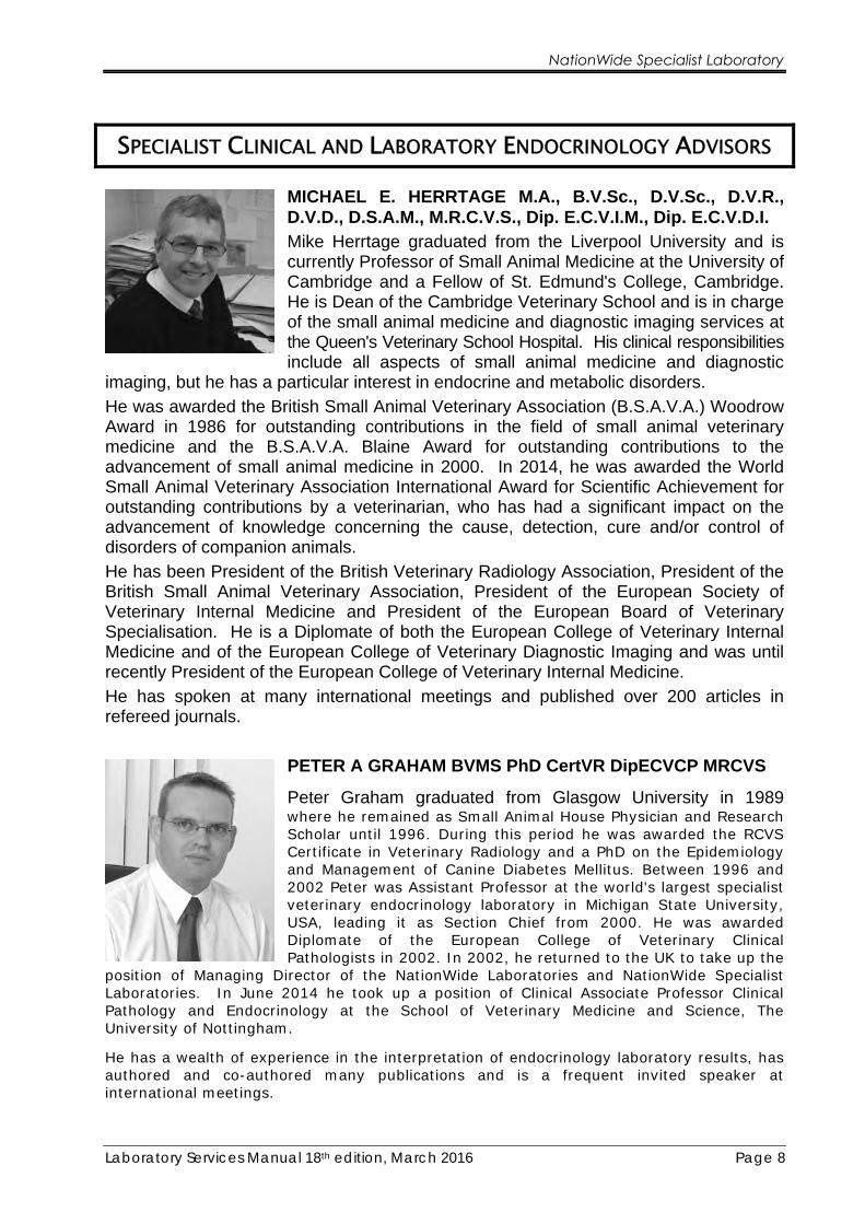
SPECIALIST CLINICAL AND LABORATORY ENDOCRINOLOGY ADVISORS
MICHAEL E. HERRTAGE M.A., B.V.Sc., D.V.Sc., D.V.R., D.V.D., D.S.A.M., M.R.C.V.S., Dip. E.C.V.I.M., Dip. E.C.V.D.I. Mike Herrtage graduated from the Liverpool University and is currently Professor of Small Animal Medicine at the University of Cambridge and a Fellow of St. Edmund's College, Cambridge. He is Dean of the Cambridge Veterinary School and is in charge of the small animal medicine and diagnostic imaging services at the Queen's Veterinary School Hospital. His clinical responsibilities include all aspects of small animal medicine and diagnostic
imaging, but he has a particular interest in endocrine and metabolic disorders.
He was awarded the British Small Animal Veterinary Association (B.S.A.V.A.) Woodrow Award in 1986 for outstanding contributions in the field of small animal veterinary medicine and the B.S.A.V.A. Blaine Award for outstanding contributions to the advancement of small animal medicine in 2000. In 2014, he was awarded the World Small Animal Veterinary Association International Award for Scientific Achievement for outstanding contributions by a veterinarian, who has had a significant impact on the advancement of knowledge concerning the cause, detection, cure and/or control of disorders of companion animals.
He has been President of the British Veterinary Radiology Association, President of the British Small Animal Veterinary Association, President of the European Society of Veterinary Internal Medicine and President of the European Board of Veterinary Specialisation. He is a Diplomate of both the European College of Veterinary Internal Medicine and of the European College of Veterinary Diagnostic Imaging and was until recently President of the European College of Veterinary Internal Medicine.
He has spoken at many international meetings and published over 200 articles in refereed journals.
PETER A GRAHAM BVMS PhD CertVR DipECVCP MRCVS
Peter Graham graduated from Glasgow University in 1989 where he remained as Small Animal House Physician and Research Scholar until 1996. During this period he was awarded the RCVS Certificate in Veterinary Radiology and a PhD on the Epidemiology and Management of Canine Diabetes Mellitus. Between 1996 and 2002 Peter was Assistant Professor at the world's largest specialist veterinary endocrinology laboratory in Michigan State University, USA, leading it as Section Chief from 2000. He was awarded Diplomate of the European College of Veterinary Clinical Pathologists in 2002. In 2002, he returned to the UK to take up the
position of Managing Director of the NationWide Laboratories and NationWide Specialist Laboratories. In June 2014 he took up a position of Clinical Associate Professor Clinical Pathology and Endocrinology at the School of Veterinary Medicine and Science, The University of Nottingham.
He has a wealth of experience in the interpretation of endocrinology laboratory results, has authored and co-authored many publications and is a frequent invited speaker at international meetings.
NationWide Specialist Laboratory
Laboratory Services Manual 18th edition, March 2016 Page 8
SPECIALIST CLINICAL AND LABORATORY ENDOCRINOLOGY ADVISORS
MICHAEL E. HERRTAGE M.A., B.V.Sc., D.V.Sc., D.V.R., D.V.D., D.S.A.M., M.R.C.V.S., Dip. E.C.V.I.M., Dip. E.C.V.D.I. Mike Herrtage graduated from the Liverpool University and is currently Professor of Small Animal Medicine at the University of Cambridge and a Fellow of St. Edmund's College, Cambridge. He is Dean of the Cambridge Veterinary School and is in charge of the small animal medicine and diagnostic imaging services at the Queen's Veterinary School Hospital. His clinical responsibilities include all aspects of small animal medicine and diagnostic
imaging, but he has a particular interest in endocrine and metabolic disorders.
He was awarded the British Small Animal Veterinary Association (B.S.A.V.A.) Woodrow Award in 1986 for outstanding contributions in the field of small animal veterinary medicine and the B.S.A.V.A. Blaine Award for outstanding contributions to the advancement of small animal medicine in 2000. In 2014, he was awarded the World Small Animal Veterinary Association International Award for Scientific Achievement for outstanding contributions by a veterinarian, who has had a significant impact on the advancement of knowledge concerning the cause, detection, cure and/or control of disorders of companion animals.
He has been President of the British Veterinary Radiology Association, President of the British Small Animal Veterinary Association, President of the European Society of Veterinary Internal Medicine and President of the European Board of Veterinary Specialisation. He is a Diplomate of both the European College of Veterinary Internal Medicine and of the European College of Veterinary Diagnostic Imaging and was until recently President of the European College of Veterinary Internal Medicine.
He has spoken at many international meetings and published over 200 articles in refereed journals.
PETER A GRAHAM BVMS PhD CertVR DipECVCP MRCVS
Peter Graham graduated from Glasgow University in 1989 where he remained as Small Animal House Physician and Research Scholar until 1996. During this period he was awarded the RCVS Certificate in Veterinary Radiology and a PhD on the Epidemiology and Management of Canine Diabetes Mellitus. Between 1996 and 2002 Peter was Assistant Professor at the world's largest specialist veterinary endocrinology laboratory in Michigan State University, USA, leading it as Section Chief from 2000. He was awarded Diplomate of the European College of Veterinary Clinical Pathologists in 2002. In 2002, he returned to the UK to take up the
position of Managing Director of the NationWide Laboratories and NationWide Specialist Laboratories. In June 2014 he took up a position of Clinical Associate Professor Clinical Pathology and Endocrinology at the School of Veterinary Medicine and Science, The University of Nottingham.
He has a wealth of experience in the interpretation of endocrinology laboratory results, has authored and co-authored many publications and is a frequent invited speaker at international meetings.

NationWide Specialist Laboratory
Laboratory Services Manual 18th edition, March 2016 Page 9
ABOUT THIS MANUAL
This information manual describes the testing procedures available for a variety of endocrine diseases, pregnancy testing and therapeutic drug monitoring. The majority of the tests refer to canine, feline and equine submissions but we also have experience in other species. Please call if you have questions about the application of a measurement technique in a particular species.
Copyright: © NationWide Specialist Laboratory 2016
Laboratory Services Manual 18th edition, March 2016 Page 10
THYROID FUNCTION
CANINE HYPOTHYROIDISM
There are a great many factors affecting thyroid hormone levels in dogs. It is essential that a thorough clinical examination is performed and history taken prior to any diagnostic testing. The following factors are also very important to consider when carrying out thyroid function tests.
• Concurrent Therapy – Certain therapies can affect thyroid hormone results and complicate their interpretation. Glucocorticoid and barbiturate medications often cause low total T4 (TT4) concentrations. Where possible, it helps to discontinue these therapies for 1 month prior to thyroid diagnostic testing. When clinical circumstances prevent withdrawal, a diagnostic panel including FT4ED (see below) should be selected. Sulphonamide products can cause a reversible hypothyroidism during their use and, consequently, thyroid diagnostic testing should be postponed until 3 weeks after they have been discontinued.
• Breed Specific Ranges – Sight hounds are known to have lower TT4 levels compared to other breeds. The diagnosis of hypothyroidism in these breeds should be made cautiously and more reliance should be placed on other measures of thyroid function that are less influenced by breed such as TSH (or total T3).
• Low T4 State of Medical Illness (Sick Euthyroid Syndrome: SES) – Dogs with non-thyroidal illnesses will often have low serum TT4 as part of their physiological response to that illness. This is not a T4 deficient state and thyroid supplementation is not appropriate. Instead, it is a reflection of the mechanism used to control metabolic rate during illness that is believed to improve the chances of survival. The effect of non-thyroidal illness on FT4ED values is less common and less dramatic. Ill dogs will often have depressed TT4 levels so the use of additional or alternative thyroid function tests along with careful evaluation of clinical signs and assessment of the likelihood of non-thyroidal illness are important in distinguishing the true hypothyroid dogs from those that are just responding to a non-thyroidal illness.
We recommend that the investigation of possible thyroid disease or dysfunction be performed using panels of several tests rather than individual tests. The diagnostic power of a group of thyroid tests is much greater than that of any available single test. An alternative to the panel approach is the dynamic test approach. Our panels are made up of selections of the following tests: total T4, free T4 by equilibrium dialysis, thyroid stimulating hormone, thyroglobulin antibody and thyroid hormone antibodies.

THYROID FUNCTION
Laboratory Services Manual 18th edition, March 2016 Page 11
Individual tests
Total T4 (TT4) – Most dogs with hypothyroidism would be expected to have a low TT4 (~90% of them) making this a test with high, but slightly less than perfect, diagnostic sensitivity. On the other hand, the effects of non-thyroidal illness on TT4 means that many dogs with normal thyroid function will also have a low TT4 making the test poorly specific. Depending on the type of population sampled, up to 25% of dogs with normal thyroid function will yield a low TT4. This poor diagnostic specificity and less than perfect sensitivity means that TT4 has limited value as a stand-alone test for hypothyroidism. Diagnostic power is improved by combining it with TSH measurement or by performing a dynamic response test.
Total T4 also has a role to play in monitoring thyroid therapy where it can be used alone (if hours post-pill and dose frequency are recorded) or ideally in combination with TSH.
Free T4 by Equilibrium Dialysis (FT4ED) Almost all (>99.9%) of circulating T4 is bound to carrier proteins leaving only a tiny fraction available to interact with tissues. This free fraction can be measured in an ultra sensitive radioimmunoassay following an equilibrium dialysis step. The analysis of FT4ED is the most accurate way of assessing the physiologically important thyroid status of an animal. Samples are dialysed, separating FT4 from serum proteins and protein bound T4. In most cases, TT4 and FT4ED will be highly correlated. The specific circumstances in which they are not are when we would recommend FT4ED as the thyroid hormone test of choice. It would be an advantage to measure FT4ED instead of, or in addition to, TT4 in the following situations:
• Non-thyroidal illness: one of the contributing mechanisms to the low TT4 we see in non-thyroidal illness is an alteration in thyroid hormone-protein binding. Although TT4 concentrations may be greatly reduced, the lower protein affinity for T4 means a higher fraction is available as free hormone and the FT4ED concentration usually remains within the reference range. This makes FT4ED a good test for distinguishing non-thyroidal illness from true hypothyroidism as the cause of a low TT4.
• Concurrent therapies: part of the effect on certain therapies on TT4 is mediated through thyroid hormone-protein binding meaning that FT4ED is less commonly and less dramatically affected by concurrent therapy. FT4ED is the analysis of choice when glucocorticoid or barbiturate therapies cannot be withdrawn prior to embarking on a thyroid diagnostic investigation.
• T4AA: the presence of T4 cross-reacting antibodies (T4AA) in the patient’s serum will interfere with TT4 measurement causing false high values. The FT4ED procedure is unaffected by these antibodies because they are removed by the dialysis step. For an accurate estimation of thyroid status in a dog with T4AA, FT4ED is required. T4AA are present in the serum of approximately 10% of hypothyroid dogs as part of the thyroid pathologic process.
Canine TSH (cTSH) We expect serum concentrations of cTSH to be high in animals with primary hypothyroidism because the negative feedback effect of thyroid hormones on pituitary production of TSH is lost. Indeed, this is the case most of the time but the diagnostic sensitivity is less than ideal. About 80-85% of hypothyroid dogs will have the expected high cTSH, unfortunately, leaving a proportion that will not. Conversely, cTSH measurement has good diagnostic specificity (up to 100%) meaning that false positives
THYROID FUNCTION
Laboratory Services Manual 18th edition, March 2016 Page 12
are rare. The combination of thyroid hormone analysis with cTSH measurement makes the most of the advantages of the individual tests while minimizing their deficiencies (see flow chart for interpretation of TT4 & cTSH).
Canine TSH may be measured at 30 minutes as part of a TRH stimulation test in the diagnosis of secondary hypothyroidism. Normal dogs should increase cTSH by at least 0.4 ng/mL.
Total T3 (TT3) The analysis of TT3 is of little value in the diagnosis of hypothyroidism principally because of the high prevalence of cross-reacting T3 autoantibodies (T3AA) in hypothyroid dogs. These antibodies cause false results to be generated in T3 assays.
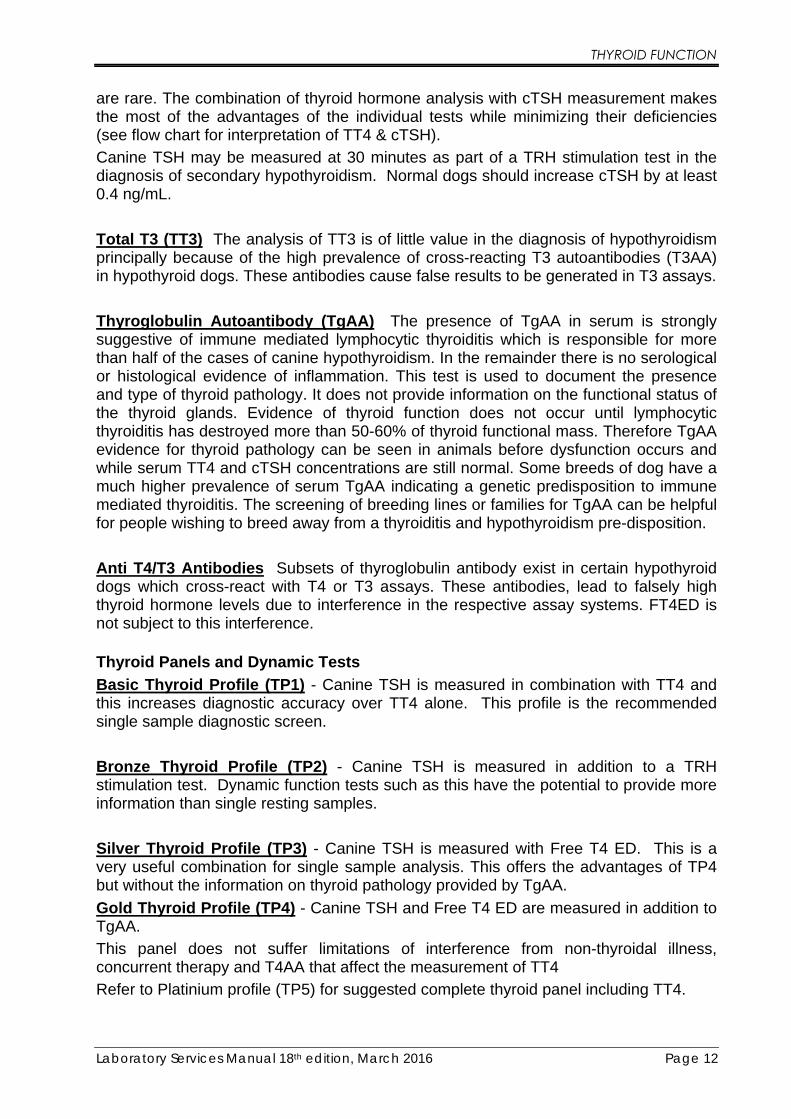
Thyroglobulin Autoantibody (TgAA) The presence of TgAA in serum is strongly suggestive of immune mediated lymphocytic thyroiditis which is responsible for more than half of the cases of canine hypothyroidism. In the remainder there is no serological or histological evidence of inflammation. This test is used to document the presence and type of thyroid pathology. It does not provide information on the functional status of the thyroid glands. Evidence of thyroid function does not occur until lymphocytic thyroiditis has destroyed more than 50-60% of thyroid functional mass. Therefore TgAA evidence for thyroid pathology can be seen in animals before dysfunction occurs and while serum TT4 and cTSH concentrations are still normal. Some breeds of dog have a much higher prevalence of serum TgAA indicating a genetic predisposition to immune mediated thyroiditis. The screening of breeding lines or families for TgAA can be helpful for people wishing to breed away from a thyroiditis and hypothyroidism pre-disposition.
Anti T4/T3 Antibodies Subsets of thyroglobulin antibody exist in certain hypothyroid dogs which cross-react with T4 or T3 assays. These antibodies, lead to falsely high thyroid hormone levels due to interference in the respective assay systems. FT4ED is not subject to this interference.
Thyroid Panels and Dynamic Tests
Basic Thyroid Profile (TP1) - Canine TSH is measured in combination with TT4 and this increases diagnostic accuracy over TT4 alone. This profile is the recommended single sample diagnostic screen.
Bronze Thyroid Profile (TP2) - Canine TSH is measured in addition to a TRH stimulation test. Dynamic function tests such as this have the potential to provide more information than single resting samples.
Silver Thyroid Profile (TP3) - Canine TSH is measured with Free T4 ED. This is a very useful combination for single sample analysis. This offers the advantages of TP4 but without the information on thyroid pathology provided by TgAA.
Gold Thyroid Profile (TP4) - Canine TSH and Free T4 ED are measured in addition to TgAA.
This panel does not suffer limitations of interference from non-thyroidal illness, concurrent therapy and T4AA that affect the measurement of TT4
Refer to Platinium profile (TP5) for suggested complete thyroid panel including TT4.
THYROID FUNCTION
Laboratory Services Manual 18th edition, March 2016 Page 12
are rare. The combination of thyroid hormone analysis with cTSH measurement makes the most of the advantages of the individual tests while minimizing their deficiencies (see flow chart for interpretation of TT4 & cTSH).
Canine TSH may be measured at 30 minutes as part of a TRH stimulation test in the diagnosis of secondary hypothyroidism. Normal dogs should increase cTSH by at least 0.4 ng/mL.
Total T3 (TT3) The analysis of TT3 is of little value in the diagnosis of hypothyroidism principally because of the high prevalence of cross-reacting T3 autoantibodies (T3AA) in hypothyroid dogs. These antibodies cause false results to be generated in T3 assays.
Thyroglobulin Autoantibody (TgAA) The presence of TgAA in serum is strongly suggestive of immune mediated lymphocytic thyroiditis which is responsible for more than half of the cases of canine hypothyroidism. In the remainder there is no serological or histological evidence of inflammation. This test is used to document the presence and type of thyroid pathology. It does not provide information on the functional status of the thyroid glands. Evidence of thyroid function does not occur until lymphocytic thyroiditis has destroyed more than 50-60% of thyroid functional mass. Therefore TgAA evidence for thyroid pathology can be seen in animals before dysfunction occurs and while serum TT4 and cTSH concentrations are still normal. Some breeds of dog have a much higher prevalence of serum TgAA indicating a genetic predisposition to immune mediated thyroiditis. The screening of breeding lines or families for TgAA can be helpful for people wishing to breed away from a thyroiditis and hypothyroidism pre-disposition.
Anti T4/T3 Antibodies Subsets of thyroglobulin antibody exist in certain hypothyroid dogs which cross-react with T4 or T3 assays. These antibodies, lead to falsely high thyroid hormone levels due to interference in the respective assay systems. FT4ED is not subject to this interference.
Thyroid Panels and Dynamic Tests
Basic Thyroid Profile (TP1) - Canine TSH is measured in combination with TT4 and this increases diagnostic accuracy over TT4 alone. This profile is the recommended single sample diagnostic screen.
Bronze Thyroid Profile (TP2) - Canine TSH is measured in addition to a TRH stimulation test. Dynamic function tests such as this have the potential to provide more information than single resting samples.
Silver Thyroid Profile (TP3) - Canine TSH is measured with Free T4 ED. This is a very useful combination for single sample analysis. This offers the advantages of TP4 but without the information on thyroid pathology provided by TgAA.
Gold Thyroid Profile (TP4) - Canine TSH and Free T4 ED are measured in addition to TgAA.
This panel does not suffer limitations of interference from non-thyroidal illness, concurrent therapy and T4AA that affect the measurement of TT4
Refer to Platinium profile (TP5) for suggested complete thyroid panel including TT4.

THYROID FUNCTION
Laboratory Services Manual 18th edition, March 2016 Page 13
Platinium Thyroid Profile (TP5) – TT4, Canine TSH, Free T4 ED and TgAA. This panel provides the most comprehensive information available from a single sample. The TgAA identifies thyroid pathology and the TT4, FT4ED and TSH provide a complete picture of thyroid function.
Copper Thyroid Profile (TP6) – TT4, Canine TSH and Free T4 ED. This panel provides the most comprehensive information available from a single sample at a more competitive price as TGAA is not measured and the possible cause of the thyroid pathology may not be considered important.
TSH Stimulation Test
Unfortunately, pharmaceutical grade bovine TSH is no longer available. Chemical grade bovine TSH is available but extreme care should be taken if this product is used as there may be a risk of adverse reaction. A commercial pharmaceutical recombinant human TSH has been demonstrated to be useful for TSH stimulation testing in the dog. However, in its present form, it is very expensive.
1. Take blood sample for basal TT4 concentration.
2. Inject 0.1IU/kg TSH i/v. or 100 to 150 ug/dog human recombinant (rh)TSH
3. Take a second blood sample 4 - 6 hours later for post TT4 concentration.
Interpretation
T4 levels in a normal dog should increase by 1.5 - 2.0 times the basal concentration to reach a value above 26 nmol/L.
Greyhound Thyroid Panels Greyhounds and other sighthounds (e.g. Saluki’s) have total serum thyroxine (TT4) levels that are lower than those of other breeds of dog. Thyroid investigation is not uncommon in greyhounds as part of the work up for poor performance, behavioural change, bald thighs and fertility concerns. NationWide Specialist Laboratory have put together profiles of thyroid tests specifically for the greyhound breed.
Greyhound Thyroid Investigation (GTH1) – Sensitive TT4, TT3, TSH and TGAA
Greyhound Thyroid Monitoring (GTH2) – Sensitive TT4, TT3 and Canine TSH.

THYROID FUNCTION
Laboratory Services Manual 18th edition, March 2016 Page 14
TRH Stimulation Test 1. Take blood sample for basal TT4 concentration.
2. Inject TRH i/v slowly over one minute.
3. 1 - 5 kg 100ug TRH
5 - 30 kg 200ug TRH
>30 kg 300ug TRH
4. Take a second blood sample 4 - 6 hours later for post TT4 concentration.
Interpretation TT4 levels in a normal dog should increase by about 1.2 times the basal concentration to reach a value above 25 nmol/L. If the pre stimulation sample is above 25 nmol/L the dog is likely to be normal, regardless of the post stimulation TT4.
Hypothyroid dogs usually show low basal TT4 levels, which fail to respond to TRH or stimulate to a value below 25 nmol/L. Sometimes a high/normal basal concentration may be seen which fails to increase 1.2 times. On these occasions it is likely that the thyroid gland is being stimulated maximally and cannot respond further (in the absence of T4AA).
Diagnosis of secondary hypothyroidism can be attempted by measuring cTSH at zero and 30 minutes post TRH stimulation. Normal dogs should increase cTSH by at least 0.4 ng/mL.
Monitoring Therapy Thyforon® (Dechra Veterinary Products), Soloxine® (Virbac) and Leventa® (MSD) are the only licensed veterinary preparations of levothyroxine. The initial recommended dose varies by datasheet but is around 20ug/kg daily. However, a large proportion of hypothyroid dogs can be successfully treated with a lower dose. TT4 levels should be monitored after several weeks of therapy to determine whether a dose change would be appropriate. For animals receiving therapy twice daily, the time post pill at which the monitoring sample is taken is less important than that it is recorded. TT4 levels should ideally be between 30 - 47 nmol/L at around the time of expected peak concentrations (3 hours post pill) and be above 19.0 nmol/L by the time the next pill is due (trough concentration). The half-life of TT4 in the dog can vary but is in the region of 10 to 14 hours.
The measurement of both TT4 and cTSH is recommended in monitoring thyroid therapy in dogs. cTSH reflects the adequacy of thyroid replacement therapy in the preceding days, not just the day of the test. This can help identify inconsistencies in dosing and also prevent inappropriate management decisions being made based on unrepresentative single day TT4 results.

THYR
OID
FUN
CTIO
N
Labo
rato
ry S
ervi
ces M
anua
l 18t
h edi
tion,
Mar
ch 2
016
Pa
ge 1
5
DIAG
NOSI
NG C
ANIN
E HY
POTH
YROI
DISM
FLO
WCH
ART
T4
/ cT
SH a
ssay
Nor
mal
T4
25-8
0 nm
ol/l
Hig
h T4
80 n
mol
/l Sign
ifica
nce
uncl
ear.
Con
side
r:-O
estru
s?H
yper
thyr
oidi
sm?
Thyr
oxin
e tre
atm
ent?
Nor
mal
cTS
H<0
.6 n
g/m
lIn
crea
sed
cTSH
>0.6
ng/
ml
Nor
mal
cTS
H<0
.6 n
g/m
lIn
crea
sed
cTSH
>0.6
ng/
ml
Hyp
othy
roid
ism
susp
ecte
d on
bas
is o
f his
tory
and
clin
ical
sign
s
Low
T4
<15n
mol
/l
Hyp
othy
roid
ism
ver
y un
likel
yH
ypot
hyro
idis
m u
nlik
ely
Hyp
othy
roid
ism
ver
y lik
ely
Hyp
othy
roid
ism
like
ly
Ant
i-T4
antib
odie
s
Hig
h tit
reIn
dex
> 2.
0Lo
w ti
treIn
dex
< 2.
0
Che
ck h
isto
ry fo
r rec
ent a
dmin
istra
tion
of th
yros
uppr
essi
ve d
rugs
e.g
pred
niso
lone
, pot
entia
ted
sulp
hona
mid
e
Rou
tine
haem
atol
ogy
and
bioc
hem
istr
y to
exc
lude
oth
er sy
stem
ic d
isea
ses
Hyp
erch
oles
tero
laem
ia, m
ild a
naem
ia a
nd m
ild in
crea
ses i
n liv
er e
nzym
es m
ay b
e se
en w
ith h
ypot
hyro
idis
m
Sign
ifica
nce
uncl
ear
Con
side
r:-Si
ck e
uthy
roid
ism
?N
orm
al b
reed
var
iatio
n?Ea
rly h
ypot
hyro
idis
m?
Hyp
othy
roid
ism
still
susp
ecte
d
Con
side
r ret
estin
g af
ter 3
mon
ths o
r dy
nam
ic th
yroi
d fu
nctio
n te
st (T
RH
or T
SH st
imul
atio
n te
st)
Nor
mal
cTS
H<0
.6 n
g/m
lIn
crea
sed
cTSH
>0.6
ng/
ml
Low
-nor
mal
T4
15-2
5 nm
ol/l
Sick
eut
hyro
idis
m ?
Hyp
othy
roid
ism
?Hyp
othy
roid
ism
ver
y lik
ely
Furth
er in
vest
igat
ions
of o
ther
dis
ease
s
Not
esD
ata
base
d on
SC
L-LC
G B
iosc
ienc
es re
fere
nce
valu
esBe
aw
are
that
adm
inis
tratio
n of
TSH
can
cau
se fa
tal a
naph
ylac
tic re
actio
nsFu
rthe
r rea
ding
Ram
sey,
I.K
. (19
97) D
iagn
osin
g ca
nine
hyp
othy
roid
ism
. In
Prac
tice
19: 3
71
See
als
o: In
Pra
ctic
e 20
09;3
1:77
-82
Can
ine
hypo
thyr
oidi
sm: d
iagn
osis
and
ther
apy
– P
eter
A G
raha
m

THYROID FUNCTION
Laboratory Services Manual 18th edition, March 2016 Page 16
FELINE HYPERTHYROIDISM (THYROTOXICOSIS)
Individual Tests
Total T4 - TT4 is generally used as the main diagnostic test for hyperthyroidism and to monitor T4 levels post treatment. TT4 should be used as the first screening test. If TT4 is raised (> 60 nmol/L) it is very likely the cat is hyperthyroid. If TT4 is within the reference range then further tests may be helpful in ruling out or confirming hyperthyroidism. Often hyperthyroid cats with concurrent non-thyroidal illness will have normal TT4 concentrations (occult hyperthyroidism). In these cases the detection of a palpable thyroid nodule will be a strong indicator that hyperthyroidism should still be suspected and investigated with FT4ED or a dynamic function test. If TT4 is in the lower end of the reference range (<30 nmol/L) it is unlikely that the cat is hyperthyroid. Euthyroid cats with significant non-thyroidal illness will have low-normal or subnormal TT4 concentrations; generally, the more severe the illness the lower the TT4.
Free T4 by Equilibrium Dialysis (FT4ED) - FT4ED is the most accurate way to measure free T4 and has greater diagnostic sensitivity for hyperthyroidism in sick animals where the TT4 may be depressed into the reference range. FT4ED is less affected by altered binding protein characteristics seen in non-thyroidal illnesses as the samples are dialysed prior to assay. FT4ED levels are elevated in hyperthyroid cats that have TT4 levels within the normal range (19 - 65 nmol/L). Care is needed when using FT4ED as a stand-alone test because it may be elevated in sick euthyroid cats. To mitigate this risk of false positives, FT4ED should be measured in conjunction with TT4 and also when there are appropriate clinical signs such as a palpable thyroid nodule.
Thyrotropin (TSH) – Using the same assay as is used to measure canine TSH there is sufficient cross-reactivity for us to detect increased levels of feline TSH. This can be a helpful analyte in the identification of iatrogenic hypothyroidism following treatment of hyperthyroidism and in the less common presentation of naturally occurring feline hypothyroidism (both congenital +/- goitre and inflammatory forms have been recognised). The combination of azotaemia and iatrogenic hypothyroidism has been shown have a detrimental effect on the survival of treated hyperthyroid cats.
Thyroid Panels and Dynamic Tests
Feline Hyperthyroid Profile – The combination of TT4 and FT4ED offers the advantages of improved diagnostic sensitivity (less false negatives) over TT4 alone and improved diagnostic specificity (less false positives) compared to FT4ED alone.
Feline Hyperthyroid Profile – Silver (Total T4, ALT, ALP, Total Protein, Urea, Creatinine, Phosphorus). Specific biochemistry tests in addition to TT4.
Feline Hyperthyroid Profile – Gold (As silver plus FT4ED to improve diagnostic sensitivity and specificity).

THYROID FUNCTION
Laboratory Services Manual 18th edition, March 2016 Page 17
T3 Suppression Test
This is a useful test for diagnosing borderline cases of hyperthyroidism where the TT4 is consistently within the normal range. It is very important to measure Total T3 (TT3) as well to ensure the cat actually absorbed the T3 dose.
1. Collect basal blood sample for TT4 and TT3. Store separated serum frozen until
protocol completed and submit with post-sample
2. Administer T3 (Tertroxin) orally every 8 hours for a total of 7 doses according to body weight:
cats <5 kg 20 micrograms of Tertroxin
cats >5 kg 30 micrograms of Tertroxin
3. Collect second blood sample 2 - 6 hours after final dose
4. Label both samples clearly and request TT4 and TT3.
Interpretation
TT3 suppression normal cats usually show at least 50% reduction in TT4 levels following suppression. Hyperthyroid cats generally show limited suppression. TT3 should be increase indicating the T3 tablets have been absorbed.
TRH Stimulation Test
Can be useful in cases of mild or borderline hyperthyroidism.
1. Collect basal blood sample.
2. Administer 100 μg/kg i/v slowly over one minute
3. Collect second blood sample 4 hours later
4. Label samples clearly and request TT4.
Interpretation
Normal cats increase 1.5 - 2 times following stimulation.
Hyperthyroid cats generally show little or no rise in TT4 levels from a high or high-normal baseline.

THYROID FUNCTION
Laboratory Services Manual 18th edition, March 2016 Page 18
FELINE HYPOTHYROIDISM
Feline hypothyroidism is considered to be a rare condition. Options for diagnosis include the measurement of serum TT4 or Free T4 and TSH (TP1 or TP3 – see canine hypothyroidism), or a TRH stimulation test. A TSH stimulation test would be ideal if a TSH preparation is available (including recombinant human TSH; Thyrogen®, Genzyme; 0.025 to 0.200 mg IV of rhTSH).
The most common form of feline hypothyroidism is iatrogenic following treatment of hyperthyroidism. Less commonly we recognise naturally occurring feline hypothyroidism (both congenital +/- goitre and inflammatory forms exist). The combination of azotaemia and iatrogenic hypothyroidism has been shown to have a detrimental effect on the survival of treated hyperthyroid cats.
TRH Stimulation Test
1. Collect blood for basal TT4 concentration
2. Inject 100 μg TRH i/v slowly over one minute. Side effects including salivation and vomiting can be seen occasionally
3. Collect second blood sample 4 - 6 hours later
4. Label samples clearly and request TT4
Interpretation Normal cats - post stimulation result is expected to be about 1.5 - 2 times greater than the basal T4 concentration.
Monitoring of feline hyperthyroidism is detailed at the end of the booklet under endocrine therapeutic monitoring.
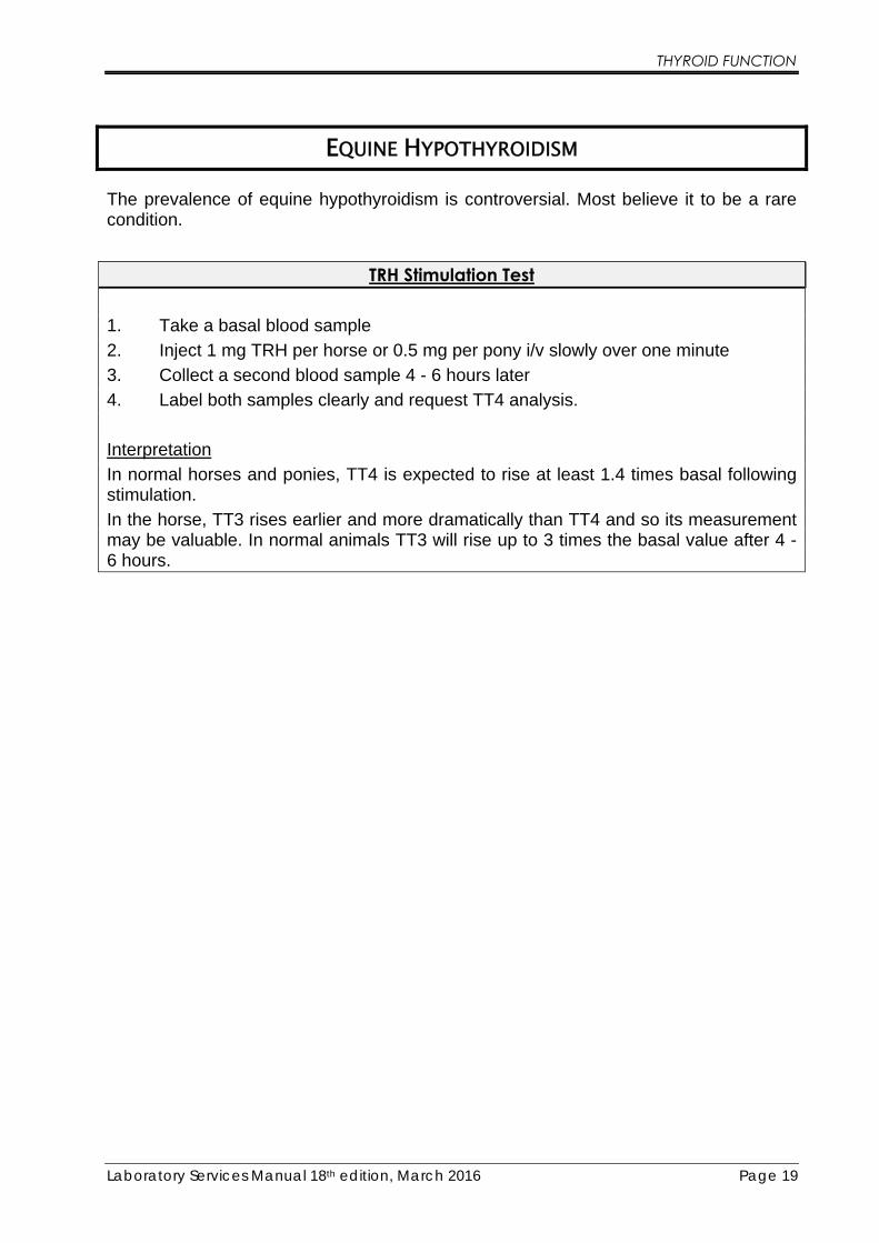
THYROID FUNCTION
Laboratory Services Manual 18th edition, March 2016 Page 19
EQUINE HYPOTHYROIDISM
The prevalence of equine hypothyroidism is controversial. Most believe it to be a rare condition.
TRH Stimulation Test
1. Take a basal blood sample
2. Inject 1 mg TRH per horse or 0.5 mg per pony i/v slowly over one minute
3. Collect a second blood sample 4 - 6 hours later
4. Label both samples clearly and request TT4 analysis.
Interpretation In normal horses and ponies, TT4 is expected to rise at least 1.4 times basal following stimulation.
In the horse, TT3 rises earlier and more dramatically than TT4 and so its measurement may be valuable. In normal animals TT3 will rise up to 3 times the basal value after 4 - 6 hours.

Laboratory Services Manual 18th edition, March 2016 Page 20
ADRENOCORTICAL FUNCTION
CANINE HYPERADRENOCORTICISM (HAC - CUSHING’S DISEASE)
Hyperadrenocorticism occurs in middle–aged and geriatric dogs. In around 80% of cases it is pituitary-dependent (PDH). In the remainder, the disease results from primary adrenal-dependent neoplasia or hyperplasia (ADH).
Individual tests
Cortisol - Basal cortisol levels are of no value in diagnosing HAC. There is significant overlap in cortisol concentration among healthy, stressed, sick and affected dogs.
Cortisol/Creatinine Ratio (CCR) - This is a very sensitive test to exclude HAC (very few false negatives) but must not be used to diagnose HAC as it is very poorly specific and non-adrenal illness commonly gives a positive result. A morning urine sample is collected in the animal’s home environment. This reflects the cortisol released over several hours. Normal dogs will have a CCR less than 30 x 10-6.
Dynamic test protocols
Which test should be used to diagnose HAC reliably in the dog? Unfortunately, there is no perfect diagnostic test or test protocol for hyperadrenocorticism. The line between physiologic adrenal stress responses to non-adrenal illness and pathologic hyperfunction of the pituitary-adrenal axis can be fine indeed. As a consequence, false positive and false negative test results occur. The different available diagnostic protocols available have different properties, advantages and disadvantages but none is perfect. In establishing confidence that hyperadrenocorticism is confirmed or ruled out more than one diagnostic test protocol may be required.
ACTH stimulation is a good screening test in the first instance and is the test of choice for diagnosing iatrogenic Cushing’s and monitoring anti-adrenal therapy. It has a lower false positive rate than the low-dose dexamethasone suppression test but a significant false negative rate. It will reliably diagnose about 85% of PDH cases but only 50% of ADH cases. It is quick and simple to perform and is less affected by stress and non-adrenal illness. The initial values are useful as a reference to monitor effectiveness of treatment.
Low-dose Dexamethasone screening test is more sensitive than the ACTH stimulation test in confirming HAC. Diagnostic sensitivity is almost perfect (>95%) meaning that false negative results very seldom occur giving us tremendous confidence in a negative test result. Unfortunately, false positive results are common especially when there is concurrent non-adrenal illness or other sources of stress. Positive results of the low-dose dexamethasone test should be regarded with suspicion in dogs known to have significant non-adrenal illness. Ideally, the test should be postponed until any identified

ADRENOCORTICAL FUNCTION
Laboratory Services Manual 18th edition, March 2016 Page 21
non-adrenal illnesses have been resolved or stabilised. See “Diagnosing Canine Hyperadrenocorticism” flowchart (page 25).
17-hydroxyprogesterone measurement in the diagnosis of adrenocortical disease
Certain functional adrenocortical tumours do not have cortisol as their principle secretory product but instead the tumour has developed in such a way that it is adrenal steroid precursors that are released into the circulation. Many dogs with this type of functional adrenal tumour will have clinical signs suggestive of hyperadrenocorticism. A common cortisol response to ACTH in these cases is a “flat-line, mid-range” pattern. Measuring 17OHP before and after ACTH stimulation (same sample as you would take for cortisol) can be very helpful in identifying these tumours.
Additionally, in cases suspected of hyperadrenocorticism which do demonstrate an exaggerated cortisol response to ACTH, the additional measurement of 17OHP can help improve the confidence in ruling hyperadrenocorticism in or out. Less than 10% of dogs with classic hyperadrenocorticism would be expected to have post-ACTH 17OHP <4.5 nmol/L (Chapman et al, 2003 Veterinary Record, 153(25):771-5) and fewer than 10% of dogs which do not have classic hyperadrenocorticism would be expected to have post-ACTH 17OHP >16.7 nmol/L.
Consider measuring 17OHP in addition to cortisol when:
• Flat-line, mid-range cortisol response to ACTH
o Investigate functional adrenocortical tumour
• Mildly exaggerated cortisol response to ACTH and few appropriate clinical signs of HAC
o Look for post-ACTH <4.5nmol/L to improve rule-out
• Normal cortisol response to ACTH and clinical signs consistent with HAC
o Investigate “atypical” hyperadrenocorticism (Ristic et al 2002 JVIM 16(4):433-9) and “Alopecia X” (p32/33)
17OHP may also be useful in ruling out HAC where an exaggerated Cortisol response is seen which is mainly due to stress, often associated with other illness. The Cortisol levels may be borderline or high but the 17OHP response to ACTH is normal.

ADRENOCORTICAL FUNCTION
Laboratory Services Manual 18th edition, March 2016 Page 22
ACTH Stimulation Test 1. Collect basal blood sample.
2. Immediately inject 0.25 mg synthetic ACTH (Synacthen*) i/v or i/m
3. Collect a second blood sample one hour later.
4. Label samples clearly and request Cortisol analysis.
* If Synacthen is unavailable Dechra Veterinary Products Ltd have an alternative drug to Synacthen available on special prescription, contact [email protected] for details.
Interpretation Normal dogs will show an increase in cortisol levels of up to 450 nmol/L post stimulation.
An exaggerated response is expected in animals with PDH and cortisol concentrations rise above 600 nmol/L and often above 1000 nmol/L.
Dogs with an adrenal tumour often have basal cortisol above 250 nmol/L with little or no change after stimulation. However the ACTH stimulation test is not as sensitive at detecting adrenal tumours and negative results should be confirmed with a low dose dexamethasone test when strong clinical suspicion remains.
Low Dose Dexamethasone Screening Test
1. Collect a basal blood sample
2. Inject 0.01 mg/kg i/v of Dexamethasone.
3. Collect two further blood samples 3 hours and 8 hours later
4. Label sample times clearly on the tubes and request cortisol.
Interpretation
The low-dose dexamethasone screening test is interpreted in two stages. Firstly, the presence or absence of hyperadrenocorticism is established by examining the 8-hour result. A value above 40 nmol/L is positive. The next step applies in the positive cases and checks for evidence of suppression sufficient to identify the source of hyperadrenocorticism. Greater than 50% suppression from the baseline value at either 3 or 8 hours is consistent with pituitary dependent disease. When there is no, or minimal, evidence of suppression it is necessary to follow-up with a differentiation test to identify the source (high-dose dexamethasone suppression test, endogenous (plasma) ACTH, adrenal ultrasonography). Both dogs with an adrenal tumour and some dexamethasone “resistant” pituitary cases will not depress by 50%. In about 60% of hyperadrenocorticism cases, we will obtain differentiation from the low-dose test alone with no need to move on to the differentiation tests.

ADRENOCORTICAL FUNCTION
Laboratory Services Manual 18th edition, March 2016 Page 23
High Dose Dexamethasone Suppression Test
This test is used to help distinguish between PDH and an adrenal tumour. It must only be used after Cushing’s disease has been confirmed by other tests (low dose dex. or ACTH stim.)
The disadvantages to the HDDST are the 8 hour duration and that a small proportion of pituitary dependent cases will fail to suppress even at substantial doses of dexamethasone. An alternative differentiation test is the endogenous (plasma) ACTH which has the advantage of being a single point test.
1. Collect a basal blood sample.
2. Inject 0.1 - 1.0 mg/kg i/v of Dexamethasone.
3. Collect two further blood samples 3 hours and 8 hours later
4. Label sample times clearly on the tubes and request cortisol.
Interpretation
Any suppression greater than 50% of the baseline concentration indicates a pituitary source. A failure to suppress by 50% is consistent with either an adrenal tumour or a “dexamethasone-resistant” pituitary lesion. When no suppression is observed, repeating the test with a higher dose of dexamethasone or diagnostic imaging of the adrenals and/or pituitary could be considered.

ADRENOCORTICAL FUNCTION
Laboratory Services Manual 18th edition, March 2016 Page 24
Endogenous (plasma) ACTH
Endogenous ACTH may be used as a reliable tool for differentiating PDH from ADH when hyperadrenocorticism has already been confirmed by ACTH stimulation test or low-dose dexamethasone screening test. However, it has no value as a diagnostic test for hyperadrenocorticism in dogs and cats. Special sample handling procedures apply.
ACTH Sample Preparation
Sample MUST be EDTA plasma or EDTA plasma with Aprotinin. 1. Request a transport pack and follow the instructions that come with the pack for
sample preparation.
2. Take the blood sample into the cooled Aprotinin EDTA tube (supplied in pack) which has been cooled in the fridge.
3. Mix very well but gently at least 10 times and centrifuge as quickly as possible.
4. Transfer the plasma into a cooled plastic (not glass!) PLAIN tube kept in the fridge.
5. Immediately freeze (<-10°C) the plasma sample and keep frozen until dispatch in the transport pack. Freeze the sample first, separately, before placing in the frozen pack for transport.
6. Mark the tube with “EDTA plasma” and animal/owner name.
Cortisol/Creatinine Ratio (CCR) with Dexamethasone Suppression Test When Cushing’s disease is strongly suspected, this test can be used when the dog is easily stressed or difficult to sample. The owner does all the sampling at home.
1. Day 1, collect a first morning urine sample.
2. Mix the urine and add some to Sample Tube 1, place in the fridge until dispatch.
3. Day 2, collect a first morning urine sample.
4. Mix the urine and add some to Sample Tube 2, place in the fridge until dispatch.
5. Immediately after urine collection, note the time and give the dog the required number of Dexamethasone tablets (dose = 0.1mg/kg).
6. 8 hours later give the dog a 2nd set of Dexamethasone tablets.
7. 16 hours later give the dog a 3rd set of Dexamethasone tablets.
8. Day 3, collect a first morning urine sample.
9. Mix the urine and add some to Sample Tube 3, place in the fridge until dispatch.
10. Send all 3 urine samples to the laboratory for CCR.
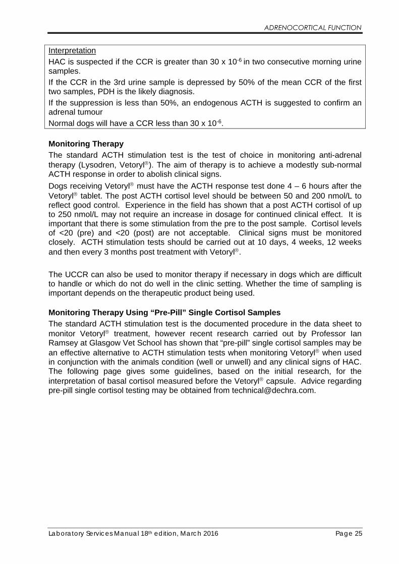
ADRENOCORTICAL FUNCTION
Laboratory Services Manual 18th edition, March 2016 Page 25
Interpretation
HAC is suspected if the CCR is greater than 30 x 10-6 in two consecutive morning urine samples.
If the CCR in the 3rd urine sample is depressed by 50% of the mean CCR of the first two samples, PDH is the likely diagnosis.
If the suppression is less than 50%, an endogenous ACTH is suggested to confirm an adrenal tumour
Normal dogs will have a CCR less than 30 x 10-6.
Monitoring Therapy The standard ACTH stimulation test is the test of choice in monitoring anti-adrenal therapy (Lysodren, Vetoryl). The aim of therapy is to achieve a modestly sub-normal ACTH response in order to abolish clinical signs.
Dogs receiving Vetoryl must have the ACTH response test done 4 – 6 hours after the Vetoryl tablet. The post ACTH cortisol level should be between 50 and 200 nmol/L to reflect good control. Experience in the field has shown that a post ACTH cortisol of up to 250 nmol/L may not require an increase in dosage for continued clinical effect. It is important that there is some stimulation from the pre to the post sample. Cortisol levels of <20 (pre) and <20 (post) are not acceptable. Clinical signs must be monitored closely. ACTH stimulation tests should be carried out at 10 days, 4 weeks, 12 weeks and then every 3 months post treatment with Vetoryl.
The UCCR can also be used to monitor therapy if necessary in dogs which are difficult to handle or which do not do well in the clinic setting. Whether the time of sampling is important depends on the therapeutic product being used.
Monitoring Therapy Using “Pre-Pill” Single Cortisol Samples The standard ACTH stimulation test is the documented procedure in the data sheet to monitor Vetoryl treatment, however recent research carried out by Professor Ian Ramsey at Glasgow Vet School has shown that “pre-pill” single cortisol samples may be an effective alternative to ACTH stimulation tests when monitoring Vetoryl when used in conjunction with the animals condition (well or unwell) and any clinical signs of HAC. The following page gives some guidelines, based on the initial research, for the interpretation of basal cortisol measured before the Vetoryl capsule. Advice regarding pre-pill single cortisol testing may be obtained from [email protected].

Un
we
ll
Res
tart
tri
lost
ane
at lo
we
r d
ose
on
ce w
ell
an
d c
ort
iso
l >4
0 n
mo
l/l (
<1
.4 m
g/d
l)
Un
like
ly t
o b
e h
ypo
cort
iso
lism
. In
vest
igat
e o
ther
cau
ses
We
ll
No
clin
ical
si
gns*
of
HA
C
Dec
reas
e d
ose
b
y 1
0-2
0%
No
ch
ange
in
do
se
Clin
ical
sig
ns*
o
f H
AC
Rar
e re
sult
, re
pea
t te
st
and
re-
eval
uat
e c
ase
Incr
ease
do
se
by
10
-20
%
Me
asu
re
po
st p
ill
cort
iso
l
Re
che
ck in
1
mo
nth
Po
st p
ill <
40
nm
ol/
l (<
1.4
mg/
dl)
d
ivid
e d
aily
do
se
Po
st p
ill >
40
nm
ol/
l (>
1.4
mg/
dl)
in
crea
se d
ose
20
-50
%
His
tory
an
d c
linic
al
exam
inat
ion
STO
P
trilo
stan
e
Re
che
ck in
3
mo
nth
s
Smal
l in
crea
se (
10
-20
%)
in d
ose
can
b
e co
nsi
der
ed w
ith
ow
ner
Pre
-pill
co
rtis
ol
<40
nm
ol/
l
>40
nm
ol/
l
40
-14
0 n
mo
l/l
>14
0 n
mo
l/l
<40
nm
ol/
l
40
-14
0 n
mo
l/l
>14
0 n
mo
l/l
<40
nm
ol/
l
Inte
rpre
tati
on
of
cort
iso
l mea
sure
d
bef
ore
an
d a
fter
tri
lost
ane
*PU
/PD
sh
ou
ld r
eso
lve
wit
hin
a
mo
nth
of
star
tin
g th
e co
rrec
t d
ose
, al
op
ecia
may
tak
e 3
to
4 m
on
ths
© Ia
n R
amse
y, U
niv
ersi
ty o
f G
lasg
ow
Un
we
ll
Res
tart
tri
lost
ane
at lo
we
r d
ose
on
ce w
ell
an
d c
ort
iso
l >4
0 n
mo
l/l (
<1
.4 m
g/d
l)
Un
like
ly t
o b
e h
ypo
cort
iso
lism
. In
vest
igat
e o
ther
cau
ses
We
ll
No
clin
ical
si
gns*
of
HA
C
Dec
reas
e d
ose
b
y 1
0-2
0%
No
ch
ange
in
do
se
Clin
ical
sig
ns*
o
f H
AC
Rar
e re
sult
, re
pea
t te
st
and
re-
eval
uat
e c
ase
Incr
ease
do
se
by
10
-20
%
Me
asu
re
po
st p
ill
cort
iso
l
Re
che
ck in
1
mo
nth
Po
st p
ill <
40
nm
ol/
l (<
1.4
mg/
dl)
d
ivid
e d
aily
do
se
Po
st p
ill >
40
nm
ol/
l (>
1.4
mg/
dl)
in
crea
se d
ose
20
-50
%
His
tory
an
d c
linic
al
exam
inat
ion
STO
P
trilo
stan
e
Re
che
ck in
3
mo
nth
s
Smal
l in
crea
se (
10
-20
%)
in d
ose
can
b
e co
nsi
der
ed w
ith
ow
ner
Pre
-pill
co
rtis
ol
<40
nm
ol/
l
>40
nm
ol/
l
40
-14
0 n
mo
l/l
>14
0 n
mo
l/l
<40
nm
ol/
l
40
-14
0 n
mo
l/l
>14
0 n
mo
l/l
<40
nm
ol/
l
Inte
rpre
tati
on
of
cort
iso
l mea
sure
d
bef
ore
an
d a
fter
tri
lost
ane
*PU
/PD
sh
ou
ld r
eso
lve
wit
hin
a
mo
nth
of
star
tin
g th
e co
rrec
t d
ose
, al
op
ecia
may
tak
e 3
to
4 m
on
ths
© Ia
n R
amse
y, U
niv
ersi
ty o
f G
lasg
ow

Labo
rato
ry S
ervi
ces M
anua
l 18t
h edi
tion,
Mar
ch 2
016
Page
27
DIAG
NOSI
NG C
ANIN
E HY
PERA
DREN
OCOR
TICI
SM F
LOW
CHAR
T
Ro
uti
ne
hae
mat
olo
gy
Ro
uti
ne
bio
chem
istr
y
AC
TH
sti
mu
lati
on
tes
t
Lo
w d
ose
dex
amet
has
on
e su
ppre
ssio
n t
est
Det
erm
ine
if a
dre
nal
or
pit
uit
ary
dep
end
ent
Rad
iogr
aph
y, u
ltra
son
ogr
aph
y, e
ndo
gen
ous
AC
TH
17-O
H p
roge
ster
on
e (p
re /
po
st A
CT
H)
and
/ o
r u
rin
e co
rtis
ol c
reat
inin
e ra
tio
His
tory
an
d p
hysi
cal
exam
inat
ion
Ano
rexi
a, v
om
itin
g, d
iarr
hoea
and
pru
ritu
sar
e al
l u
nco
mm
on
in H
AC
.
Uri
nal
ysi
s (i
ncl
ud
ing
spec
ific
gra
vit
y)
Spe
cifi
c gr
avit
y 1
.00
1 -
1.0
30, +
/-pr
ote
in, b
loo
d.
If S
.G >
1.0
30
the
n H
AC
unl
ikel
yco
nsid
er o
ther
dif
fere
ntia
ls f
irst
Incr
ease
d A
P,
AL
T c
hole
ster
ol,
glu
cose
Dec
reas
ed u
rea
If A
P i
s no
rmal
then
HA
C i
s u
nlik
ely
cons
ider
oth
er d
iffe
rent
ials
fir
st
If l
ymph
ocy
te c
ou
nt i
s gr
eate
r th
an 1
.5 x
10
9 /l
then
H
AC
is
unl
ikel
y: c
ons
ider
oth
er d
iffe
rent
ials
fir
stIn
crea
sed
neu
tro
phil
and
occ
asio
nall
y R
BC
cou
nts
Dec
reas
edeo
sino
phil
and
lym
pho
cyte
co
unt
s
Po
st A
CT
H c
ort
iso
l co
nce
ntr
ati
on
> 6
00
nm
ol/
lH
AC
ver
y li
kel
y.
Po
st A
CT
H c
ort
iso
l co
ncen
trat
ion
<6
00
nm
ol/l
HA
C n
ot
excl
ude
dP
ost
AC
TH
co
rtis
ol c
on
cen
tra
tio
n <
40
con
sid
eria
tro
gen
icH
AC
If 8
ho
ur
cort
iso
l > 4
0n
mo
l/l
HA
C v
ery
lik
ely
If 8
ho
ur
cort
iso
l <
40
nmo
l/l
HA
C u
nlik
ely
Dia
gno
sis
con
firm
ed i
f cl
inic
al s
ign
s an
d
rou
tin
e la
bo
rato
ry t
ests
are
co
nsi
sten
t an
d
oth
er d
iffe
ren
tial
s ex
clu
ded
If c
lin
ica
l sig
ns
an
d r
ou
tin
e b
loo
d a
nd
uri
ne
test
s st
ill
con
sist
ent
Po
lydi
psia
, po
lyu
ria,
po
lyph
agia
, wei
ght
gain
, de
crea
sed
exer
icse
tole
ranc
e, i
ncr
ease
d p
anti
ng,
sym
met
rica
l no
n-pr
uri
tic
alo
peci
a,
pot
bell
ied,
ski
n th
inni
ng,
calc
ino
sis
cuti
s
©I.
Ram
sey
(20
02
)

ADRENOCORTICAL FUNCTION
Laboratory Services Manual 18th edition, March 2016 Page 28
CANINE HYPOADRENOCORTICISM - ADDISON’S DISEASE
Diagnosis Primary hypoadrenocorticism results when more than 90% of both adrenal cortices are destroyed and this leads to a clinical deficiency of all adrenal hormones. The ACTH stimulation test will provide confirmation of Addison’s. It is also useful to measure the Sodium/Potassium ratio as evidence for mineralocorticoid deficiency. A low Na:K ratio may not be seen in some cases of primary hypoadrenocorticism until a very advanced stage of the disease. That is, an animal can have confirmed primary hypoadrenocorticism without abnormal electrolytes. A diagnosis of “atypical” or “secondary” hypoadrenocorticism can only be made if failure to produce aldosterone (see below) is confirmed in addition to failure to produce cortisol.
ACTH Stimulation Test 1. Collect basal blood sample.
2. Immediately inject 0.25 mg synthetic ACTH (Synacthen*) i/v or i/m
3. Collect a second blood sample one hour later.
4. Label samples clearly and request Cortisol analysis.
* If Synacthen is unavailable Dechra Veterinary Products Ltd have an alternative drug to Synacthen available on special prescription, contact [email protected] for details.
Interpretation
The basal cortisol will be low with little or no response after ACTH. A classic Addison’s ACTH response test would be Pre cortisol = <20 nmol/L, Post cortisol = <20 nmol/L. False positive diagnoses occur in animals which have been receiving glucocorticoid therapy recently including oral, injectable and topical (eye, ear and skin) preparations. For confirmation of primary hypoadrenocorticism in dogs which have been receiving glucocorticoid therapy, consider measuring aldosterone instead of, or in addition to, cortisol.
Prednisolone, prednisone and hydrocortisone will cross-react in the cortisol assay and give falsely elevated results. Such therapies should be avoided in the 24 to 36 hours prior to the test. Dexamethasone does not have this effect so it can safely be used prior to confirmation of diagnosis without causing assay interference. However, prolonged use (several days) of dexamethasone prior to performing the ACTH response test can increase the risk of false positive diagnosis and require the measurement of aldosterone for confirmation.
Endogenous (plasma) ACTH ACTH concentrations can be useful in distinguishing primary from secondary hypoadrenocorticism (see differentiation in Canine hyperadrenocorticism section). Dogs with primary Addison’s have very high levels of ACTH, usually greater than 500 pg/mL. Dogs with secondary Addison’s have low or undetectable (< 5.0 pg/mL) levels of ACTH.

ADRENOCORTICAL FUNCTION
Laboratory Services Manual 18th edition, March 2016 Page 29
Aldosterone
Aldosterone measurement may also be used to differentiate between primary and secondary Addison’s and this can be performed on the same samples used to measure the cortisol, with no difference in sample handling procedures.
In primary hypoadrenocorticism, there will be failure of both glucocorticoid and mineralocorticoid production. In secondary hypoadrenocorticism (pituitary ACTH production failure, exogenous steroids) glucocorticoid production will have failed but mineralocorticoid production is unaffected. The trophic support for mineralocorticoid production comes from the renin-angiotensin system not ACTH. However, there is a sufficient aldosterone response to ACTH stimulation to use this as a dynamic diagnostic test for mineralocorticoid production which has advantages over measuring baseline aldosterone alone.
Measuring aldosterone in addition to cortisol is important when it is necessary to perform an ACTH stimulation test to confirm a diagnosis of hypoadrenocorticism after an animal has already received symptomatic glucocorticoid therapy. It is also essential if a diagnosis of atypical or secondary hypoadrenocorticism is being considered (unless cACTH is measured).
Monitoring therapy The laboratory monitoring of primary hypoadrenocorticism focuses on electrolyte status. There is no value in repeating ACTH stimulation tests in animals which have failed adrenal glands. The artefactual cortisol concentrations generated from the cross-reaction of prednisolone in the assay are not biologically equivalent and so a cortisol assay cannot assess the adequacy of glucocorticoid therapy. Biologically equivalent cortisol results can be obtained only during periods that hydrocortisone is the exclusive corticosteroid therapy.
The monitoring of secondary hypoadrenocorticism is based mostly on clinical response with periodic attempts to find the lowest glucocorticoid dose at which the animal appears well. Liver enzyme analysis can help identify steroid hepatopathy resulting from glucocorticoid excess.
Zycortal® (Dechra Veterinary Products) is indicated for replacement therapy for mineralocorticoid deficiency in dogs with primary hypoadrenocorticism. Zycortal contains desoxycortone pivalate (DOCP) – an aldosterone analogue with purely mineralocorticoid effect. Zycortal is available as a prolonged-release suspension for subcutaneous injection and the dosing interval is approximately 25 days. The exact dose to be given and the interval between treatments depend on the dog’s clinical response as well as sodium and potassium levels. A glucocorticoid (e.g. prednisolone) is also needed daily to replace the missing cortisol. To obtain a Monitoring and Dose Adjustment Flowchart, or for further advice, contact Dechra by e-mailing [email protected].
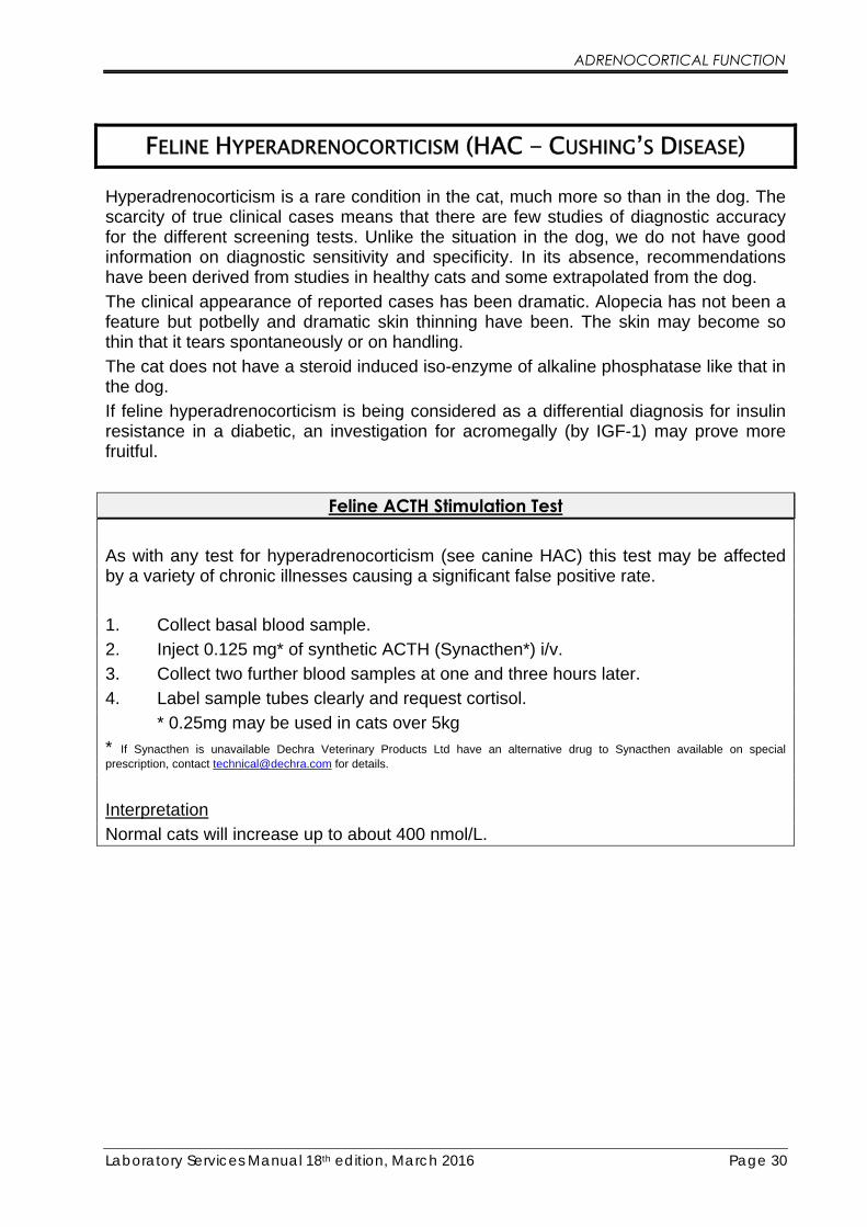
ADRENOCORTICAL FUNCTION
Laboratory Services Manual 18th edition, March 2016 Page 30
FELINE HYPERADRENOCORTICISM (HAC - CUSHING’S DISEASE)
Hyperadrenocorticism is a rare condition in the cat, much more so than in the dog. The scarcity of true clinical cases means that there are few studies of diagnostic accuracy for the different screening tests. Unlike the situation in the dog, we do not have good information on diagnostic sensitivity and specificity. In its absence, recommendations have been derived from studies in healthy cats and some extrapolated from the dog.
The clinical appearance of reported cases has been dramatic. Alopecia has not been a feature but potbelly and dramatic skin thinning have been. The skin may become so thin that it tears spontaneously or on handling.
The cat does not have a steroid induced iso-enzyme of alkaline phosphatase like that in the dog.
If feline hyperadrenocorticism is being considered as a differential diagnosis for insulin resistance in a diabetic, an investigation for acromegally (by IGF-1) may prove more fruitful.
Feline ACTH Stimulation Test
As with any test for hyperadrenocorticism (see canine HAC) this test may be affected by a variety of chronic illnesses causing a significant false positive rate.
1. Collect basal blood sample.
2. Inject 0.125 mg* of synthetic ACTH (Synacthen*) i/v.
3. Collect two further blood samples at one and three hours later.
4. Label sample tubes clearly and request cortisol.
* 0.25mg may be used in cats over 5kg
* If Synacthen is unavailable Dechra Veterinary Products Ltd have an alternative drug to Synacthen available on special prescription, contact [email protected] for details.
Interpretation
Normal cats will increase up to about 400 nmol/L.

ADRENOCORTICAL FUNCTION
Laboratory Services Manual 18th edition, March 2016 Page 31
Feline Combined High-Dose Dexamethasone/ACTH Test
Because more than one diagnostic endpoint is assessed, this test may be more accurate than an ACTH stimulation test alone.
1. Collect basal blood sample.
2. Inject 0.1mg/kg Dexamethasone i/v.
3. Collect a second blood sample at 2 hours.
4. Immediately inject 0.125 mg of synthetic ACTH (Synacthen*) i/v.
5. Collect a third blood sample at 3 hours (1 hour after ACTH).
6. Label sample tubes clearly and request cortisol.
* If Synacthen is unavailable Dechra Veterinary Products Ltd have an alternative drug to Synacthen available on special prescription, contact [email protected] for details.
Interpretation
Normal cats show at least 50% suppression to a value <40nmol/L after dexamethasone and normal cortisol response after ACTH stimulation (up to 400 nmol/L).
Cats with HAC show little suppression after dexamethasone and an exaggerated response after ACTH.
ACTH - Endogenous ACTH may be used to assist in the diagnosis of feline HAC and is useful in differentiating PDH and ADH but it should not be used as a diagnostic test in the first instance. Special sample handling procedures are required (see canine section, page 24).
Cortisol/Creatinine Ratio (CCR) - This is a very sensitive test to exclude HAC but must not be used to diagnose HAC, as it is very poorly specific because non-adrenal illness will commonly cause a positive result.
A morning urine sample is collected and this reflects the cortisol release over several hours. Samples taken from a litter tray in the home environment will have a lower false positive rate than those obtained from animals in the clinic. Non-absorptive material such as gravel, glass beads etc should be used as litter.
Normal cats have a CCR less than 10 x 10-6.
Cortisol/Creatinine Ratio (CCR) with Dexamethasone Suppression Test When Cushing’s disease is strongly suspected, this is a test which is especially useful in cats, as they usually get very stressed when visiting the vet. The owner does all the sampling at home and samples are sent to the lab together.
The procedure is the same as for the dog CCR/Dex. Test (page 23), except that the litter in the litter tray is replaced with washed and dried aquarium gravel and the urine samples collected with a plastic pipette. Wash the tray and gravel thoroughly between samples.

ADRENOCORTICAL FUNCTION
Laboratory Services Manual 18th edition, March 2016 Page 32
GUINEA PIG HYPERADRENOCORTICISM (HAC - CUSHING’S DISEASE)
Hyperadrenocorticism has been reported in Guinea Pigs. Affected animals display non-puritic hair loss, thin skin, polydipsia, weight loss and decreased activity. The investigation of suspected cases is now much easier because of the availability of salivary cortisol measurement at NationWide Specialist Laboratories. Saliva samples can be collected on several large plain cotton buds before and after intramuscular injection of ACTH. The saliva cortisol approach eliminates the difficulties of multiple blood sampling in this small species and avoids unnecessary stress for the animal. Please call for details on taking the saliva samples.
Guinea Pig ACTH Stimulation Test
1. Collect basal saliva sample.
2. Inject 20 IU* of synthetic ACTH (Synacthen depot*) i/v.
3. Collect a further saliva sample four hours later.
4. Place the saliva swabs in separate tubes and label clearly.
5. Request saliva cortisol analysis
* If Synacthen is unavailable Dechra Veterinary Products Ltd have an alternative drug to Synacthen available on special prescription, contact [email protected] for details.
Interpretation
Normal guinea pigs will increase up to about 725** nmol/L.
** Provisional range taken from Vet Record, June 23, 2007 p878-880

ADRENOCORTICAL FUNCTION
Laboratory Services Manual 18th edition, March 2016 Page 33
EQUINE PITUITARY PARS INTERMEDIA DYSFUNCTION (PPID)
Equine Cushing’s Disease (ECD) or Equine Cushing’s Syndrome (ECS) PPID is commonly known as equine cushing’s disease/syndrome (ECD/ECS) and is caused by hyperplasia of the pituitary pars intermedia. This produces excess secretions of a variety of closely related peptides derived from pro-opiomelanocortin including alpha-MSH, B-endorphin and CLIP, and smaller amounts of ACTH, which results in increased adrenocortical production of cortisol. It is a disease of the older horse, usually >15 years. The most obvious clinical sign is the characteristic long curly coat (hirsutism) and abnormal shedding patterns. Other common clinical signs include weight loss, weight distribution (pot belly, loss of epaxial musculature, increased supraorbital fat) lethargy, laminitis, polyuria, polydipsia and hyperhidrosis. Single basal cortisol levels are often within or below the normal range unless the animal is acutely stressed due to pain possibly caused by an attack of laminitis. Insulin levels are frequently raised but insulin alone should not be used to diagnose ECS. Endogenous ACTH is considered to be a very sensitive test and can be done alone or in conjunction with either the regular overnight Dexamethasone test or the combined TRH/overnight Dexamethasone test.
Overnight Dexamethasone Suppression Test 1. Take a basal blood sample at about 17.00 hours (5 pm). 2. Inject 0.04 mg/kg Dexamethasone i/m. 3. Take a further blood sample 20 hours later. 4. Label samples clearly and request cortisol. Interpretation Normal horses should suppress cortisol levels to below 30 nmol/L and ideally <20 nmol/L. Horses with high baseline values (>150 nmol/L) usually have less suppression and values below 40nmol/L would be considered normal. Important Note: Normal horses may give false positive results if the test is done in the autumn months (August, September, October).
Insulin - The analysis of Insulin on a basal serum sample is very useful as this detects the presence of peripheral insulin resistance. Insulin levels are often very high (>250 μU/mL) in ECS. Insulin levels in equine metabolic syndrome (EMS) are raised but there can be considerable variation over a 24-hour period.
Normal horses and ponies have insulin levels less than 65 μU/mL.
ACTH - In horses and ponies a single sample analysed for ACTH has been shown to be a very sensitive (100%) test for equine ECS. Normal ACTH levels vary according to the time of year and are significantly increased in the autumn (August, September, October) so this must be taken into account when interpreting results for the diagnosis

ADRENOCORTICAL FUNCTION
Laboratory Services Manual 18th edition, March 2016 Page 34
of ECS. However very high levels (>300pg/mL) are still highly suggestive of ECS and a normal level in the autumn (<100 pg/mL) is more likely to be truly negative. Horses with EMS may have elevated ACTH due to the stress of their laminitic condition but levels will still not be as high as expected with ECS.
Samples for equine ACTH estimation can be submitted without the use of a freezer pack, simply collect the sample into an EDTA tube (plastic if possible), mix very well and centrifuge to separate off the plasma and send to the lab within 24 hours of collection.
Following extensive sample comparisions using our very specialised eACTH RIA assay we are now able to accept samples as WHOLE BLOOD EDTA submitted through the post and received at the lab within 24 hours of collection.
Cortisol/Creatinine Ratio (CCR) - This can be useful as a screening test to exclude ECS but must not be used to diagnose ECS as it is not very specific and other factors such as exercise may give a positive result. A morning urine sample is collected for analysis.
Normal horses have a CCR less than 20 x 10-6.

ADRENOCORTICAL FUNCTION
Laboratory Services Manual 18th edition, March 2016 Page 35
EQUINE METABOLIC SYNDROME (EMS)
Equine Metabolic Syndrome (EMS) EMS was often previously referred to as Atypical Equine Cushing’s disease or Peripheral Cushing’s disease. EMS is a syndrome of obesity, insulin resistance (IR) and laminitis. Obesity is not a significant factor on its own and appears to be strongly linked to genetic predisposition especially in some of the susceptible British breeds of ponies. Obesity includes abnormal weight distribution including “cresty” neck or bulging supraorbital fat pads. Insulin resistance is the crucial factor in EMS and results in significant hyperinsulinaemia. Laminitis is the most common disease expression associated with EMS and is often seasonal, occurring during the summer months when animals have access to lush summer pastures. The spectrum of EMS varies from overly obese horses that become insulin resistant during the summer months resulting in seasonal hyperinsulinaemia and increased risk of summer pasture associated laminitis to the other end of the spectrum where insulin resistance and hyperinsulinaemia are present all year round and laminitis can occur at any time. Hirsutism or abnormal coat shedding are not usually found in EMS and this helps in the differentiation with Equine Cushing’s Syndrome (ECS). Horses are also usually younger than those with ECS. Basal cortisol levels may be significantly increased above the reference range. Endogenous ACTH is expected to be within the normal range or borderline high. Animals test negative on an overnight Dexamethasone test, so this is a useful test to differentiate EMS and ECS except in the autumn when false positives may occur due to seasonal variation.
Diagnosis of EMS is obtained by confirming insulin resistance and showing hyperinsulinaemia in a basal serum Insulin sample ideally taken in the morning before feeding (fasted sample). Insulin levels are usually raised on basal samples, but if there is doubt, a dynamic test of insulin resistance can be used, e.g. the combined glucose-insulin test. Ensure horse is fasted or not fed any carbohydrate prior to collection of samples and the horse has not exercised that day. Elevated insulin values without having fed a carbohydrate meal is likely to be caused by insulin resistance.

ADRENOCORTICAL FUNCTION
Laboratory Services Manual 18th edition, March 2016 Page 36
Combined Glucose/Insulin Test Fast the animal overnight and begin the test at about 9am. 1. Take basal blood sample and measure glucose with handheld glucometer. 2. Inject bolus of glucose (150mg/kg); use 40% or 50% glucose. 3. Immediately after glucose administration, give 0.1IU of soluble Insulin/kg. 4. Collect blood samples and analyse with glucometer at the following times after
completion of Insulin administration: 1,5,25,35,45,60,75,90,105,120,135,150 minutes.
Interpretation Blood glucose concentration should return to baseline within 45 minutes in healthy animals. If blood glucose remains elevated for longer than 45 minutes then some degree of insulin resistance is present. The test may be shortened to just 60 minutes if the glucose concentration has returned to normal.
ADRENAL DISEASE IN FERRETS
Adrenal disease is commonly seen in ferrets over 3 years old and is caused by adrenal gland tumours often associated with the left adrenal gland (60% - 70%). Females appear to be more affected than males (70%). Clinical signs include significant hair loss, muscle wastage, aggression and vulval enlargement in females. Diagnosis can be confirmed by assaying the adrenal sex hormones, oestradiol (E2), 17-Hydroxy-Progesterone (OHP) and cortisol (Ferret Adrenal Profile)
Additional diagnostic information may also be obtained by adding Androstenedione to the above hormones (Ferret Adrenal Profile PLUS). Cortisol alone is not a good diagnostic test for ferret adrenal neoplasia. Often the tumour has differentiated in such a way that cortisol is not its principal product.
ADRENAL SEX HORMONE IMBALANCE IN DOGS (SHAP)
Adrenal sex hormone imbalance or congenital adrenal hyperplasia-like syndrome is a recently described disorder in dogs. The clinical signs are symmetrical alopecia and hyperpigmentation on the rump, perineum, caudal thighs, neck, tail and trunk, other areas being generally spared. Presentation is similar to the previously described growth hormone (GH) responsive alopecia and it is now thought that although decreased levels of GH may be a contributing factor, these are secondary to the sex hormone imbalance. The condition is thought to be due to a partial deficiency in glucocorticoid synthesis which results in the accumulation of the adrenal sex hormones that are precursors for these steroids. Unfortunately, such enzyme deficiencies have yet to be demonstrated in-vitro. Certain breeds are predisposed - Pomeranians and Chow Chows.

ADRENOCORTICAL FUNCTION
Laboratory Services Manual 18th edition, March 2016 Page 37
The pathophysiology of this presentation is somewhat controversial. Many of the previously described therapy responsive dermatopathies (GH responsive, castration response, Lysodren responsive, melatonin responsive etc) have very similar clinical presentations and perhaps their response to those therapies is non-specific. Realisation of these similarities has lead to the coining of the term “Alopecia X”.
Adrenal sex hormones are also useful in assessing male dogs who are attractive to other dogs (male and female).
Recent studies have suggested that some dogs with hyperadrenocorticism (particularly those with adrenal neoplasia) do not test positive by conventional ACTH stim. test, but will show elevated levels of sex hormones, including OHP. It is suggested these dogs are also tested using the SHAP profile.
In some cases, SHAP testing may also be used to help identify stress responses which give false positive ACTH stimulation test results. The OHP levels may be less affected than the cortisol levels. Less than 10% of dogs with hyperadrenocorticism would be expected to have post ACTH OHP values <4.5 nmol/L (Chapman et al, Veterinary Record, December 2003).
SHAP Testing Protocol Adrenal sex hormone status is investigated using a standard ACTH stimulation test (Page 22).
17-OH-Progesterone (OHP) has been identified as a sensitive marker and this along with Cortisol is measured before and after ACTH administration.
Interpretation
In normal dogs the basal OHP level is less than 3.0 nmol/L (usually undetectable (<1.0 nmol/L)) and after stimulation with ACTH normal dogs show an increase up to 8.0 nmol/L.
Dogs with a possible sex hormone imbalance often have a raised basal OHP level and show a significant increase after stimulation.
Dogs with HAC show a significant increase after stimulation. An exaggerated cortisol response could indicate classic hyperadrenocorticism.
CANINE FULL ADRENAL SEX HORMONE PROFILE
In addition to the 17-OH-Progesterone (OHP), we are now able to offer an expanded adrenal steroid hormone profile which includes Cortisol, OHP, Progesterone, E2, Testosterone and Androstenedione. This gives a complete picture of the adrenal status
A standard ACTH stimulation test (Page 22) should be performed. At least 2.0 mLs of serum or plasma is required to carry out all the analysis.

ADRENOCORTICAL FUNCTION
Laboratory Services Manual 18th edition, March 2016 Page 38
ALDOSTERONE
Aldosterone is the most important mineralocorticoid and it is produced by the zona glomerulosa of the adrenal cortex. Release of aldosterone is mainly controlled by the renin-angiotensin system (RAS) and by potassium levels in the blood.
Aldosterone levels may be useful in the differential diagnosis of primary and secondary Addison’s and to assess the degree of destruction of the mineralocorticoid areas of the adrenals. An ACTH stimulation test should be performed and cortisol and aldosterone measured in both samples.
Hyperaldosteronism may be primary or secondary. Primary hyperaldosteronism (Conn’s syndrome) is rare in the dog and cat and is usually caused by a small, solitary, aldosterone-producing adenoma of the adrenal cortex. Secondary hyperaldosteronism is more common and is caused by continued stimulation of the RAS.
Persistently low serum potassium levels <3.0 mmol/L (Hypokalaemia) may be due to hyperaldosteronism (aldosteronoma).
Because exogenous glucocorticoids will have little or no effect on the RAS, aldosterone production is not affected by them. By measuring aldosterone response to ACTH instead of (or in addition to) cortisol we can confirm primary hypoadrenocorticism in dogs that have already had several or more days of glucocorticoid therapy. Measuring only cortisol in the same circumstance risks a false positive diagnosis of hypoadrenocorticism because of the negative feedback effects of exogenous steroid therapy (including topical skin, eye and ear preparations) on endogenous glucocorticoid production.
Aldosterone levels can be measured in the same samples as cortisol and the measurement of aldosterone pre- and post- ACTH stimulation provides the most information on the mineralocorticoid producing abilities of the adrenals (see Hypoadrenocorticism: Diagnosis).
The principle uses of aldosterone analysis therefore are:
• Confirming primary hypoadrenocorticism in an animal which has already been receiving therapy prior to diagnosis
• Differentiating primary from secondary hypoadrenocorticism
• Investigating hypokalaemia (considering aldosteronoma)
• Assessing renin-angiotensin dependency in cardiac patients prior to ACE inhibition
• Assessing mineralocorticoid effect of anti-adrenal therapy (e.g. Lysodren, Vetoryl)

ADRENOCORTICAL FUNCTION
Laboratory Services Manual 18th edition, March 2016 Page 39
Example Aldosterone and Cortisol Interpretation:
Cortisol (nmol/L) Aldosterone (pmol/L)
Pre ACTH <20 <20
Post ACTH <20 <20
Consistent with primary hypoadrenocorticism (adrenocortical destruction)* – even if already received symptomatic glucocorticoid
therapy
Cortisol (nmol/L) Aldosterone (pmol/L)
Pre ACTH <20 Normal
Post ACTH <20 Normal
Consistent with secondary (pituitary) hypoadrenocorticism OR
Exogenous glucocorticoids (including topical skin, eye and ear preparations)
* In the case of trilostane (Vetoryl®) therapy, the ACTH response test results reflect the status of adrenal steroidogenesis only at the time of the test. Because of the temporary and reversible action of trilostane, the response may be greater later in the day, i.e., unlike in the case of mitotane, a flat aldosterone response does not necessarily mean that the trilostane patient is absolutely unable to make aldosterone.

Laboratory Services Manual 18th edition, March 2016 Page 40
MALE REPRODUCTIVE FUNCTION
CRYPTORCHID/TESTICULAR FUNCTION
Testosterone Testosterone may be analysed in a single sample if the presence of a whole testicle is suspected. Where testicular remnants or incomplete castration may have occurred it is better to do the hCG stimulation test (listed below).
hCG Stimulation Test The hCG stimulation test may be used to identify cryptorchidism or testicular function in virtually any species. The following procedure may be used as a guideline; the product data sheet should be consulted for exact doses as required (*).
1. Take basal blood sample
2. Inject hCG i/v (Chorulon, Intervet) according to species:
Equine: 6000 IU
Canine: 200 - 500 IU*
Feline: 100 - 500 IU*
3. Take a second blood sample 30 minutes to 2 hours later.
4. Label both samples clearly and request testosterone.
Interpretation
Animals that have been castrated successfully have low basal levels of testosterone which show little or no increase in testosterone levels following the administration of hCG . Cryptorchid animals show increased basal levels of testosterone which usually increase following the administration of hCG. Animals with normal testicular function should have basal testosterone levels within the relevant normal range and stimulate adequately post injection of hCG.
Oestrone Sulphate (O-SO4) - Equine test for cryptorchidism (Rig Test).
Rig is a behavioural term used to describe a castrated animal showing unexpected male behaviour. A true rig is an animal whose unexpected male behaviour is confirmed to be due to testicular tissue. A false rig is an animal whose unexpected male behaviour is confirmed not due to testicular tissue. Oestrone Sulphate is a single sample test to distinguish between cryptorchids, true and false rigs. The O-SO4 is very accurate but any borderline results will need to be confirmed using the hCG stimulation test. O-SO4 must not be used in donkeys or animals under three years old.

MALE REPRODUCTIVE FUNCTION
Laboratory Services Manual 18th edition, March 2016 Page 41
Note: Both O-SO4 and the hCG stimulation test should be used on any animals likely to be the subject of any dispute or court case.
AMH (Anti-Mullerian Hormone) AMH may also be used as a cryptorchid test to detect testicular tissue and may also be used in young animals under 3 years old. AMH levels should be very low (undetectable) in correctly castrated animals. Other tests (as above) should also be used on any animals likely to be the subject of any dispute or court case.

Laboratory Services Manual 18th edition, March 2016 Page 42
FEMALE REPRODUCTIVE FUNCTION
OVULATION DETECTION
Ovulation Detection in the Bitch The use of quantitative blood progesterone estimations at specific times after the start of vulval bleeding can help to determine the best time for mating. This is very useful in bitches with reduced fertility or those who have “missed” on previous occasions due to unknown reasons.
Progesterone (quantitative)
A blood sample should be taken about day 7 post vulval bleeding. In most bitches the progesterone concentration will be <3 nmol/L. If the progesterone level is above 6 nmol/L, functional luteinisation has occurred and depending on the actual level we can advise when to sample again if required.
Interpretation
Progesterone <1.0 - 3.0 nmol/L Resample 3 - 4 days
Progesterone >6 nmol/L Indicates functional luteinisation, resample again if required
Progesterone >20 nmol/L Ovulation has occurred, resample again if required
Progesterone 16 - 20 nmol/L Mate within 33 - 57 hours
Progesterone 20 - 38 nmol/L Mate within 9 - 33 hours
Progesterone >38 nmol/L Mate within 9 hours
Sample required – 0.5 mL serum (NO GEL TUBES) or heparin plasma.
Ovulation Detection in Other Species Progesterone may be used to identify that ovulation has taken place in a wide variety of species by serial sampling to test progesterone concentration. Females in anoestrus will normally have undetectable progesterone levels. When ovulation occurs the progesterone rises and this rise can be detected with a sensitive quantitative progesterone.

FEMALE REPRODUCTIVE FUNCTION
Laboratory Services Manual 18th edition, March 2016 Page 43
OVARIAN TISSUE DETECTION
Oestradiol (E2) Oestradiol may be used to detect the onset of oestrus in females or to detect ovarian tissue in spayed females. A single sample may only be required if the animal is showing signs of oestrus at the time of sampling. In spayed animals it may be necessary to do a GnRH or hCG stimulation test to stimulate any ovarian tissue to produce E2. Males with suspected sertoli cell tumours should be screened with E2.
GnRH Stimulation Test 1. Take a basal blood sample.
2. Inject i/v 0.32 µg Buserelin (Receptal®, Intervet SP).
(A very small volume of drug required (80µL) therefore it is advisable to
dilute the drug.)
1:10 with sterile water for injection and then inject 0.8mL).
3. Take a further blood sample 3 hours later.
4. Label tubes clearly and request Oestradiol.
Interpretation
Males and females in anoestrus should have undetectable (<10.0 pmol/L) levels of E2.
Ovarian activity or abnormal testicular activity will result in increased levels of E2.
Sertoli cell tumours in male dogs normally produce significant levels of oestrogens.
Each case must be carefully assessed and interpretation given according to clinical history.
Canine hCG Stimulation Test
1. Take a basal blood sample.
2. Inject i/v 200 - 500 IU hCG (Chorulon®; Intervet SP).
3. Take a further blood sample at 90 – 120 minutes
4. Label tubes clearly and request Oestradiol.
Interpretation
Basal oestradiol levels should be <10.0 pmol/L.
Levels greater than 10.0 pmol post hCG are suggestive of ovarian remnants.

FEMALE REPRODUCTIVE FUNCTION
Laboratory Services Manual 18th edition, March 2016 Page 44
Feline hCG Stimulation Test
This test is reported as being more effective at detecting ovarian remnants in queens. Testing should be done between one and three days after the onset of oestrous behaviour. The administration of hCG is to induce ovulation.
1. Take a basal blood sample (do not use serum gel tubes).
2. Inject i/m 500 IU hCG (Chorulon®; Intervet SP).
3. 7 days later take a further blood sample (do not use serum gel tubes).
4. Label tubes clearly and request Progesterone.
Interpretation
Basal progesterone levels should be <3.0 nmol/L.
After 7 days progesterone levels increase significantly above 3.0 nmol/L and often to >15.0 nmol/L in queens with ovarian remnants. Luteal tissue may be easier to see at exploratory surgery
AMH (Anti-Mullerian Hormone) Neutered status can be very difficult to determine, especially in animals with an unknown history such as strays or rescue animals. AMH is a very useful test to determine spay status in both cats and dogs. Our studies have suggested it is likely to be more reliable than performing the GnRH stimulation test where no signs of oestrus are present at the time of sampling. AMH levels remain constant throughout the cycle. Animals that have been neutered will have undetectable levels of AMH (< 0.04 ng/mL).
FEMALE REPRODUCTIVE FUNCTION
Laboratory Services Manual 18th edition, March 2016 Page 45
EQUINE INHIBIN
Inhibin is a polypeptide hormone secreted by ovarian granulosa cells in females and testicular sertoli cells in males. It selectively suppresses the secretion of pituitary FSH and also has local paracrine actions in the gonads.
The most common ovarian tumour in the mare is the granulosa cell tumour (GCT). Clinical diagnosis of GCTs is usually based on behavioural abnormalities such as prolonged anoestrus, stallion like behaviour, persistent oestrus and nymphomania and are often difficult to handle. Unilateral ovarian enlargement with atrophy of the other ovary is also a common finding.
Testosterone is elevated in approximately 54% of mares with GCTs and usually only those showing stallion-like behaviour. Mares showing anoestrous or persistent oestrous may have normal testosterone concentrations. Progesterone concentrations are almost invariably low
(< 1.0 nmol/L) in mares with GCTs and as mares are usually acyclic, the detection of an elevated progesterone (>3.0 nmol/L) suggests that a GCT is unlikely to be present. Oestradiol levels are very variable and are not suitable as an aid to the detection of GCTs. Inhibin concentrations have been found to be elevated above normal values in approximately 87% of mares with GCTs and therefore appears to be a much more accurate indicator of the presence of a GCT than testosterone alone. The measurement of progesterone, testosterone and inhibin is suggested and we offer this as a GCT profile.
EQUINE AMH
Anti-Mullerian Hormone (AMH) is produced by granulosa cells in the equine ovary and in normal mares this level is very low (usually less than 1.0 ng/mL). AMH levels in normal mares do not significantly change during oestrus or pregnancy, unlike Inhibin, and this makes it clinically more useful in these cases.
Mares with GCTs have greatly increased levels of AMH (usually greater than 25ng/mL) and this makes it a very useful diagnostic tool in addition to Inhibin to detect the presence of GCTs. AMH analysis is especially important in pregnant or cycling mares where Inhibin and Testosterone levels are often raised as part of the normal cycle and in pregnancy.
We offer AMH analysis on its own, or as part of our GCT Profile which includes AMH and Progesterone.

Laboratory Services Manual 18th edition, March 2016 Page 46
PREGNANCY TESTS
EQUINE PROGESTERONE (EARLY INDICATIVE PREGNANCY TEST)
Quantitative progesterone is measured in a serum sample taken 18 - 25 days post service. Progesterone levels >13.0 nmol/L are highly suggestive of pregnancy at this time, levels <3.0 nmnol/L are likely to be non-pregnant. This is very useful for thoroughbred mares, where time is critical and who need to be returned to the stallion for further mating.
Progesterone can also be used in other ruminants that cycle every 21 days.
Note: Bovine samples for progesterone must be taken as clotted blood and separated as soon as possible. Heparinised bovine blood samples will have falsely low progesterone levels due to the red blood cells metabolising the progesterone.
EQUINE PREGNANT MARE SERUM GONADOTROPHIN (PMSG)
Note: SERUM sample MUST be used for PMSG
PMSG is produced from the endometrial cups from about day 40 - 120 post covering and is the next available pregnancy test. PMSG is prone to false positives as, if the foal dies, the endometrial cups continue to produce PMSG for some time. O-SO4 should be used to confirm pregnancy and the presence of a live foal after 100 days.
EQUINE OESTRONE SULPHATE PREGNANCY TEST
Oestrone Sulphate can be used as a very sensitive pregnancy test in horses and demonstrates the presence of a live foal, if the foal dies the O-SO4 decreases very rapidly. O-SO4 starts to rise from about 80 days post covering, increases steadily to above 25 ng/mL and remains detectable until very close to term.
O-SO4 may also be used in cows and donkeys as a late pregnancy test and in goats to distinguish between true pregnancy and “cloudburst”. Pigs have an early O-SO4 peak about 25-35 days and again at 80-110 days.
TOTAL URINARY OESTROGENS (TUE)
TUE may be used as a late pregnancy test (>100 days gestation) as there are very large concentrations of a variety of oestrogens excreted in the urine in late pregnancy. This is also applicable to a wide range of species.

PREGNANCY TESTS
Laboratory Services Manual 18th edition, March 2016 Page 47
SUMMARY TABLE OF EQUINE PREGNANCY TESTS
Days Gestation
Test Use
18 - 25 Progesterone (quantitative)
Early - first indication
40 - 120 PMSG (quantitative) Next - good indication
>100 Oestrone Sulphate Middle/Late - very accurate
>100 Total Urinary Oestrogens
Middle/Late - urine test
CANINE PREGNANCY TEST - RELAXIN
Non-pregnant bitches have significant levels of progesterone as part of their normal cycle (metoestrus), therefore progesterone alone cannot be used as a canine pregnancy test but may be used to rule out pregnancy where very low levels are detected
Relaxin is very specific to pregnancy and so can be used as an accurate pregnancy test. Following conception relaxin rises to detectable levels from about 21 - 28 days. It is advisable to take blood samples at around 28 days or more post mating as this will allow for differences in conception dates.
Sample required is 0.5mL HEPARIN plasma (EDTA plasma MUST NOT be used).
FELINE PREGNANCY TEST - RELAXIN
Progesterone may be analysed but this will only indicate if the cat is non-pregnant (Progesterone <3.0 nmol/L). Recent work has shown that feline relaxin may be used as a pregnancy test in the cat. Samples should be taken at least 31 days post mating and heparin plasma or serum is required.
FAECAL PREGNANCY DETECTION
We have developed methods to determine pregnancy in wild cats such as lions, leopards and tigers using the measurement of progesterone in faecal samples. Please ask for details.

Laboratory Services Manual 18th edition, March 2016 Page 48
GASTROINTESTINAL AND PANCREATIC
TRYPSIN-LIKE IMMUNOREACTIVITY (TLI)
Serum TLI will detect exocrine pancreatic insufficiency (EPI) in dogs and cats in a single blood sample and this remains the test of choice for EPI in both species. Samples must be serum and the animals need to be fasted for at least 6 hours and preferably overnight. Acute pancreatitis gives rise to very high levels due to increased amounts of trypsinogen released into the general circulation. It is very important to sample suspected cases of pancreatitis as early as possible to obtain an accurate diagnosis, as the levels of TLI decrease as the condition progresses.
Interpretation
Animals with EPI have very low levels of TLI. Animals with pancreatitis may have very high levels of TLI.
LIPASE IPLA
NationWide Specialist Laboratory are using a new improved method for pancreatic lipase activity which strongly correlates with cPLI in dogs and fPLI in cats.
In a study of 64 canine samples we derived cut-off values for the detection of pancreatic inflammation by comparison to Pancreatic Lipase Immunoreactivity (cPLI). This study confirmed excellent diagnostic agreement between “Lipase iPLA” and cPLI
• Overall agreement between “Lipase iPLA” and cPLI™ of 92%
• All positive cPLI cases (>400 ug/L; n=18) had “Lipase iPLA” > 95 IU/L
• All negative “Lipase iPLA” (<45 IU/L; n=30) cases had negative cPLI (<200 ug/L)
• 11 of 12 equivocal cPLI results (200 - 400 ug/L) were equivocal by “Lipase iPLA”(45 – 95 IU/L)
• Based on this study, cPLI appears to offer little diagnostic advantage over
“Lipase iPLA”
• A similar smaller comparison study against fPLI yielded a cut-off of 25 IU/L
Lipase iPLA (IU/L) cPLI/fPLI (ug/L)
Canine normal <45 <200
Canine probable pancreatic inflammation >95 >400
Feline normal <25 <3.6
cPLI = IDEXX Spec cPL™
fPLI = IDEXX Spec cPL™
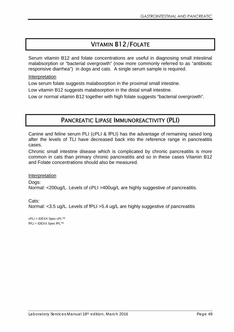
GASTROINTESTINAL AND PANCREATIC
Laboratory Services Manual 18th edition, March 2016 Page 49
VITAMIN B12/FOLATE
Serum vitamin B12 and folate concentrations are useful in diagnosing small intestinal malabsorption or “bacterial overgrowth” (now more commonly referred to as “antibiotic responsive diarrhea”) in dogs and cats. A single serum sample is required.
Interpretation
Low serum folate suggests malabsorption in the proximal small intestine.
Low vitamin B12 suggests malabsorption in the distal small intestine.
Low or normal vitamin B12 together with high folate suggests “bacterial overgrowth”.
PANCREATIC LIPASE IMMUNOREACTIVITY (PLI)
Canine and feline serum PLI (cPLI & fPLI) has the advantage of remaining raised long after the levels of TLI have decreased back into the reference range in pancreatitis cases.
Chronic small intestine disease which is complicated by chronic pancreatitis is more common in cats than primary chronic pancreatitis and so in these cases Vitamin B12 and Folate concentrations should also be measured.
Interpretation
Dogs: Normal: <200ug/L. Levels of cPLI >400ug/L are highly suggestive of pancreatitis.
Cats: Normal: <3.5 ug/L. Levels of fPLI >5.4 ug/L are highly suggestive of pancreatitis
cPLI = IDEXX Spec cPL™
fPLI = IDEXX Spec fPL™

GASTROINTESTINAL AND PANCREATIC
Laboratory Services Manual 18th edition, March 2016 Page 50
INSULIN
Insulin levels in normal fasting dogs are less than 29 μU/mL. To investigate hypoglycaemia, samples for insulin assay should be taken during a period when the blood sugar is known to be low. If the insulin levels are greater than 29 μU/mL this is considered to be excessive secretion. Guidelines for the interpretation of fasting insulin are given below.
Insulin greater than 29 μU/mL
High insulin, insulinoma very likely
Insulin 16 – 29 μU/mL High/normal insulin, insulinoma likely
Insulin 8.3 - 16 0μU/mL Low/normal insulin, insulinoma possible if glucose very low
Insulin less than 8.3 μU/mL Undetectable/low insulin - insulinoma very unlikely
Repeat estimation of borderline results may be required to confirm the diagnosis of insulinoma. Elevated insulin in combination with normal or elevated glucose is consistent with insulin resistance rather than insulinoma.
Insulin/Glucose Ratio (IGR)
The IGR is useful when absolute hyperinsulinaemia (insulin >29 μU/mL) is not present and the insulin levels alone are not diagnostic. Glucose and insulin are measured on the same sample.
Interpretation
IGR of >7.0 U/mol is consistent with insulinoma when glucose is low or low-normal. An elevated ratio in an animal with high normal or high glucose indicates insulin insensitivity.
Amended Insulin/Glucose Ratio (AIGR) The AIGR is reported to be more helpful in some cases and is calculated on the assumption that insulin levels should be undetectable if the blood glucose falls below 30 mg/dL (1.7 mmol/L).
Note: If glucose is <30mg/dL then the divisor becomes 1.0.
GASTROINTESTINAL AND PANCREATIC
Laboratory Services Manual 18th edition, March 2016 Page 51
Interpretation
AIGR of >50 is consistent with insulinoma when blood glucose is subnormal.
Equine Insulin
Horses with PIA and “peripheral hyperadrenocorticism” often have very high serum insulin levels due to peripheral insulin resistance.
FRUCTOSAMINE
Fructosamine is useful to assess long-term glycaemic control in dogs and cats. It is a measure of plasma proteins which have undergone non-enzymatic glycation and is therefore related to the mean blood glucose concentration over the previous 1 - 3 weeks.
It is important that the fructosamine normal reference range is not used when monitoring diabetic patients as this is much lower. Results within the reference range usually indicate that the diabetic patient is likely to be experiencing significant periods of hypoglycaemia.
Haemolysed samples must not be submitted for the assay of fructosamine as this gives falsely elevated results.
Feline fructosamine levels greater than 550 μmol/L are consistent with poor diabetic control.
Canine fructosamine levels greater than 450 μmol/L are consistent with poor diabetic control.

Laboratory Services Manual 18th edition, March 2016 Page 52
CALCIUM DISORDERS
PARATHYROID HORMONE (PTH) AND IONISED CALCIUM (ICA)
Parathyroid hormone (PTH) and ionised calcium (iCa) measurements are the mainstay of calcium investigations and their combined measurement should be considered in both hypo- and hypercalcaemic situations where the cause is not immediately obvious. Neither total nor albumin corrected calcium measurements give a true reflection of calcium status in all cases. This is particularly so in animals with compromised renal function.
The combination of iCa and PTH measurement allow us to distinguish between four main categories of calcium regulation disorders:
1. Primary hyperparathyroidism (parathyroid dependent hypercalcaemia) – functional parathyroid neoplasia
2. Parathyroid suppression (parathyroid independent hypercalcaemia) – hypercalcaemia of malignancy, vitamin D toxicosis (cholecalciferol rodenticide, calcitriol, Dovonex), feline idiopathic hypercalcaemia* and granulomatous disease processes.
3. Primary hypoparathyroidism (parathyroid dependent hypocalcaemia) – parathyroid gland destruction by inflammation or surgery
4. Secondary hyperparathyroidism (parathyroid independent hypocalcaemia) – renal failure, calcium losses (e.g. pancreatitis, equine diarrhoea/colic), dietary deficiency, rickets, hyperadrenocorticism, hyperthyroidism.
*As it’s name suggests, there is currently limited understanding of feline idiopathic hypercalcaemia. In some studies there is a suggested association with urinary acidifying diets. In others, calcium oxalate crystalluria has been reported and some amelioration achieved by a change to high fibre diets. Some authors believe the condition to be relatively benign, while others believe that the condition promotes the onset of renal dysfunction. Low-dose-glucocorticoids have been suggested as a therapy if there is concern about progressive disease.

CALCIUM DISORDERS
Laboratory Services Manual 18th edition, March 2016 Page 53
PTH/Ionised Calcium Interpretation Chart
Serum ionised calcium →
Pla
sma
para
thyr
oid
ho
rmo
ne→
NORMALNORMALNORMAL
1º Hyper PTH
PTH independent hypercalcemia
(e.g. malignancy,vitamin D toxicity)
1º Hypo PTH
2º Hyper PTH(usu. renal or nutritional)
The chart illustrates the diagnostic categorisation of calcium disorders using PTH and iCa alone. The chart as depicted applies well to dogs but the lower and smaller feline PTH reference range makes interpretation sometimes a little less clear-cut in cats.
Sample preparation is critical for the accurate analysis of PTH, see procedure below.
An ionised calcium sample should also be taken at the same time as the PTH sample as this gives an accurate indication of the calcium status at that time.
PTH Sample Preparation
Submitted sample must be EDTA plasma or EDTA plasma with Aprotinin.
1. Request a transport pack for delivery to the laboratory.
2. Take the blood sample into a fridge cooled EDTA or Aprotinin EDTA tube (supplied in pack). Note: To avoid agglutination of samples in the assay, if possible, half fill several EDTA tubes with blood to ensure a high concentration of EDTA, especially if very high levels of calcium have been observed.
3. Mix very well but gently at least 10 times and centrifuge as quickly as possible. Note: If the Aprotinin EDTA tube will not fit into your centrifuge, then carefully transfer the well mixed blood into cooled tubes that do fit into the centrifuge.
4. Transfer the plasma into a cooled plastic (not glass!) PLAIN (or EDTA tube if agglutination has occurred in a previous sample) which has been cooled in the fridge.
5. Immediately freeze (<-10°C) the plasma sample and keep frozen until dispatch in the transport pack. Freeze the plasma sample first, separately, before placing in the frozen pack for transport. Samples are stable for several weeks frozen.
6. Mark the tube with “EDTA plasma” and animal/owner name.

CALCIUM DISORDERS
Laboratory Services Manual 18th edition, March 2016 Page 54
Ionised Calcium (iCa)
Submitted sample should be separated serum. EDTA plasma is not acceptable, the EDTA would chelate all the available calcium making it unavailable for analysis.
If possible take the sample at the same time as the PTH sample.
Collection technique
1. Obtain a blood sample in a plain non-gel tube and fill to the brim with blood.
2. Screw the lid down so no air enters the tube and allow to clot at room temperature for at least 30 minutes.
3. Spin the sample and separate the serum into another plain tube, take care to leave as small an air gap as possible.
4. Mark the tube with “Serum” and animal/owner name
5. If possible do not freeze the iCa sample, store in the fridge and send with the PTH sample outside the pack.
6. The laboratory will apply a correction formula to the result to take account of the changing pH that will have occurred due to the exposure to air. This will result in an estimated iCa at a standardised pH.
PARATHYROID HORMONE RELATED PEPTIDE (PTHRP)
PTHrP may be used for the differential diagnosis of hypercalcaemia of unknown origin where other diagnostic tests have not identified the aetiology of the hypercalcaemia but where PTH and iCa suggest parathyroid independent hypercalcaemia. PTHrP is a hormone that can be produced by dogs, cats and horses with several different types of tumours and is considered to be the underlying cause of hypercalcaemia of malignancy in many but not all cases. Almost all anal gland apocrine neoplasia, the majority of hypercalcaemic lymphomas and smaller percentages of myeloma and carcinomas are PTHrP positive. Circulating levels of PTHrP in normal dogs are almost undetectable (< 0.5 pmol/L). Levels greater than 2.0 pmol/L are considered significant in dogs. Cats appear to show similar values.
Please note: SAMPLE PREPARATION IS CRITICAL FOR THE ACCURATE ANALYSIS OF PTHrP (see procedure for PTH, page 53). THE APROTININ EDTA TUBE SUPPLIED WITH THE TRANSPORT PACK SHOULD BE USED FOR THE COLLECTION OF PTHrP SAMPLES.

CALCIUM DISORDERS
Laboratory Services Manual 18th edition, March 2016 Page 55
VITAMIN D METABOLITES
Two forms of vitamin D are measurable in veterinary serum samples: 25-hydroxyvitamin D (25OHD) and 1,25-dihydroxyvitamin D (calcitriol).
25OHD is produced by the liver and its concentration parallels that of available vitamin D that may be dietary in origin (cholecalciferol (D3), ergocalciferol (D2)) or produced in the skin of certain animals under the influence of UV light. Because the enzymatic hydroxylation of vitamin D in the liver depends almost entirely on the availability of the substrate, this test is an excellent marker of overall vitamin D status. It can be used to diagnose both conditions of vitamin D deficiency and excess and therefore is valuable in the investigation of both hyper- and hypo-calcaemic disorders. 25OHD is also the analyte of choice in verifying adequacy of UV light exposure in reptilian species. Other situations in which we can expect abnormal 25OHD results would include dietary deficiencies, malabsorption syndromes and calciferol-rodenticide toxicities.
Calcitriol is produced by renal tubular cells as a result of enzymatic action (1α-hydroxylase) on 25OHD substrate. The rate of this process is controlled by PTH concentrations (increasing PTH causes increased 1α-hydroxylase activity). Calcitriol is the most biologically potent form of vitamin D and its principle activities are directed at increasing serum calcium concentrations, including increased intestinal uptake of calcium. The failure of renal tubular cells to generate calcitriol in renal disease is one of the contributing mechanisms to renal secondary hyperparathyroidism (and “rubber jaw”). The measurement of calcitriol may be of some value in understanding, renal tubular function and the effects of PTH on the vitamin D system.
In dogs, cats and most veterinary species, very little passive intestinal absorption of calcium occurs. In these species, intestinal calcium absorption is almost exclusively mediated by vitamin D. The situation is different in horses, rabbits (and the hippopotamus!), in which there is significantly more passive absorption of calcium and calcium status is controlled more by renal losses. In these species, the effects of renal disease on calcium status will be different than in most common mammalian species.
Recent research has suggested that low concentrations of 25OHD may be a risk factor for congestive heart failure (CHF) in dogs. There is extensive evidence that Vitamin D has cardioprotective actions by suppressing the renin-angiotensin-aldosterone system, decreasing myocardial hypertrophy, inhibiting pro-imflammatory cytokines and improving endothelial dysfunction and atherosclerosis. (Kraus, Rassnick, Wakshlag et al, J Vet Intern Med, 2014, 28, 109 – 115)

Laboratory Services Manual 18th edition, March 2016 Page 56
OTHER ENDOCRINE
INSULIN-LIKE GROWTH FACTOR 1 (IGF-1)
IGF-1 levels correlate very closely with Growth Hormone (GH) secretion and this enables an estimation of the GH status to be made in dogs and cats. IGF-1 is very stable and no special sampling procedures are required. IGF-1 may be used to diagnose pituitary dwarfism and acromegaly in cats and dogs and in monitoring dietary sufficiency (objective documentation of malnourishment). When sending samples for pituitary dwarfism it is advisable to submit samples from siblings as well for comparision. This is especially important when sampling small or “dwarf” breeds of dogs and cats.
ERYTHROPOIETIN (EPO)
Erythropoietin (EPO) plays a key role in the regulation of red cell mass and erythrocyte production. EPO may be used to aid the differential diagnosis of polycythemia and non-regenerative anaemia. Raised levels of EPO occur in secondary polycythemias and renal tumours. Low/normal levels may occur in renal failure and polycythemia vera.
GASTRIN (SPECIAL REQUEST ASSAY)
Gastrin measurement is useful in the investigation of chronic vomiting in dogs where gastrinoma is considered a differential diagnosis. High fasting serum concentrations or an exaggerated response to feeding can indicate Gastrinoma in the dog.
“False” elevation in Gastrin can occur with certain medications including proton-pump inhibitors (e.g. Omeprazole) and in chronic renal failure.
Sample Preparation for the Assay of Serum Gastrin Fast the animals overnight or for 12 hours or more.
Take the blood into a plain tube with no additive and allow to clot and centrifuge and separate off the serum within 1 hour of collection.
The samples may be stored for up to 8 hours at 2-8°C or below -20°C for up to 2 weeks. Avoid freeze-thaw cycles and keep frozen until assayed.
Samples must be sent to the laboratory frozen and courier is essential.
Lipaemic samples may interfere with the assay.
At least 1.0 mL of Serum is required for the assay.

OTHER ENDOCRINE
Laboratory Services Manual 18th edition, March 2016 Page 57
RENIN (SPECIAL REQUEST ASSAY)
Renin is released by the juxtaglomerular apparatus of the kidney in response to a decrease in arterial blood pressure (including decreased blood volume) or a reduced Na content of glomerular ultrafiltrate. It is the first part in the Renin-Angiotensin-Aldosterone-System
Renin activates Angiotensin by cleaving Angiotensinogen to release Angiotensin I which through the action of Angiotensin converting enzyme (ACE) produces Angiotensin II. In turn, Angiotensin II, stimulates the release of aldosterone from the adrenal cortex. The action of aldosterone should promote the resorption and retention of Na and restore blood volume.
The measurement of Renin can be particularly useful in the investigation of hypokalaemia and hypertension.
In conditions where there is an elevated aldosterone it is helpful to discover if that is “appropriate” and is the result of Renin activation or “inappropriate” and likely to be autonomous such as functional adrenal cortex neoplasia (aldosteronoma).
The measurement of Renin is therefore essential in the investigation of hypertension or situations of elevated aldosterone.
We now offer a full Hypotension/Hypokalaemia profile comprising Plasma RENIN, Aldosterone and Na/K ratio including the calculation of Aldo/Renin ratio.
Sample preparation for the assay of plasma Renin Blood must be collected into pre-chilled tubes containing EDTA as an anticoagulant.
HEPARIN must not be used as this interferes with the assay.
The samples must be kept cold and ideally centrifuged in a refrigerated centrifuge to separate the plasma. The plasma samples must then be aliquoted and frozen at less than - 10°C until assayed.
Samples must be sent to the laboratory frozen and courier is essential. Haemolysed or lipaemic samples must not be used in the assay so great care is needed when blood sampling to ensure samples do not become haemolysed.
At least 2.0 mL of EDTA plasma MUST be sent for the analysis of Renin otherwise it may not be possible to get a valid result.

OTHER ENDOCRINE
Laboratory Services Manual 18th edition, March 2016 Page 58
GLUCAGON (SPECIAL REQUEST ASSAY)
The measurement of glucagon is helpful in the investigation of unusual cases of insulin resistant diabetes mellitus and in clinical research situations focused on factors affecting glucose metabolism.
Glucagonomas have been identified in dogs. A dermatological presentation has been associated with Glucagonoma in humans also known as necrolytic migratory erythema or hepato-cutaneous syndrome. In dogs, this skin condition usually has a hepatic origin.
Sample Preparation for the Assay of Plasma Glucagon Blood must be collected into iced glass tubes containing EDTA as an anticoagulant. The tubes must be immersed in an ice-bath at 2 – 8°C throughout the entire collection and handling procedure.
Immediately following collection add 5000 Kallikrein Inactivating Units (KIU) per 10mL of whole blood in the form of Trayslol (500uL) or Aprotinin (1mg). Mix gently but thoroughly and keep chilled (on ice) until separation.
Centrifuge the samples in a refrigerated centrifuge at 2000g to separate the plasma. Separate the plasma using a glass pasteur pipette and transfer the plasma into glass tubes (aliquots of 0.6mL required) and freeze immediately. Store the plasma frozen at less than -10°C until assayed.
Samples must be transported to the laboratory frozen and remain frozen until assayed. Dry ice is recommended. Lipaemic and haemolysed samples may affect the assay so great care is needed when blood sampling to ensure samples do not become haemolysed. Courier transport is essential.
At least 1mL of EDTA plasma is required for the assay.
PLEASE NOTE AT THE CURRENT TIME WE ARE UNABLE TO PERFORM GLUCAGON ANALYSIS DUE TO THE WITHDRAWAL OF THE METHOD WE HAD VALIDATED.
WE WILL BE LOOKING AT OTHER METHODS FOR THE FUTURE.

OTHER ENDOCRINE
Laboratory Services Manual 18th edition, March 2016 Page 59
NT-PROBNP – CANINE/FELINE CARDIAC MARKER
Heart disease in dogs and cats may be congenital (present at birth) or acquired. Acquired heart disease is more common and tends to occur as the animal gets older. Often the owner does not notice as severe clinical signs may not arise during gradual deterioration of cardiac function.
NT-pro-BNP will be a useful screen where subtle clinical signs are present, as part of a geriatric profile or a health check for breeds who have a history of cardiac problems.
Canine Interpretation
NT-pro-BNP levels less than 900 pmol/L are found in normal healthy dogs and it is unlikely that the clinical signs are related to cardiac failure.
NT-pro-BNP levels between 900 and 1800 pmol/L are in the suspect range and in the absence of a murmur or common clinical signs, heart disease is unlikely.
NT-pro-BNP levels greater than 1800 pmol/L Heart disease is likely. Further cardiac workup or specialist cardiac referral suggested.
Feline Interpretation
NT-pro-BNP levels less than 100 pmol/L are found in normal healthy cats and it is unlikely that the clinical signs are related to cardiac failure.
NT-pro-BNP levels between 100 and 270 pmol/L are in the suspect range and in the absence of a murmur or common clinical signs, heart disease is unlikely.
NT-pro-BNP levels greater than 270 pmol/L Heart disease is likely. Further cardiac workup or specialist cardiac referral suggested.
In cats and dogs NT-pro-BNP levels may decrease with cardiac medication. Arrhythmias and the presence of pulmonary hypertension may result in higher NT-pro-BNP levels. Azotemic animals may have increased NT-pro-BNP levels.
Please note very low values are suggestive of inappropriate sampling or sample degradation and a repeat sample should be submitted.
NT-pro-BNP Sample Preparation Sample must be at least 0.5mL of separated EDTA PLASMA
SAMPLES MUST BE SEPARATED FROM THE RED BLOOD CELLS WITHIN 10 MINUTES .
Send to lab immediately (samples are stable for 24 hours at room temperature).
If there is going to be any delay then send to the lab on ice packs.
Do not send lipaemic or haemolysed samples.

OTHER ENDOCRINE
Laboratory Services Manual 18th edition, March 2016 Page 60
CANINE BENIGN PROSTATE HYPERPLASIA (BPH)
BPH appears to affect over 50% of entire male dogs over 5 years old. There is an increase in prostate cell size and cell numbers. Clinical signs suggestive of BPH are constipation, dysuria, haematuria, pain, lameness, behavioural problems, infertility and haematospermia. A simple blood test is now available and can provide a reliable alternative to rectal palpation. A single serum sample overcomes the many pitfalls of monitoring by rectal palpation such as the unpleasantness for dog, owner and vet, the awkwardness in large and minature breeds, difficulty in distinguishing normal from abnormal and the fact that BPH must be severe to detect accurately by palpation.
Canine Prostate Specific Esterase (CPSE) is a major protein secreted by the prostate and represents more than 90% of the proteins found in seminal fluid. CPSE passes into the bloodstream following either an increase in its production or an alteration in the blood-glandular barrier caused by changes in glandular structure.
Interpretation
Normal: < 50 ng/mL. Strong suspicion of BPH: > 70 ng/mL
BPH Sample Preparation
Sample must be SERUM

Laboratory Services Manual 18th edition, March 2016 Page 61
THERAPEUTIC DRUG MONITORING
PHENOBARBITONE
Phenobarbitone should be given for about 2 - 3 weeks to allow for serum levels to stabilise before checking therapeutic concentrations.
Monitoring of therapeutic levels of phenobarbitone should be assessed at 8 - 12 hours post treatment. Peak levels occur about 5 hours post treatment.
Therapeutic levels for phenobarbitone are 15 - 40 μg/mL
SERUM GEL TUBES MUST NOT BE USED FOR PHENOBARBITONE ANALYSIS
POTASSIUM BROMIDE
Serum levels of potassium bromide may take at least 125 days to stabilise due to the long half life of the drug. Monitoring of therapeutic levels of potassium bromide can be assessed at any time post dosing.
Therapeutic levels for potassium bromide are 0.5 - 1.9 mg/mL. Young healthy dogs may tolerate levels up to 2.5 mg/mL and most dogs will tolerate levels up to 1.5 mg/mL.
SERUM GEL TUBES MUST NOT BE USED. EXTRA CARE IS NEEDED TO AVOID HAEMOLYSIS AS THIS SEVERLY ELEVATES THE RESULTS. SAMPLES MUST BE CENTRIFUGED AND SEPARATED BEFORE DESPATCH. WE SUGGEST THE USE OF LITHIUM HEPARIN SAMPLES AS THIS ALLOWS IMMEDIATE CENTRIFUGATION.
Potassium Bromide samples are reported with a haemolysis index where appropriate.
LEVETIRACETAM (KEPPRA®) Levetiracetam can be administered in addition to potassium bromide and phenobarbitone to further help in the reduction of seizure frequency and duration. The use of Levetiracetam is not associated with additional side effects and may help in cases of liver toxicity as the dose of KBr may be reduced as part of combination therapy.
A therapeutic range has not been established for the dog or cat but the human therapeutic range for Levetiracetam of 29 – 123 umol/L is considered appropriate. A steady state is not achieved with Levetiracetam, because accumulation does not occur. However the pharmacodynamic effect is believed to outlive the known half life. The risk of toxicity is believed to be low.
SERUM GEL TUBES MUST NOT BE USED FOR LEVETIRACETAM ANALYSIS

THERAPEUTIC DRUG MONITORING
Laboratory Services Manual 18th edition, March 2016 Page 62
DIGOXIN
Digoxin should be given for about 3 - 5 days to allow for serum levels to stabilise before checking therapeutic concentrations.
Monitoring of therapeutic levels of digoxin should be assessed at 8 hours post treatment.
Digoxin concentrations of 0.8 - 2.0 ng/mL are required post treatment.
SERUM GEL TUBES MUST NOT BE USED FOR DIGOXIN ANALYSIS
CYCLOSPORINE
Cyclosporine has been shown to be useful in the treatment of anal furunculosis (AF) and atopy in dogs.
Monitoring of cyclosporine levels during treatment may be useful. Approx 30 days should be allowed to reach stable trough levels using combination therapy. Approx 17 days should be allowed to reach stable trough levels using Cyclosporine alone.
Therapeutic levels of Cyclosporine about 12 hours post pill are expected to be between 300–400 ug/L for AF but lower for atopy.
Sample requirement is at least 2.0 mL of whole EDTA blood.

Laboratory Services Manual 18th edition, March 2016 Page 63
ENDOCRINE THERAPEUTIC MONITORING
BASIC VETORYL MONITOR
ACTH Stimulation and Na:K ratio
In order to monitor Vetoryl it is important to know what effect the medication is having on adrenal function. The test is performed 4-6 hours after the medication when Trilostane is at its greatest level of activity. By sampling at this time we know what the greatest level of adrenal suppression will be. It is likely that later in the day, suppression will be less. There are circumstances when investigating problem cases, that it may be appropriate to perform the ACTH stimulation test outside this period on the guidance of a specialist or Dechra Technical Support (01939 211200).
Vetoryl will inhibit both glucocorticoid and mineralocorticoid production. Sodium and potassium are included because they will help identify whether there is too much suppression of the adrenal glands.
In interpreting the ACTH stimulation test 4-6 hours post Vetoryl Capsule, we are looking for evidence of suppressed steroidogenesis, that is, post-ACTH cortisol concentrations <250nmol/L that also show an increase over baseline levels. The failure of ACTH to stimulate an increase in cortisol concentrations suggests that consideration be given to lowering the dose or withholding medication.
Please note all the following diagnostic and treatment flowcharts are reproduced by kind permission of Dechra Veterinary Products
VETORYL MONITOR PLUS
ACTH Stimulation, Na:K ratio, Alkaline Phosphatase (AP), Alanine Aminotransferase (ALT), Urea, Creatinine, Phosphorus, Total Protein, Albumin, Globulin, Cholesterol and Glucose
The Vetoryl datasheet recommends regular monitoring particularly during the early phase of therapy that includes ACTH stimulation and biochemistry including electrolytes.
The Vetoryl Monitor Plus includes the ACTH stimulation and Sodium:Potassium ratio but adds a tailored selection of other biochemistry tests.

ENDOCRINE THERAPEUTIC MONITORING
Laboratory Services Manual 18th edition, March 2016 Page 64
Alkaline phosphatase (AP)
Alanine aminotransferase (ALT)
Both AP and ALT are usually elevated in untreated hyperadrenocorticism. AP is elevated in dogs because of induction of a steroid induced isoenzyme and ALT is elevated because of a “steroid hepatopathy”. By measuring these enzymes, it is possible to assess improvements in liver pathology and circulating steroid concentrations. The Vetoryl datasheet recommends that dogs should be monitored at regular intervals for primary hepatic disease.
Urea
Creatinine
Phosphorus
The measurement of urea, creatinine and phosphorus will assist in the identification of renal disease (and iatrogenic hypoadrenocorticism). Renal dysfunction is not uncommon in geriatric dogs. The Vetoryl datasheet recommends that dogs should be monitored at regular intervals for renal disease and that the product should not be used in dogs with renal insufficiency.
Total protein
Albumin
Globulin (calculated)
The measurement of proteins aids in the assessment of hydration status in dogs that may be polydipsic. Albumin and globulin measurement can help identify late stage (protein-losing) glomerulopathies and inflammatory disorders that can affect dogs with hyperadrenocorticism
Cholesterol Cholesterol is commonly elevated in dogs with untreated hyperadrenocorticism. Measurement during therapeutic monitoring provides information on whether deranged lipid metabolism has been corrected.
Glucose Diabetes mellitus is not uncommon in dogs with both untreated and treated hyperadrenocorticism. The Vetoryl datasheet recommends that dogs are monitored at regular intervals for diabetes mellitus.
Monitoring Therapy Using “Pre-Pill” Single Cortisol Samples, see page 26 for details.

BID dosing required Rule out concurrent conditions
* If the post-ACTH cortisol is >250 nmol/l and the clinical signs of HAC are marked then you may consider a dose increase at this stage. Increase dose by smallest increment possible.
TreaTMenT and MoniToring of HyperadrenocorTicisM
Clinical examination ACTH stim test 4 hours post morning capsule
Biochemistry including electrolytes
Day 10
Post-ACTH serum cortisol <50 nmol/l and clinically well
Assess degree of clinical improvement (for further information see clinical signs
timeline information)
Clinical examinationACTH stim test 4 hours post-capsuleBiochemistry including electrolytes
≥28 daysStop Vetoryl® treatment
Confirm whether clinical signs are due to hypoadrenocorticism with ACTH stim test and
analysis of serum electrolytes (in particular Na and K).
Treat symptomatically as required e.g. • dexamethasone to treat hypocortisolaemia
• IV 0.9% NaCl to resolve hyperkalaemia
Post-ACTH serum cortisol >50 nmol/l* and clinically well
Stop treatment Break for 7 daysRestart at lower dose reTurn To day 1
Continue treatment at current dose
Showing clinical signs consistent with:1. corticosteroid withdrawal syndrome “relative” cortisol deficiency characterised by weakness, lethargy, stiff gait,
anorexia, fever during first 10 days of therapy.
2. Hypoadrenocorticism e.g. anorexia, lethargy/depression, weakness, shaking/shivering, vomiting, diarrhoea,
bradycardia, collapse.
Start Vetoryl® Hard CapsulesGive in morning with food
Start as per guideline table on datasheet
Day 1
Continue on current dose but monitor
clinical signs carefully for recurrence
Very little or no improvement Significant improvement
Post-ACTH serum cortisol <200 nmol/l
Post-ACTH serum cortisol >200 nmol/l
Post-ACTH serum cortisol <50 nmol/l and
clinically well
Continue to monitor as per datasheet recommendations. Perform ACTH stim test
4 hours post morning capsule
Post-ACTH serum cortisol >250 nmol/l*
Post-ACTH serum cortisol 50-200 nmol/l
Post-ACTH serum cortisol 200-250 nmol/l
Stop treatment for 7 days.
Then restart at lower dose
reTurn To day 1
Increase morning dose
reTurn To day 1
Continue treatment at current dose
Contact Dechra Technical Support for
advice on dosing
Increase once daily dose and
reTurn To day 1
22-24 hr ACTH Stim Test
22-24hr Post-ACTH serum cortisol >200
nmol/l
Post-ACTH serum cortisol <200 nmol/l

Laboratory Services Manual 18th edition, March 2016 Page 66
BASIC THYFORON
Thyroxine (T4) The measurement of thyroxine (T4) at a single point in time. It is important to know that a dog receiving Thyforon® therapy is absorbing the medication appropriately. Insufficient absorption or insufficient dose will lead to treatment failure and over-dose can lead to clinical signs of hyperthyroidism. When a T4 measurement is made, it is important to record the hours post-pill at which the sample was taken to be able to appropriately interpret the results.
THYFORON MONITOR PLUS
Thyroxine (T4) and Thyrotropin (TSH) The measurement of thyroxine (T4) and thyrotropin (TSH) at a single point in time. It is important to know that a dog receiving Thyforon® therapy is absorbing the medication appropriately. Insufficient absorption or insufficient dose will lead to treatment failure and over-dose can lead to clinical signs of hyperthyroidism. When a T4 measurement is made, it is important to record the hours post-pill at which the sample was taken to be able to appropriately interpret the results.
Thyrotropin (TSH) levels take several days to change in response to changes in thyroxine (T4) therapy, they therefore are able to reflect the adequacy of therapy in the days preceding the test. TSH could be described as the “fructosamine of hypothyroidism”.
The combined measurement of T4 and TSH will help in the identification of compliance failure and reduce unnecessary dose adjustments that could arise from reliance on T4 measurement alone on an “unrepresentative” day.
BASIC FELIMAZOLE
Thyroxine (T4) The measurement of single thyroxine (T4) concentration in hyperthyroid cats allows us to assess the adequacy of therapy and to make appropriate dose adjustments. As long as the cat is receiving medication on its regular schedule the interval post-pill at which the sample is taken is not important.

ENDOCRINE THERAPEUTIC MONITORING
Laboratory Services Manual 18th edition, March 2016 Page 67
FELIMAZOLE MONITOR PLUS
Thyroxine (T4), Alkaline Phosphatase (AP), Alanine aminotransferase (ALT), Urea, Creatinine and Phosphorus
Felimazole monitor plus includes the measurement of thyroxine (T4) but also includes specific biochemistry tests.
Alkaline phosphatase (AP)
Alanine aminotransferase (ALT)
Both AP and ALT are commonly elevated in untreated hyperthyroidism. By measuring these enzymes, it is possible to assess improvements due to therapy and to identify any underlying primary hepatic disease.
Urea
Creatinine
Phosphorus
The measurement of urea, creatinine and phosphorus will assist in the identification of renal disease. The “unmasking” of previously subclinical renal insufficiency is not uncommon following all forms of anti-thyroid therapy. By monitoring these parameters, it is possible to better navigate the balance between hyperthyroidism and renal insufficiency in case this situation should arise.

Hyp
ert
hyro
idis
m s
usp
ecte
d b
ased
on h
isto
ry, p
hysic
al exam
inatio
n �
nd
ing
s +
/-
sup
po
rtiv
e c
hang
es o
n r
outine b
iochem
istr
y
Measure
To
tal T
4
Tota
l T
4 a
bo
ve r
efe
rence inte
rval
Tota
l T
4 w
ithin
refe
rence inte
rval
Ap
pro
xim
ate
ly 1
0%
of hyp
ert
hyro
id c
ats
have s
eru
m T
ota
l T
4 c
oncentr
atio
n w
ithin
the
refe
rence inte
rval d
ue t
o:
i) F
luctu
atio
n fro
m a
bo
ve t
o w
ithin
refe
rence inte
rval in
early/m
ildly
affecte
d c
ases
ii) T
4 s
up
pre
ssio
n d
ue t
o c
oncurr
ent
no
n-t
hyro
idal ill
ness (N
TI)
If s
till
susp
ect
hyp
ert
hyro
idis
m…
Fre
e T
4 w
ithin
refe
rence inte
rval
Fre
e T
4 a
bo
ve r
efe
rence inte
rval
EIT
HE
R
If s
usp
ect
early o
r m
ildly
affecte
d c
ase:
Rete
st
Tota
l T
4 2
- 4
weeks late
r, o
r w
hen
mo
re o
vert
clin
ical sig
ns d
evelo
p
If s
usp
ect
NT
I: Id
entify
and
tre
at
(if p
ossib
le) b
efo
re r
ete
sting
To
tal T
4
Hyp
ert
hyro
idis
m c
on
!rm
ed
Hyp
ert
hyro
idis
m lik
ely
Tota
l T
4 in u
pp
er
half o
f
refe
rence inte
rval
Hyp
ert
hyro
idis
m u
nlik
ely
NT
I lik
ely
Up
to
20%
of sic
k e
uth
yro
id c
ats
have
ele
vate
d F
ree T
4 c
oncentr
atio
ns
Tota
l T
4 in lo
wer
half o
f,
or
belo
w, th
e r
efe
rence inte
rval
Co
nsid
er
NT
I as c
ause o
f o
bserv
ed
clin
ical sig
ns e
.g. g
astr
oin
testinal d
isease, neo
pla
sia
If s
till
susp
ect
hyp
ert
hyro
idis
m:
Co
nsid
er
measuring
TS
H c
oncentr
atio
n, usin
g c
anin
e T
SH
assay
(co
nta
ct
yo
ur
dia
gno
stic lab
ora
tory
or
Dechra
Vete
rinary
Pro
ducts
fo
r fu
rther
info
rmatio
n)
OR
Co
nsid
er
refe
rral fo
r th
yro
id s
cin
tig
rap
hy
Hyp
ert
hyro
idis
m u
nlik
ely
OR
Measure
Fre
e T
4 a
nd
To
tal T
4 in s
am
e
blo
od
sam
ple
Dia
gn
osis
of
felin
e h
yp
ert
hyro
idis
m
Hyp
ert
hyro
idis
m s
usp
ecte
d b
ased
on h
isto
ry, p
hysic
al exam
inatio
n �
nd
ing
s +
/-
sup
po
rtiv
e c
hang
es o
n r
outine b
iochem
istr
y
Measure
To
tal T
4
Tota
l T
4 a
bo
ve r
efe
rence inte
rval
Tota
l T
4 w
ithin
refe
rence inte
rval
Ap
pro
xim
ate
ly 1
0%
of hyp
ert
hyro
id c
ats
have s
eru
m T
ota
l T
4 c
oncentr
atio
n w
ithin
the
refe
rence inte
rval d
ue t
o:
i) F
luctu
atio
n fro
m a
bo
ve t
o w
ithin
refe
rence inte
rval in
early/m
ildly
affecte
d c
ases
ii) T
4 s
up
pre
ssio
n d
ue t
o c
oncurr
ent
no
n-t
hyro
idal ill
ness (N
TI)
If s
till
susp
ect
hyp
ert
hyro
idis
m…
Fre
e T
4 w
ithin
refe
rence inte
rval
Fre
e T
4 a
bo
ve r
efe
rence inte
rval
EIT
HE
R
If s
usp
ect
early o
r m
ildly
affecte
d c
ase:
Rete
st
Tota
l T
4 2
- 4
weeks late
r, o
r w
hen
mo
re o
vert
clin
ical sig
ns d
evelo
p
If s
usp
ect
NT
I: Id
entify
and
tre
at
(if p
ossib
le) b
efo
re r
ete
sting
To
tal T
4
Hyp
ert
hyro
idis
m c
on
!rm
ed
Hyp
ert
hyro
idis
m lik
ely
Tota
l T
4 in u
pp
er
half o
f
refe
rence inte
rval
Hyp
ert
hyro
idis
m u
nlik
ely
NT
I lik
ely
Up
to
20%
of sic
k e
uth
yro
id c
ats
have
ele
vate
d F
ree T
4 c
oncentr
atio
ns
Tota
l T
4 in lo
wer
half o
f,
or
belo
w, th
e r
efe
rence inte
rval
Co
nsid
er
NT
I as c
ause o
f o
bserv
ed
clin
ical sig
ns e
.g. g
astr
oin
testinal d
isease, neo
pla
sia
If s
till
susp
ect
hyp
ert
hyro
idis
m:
Co
nsid
er
measuring
TS
H c
oncentr
atio
n, usin
g c
anin
e T
SH
assay
(co
nta
ct
yo
ur
dia
gno
stic lab
ora
tory
or
Dechra
Vete
rinary
Pro
ducts
fo
r fu
rther
info
rmatio
n)
OR
Co
nsid
er
refe
rral fo
r th
yro
id s
cin
tig
rap
hy
Hyp
ert
hyro
idis
m u
nlik
ely
OR
Measure
Fre
e T
4 a
nd
To
tal T
4 in s
am
e
blo
od
sam
ple
Dia
gn
osis
of
felin
e h
yp
ert
hyro
idis
m

Hyp
erth
yroi
dism
di
agno
sed
Tota
l T4,
Hae
mat
olog
y an
d B
ioch
emis
try
shou
ld b
e as
sess
ed a
fter
3 w
eeks
, 6
wee
ks, 1
0 w
eeks
, 20
wee
ks, a
nd th
erea
fter
eve
ry 3
mon
ths
Aim
for
a To
tal T
4 co
ncen
trat
ion
in th
e LO
WE
R H
ALF
of t
he re
fere
nce
inte
rval
(<
35 n
mol
/l ap
prox
.) an
d A
VO
ID IA
TR
OG
EN
IC H
YP
OT
HY
RO
IDIS
MTi
min
g of
Tot
al T
4 sa
mpl
ing
is n
ot im
port
ant
Reg
ular
mon
itorin
g of
blo
od p
ress
ure
is a
lso
reco
mm
ende
d
Tota
l T4
in lo
wer
hal
f of r
efer
ence
inte
rval
Con
tinue
to m
onito
r as
per
dat
ashe
et re
com
men
datio
nsM
aint
ain
appr
opria
te T
otal
T4
conc
entr
atio
n us
ing
the
low
est p
ossi
ble
dose
of F
elim
azol
e
Non
-azo
taem
icA
zota
emic
Tota
l T4
belo
w re
fere
nce
inte
rval
Red
uce
Felim
azol
e do
se b
y 2.
5 m
g p
er d
ay
Tota
l T4
in u
ppe
r ha
lf of
refe
renc
e in
terv
al o
r ab
ove
refe
renc
e in
terv
al
Che
ck o
wne
r co
mp
lianc
e. If
no
issu
es th
en:
Incr
ease
Fel
imaz
ole
dose
by
2.5
mg
per
day
OR
If To
tal T
4 in
up
per
half
of re
fere
nce
inte
rval
and
sig
nific
antly
de
crea
sed
from
pre
viou
s te
st, c
ould
con
tinue
cur
rent
dos
e an
d
reas
sess
at n
ext t
est
NB
: If u
nsta
ble
azot
aem
ia w
ith m
oder
ate
to m
arke
d cl
inic
al
sign
s of
CK
D (I
RIS
Sta
ge 3
- 4
), st
ill a
im to
mai
ntai
n To
tal T
4 w
ithin
upp
er h
alf o
f ref
eren
ce in
terv
al if
pos
sibl
e
Non
-azo
taem
ic o
r IR
IS S
tage
1 -
2 C
hron
ic K
idne
y D
isea
se (C
KD
)A
zota
emic
or
IRIS
Sta
ge 3
- 4
Chr
onic
Kid
ney
Dis
ease
(CK
D)
Use
of F
elim
azol
e in
cat
s w
ith C
KD
sho
uld
be s
ubje
ct to
car
eful
ris
k : b
enefi
t ass
essm
ent.
Ren
al fu
nctio
n sh
ould
be
mon
itore
d cl
osel
y af
ter
onse
t of F
elim
azol
e th
erap
yG
rad
ually
intr
oduc
e Fe
limaz
ole,
sta
rtin
g w
ith th
e lo
wes
t pos
sibl
e do
se
Eva
luat
e an
d m
anag
e C
KD
as
per
IRIS
gui
delin
es (w
ww
.iris
-kid
ney.
com
)
Use
of F
elim
azol
e in
cat
s w
ith C
KD
sho
uld
be s
ubje
ct to
car
eful
ris
k : b
enefi
t ass
essm
ent.
Ren
al fu
nctio
n sh
ould
be
mon
itore
d cl
osel
y af
ter
onse
t of F
elim
azol
e th
erap
yS
tart
Fel
imaz
ole
at 5
mg/
day,
ad
min
iste
red
as
2.5
mg
BID
If, fo
r re
ason
s of
com
plia
nce,
SID
dos
ing
with
a 5
mg
tab
let i
s p
refe
rabl
e, th
en th
is is
acc
epta
ble
(alth
ough
the
2.5
mg
tabl
et g
iven
twic
e da
ily m
ay b
e m
ore
effic
acio
us in
the
shor
t ter
m)
Eva
luat
e an
d m
anag
e C
KD
as
per
IRIS
gui
delin
es (w
ww
.iris
-kid
ney.
com
)
Con
tinue
cur
rent
dos
e
DO
NO
T a
djus
t Fel
imaz
ole
dose
– m
aint
ain
Tota
l T4
in
low
er h
alf o
f ref
eren
ce in
terv
al
Eva
luat
e an
d m
anag
e C
KD
as
per
IRIS
gui
delin
es
Sta
ble
azot
aem
ia, n
o to
mild
cl
inic
al s
igns
of C
KD
(IR
IS S
tage
1 -
3)
Red
uce
Felim
azol
e do
se b
y 2.
5 m
g pe
r da
y b
ut a
im to
m
aint
ain
Tota
l T4
with
in u
ppe
r ha
lf of
refe
renc
e in
terv
al if
po
ssib
le
Eva
luat
e an
d m
anag
e C
KD
as
per
IRIS
gui
del
ines
Uns
tabl
e az
otae
mia
, m
oder
ate
to m
arke
d cl
inic
al
sign
s of
CK
D (I
RIS
Sta
ge
3 -
4)
Felim
azo
le® t
reat
men
t &
mo
nito
ring
flo
wch
art

Laboratory Services Manual 18th edition, March 2016 Page 70
SEROLOGY/IMMUNOLOGY
ACETYLCHOLINE RECEPTOR ANTIBODIES (ACRAB)
Acquired canine myasthenia gravis (cMG) is an autoimmune disease of neuromuscular transmission caused by autoantibodies directed against the acetylcholine receptors at the neuromuscular junction. cMG can occur as two forms, focal and generalised. In focal MG dogs usually present with megaoesophagus as weakness affects the oesophageal, pharyngeal and facial muscles. In generalised cMG there is widespread skeletal muscle weakness which is made worse by exercise. ACRABs may only be used to diagnose the acquired form of the disease.
Positive serum titre for ACRABs is highly suggestive of acquired cMG.
A 1mL serum sample is required.
TYPE 2M MUSCLE FIBRE ANTIBODY (2MAB)
Disorders of the muscles of mastication occur relatively commonly in clinical practice and can be of myopathic or neuropathic origin. Masticatory muscles are predominantly composed of fibres designated as Type 2M. Masticatory muscle disorders (MMD) can be diagnosed by the assay of serum from affected animals to see if there are autoantibodies against Type 2M fibres.
False negative results may be obtained if immunosuppressive dosages of corticosteroids have been given for longer than 7 - 10 days in end-stage masticatory muscle myositis and in polymyositis. A muscle biopsy would be necessary for confirmation of the diagnosis in these cases.
A 1mL serum sample is required.
SARCOPTIC MANGE ANTIBODY (SMA)
It is useful to be able to make a firm diagnosis for the presence of sarcoptic mange mites as this helps to choose the right treatment regime especially where there may be breed sensitivity to some drugs. Skin scrapes often fail to detect the presence of mites due to the difficulty in actually finding the mites.
A serum sample taken 4 - 8 weeks post infection will detect the presence of sarcoptic mange antibodies (IgG). A borderline result should be repeated in two weeks.
SEROLOGY/IMMUNOLOGY
Laboratory Services Manual 18th edition, March 2016 Page 71
ALLERGY TESTING
Allergy testing is available including the most commonly found allergens either as a full panel or by using a very cost-effective “screen and expand” approach.
Please see www.allervet.co.uk or contact the laboratory for details.
Laboratory Services Manual 18th edition, March 2016 Page 72
ONCOLOGY
THYMIDINE KINASE
Serum thymidine kinase is a tumour marker for canine and feline malignant lymphoma. Thymidine kinase (TK1) is a cytosolic enzyme associated with cellular proliferation. It markedly increases in activity during the G1 and S phases of the cell cycle. Thymidine kinase enters the circulation from cycling cells or perhaps from cells dying during replication. In essence, then, TK is a marker of the volume of replicating cells in the animal and consequently an indirect measure of neoplastic burden.
The relationship between serum TK and a significant increase in cell replication allows the test to be considered as a diagnostic adjunct for neoplasia such as malignant lymphoma. However, suggestions for its use focus more on prognosis and therapeutic monitoring.
Those lymphoma patients with the highest TK activities are likely to have the highest neoplastic burden or replication rate and these may have a worse prognosis than those lymphoma patients with lower levels.
Thymidine kinase can also be used during the initial phase of chemotherapy to document a decrease from pre-treatment values, indicating the “success” of early therapy and informing the remaining protocol. Frequent monitoring of TK during or between chemotherapeutic cycles should also identify relapse (perhaps pre-clinically) or help differentiate the origin of a clinical deterioration.
Thymidine kinase may not have perfect diagnostic specificity when used as a diagnostic test for lymphoma because of the possible association with other conditions of increased cell turnover including inflammation. However, in the absence of leukocytosis the likelihood of a neoplastic origin is much increased.
Reference: VonEuler, Einarsson, Olsson, Lagerstedt and Erikson (2004) Serum Thymidine Kinase Activity in Dogs with Malignant Lymphoma: A potent marker for prognosis and monitoring the disease. Journal of Veterinary Internal Medicine 18:696-702

Laboratory Services Manual 18th edition, March 2016 Page 73
REFERENCES
A list of references used in the preparation of this manual is available on request.
Laboratory Services Manual 18th edition, March 2016 Page 73
REFERENCES
A list of references used in the preparation of this manual is available on request.

Laboratory Services Manual 18th edition, March 2016 Page 74
APPENDICES
I. CONVERSION FACTORS
ANALYTE Per litre
units CONVERSION Traditional
UNITS
ACTH pmol/L divide /L result by 0.2222 to get Trad. units pg/mL
Aldosterone pmol/L '' 2.774 '' pg/mL
Calcium mmol/L '' 0.25 '' mg/dL
Cortisol nmol/L '' 2.759 '' ng/mL
Cortisol nmol/L '' 27.59 '' ug/dL
Creatinine mmol/L '' 0.0884 '' mg/dL
Glucose mmol/L '' 0.055 '' mg/dL
IGF-1 nmol/L '' 0.131 '' ng/mL
Insulin pmol/L '' 7.175 '' uIU/mL
Parathyroid hormone pmol/L '' 0.105 '' pg/mL
Phenobarbital umol/L '' 4.306 '' ug/mL
Progesterone nmol/L '' 3.18 '' ng/mL
Testosterone nmol/L '' 3.467 '' ng/mL
Total T4 nmol/L '' 1.287 '' ng/mL
Total T4 nmol/L '' 12.87 '' ug/dL
Total T3 nmol/L '' 1.536 '' ng/mL
Total T3 nmol/L '' 0.01536 '' ug/dL
Free T4 pmol/L '' 1.287 '' pg/mL
For example, 220 nmol/L Cortisol = (220 ÷ 27.59) = 8.0 ug/dL
To convert from traditional to per litre units, multiply traditional result by factor listed above
e.g. 8.0 ug/dL Cortisol = 8.0 x 27.59 = 220 nmol/L

Assay Sample Requirements
Specialists in Endocrinology: Experts in Immunoassay.
An ideal sample volume is 1ml of separated serum or plasma.
This allows for assays to be repeated if necessary or further analysis to be added if required.
*A serum sample can also be submitted for Glucose if this has been separated with one hour of collection
Assay Sample Type
SpecialRequirements
Min Sample Volume
(ml)
Thyroid
T4 S H None 0.3
T3 S H None 0.5
TSH S H None 0.5
FT4ED S H None 0.5
Thyroid Antibodies S H None 0.2
TGAA S H None 0.2
Adrenocortical
Cortisol (includes ACTH Stim & DEX Screening) S H None 0.3
Urinary Cortisol Urine None 0.3
ACTH Endogenous (Canine/Feline) E
Frozen Separated EDTA Plasma
1.0
ACTH Endogenous (Equine) E Separated
EDTA Plasma 1.0
OHP S H None 0.3
Aldosterone S H None 1.0
Renin EFrozen Separated EDTA Plasma
2.0
Reproduction Pregnancy
Progesterone S H No Gel Tubes 0.5
Testosterone S H None 0.6
Oestradiol S H None 1.0
Oestrone Sulphate Pregnancy/Rig S H None 0.6
PMSG Serum only None 0.5
AMH (all species) S H None 0.5
Relaxin H None 0.2
Diabetes
Insulin S Starved Sample 0.3
Insulin/Glucose RatioS & Fluoride Oxalate*
Starved Sample
S = 0.3 and FO = 0.3
Insulin Antibodies S None 0.5
Fructosamine S H None 0.3
Gastrointestinal
Canine TLI S Starved Sample 0.4
Feline TLI S Starved Sample 0.2
Vitamin B12/Folate S None 1.0
Bile Acids S H Post sample 2 hrs after fatty meal
0.3
Canine and Feline PLI S Starved Sample 0.5
Assay Sample Type
SpecialRequirements
Min Sample Volume
(ml)
Calcium Disorders
Parathyroid Hormone (PTH) E Frozen EDTA Plasma 0.5
Parathyroid Hormone Related Peptide E Frozen EDTA
Plasma 1.0
Ionised Calcium Separated Serum
Anaerobic Collection. Plain tube filled to brim - no air
0.5
Vitamin D/Calcitriol S None 1.0
Serology / Immunology
Acetylcholine Receptor Antibody S None 0.5
2M Antibodies S None 0.5
Sarcoptic Mange Antibody S None 0.2
Therapeutic Drug Monitoring
Phenobarbitone S No Gel Tubes 0.3
Potassium Bromide (KBr) S H No Gel Tubes 0.3
Levetiracetam S No Gel Tubes 0.5
Digoxin S No Gel Tubes 0.5
Cyclosporin E (Whole Blood)
Do not post Friday 1.0
Miscellaneous
IGF-1 S H None 0.2
Erythropoietin S None 1.0
Thymidine Kinase S None 0.3
Gastrin SFrozen Separated Serum
1.0
Biochemistry Profiles
All Profiles S H None 1.0
Sample GuideE = EDTAS = SerumH = Heparin

Biochemistry Profiles
For all these profiles, the Total T4, Free T4 by equilibrium dialysis, Cortisol, Aldosterone and ACTH assays will be run using RIA (radioimmunoassay) methods. We believe this to be the best method to accurately measure hormones.
For further information contact NationWide Specialist LaboratoriesUnit 2, Sawston Park, London Road,Pampisford, Cambridge, CB22 3EET. 01223 493400 F. 01223 493404email: [email protected] or visit www.thehormonelab.com
Canine AddisonianProfile - ADDPSenior Canine Cushing’s Profile - CUSH
Senior Canine Profile - BK9P
Canine/Feline Pancreatic Profile Basic - PANC
Feline Addisonian Profile - BFAP
Feline Hyperthyroid Profile - Silver - BFSP
Feline Hyperthyroid Profile - Gold - BFGP
Feline Pancreatic Profile Plus - PANP
Senior Feline Profile - BFSR
Equine Kidney Profile – EKP
Equine Liver Profile – ELP
Equine Basic Profile – EGP
Equine Laminitis Profile Plus – LAMB
ACTH Stimulation Test, Aldosterone Pre and Post, Sodium, Potassium,Na:K Ratio
ACTH Stimulation Test, ALT, ALP, Total Protein, Albumin, Globulin,A:G Ratio, Urea, Creatinine, Glucose, Calcium, Phosphorus, Cholesterol
Total T4, ALT, ALP, Total Protein, Albumin, Globulin, A:G Ratio, Urea, Creatinine, Glucose, Calcium, Phosphorus, Cholesterol
Amylase, Lipase, Calcium, Albumin
Feline ACTH Stimulation Test, Aldosterone on basal sample, Sodium, Potassium, Na:K Ratio
Total T4, ALT, ALP, Total Protein, Urea,Creatinine, Phosphorus
Total T4, Free T4ED, ALT, ALP, Total Protein, Urea, Creatinine, Phosphorus
Feline TLI, Amylase, Lipase, Calcium, Albumin
Total T4, ALT, ALP, Total Protein, Albumin, Globulin, A:G Ratio, Urea, Creatinine, Glucose, Calcium, Phosphorus, Sodium, Potassium, Na:K Ratio, Fructosamine, Billirubin
Urea, Creatinine, Phosphorus, Total Protein, Albumin, Globulin, Calcium, Sodium, Potassium, Chloride
AP, AST, GGT, Bilirubin, GLDH, LDH, Bile Acid, Total Protein, Albumin, Globulin, Fibrinogen, Serum Amyloid A
AP, AST, GGT, Bilirubin, GLDH, Total Protein, Albumin, Globulin, Urea, Creatinine, Phosphorus, Calcium, CK, Bile Acid, LDH.
ACTH, Insulin, Cortisol, Glucose, Urea, Creatinine, Calcium, Phosphorus, Sodium, Potassium, Total Protein, Albumin
Pre Op Profile - BPOP
Small Animal Kidney Profile - BKPSmall Animal Biochemis-try Profile - BSAP
ALT, Urea, Creatinine, Bile Acid
Total Protein, Albumin, Globulin, A:G Ratio, Urea, Creatinine, Glucose, Calcium, Phosphorus, Sodium, Potassium, Na:K Ratio.
ALT, ALP, Total Protein, Albumin, Globulin, A:G Ratio, Urea, Creatinine, Glucose, Calcium, Phosphorus, Cholesterol, Bilirubin
Equine Basic ProfilePlus - ESP
AP, AST, GGT, Bilirubin, GLDH, Total Protein, Albumin, Globulin, Urea, Creatinine, Phosphorus, Calcium, CK, Bile Acid, LDH, Fibrinogen, Serum Amyloid A.

REPRODUCTION & PREGNANCY Progesterone Progesterone for mating Oestradiol (E2) GnRH stimulation test (2 x E2) Progesterone x 2 (Feline hcG Stim test) Testosterone hCG stimulation test (2 x Testosterone) AMH - Canine/Feline Relaxin - Canine/Feline (Heparin sample only)
THERAPEUTIC MONITORING(Do not use serum gel tubes)
Phenobarbitone Phenobarbitone Monitoring Profile
(Phenobarbitone, ALP, ALT, Albumin, GGT, GLDH, Bile Acid)
Potassium Bromide Potassium Bromide Monitoring Profile (KBR,
Urea, Creatinine, Phosphorous) Digoxin Cyclosporin Levetiracetam
GASTROINTESTINAL Canine TLI (serum) Vitamin B12/Folate (serum) Feline TLI (serum) Canine/Feline PLI (serum) Bile Acids (pre & post) Gastrin (special sampling requirements) Lipase
DIABETES Insulin Insulin/Glucose Ratio Insulin Antibodies Fructosamine
CALCIUM HOMEOSTASIS (Special Sampling)
PTH PTHrP PTH/PTHrP Ionised Calcium (ICa) PTH/ICa PTH/PTHrP/ICa 25-OH-Vitamin D 25-OH-Vitamin D Plus Calcitriol
SEROLOGY/IMMUNOLOGY ACR antibodies 2M antibodies Sarcoptic Mange antibodies Canine Prostate Specific Esterase
EQUINE SPECIALITIES Endogenous ACTH – Equine (EDTA
plasma) Laminitis Profile (ACTH, Insulin, Cortisol &
Glucose) Laminitis Profile Plus
(as above + chemistries) Equine O/N DXM Test (2 x Cort 0 & 22hrs) Oestrone Sulphate Rig hCG Stimulation (2 x Testosterone) Oestrone Sulphate Pregnancy PMSG Inhibin GCT Profile (Progesterone, AMH) AMH (Anti-Müllerian Hormone) GCT Profile Plus (Progesterone,
Testosterone, Inhibin & AMH)
MISCELLANEOUS IGF1 (serum) Others (specify): Erythropoietin Thymidine Kinase
MONITORINGPlease Circle Product Used: Vetoryl® Felimazole® Thyforon®
Vidalta® Soloxine®
Basic Monitoring Monitoring Plus
BIOCHEMISTRIES Senior Feline Profile Senior Canine Profile Feline Hyperthyroid Profile - Silver Feline Hyperthyroid Profile - Gold Feline Hyperthyroid Monitor - Basic Feline Hyperthyroid Monitor - Plus Canine Addisonian Profile Feline Addisonian Profile Small Animal Biochemistry Profile Small Animal Kidney Profile Pre Op Profile Senior Canine Cushing’s Profile Canine/Feline Pancreatic Profile Basic Feline Pancreatic Profile Plus
Owners name:Animal Name / Reference:Species:Breed:Age:Sex: Male Female Neut
Clinical Details / Current Therapy:
Sample: Serum Plasma UrineDate: Time(s):
SUPPLIES REQUESTED:
NationWide Specialist LaboratoriesAssay Request Form
Clinician:Practice:Address:
E-mail for results:
THYROID CANINE PROFILES
Platinum Profile (T4, TSH, FT4ED, TGAA) Copper Profile (FT4ED, TSH, T4) Gold Profile (TSH, FT4ED, TGAA) Silver Profile (TSH, FT4ED) Bronze Profile (TRH stim, TSH) Basic Profile (T4, TSH) Steel Profile (TGAA, FT4ED) Greyhound Thyroid Investigation
(T4, T3, TSH, TGAA) Greyhound Thyroid Monitoring
(T4, T3, TSH)
FELINE PROFILES Feline Hyperthyroid Profile - Silver
(Total T4, ALT, ALP, Total Protein, Urea, Creatinine, Phosphorus)
Feline Hyperthyroid Profile - Gold (As Silver plus FT4ED)
Feline Hyperthyroid Profile Basic (T4, FT4ED)
INDIVIDUAL TESTS Thyroxine (T4) Free T4 by dialysis (FT4ED) TSH TRH/TSH stimulation test TGAA Anti T4 Antibodies Triiodothyronine (T3)
ADRENOCORTICAL Canine ACTH stimulation test (0 & 1hr) Feline ACTH Stim Test (0, 1 & 3 hrs) Dexamethasone screening test
(0, 3 & 8hrs) ACTH (EDTA only, special transport) Feline combined HDD/ACTH test Cortisol/Creatinine Ratio (urine) Cortisol Saliva Cortisol SHAP profile (Cort x 2, OHP x 2) Aldosterone Aldosterone stim test (pre & post) Hypertension/Hypokalaemia Investigation
(Plasma Renin, Aldosterone, Na:K) Renin (EDTA plasma/special transport) Canine Addisonian Profile
(2 x Cort/Aldo, Na:K) Feline Addisonian Profile
(3 x Cort, Aldo, Na:K) Ferret Adrenal Profile (E2, Cort, OHP) Ferret Adrenal Profile Plus
(E2, Cort, Andro, OHP) Androstenedione Canine Adrenal Panel (2 x E2, TST, Cort,
OHP & Androstenedione)
Unit 2, Sawston Trade Park, London Road, Pampisford, Cambridge, CB22 3EE. Tel: 01223 493400 • Fax: 01223 493404 • Email: [email protected] Laboratories is a trading business of National Veterinary Services Ltd. a registered Company in England & Wales (Reg No. 8489997).
Registered Address: Unit 4, Jamage Industrial Estate, Talke Pits, Stoke-on-Trent, Staffordshire, ST7 1XW. VAT No.GB 169 1627 83

Poulton Laboratory: 01253 899215 Leeds Laboratory: 0113 250 7556NationWide Specialist Laboratory: 01223 493400
Laboratory Assay Schedule
Monday Tuesday Wednesday Thursday FridayT4 T4 T4 T4 T4
Cortisol Cortisol Cortisol Cortisol Cortisol
Insulin TSH OHP Insulin TSH
Vitamin B12/Folate Progesterone* Sarcoptic Mange Vitamin B12/Folate T3
Canine ACTH PMSG Feline TLI T3/T4 Antibodies
All Biochemistries All Biochemistries All Biochemistries AMH Relaxin
Ionised Calcium Ionised Calcium Ionised Calcium All Biochemistries All Biochemistries
Ionised Calcium Ionised Calcium
Assays Set up Today Include
Assays Set up Today Include
Assays Set up Today Include
Assays Set up Today Include
Assays Set up Today Include
Oestrone Sulphate Testosterone Equine ACTH TGAA IGF1 (to incubate over weekend)
FT4ED Oestradiol PTH Oestrone Sulphate
Aldosterone PTHrP FT4ED
(Erythropoietin and Androstenedionerun on alternate
Mondays) - please check with Client
Services for details
Results Available PM Results Available PM Results Available PM Results Available PM Results Available PM
IGF1 Oestrone Sulphate Testosterone Equine ACTH TGAA
FT4ED Oestradiol PTH Oestrone Sulphate
Aldosterone PTHrP FT4ED
Please NoteShould a result be needed urgently please contact Client Services as it may be possible to run the assay sooner than the above schedule.This service will incur an extra charge(usually double list price).
*Premate Progesterone -Assayed on day of receipt

Nationwide Specialist Laboratories
Unit 2
Industrial Estate
London Road
Pampisford
CAMBRIDGE
CB22 3EE
Business Reply
Licence Number
RTSZ–TYAR–UKXG
DFTTDTDA
AFATTADD
FTDTFTDD
TFFFAAFT
DFTF
Nationwide Specialist Laboratories
Unit 2
Industrial Estate
London Road
Pampisford
CAMBRIDGE
CB22 3EE
Business Reply
Licence Number
RTSZ–TYAR–UKXG
DFTTDTDA
AFATTADD
FTDTFTDD
TFFFAAFT
DFTF
Nationwide Specialist Laboratories
Unit 2
Industrial Estate
London Road
Pampisford
CAMBRIDGE
CB22 3EE
Business Reply
Licence Number
RTSZ–TYAR–UKXG
DFTTDTDA
AFATTADD
FTDTFTDD
TFFFAAFT
DFTF
UN
3373
BIO
LOG
ICA
L SU
BST
AN
CE
CAT
EGO
RY B
1191
4 R
espo
nse
Labe
ls.in
dd
103
/11/
2015
12
:30:
19
Nationwide Specialist Laboratories
Unit 2
Industrial Estate
London Road
Pampisford
CAMBRIDGE
CB22 3EE
Business Reply
Licence Number
RTSZ–TYAR–UKXG
DFTTDTDA
AFATTADD
FTDTFTDD
TFFFAAFT
DFTF
Nationwide Specialist Laboratories
Unit 2
Industrial Estate
London Road
Pampisford
CAMBRIDGE
CB22 3EE
Business Reply
Licence Number
RTSZ–TYAR–UKXG
DFTTDTDA
AFATTADD
FTDTFTDD
TFFFAAFT
DFTF
Nationwide Specialist Laboratories
Unit 2
Industrial Estate
London Road
Pampisford
CAMBRIDGE
CB22 3EE
Business Reply
Licence Number
RTSZ–TYAR–UKXG
DFTTDTDA
AFATTADD
FTDTFTDD
TFFFAAFT
DFTF
UN
3373
BIO
LOG
ICA
L SU
BST
AN
CE
CAT
EGO
RY B
1191
4 R
espo
nse
Labe
ls.in
dd
103
/11/
2015
12
:30:
19

Additional services from NWL
NationWide Laboratories (Head Office)
Tel: 01253 899215Fax: 01253 891934
Email: [email protected]. nwlabs.co.uk
Lancefield House23 Mains LaneLittle SingletonPoulton-le-FyldeLancashire FY6 7LJ
Monday to Friday: 8.30am to 5.30pm Saturday: 8.00am to 12.00pm
AddressNationWide Specialist LaboratoriesUnit 2, Sawston Trade ParkLondon Road, PampisfordCambridge, CB22 3EE
A telephone answering service is in operation when the laboratories are closed.
Trust in excellence. Trust us.
KNUTTONLABORATORYPRICE LIST
2016
Trust in excellence. Trust us.
COMPANIONANIMALPRICE LIST
2016
Trust in excellence. Trust us.
FARMANIMALPRICE LIST
2016
Trust in excellence. Trust us.
EXOTICANIMALPRICE LIST
2016
ABBEYVETERINARYSERVICESPRICE LIST
2016
Trust in excellence. Trust us.
Telephone: +44 (0)1223 493400Fax:+44 (0)1223 493404
Email:[email protected]
Website:www.thehormonelab.com
Opening timesMonday to Friday: 8am - 4.30pm