Embed Size (px)
Citation preview

Special microscopes
Light microscopes developed for specialdiagnostic, or research purposes.
Most often used types:
•Stereo microscope•Dark field microscope•Phase contrast microscope•Polarization microscope•Fluorescence microscope•Confocal scanning microscope

Stereomicroscope
1685, Cherubin d’Orléans: the first stereomicroscopeTwo monocular microscopes, their optical axis set at an angle of 14o.Light source can be under or above the studied specimen, or both.Objective lenses with low magnification (= large focus- and object-distance), Easily changeable, with revolving or stereozoom objective systemTheir eyepieces may have higher magnification.

Use of the stereomicroscope:
For studies of smaller anatomical objects visible with naked eye, but with higher resolution (example: villi of gut, papillae of tongue, parasites, etc.)1921, Nylén: surgical, or operation microscope – a stereomicroscope epuipped with movable stage
A tiny crab under a stereomicroscope

Hinselman, 1926: colposcope Usable for the examination of vagina and cervix uteri.
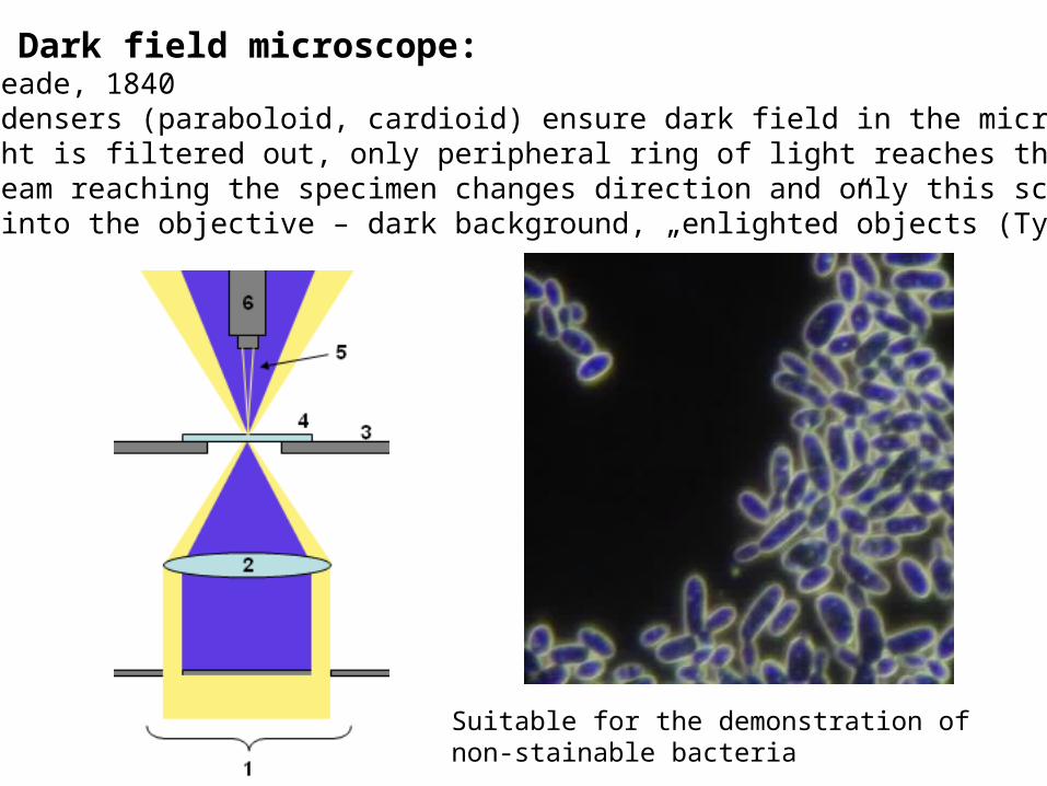
Dark field microscope:Inventor: Reade, 1840Special condensers (paraboloid, cardioid) ensure dark field in the microscope. Central light is filtered out, only peripheral ring of light reaches the specimen.The light beam reaching the specimen changes direction and only this scatteredlight gets into the objective – dark background, „enlighted”objects (Tyndall)
Suitable for the demonstration of non-stainable bacteria

Phase contrast microscope
Usage: study of non-stained, even living preparates (eg. cell cultures).
Steps of mitotic division
Inventor: Zernike, 1936 (Nobel prize, 1953)It changes differences of refractive index into light intensity differencesIn this way it enhances contrast.Below the condenser: ring shaped lamella In the focus plane of objective: partially transparent phase ringImage formation: light beams of different phase eliminate each other, light beams in the same phase enhance each other.The result is a stronger contrast.

Polarization microscope
Inventor: Talbot, 1829.Polarizator filter above the condenser.Another, movable filter – analizator – between the objective lens and the eyepieces. If the filters are in parallel position, the field is dark. But: if the preparatecontains anizotropic components, that ones selectively become visible.Usage: studying biological membranes, cross-striated muscle, diagnosis of illnesseswith crystal formation (eg. oxalate nephrosis in kidney).

Fluorescence microscopeInventor: Reichert, 1911Light source is a UV lamp. Light is directed to the specimen by a dicroic mirror.Certain molecules in the specimen are excited and emit a higher wawelength light. The dicroic mirror selectively transmits the higher wawelength light and this is contributing to the image formation.Usage: studying autofluorescence or fluorescence immunocytochmistry
Confocal scanning microscope
Light source is a laser beam, which point by point scans the surface of the specimen. Emitted light is detected point by point and used for the image formation in a computer. It makes possible optical „sectioning” of thicker samples.
Usage: studying thicker sections with optical sectioning, three-dimensional reconstruction of tissue components.

Confocal microscope
The way of the laser beamwithin the microscope

Traditional fluorescence microscopicimage
A single 2 m optical section
Five superimposed2 m sections