Embed Size (px)
Citation preview
Jour
nal o
f Cel
l Sci
ence
RESEARCH ARTICLE
Spd2 assists Spd1 in the modulation of ribonucleotide reductasearchitecture but does not regulate deoxynucleotide pools
Rasmus Vejrup-Hansen1, Oliver Fleck1,2, Katrine Landvad1, Ulrik Fahnøe1, Sebastian S. Broendum1,Ann-Sofie Schreurs3, Birthe B. Kragelund1, Antony M. Carr3, Christian Holmberg1 and Olaf Nielsen1,*
ABSTRACT
In yeasts, small intrinsically disordered proteins (IDPs) modulate
ribonucleotide reductase (RNR) activity to ensure an optimal supply
of dNTPs for DNA synthesis. The Schizosaccharomyces pombe
Spd1 protein can directly inhibit the large RNR subunit (R1), import
the small subunit (R2) into the nucleus and induce an architectural
change in the R1–R2 holocomplex. Here, we report the
characterization of Spd2, a protein with sequence similarity to
Spd1. We show that Spd2 is a CRL4Cdt2-controlled IDP that
functions together with Spd1 in the DNA damage response and in
modulation of RNR architecture. However, Spd2 does not regulate
dNTP pools and R2 nuclear import. Furthermore, deletion of spd2
only weakly suppresses the Rad3ATR checkpoint dependency of
CRL4Cdt2 mutants. However, when we raised intracellular dNTP
pools by inactivation of RNR feedback inhibition, deletion of spd2
could suppress the checkpoint dependency of CRL4Cdt2 mutant
cells to the same extent as deletion of spd1. Collectively, these
observations suggest that Spd1 on its own regulates dNTP pools,
whereas in combination with Spd2 it modulates RNR architecture
and sensitizes cells to DNA damage.
KEY WORDS: Genome stability, Ribonucleotide reductase,
CRL4Cdt2 ubiquitin ligase, Intrinsically disordered proteins,
S. pombe, Cancer model
INTRODUCTIONCorrect regulation of the levels of the building blocks of DNA is
emerging as a pre-requisite for the maintenance of genome
integrity. Cancer development is presumed to be initiated by
oncogenic mutations that cause replication stress, thereby
increasing the risk of replication fork collapse and the
subsequent formation of DNA lesions (Halazonetis et al.,
2008). This type of oncogenic replication stress has been
reported to be accompanied by a significant reduction in
cellular deoxynucleoside triphosphate (dNTP) pools, possibly
due to an imbalance between nucleotide synthesis and DNA
replication activity (Beck et al., 2012; Bester et al., 2011).
Furthermore, such oncogenic S phase problems, including shorter
inter-origin distances and slower fork migration, can be reversed
by an exogenous supply of nucleosides, suggesting that
insufficient levels of the building blocks of DNA might play an
important causal role in the early stages of tumor development.
The rate-limiting step in dNTP production is catalyzed by the
essential and highly conserved enzyme ribonucleotide reductase
(RNR), which reduces ribonucleoside diphosphates to their
corresponding deoxy forms. Eukaryotic cells use class Ia RNR,
a heteromeric enzyme consisting of up to three copies of two
different dimeric subunits, R1 and R2. The catalytic activity
resides in the large R1 subunit, whereas the smaller R2 subunit
donates reducing power to the reaction from a diferric tyrosyl
radical (Nordlund and Reichard, 2006).
Consistent with deoxynucleotide pools being important for the
maintenance of genome integrity, cellular RNR function is tightly
regulated at several levels. Expression of both subunits is induced
under conditions in which DNA synthesis is required (i.e. in S
phase or after DNA damage) (Elledge et al., 1993). Furthermore,
the activity of RNR is regulated by two intricate feedback-control
mechanisms (Reichard, 2010). The R1 subunit contains two
allosteric effector-binding sites, the specificity site and the
activity site. The specificity site ensures a balanced supply of
the four DNA building blocks, by binding dNDP species that are
present in excess, thereby remodeling the catalytic site towards
other base substrates. The activity site represents a molecular
switch that regulates the overall activity of the enzyme by
monitoring the cellular dATP:ATP ratio; RNR is inactive when
dATP is bound, and it becomes activated when dATP is replaced
by ATP. Recent structural studies suggest that the inactive form
of the enzyme is a hexameric ring consisting of three R1 dimers
with an R2 dimer embedded in the middle (Fairman et al., 2011).
Less is known about the structure of the active ATP-bound form,
but the molecular interaction between the R1 and R2 subunits
appears to be altered relative to that of the dATP-bound complex,
and the ATP-bound form might contain additional R2 dimers
(Hofer et al., 2012). RNR can be locked genetically in its active
configuration by replacing Asp57 in the activity site with Asn.
Mammalian cell lines harboring this D57N-mutated version of
R1 have highly increased dNTP pools, indicating that a large
proportion of RNR complexes is normally in the inhibited form
in vivo (Weinberg et al., 1981).
In yeasts, RNR activity is additionally regulated by a group of
small intrinsically disordered proteins (IDPs). In fission yeast, the
Spd1 protein can inhibit RNR by two different mechanisms. It
can sequester the R2 subunit in the nucleus, away from the R1
subunit, which is mainly cytosolic (Liu et al., 2003), and it can
also directly bind to and inhibit the R1 subunit (Hakansson et al.,
2006). We reported recently that Spd1 can modulate the RNR
complex in a third fashion; if the R1 and R2 subunits are tagged
with different fluorescent proteins, a fluorescence resonance
1Department of Biology, University of Copenhagen, Ole Maaløes Vej 5, 2200Copenhagen N., Denmark. 2NWCR Institute, School of Biological Sciences,Bangor University, Bangor, Gwynedd, LL57 2UW, UK. 3Genome Damage andStability Centre, School of Life Sciences, University of Sussex, Falmer, Brighton,East Sussex BN1 9RQ, UK.
*Author for correspondence ([email protected])
Received 2 August 2013; Accepted 14 February 2014
� 2014. Published by The Company of Biologists Ltd | Journal of Cell Science (2014) 127, 2460–2470 doi:10.1242/jcs.139816
2460

Jour
nal o
f Cel
l Sci
ence
energy transfer (FRET) reaction can be observed between them,and this signal is absent in Dspd1 cells (Nestoras et al., 2010). It is
unclear how the underlying Spd1-mediated change in themolecular interactions between R1 and R2 affects RNRfunction. Interestingly, these three molecular functions of Spd1can be separated genetically, suggesting that the protein mediates
them by different molecular mechanisms (Nestoras et al., 2010).The limited sequence conservation of RNR-inhibitory IDPs
has, thus far, precluded resolution of the issue of whether
mammalian counterparts exist. However, in the distantly relatedbudding yeast Saccharomyces cerevisiae, two Spd1-related IDPs,Dif1 and Sml1, sequester R2 in the nucleus or inhibit R1,
respectively. Interestingly, synteny analysis suggests that thesetwo genes arose through genome duplication of a commonancestor with both functions, similar to S. pombe Spd1 (Lee et al.,
2008). A third potential Spd1-related protein, Hug1, is alsopredicted in the budding yeast genome, but no function has yetbeen assigned to this protein (Basrai et al., 1999).
When fission yeast cells undergo DNA replication or repair,
the Spd1 protein becomes degraded by CRL4Cdt2-mediatedubiquitylation (Holmberg et al., 2005; Liu et al., 2003), a processthat occurs on chromatin-associated PCNA (Salguero et al., 2012).
Spd1 degradation also requires the Csn1 and Csn2 subunits of theCOP9 signalosome (Liu et al., 2003). The CRL4Cdt2 E3 ubiquitinligase is activated by transcriptional induction of the Cdt2 substrate
adaptor, which becomes expressed in unperturbed S phase by theMluI cell-cycle box (MCB) transcription complex. Following DNAdamage, Cdt2 is induced by a Rad3ATR-dependent pathway (Liu
et al., 2005; Moss et al., 2010).CRL4Cdt2-defective cells undergo DNA replication in the
presence of Spd1, and this gives rise to severe S phase stress;replication proceeds slowly, with concomitant activation of the
Rad3ATR checkpoint, which becomes essential for cell survivalunder these conditions. Furthermore, such cells are hypersensitiveto DNA damaging agents, are defective in double-strand break
(DSB) repair by homologous recombination, display .20-foldincrease in spontaneous mutation rates, and are also completelyunable to undergo pre-meiotic S phase (Holmberg et al., 2005;
Liu et al., 2005; Liu et al., 2003; Moss et al., 2010). Therequirement for a functional Rad3ATR pathway and the defects inrecombination and pre-meiotic S phase are all fully reversed bydeletion of the spd1 gene or by overexpression of fission yeast R2
(Suc22R2), suggesting that these phenotypes are caused by Spd1-mediated RNR inhibition. By contrast, the increased mutationrates and sensitivity to DNA damage are only partially suppressed
by Spd1 loss, indicating that deregulation of other CRL4Cdt2-controlled processes also contributes to these phenotypes.
The systematic genetic analysis of spd1 has shown that certain
mutants that are defective in nuclear import of the RNR R2 subunitor in the FRET reaction between R1 and R2 still require theRad3ATR pathway for survival when the Spd1 degradation pathway
is inactivated (Nestoras et al., 2010). These observations areconsistent with a model where Spd1 causes checkpoint activationby inhibiting dNTP formation by direct binding to RNR. However,we recently reported that Spd1 accumulation can cause checkpoint
activation even in cells that have highly elevated dNTP pools,owing to inactivation of RNR feedback inhibition (Fleck et al.,2013). Hence, Spd1 might also cause checkpoint activation
independently of deoxynucleotide synthesis.In the present study, we have characterized Spd2, an Spd1-
related putative RNR inhibitor that was identified in a
comparative genome sequencing study (Rhind et al., 2011). We
show that Spd2, similar to Spd1, is an IDP that becomes degradedby CRL4Cdt2 ubiquitylation following inhibition of RNR by
hydroxyurea. Similar to Dspd1, deletion of spd2 can rescue thedamage sensitivity of CRL4Cdt2 mutants, and Spd2, like Spd1, isrequired for the FRET signal between R1 and R2. However, Spd2does not share the ability of Spd1 to sequester the R2 subunit in
the nucleus, and, surprisingly, it does not seem to affect cellulardNTP pools. These observations support a model where Spd2 canassist Spd1 in an S phase inhibitory pathway that functions
independently of dNTP formation.
RESULTSSpd2 – a new RNR inhibitor homologThe genomes of four Schizosaccharomyces species have recentlybeen sequenced (Rhind et al., 2011). The availability of the
sequences of three additional fission yeast species allowed theidentification of .100 new conserved open reading frames in theS. pombe genome. One of these proteins, Spd2, shows limitedsimilarity to Spd1 (19% overall sequence identity) and other RNR
inhibitors (Fig. 1).In general, the homology among RNR inhibitors is limited to a
number of short motifs. The Spd1 and Spd2 orthologs also appear
to have diverged considerably, but are very well conserved withinthe Spd1 and Spd2 subgroups (Fig. 1A). The Spd1 and Spd2families share a small domain near their C-terminal ends, which
is absent from the other known RNR inhibitors. We will refer tothis as the Spd domain (Fig. 1B).
The Spd1 and Spd2 orthologs of the four fission yeast species
all contain a Hug domain (Fig. 1A), which is also present in the S.
cerevisiae Hug1 and Dif1 proteins and in Ashbya gossypii
Aer122c, but is absent from budding yeast Sml1 (Fig. 1B). TheHug domain of Dif1 is required for nuclear import of the small
RNR subunit (Lee et al., 2008; Wu and Huang, 2008), and geneticanalysis suggests that the Hug domain of Spd1 might have asimilar function (Nestoras et al., 2010). The N-terminal part of
the Hug domain in Spd1 was recently shown to constitute aPCNA-interacting protein (PIP) degron, mediating binding toPCNA, which is a prerequisite for CRL4Cdt2-dependent
ubiquitylation and subsequent degradation of Spd1 (Salgueroet al., 2012). This motif is present in Spd2 (Fig. 1), suggestingthat the protein is degraded by a similar mechanism (see below).
The CRL4Cdt2 degradation pathway is absent from budding
yeast. Instead, degradation of Sml1 and Dif1 during S phase or inresponse to DNA damage is triggered by Dun1-mediatedphosphorylation of a phospho-degron referred to as the Sml1
domain (Lee et al., 2008; Uchiki et al., 2004; Wu and Huang,2008; Zhao and Rothstein, 2002). Consistent with a differentdegradation mechanism in fission yeast, this Sml1 degron is
absent in both Spd1 and Spd2 (Fig. 1B).Finally, Sml1 can bind to and inhibit the large RNR subunit
through its Rnr1 domain (Zhao et al., 2000). Although it has been
shown that S. pombe Spd1 also can bind to the large RNR subunit(Hakansson et al., 2006), conservation of the Rnr1 domain inSpd1 is limited to a short stretch of amino acid residues. In Spd2,the conservation of the Rnr1 domain is even more limited, if not
absent (Fig. 1B).
Spd2 is an IDPSimilar to Spd1 and Sml1 (Danielsson et al., 2008; Nestoras et al.,2010), the sequence of Spd2 has the typical characteristics of anIDP – a high content of charged residues (29%) and a low
aliphatic index (67.94; for reference, myoglobin595.1). These
RESEARCH ARTICLE Journal of Cell Science (2014) 127, 2460–2470 doi:10.1242/jcs.139816
2461
Jour
nal o
f Cel
l Sci
ence
proteins lack well-structured three-dimensional folds and aredistinctive in having regions that form lowly populated, transientstructures and/or contain conserved sequence motifs (Tompa,2002). Importantly, these local features are central to target
recognition, and the flexibility of the IDPs is essential for bindingto more than one partner. To investigate the disordercharacteristics of Spd2 by spectroscopy, recombinant Spd2 was
produced in Escherichia coli and purified to .98% homogeneity(Fig. 2A). A far-UV circular dichroism spectrum of Spd2 showedno signs of pronounced secondary structure elements, with little
negative ellipticity in the 210–220 nm range (Fig. 2B). Instead, a
large negative ellipticity with a maximum at 199 nm suggested adisordered protein with little or no secondary structure. In supportof this, a 15N,1H-heteronuclear single quantum coherence
(HSQC) nuclear magnetic resonance (NMR) spectrum of 15N-labeled Spd2, recorded at 10 C, showed a narrow dispersion ofsignals in the 1H dimension, further suggesting that the protein is
disordered with no globular fold (Fig. 2C). Unlike Spd1, thepeaks of the NMR spectrum of Spd2 were of almost equalintensity, revealing the higher solubility of this protein compared
with Spd1 (Nestoras et al., 2010; data not shown). Thus, Spd2possesses hallmarks of an IDP (Tompa, 2002), with low-complexity sequence, a lack of secondary structure elements ina far-UV circular dichroism spectrum and a collapsed NMR
spectrum.
Spd1 and Spd2 both inhibit S phaseWe next constructed a strain deleted for the spd2 gene. Similarlyto Dspd1 cells, this Dspd2 strain showed no obvious phenotypicdifferences to wild-type cells with regard to cell shape and growth
rate (data not shown). Spd1 was originally identified as a proteinthat inhibited S phase progression when overexpressed, therebycausing cell elongation (Woollard et al., 1996). We found that
overexpression of Spd2 similarly caused cell elongation(Fig. 3A). The elongation of cells by overexpression of Spd2did not require functional Spd1 and vice versa.
Next, we used flow cytometry (FACS) to monitor the cell-
cycle distribution in cells overexpressing Spd1 or Spd2. The
Fig. 1. Spd2 is homologous to Spd1 and other known RNR inhibitors.(A) Comparison of amino acid sequences of Spd1 and Spd2 orthologs of thefour Schizosaccharomyces species S. pombe (Spo), S. cryophilus (Scr), S.octosporus (Soc) and S. japonicus (Sja). Residues are highlighted in blackand grey when they are identical and similar, respectively, between Spd1 andSpd2 orthologs. Amino acid residues in dark and light blue indicateconservation predominantly in Spd1 orthologs, and in dark and light redpredominantly in Spd2 orthologs. The Hug domain (*) and the Spd domain(**) are boxed. Amino acids defining the Spd1 PIP degron (Q-x-x-L-x-x-x-x-x-x-x-R) (Salguero et al., 2012) are marked above the aligned sequences.(B) Alignment of Spd2 with other fungal RNR inhibitors. Identical and similaramino acid residues are highlighted in, respectively, black and gray. Hug,Sml and Rnr1 domains, as defined previously (Lee et al., 2008), are boxed.Spd1 and Spd2 share a unique domain close to the C-terminus, which wetermed the Spd domain.
Fig. 2. Structural properties of Spd2. (A) Spd2 was purified tohomogeneity, and the mobility was investigated by electrophoresis. Lane 1,marker; lane 2, purified Spd2. (B) A far-UV circular dichroism spectrum ofpurified Spd2, showing a large negative ellipticity with a maximum at 199 nm,suggesting a disordered protein with little or no secondary structure. (C) AnNMR spectrum of purified Spd2 showing that only a small dispersion ofsignals was observed in the 1H- dimension, further suggesting that theprotein is disordered with no globular fold.
RESEARCH ARTICLE Journal of Cell Science (2014) 127, 2460–2470 doi:10.1242/jcs.139816
2462
Jour
nal o
f Cel
l Sci
ence
interpretation of FACS data in fission yeast is complicated by thefact that G1 and S phase cells are still attached to their sisters,
thus giving rise to a 2C signal. However, by measuring the DNAsignal width, it is possible to discriminate G1 and S cells from G2cells (Knutsen et al., 2011), thus giving a more precise estimate ofthe S phase population. By using this method, we found that
overexpression of Spd2 caused a strong increase in the number ofcells undergoing S phase, suggesting that the protein – similarlyto Spd1 – delays DNA replication (Fig. 3B; supplementary
material Fig. S1). Again, Spd1 and Spd2 could cause thisphenotype independently of one another.
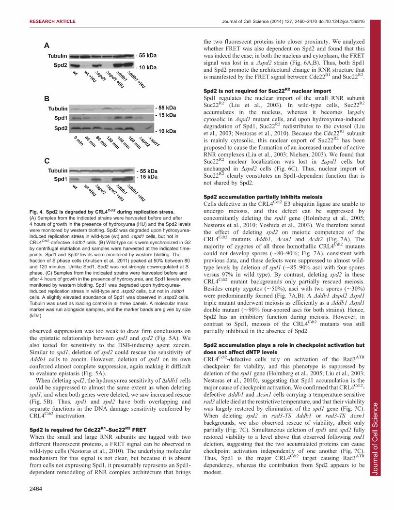
Spd2 is a CRL4Cdt2 targetSpd1 is a target of the CRL4Cdt2 E3 ubiquitin ligase (Holmberg et al.,2005; Liu et al., 2005; Liu et al., 2003), and we next investigatedwhether this was also the case for Spd2. Because we found that
epitope tagging of Spd1 adversely affected its functionality (data notshown), we raised antibodies against recombinant Spd1 and Spd2proteins. Similar to Spd1, Spd2 was degraded in response to
treatment with hydroxyurea, and this response was completelyabolished in CRL4Cdt2-defective Dddb1 cells, where the steady-statelevel of Spd2 was also higher than that observed in wild-type cells
(Fig. 4A). Furthermore, deletion of spd2 suppressed the elongated-cell phenotype and improved the growth rate of CRL4Cdt2-defectiveDddb1 cells to approximately the same extent as did spd1 deletion
(data not shown). Hence, consistent with the presence of a PIPdegron in the protein (Fig. 1A), we conclude that Spd2, like Spd1, isa target for CRL4Cdt2-mediated ubiquitylation.
The degradation of Spd2 appeared to be independent of Spd1and vice versa (Fig. 4A,C). Deletion of spd2 apparently caused a
modestly elevated level of Spd1 in asynchronously growing cells(Fig. 4C). Consistent with this, we found that spd1 mRNA levelswere 1.5-fold increased in exponentially growing Dspd2 cellscompared with wild-type cells (supplementary material Fig. S2).
Spd1 becomes degraded as cells enter S phase, because theCRL4 substrate adaptor Cdt2 is controlled by the S-phase-specifictranscription complex MCB (Liu et al., 2005). We synchronized
wild-type cells by centrifugal elutriation and monitored the levelsof Spd1 and Spd2 as cells progressed through S phase (Fig. 4B).As observed previously (Liu et al., 2003), Spd1 became
downregulated as cells entered S phase, but for Spd2, we couldnot detect a similar robust reduction.
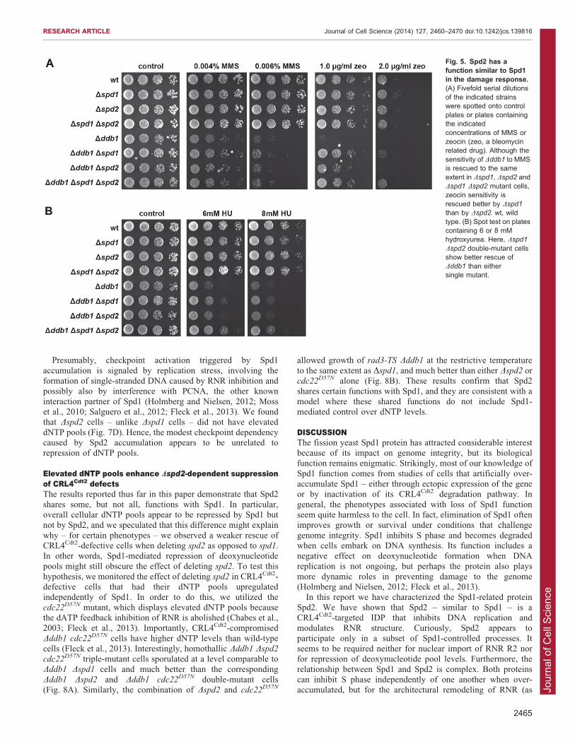
Spd2 functions in the DNA damage responseCRL4Cdt2-defective cells are sensitive to the RNR inhibitorhydroxyurea and the DNA alkylating agent methyl methanesulphonate (MMS) (Holmberg et al., 2005; Zolezzi et al., 2002),
and this phenotype can be partially suppressed by concomitantlydeleting the spd1 gene, suggesting that the accumulation ofexcess Spd1 protein contributes to the observed drug sensitivity.
We therefore investigated whether deletion of the spd2 genecould also affect the damage sensitivity of CRL4Cdt2-defectiveDddb1 cells. Rescue of the MMS sensitivity of Dddb1 occurred to
a similar modest extent in both Dddb1 Dspd1 and Dddb1 Dspd2
double mutants, and there was no further suppression of thephenotype in the Dddb1 Dspd1 Dspd2 triple mutant. However, the
Fig. 3. Overexpression of Spd2 inhibits S phase.(A) Overexpressing either Spd1 or Spd2 leads to theaccumulation of a subset of cells with elongated andaberrant cell shapes. The effect of Spd2overexpression is not dependent on a functionalspd1+ gene and vice versa. Cells were grown to mid-log phase in minimal medium with or without thiaminefor 24 hours before pictures were taken. wt, wild type.Scale bar: 6 mm. (B) Quantification of FACS analysis ofcells overexpressing Spd1 or Spd2. Cell-cycledistribution was analyzed as reported previously(Knutsen et al., 2011). S1, the fraction of S phase cellswith one nucleus; S2, binucleate cells (as defined bythe width of the DNA signal) undergoing replication.When overexpressing either Spd1 or Spd2, theproportion of S phase cells increased, in particular S1cells. Representative examples of this quantificationmethod are given in supplementary material Fig. S1.
RESEARCH ARTICLE Journal of Cell Science (2014) 127, 2460–2470 doi:10.1242/jcs.139816
2463
Jour
nal o
f Cel
l Sci
ence
observed suppression was too weak to draw firm conclusions onthe epistatic relationship between spd1 and spd2 (Fig. 5A). Wealso tested for sensitivity to the DSB-inducing agent zeocin.
Similar to spd1, deletion of spd2 could rescue the sensitivity ofDddb1 cells to zeocin. However, deletion of spd1 on its ownconferred almost complete suppression, again making it difficult
to evaluate epistasis (Fig. 5A).When deleting spd2, the hydroxyurea sensitivity of Dddb1 cells
could be suppressed to almost the same extent as when deleting
spd1, and when both genes were deleted, we saw increased rescue(Fig. 5B). Thus, spd1 and spd2 have both overlapping andseparate functions in the DNA damage sensitivity conferred byCRL4Cdt2 inactivation.
Spd2 is required for Cdc22R1–Suc22R2 FRETWhen the small and large RNR subunits are tagged with two
different fluorescent proteins, a FRET signal can be observed inwild-type cells (Nestoras et al., 2010). The underlying molecularmechanism for this signal is not clear, but because it is absent
from cells not expressing Spd1, it presumably represents an Spd1-dependent remodeling of RNR complex architecture that brings
the two fluorescent proteins into closer proximity. We analyzedwhether FRET was also dependent on Spd2 and found that this
was indeed the case; in both the nucleus and cytoplasm, the FRETsignal was lost in a Dspd2 strain (Fig. 6A,B). Thus, both Spd1and Spd2 promote the architectural change in RNR structure thatis manifested by the FRET signal between Cdc22R1 and Suc22R2.
Spd2 is not required for Suc22R2 nuclear importSpd1 regulates the nuclear import of the small RNR subunit
Suc22R2 (Liu et al., 2003). In wild-type cells, Suc22R2
accumulates in the nucleus, whereas it becomes largelycytosolic in Dspd1 mutant cells, and upon hydroxyurea-induced
degradation of Spd1, Suc22R2 redistributes to the cytosol (Liuet al., 2003; Nestoras et al., 2010). Because the Cdc22R1 subunitis mainly cytosolic, this nuclear export of Suc22R2 has been
proposed to cause the formation of an increased number of activeRNR complexes (Liu et al., 2003; Nielsen, 2003). We found thatSuc22R2 nuclear localization was lost in Dspd1 cells butunchanged in Dspd2 cells (Fig. 6C). Thus, nuclear import of
Suc22R2 clearly constitutes an Spd1-dependent function that isnot shared by Spd2.
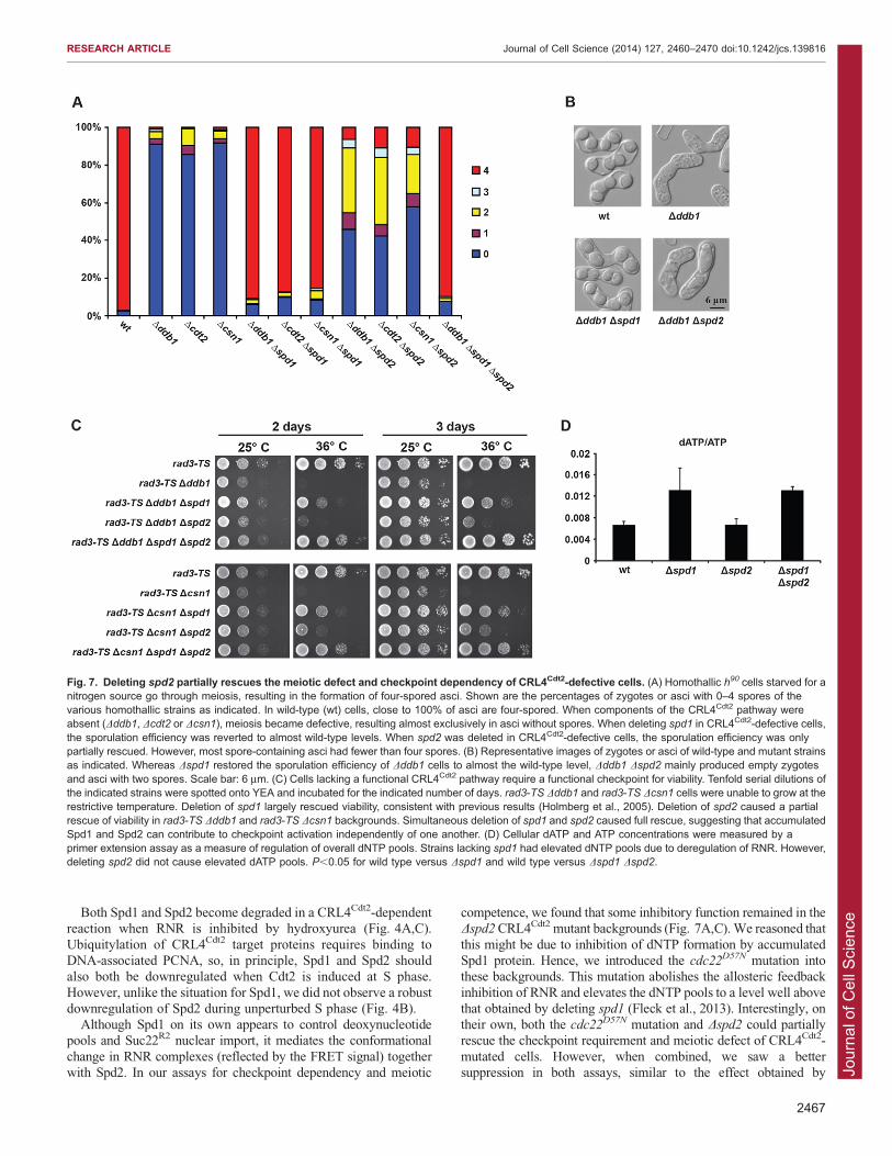
Spd2 accumulation partially inhibits meiosisCells defective in the CRL4Cdt2 E3 ubiquitin ligase are unable toundergo meiosis, and this defect can be suppressed by
concomitantly deleting the spd1 gene (Holmberg et al., 2005;Nestoras et al., 2010; Yoshida et al., 2003). We therefore testedthe effect of deleting spd2 on meiotic competence of the
CRL4Cdt2 mutants Dddb1, Dcsn1 and Dcdt2 (Fig. 7A). Themajority of zygotes of all three homothallic CRL4Cdt2 mutantscould not develop spores (,80–90%; Fig. 7A), consistent withprevious data, and these defects were suppressed to almost wild-
type levels by deletion of spd1 (,85–90% asci with four sporesversus 97% in wild type). By contrast, deleting spd2 in theseCRL4Cdt2 mutant backgrounds only partially rescued meiosis.
Besides empty zygotes (,50%), asci with two spores (,30%)were predominantly formed (Fig. 7A,B). A Dddb1 Dspd2 Dspd1
triple mutant underwent meiosis as efficiently as a Dddb1 Dspd1
double mutant (,90% four-spored asci for both strains). Hence,Spd2 has an inhibitory function during meiosis. However, incontrast to Spd1, meiosis of the CRL4Cdt2 mutants was stillpartially inhibited in the absence of Spd2.
Spd2 accumulation plays a role in checkpoint activation butdoes not affect dNTP levelsCRL4Cdt2-defective cells rely on activation of the Rad3ATR
checkpoint for viability, and this phenotype is suppressed bydeletion of the spd1 gene (Holmberg et al., 2005; Liu et al., 2003;
Nestoras et al., 2010), suggesting that Spd1 accumulation is themajor cause of checkpoint activation. We confirmed that CRL4Cdt2-defective Dddb1 and Dcsn1 cells carrying a temperature-sensitive
rad3 allele died at the restrictive temperature, and that their viabilitywas largely restored by elimination of the spd1 gene (Fig. 7C).When deleting spd2 in rad3-TS Dddb1 or rad3-TS Dcsn1
backgrounds, we also observed rescue of viability, albeit only
partially (Fig. 7C). Simultaneous deletion of spd1 and spd2 fullyrestored viability to a level above that observed following spd1
deletion, suggesting that the two accumulated proteins can cause
checkpoint activation independently of one another (Fig. 7C).Thus, Spd1 is the major CRL4Cdt2 target causing Rad3ATR
dependency, whereas the contribution from Spd2 appears to be
modest.
Fig. 4. Spd2 is degraded by CRL4Cdt2 during replication stress.(A) Samples from the indicated strains were harvested before and after4 hours of growth in the presence of hydroxyurea (HU) and the Spd2 levelswere monitored by western blotting. Spd2 was degraded upon hydroxyurea-induced replication stress in wild-type (wt) and Dspd1 cells, but not inCRL4Cdt2-defective Dddb1 cells. (B) Wild-type cells were synchronized in G2by centrifugal elutriation and samples were harvested at the indicated time-points. Spd1 and Spd2 levels were monitored by western blotting. Thefraction of S phase cells (Knutsen et al., 2011) peaked at 50% between 80and 120 minutes. Unlike Spd1, Spd2 was not strongly downregulated at Sphase. (C) Samples from the indicated strains were harvested before andafter 4 hours of growth in the presence of hydroxyurea, and Spd1 levels weremonitored by western blotting. Spd1 was degraded upon hydroxyurea-induced replication stress in wild-type and Dspd2 cells, but not in Dddb1
cells. A slightly elevated abundance of Spd1 was observed in Dspd2 cells.Tubulin was used as loading control in all three panels. A molecular massmarker was run alongside samples, and the marker bands are given by size(kDa).
RESEARCH ARTICLE Journal of Cell Science (2014) 127, 2460–2470 doi:10.1242/jcs.139816
2464
Jour
nal o
f Cel
l Sci
ence
Presumably, checkpoint activation triggered by Spd1
accumulation is signaled by replication stress, involving theformation of single-stranded DNA caused by RNR inhibition andpossibly also by interference with PCNA, the other known
interaction partner of Spd1 (Holmberg and Nielsen, 2012; Mosset al., 2010; Salguero et al., 2012; Fleck et al., 2013). We foundthat Dspd2 cells – unlike Dspd1 cells – did not have elevated
dNTP pools (Fig. 7D). Hence, the modest checkpoint dependencycaused by Spd2 accumulation appears to be unrelated torepression of dNTP pools.
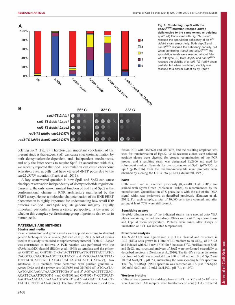
Elevated dNTP pools enhance Dspd2-dependent suppressionof CRL4Cdt2 defectsThe results reported thus far in this paper demonstrate that Spd2
shares some, but not all, functions with Spd1. In particular,overall cellular dNTP pools appear to be repressed by Spd1 butnot by Spd2, and we speculated that this difference might explain
why – for certain phenotypes – we observed a weaker rescue ofCRL4Cdt2-defective cells when deleting spd2 as opposed to spd1.In other words, Spd1-mediated repression of deoxynucleotidepools might still obscure the effect of deleting spd2. To test this
hypothesis, we monitored the effect of deleting spd2 in CRL4Cdt2-defective cells that had their dNTP pools upregulatedindependently of Spd1. In order to do this, we utilized the
cdc22D57N mutant, which displays elevated dNTP pools becausethe dATP feedback inhibition of RNR is abolished (Chabes et al.,2003; Fleck et al., 2013). Importantly, CRL4Cdt2-compromised
Dddb1 cdc22D57N cells have higher dNTP levels than wild-typecells (Fleck et al., 2013). Interestingly, homothallic Dddb1 Dspd2
cdc22D57N triple-mutant cells sporulated at a level comparable to
Dddb1 Dspd1 cells and much better than the correspondingDddb1 Dspd2 and Dddb1 cdc22D57N double-mutant cells(Fig. 8A). Similarly, the combination of Dspd2 and cdc22D57N
allowed growth of rad3-TS Dddb1 at the restrictive temperature
to the same extent as Dspd1, and much better than either Dspd2 orcdc22D57N alone (Fig. 8B). These results confirm that Spd2shares certain functions with Spd1, and they are consistent with a
model where these shared functions do not include Spd1-mediated control over dNTP levels.
DISCUSSIONThe fission yeast Spd1 protein has attracted considerable interestbecause of its impact on genome integrity, but its biologicalfunction remains enigmatic. Strikingly, most of our knowledge of
Spd1 function comes from studies of cells that artificially over-accumulate Spd1 – either through ectopic expression of the geneor by inactivation of its CRL4Cdt2 degradation pathway. In
general, the phenotypes associated with loss of Spd1 functionseem quite harmless to the cell. In fact, elimination of Spd1 oftenimproves growth or survival under conditions that challenge
genome integrity. Spd1 inhibits S phase and becomes degradedwhen cells embark on DNA synthesis. Its function includes anegative effect on deoxynucleotide formation when DNAreplication is not ongoing, but perhaps the protein also plays
more dynamic roles in preventing damage to the genome(Holmberg and Nielsen, 2012; Fleck et al., 2013).
In this report we have characterized the Spd1-related protein
Spd2. We have shown that Spd2 – similar to Spd1 – is aCRL4Cdt2-targeted IDP that inhibits DNA replication andmodulates RNR structure. Curiously, Spd2 appears to
participate only in a subset of Spd1-controlled processes. Itseems to be required neither for nuclear import of RNR R2 norfor repression of deoxynucleotide pool levels. Furthermore, the
relationship between Spd1 and Spd2 is complex. Both proteinscan inhibit S phase independently of one another when over-accumulated, but for the architectural remodeling of RNR (as
Fig. 5. Spd2 has afunction similar to Spd1in the damage response.(A) Fivefold serial dilutionsof the indicated strainswere spotted onto controlplates or plates containingthe indicatedconcentrations of MMS orzeocin (zeo, a bleomycinrelated drug). Although thesensitivity of Dddb1 to MMSis rescued to the sameextent in Dspd1, Dspd2 andDspd1 Dspd2 mutant cells,zeocin sensitivity isrescued better by Dspd1
than by Dspd2. wt, wildtype. (B) Spot test on platescontaining 6 or 8 mMhydroxyurea. Here, Dspd1
Dspd2 double-mutant cellsshow better rescue ofDddb1 than eithersingle mutant.
RESEARCH ARTICLE Journal of Cell Science (2014) 127, 2460–2470 doi:10.1242/jcs.139816
2465

Jour
nal o
f Cel
l Sci
ence
manifested by the FRET assay) they seem to cooperate.
According to a recent quantitative expression study, Spd1 andSpd2 are present at similar levels in the cell (respectively,,14,000 and ,16,000 molecules/cell, Marguerat et al., 2012), sotheir different behavior seems to be caused by different functions
rather than different concentrations.CRL4Cdt2-defective cells constitutively activate their Rad3ATR
checkpoint, presumably because Spd1 reduces dNTP pools and
interferes with other thus-far-uncharacterized functions that areimportant for genome integrity (see below). Thus, in accordancewith previous reports (Holmberg et al., 2005; Liu et al., 2003;
Nestoras et al., 2010), we found that the viability of CRL4Cdt2-defective rad3-TS cells was restored by deleting the spd1 gene(Fig. 7C). However, deletion of spd2 only caused an intermediate
suppression of viability in this assay. Hence, accumulation ofSpd2 appears to make cells much less dependent on the Rad3ATR
checkpoint pathway for survival than does Spd1 accumulation.
Furthermore, because simultaneous deletion of spd1 and spd2
caused enhanced suppression (Fig. 7C), the two proteins appearto activate the checkpoint independently of one another.
Unlike Spd1, Spd2 is not required for nuclear import ofSuc22R2. Analogous to the S. cerevisiae Dif1 protein, genetic
analysis suggests that the Hug domain of Spd1 is required fornuclear import of Suc22R2 (Nestoras et al., 2010). The Hugdomain is well conserved in Spd2 (Fig. 1), so the fact that
Spd2 is dispensable for Suc22R2 nuclear import suggests thatother regions in Spd1 are also needed. Consistent with this,mutant spd1-m2 (replacing amino acids K5, R6 and V7 with
alanines) was reported also to be defective in Suc22R2 nuclearimport (Nestoras et al., 2010). This region is absent in Spd2(Fig. 1A). The biological relevance of Spd1-mediated nuclear
Suc22R2 import is not yet known, but genetically this functioncan be separated from checkpoint activation (Nestoras et al.,2010).
Fig. 6. Deleting spd2 abolishes RNR FRET, but does notaffect Suc22 localization. (A) In wild-type (wt) cells, CFP-tagged Suc22R2 and YFP-tagged Cdc22R1 give rise to a FRETsignal, which is lost when either spd1 or spd2 is deleted. (B) TheFRET signal is lost in both the nucleus and the cytoplasm ofDspd2 cells. Data show the mean6s.d.; n58–10 cells.(C) Suc22–GFP accumulates in the nucleus in wild-type cells.The nuclear localization was intact in Dspd2 cells, but wasabolished in Dspd1 and Dspd1Dspd2 cells. Scale bars: 6 mm.
RESEARCH ARTICLE Journal of Cell Science (2014) 127, 2460–2470 doi:10.1242/jcs.139816
2466
Jour
nal o
f Cel
l Sci
ence
Both Spd1 and Spd2 become degraded in a CRL4Cdt2-dependentreaction when RNR is inhibited by hydroxyurea (Fig. 4A,C).
Ubiquitylation of CRL4Cdt2 target proteins requires binding toDNA-associated PCNA, so, in principle, Spd1 and Spd2 shouldalso both be downregulated when Cdt2 is induced at S phase.
However, unlike the situation for Spd1, we did not observe a robustdownregulation of Spd2 during unperturbed S phase (Fig. 4B).
Although Spd1 on its own appears to control deoxynucleotidepools and Suc22R2 nuclear import, it mediates the conformational
change in RNR complexes (reflected by the FRET signal) togetherwith Spd2. In our assays for checkpoint dependency and meiotic
competence, we found that some inhibitory function remained in theDspd2 CRL4Cdt2 mutant backgrounds (Fig. 7A,C). We reasoned that
this might be due to inhibition of dNTP formation by accumulatedSpd1 protein. Hence, we introduced the cdc22D57N mutation intothese backgrounds. This mutation abolishes the allosteric feedback
inhibition of RNR and elevates the dNTP pools to a level well abovethat obtained by deleting spd1 (Fleck et al., 2013). Interestingly, ontheir own, both the cdc22D57N mutation and Dspd2 could partiallyrescue the checkpoint requirement and meiotic defect of CRL4Cdt2-
mutated cells. However, when combined, we saw a bettersuppression in both assays, similar to the effect obtained by
Fig. 7. Deleting spd2 partially rescues the meiotic defect and checkpoint dependency of CRL4Cdt2-defective cells. (A) Homothallic h90 cells starved for anitrogen source go through meiosis, resulting in the formation of four-spored asci. Shown are the percentages of zygotes or asci with 0–4 spores of thevarious homothallic strains as indicated. In wild-type (wt) cells, close to 100% of asci are four-spored. When components of the CRL4Cdt2 pathway wereabsent (Dddb1, Dcdt2 or Dcsn1), meiosis became defective, resulting almost exclusively in asci without spores. When deleting spd1 in CRL4Cdt2-defective cells,the sporulation efficiency was reverted to almost wild-type levels. When spd2 was deleted in CRL4Cdt2-defective cells, the sporulation efficiency was onlypartially rescued. However, most spore-containing asci had fewer than four spores. (B) Representative images of zygotes or asci of wild-type and mutant strainsas indicated. Whereas Dspd1 restored the sporulation efficiency of Dddb1 cells to almost the wild-type level, Dddb1 Dspd2 mainly produced empty zygotesand asci with two spores. Scale bar: 6 mm. (C) Cells lacking a functional CRL4Cdt2 pathway require a functional checkpoint for viability. Tenfold serial dilutions ofthe indicated strains were spotted onto YEA and incubated for the indicated number of days. rad3-TS Dddb1 and rad3-TS Dcsn1 cells were unable to grow at therestrictive temperature. Deletion of spd1 largely rescued viability, consistent with previous results (Holmberg et al., 2005). Deletion of spd2 caused a partialrescue of viability in rad3-TS Dddb1 and rad3-TS Dcsn1 backgrounds. Simultaneous deletion of spd1 and spd2 caused full rescue, suggesting that accumulatedSpd1 and Spd2 can contribute to checkpoint activation independently of one another. (D) Cellular dATP and ATP concentrations were measured by aprimer extension assay as a measure of regulation of overall dNTP pools. Strains lacking spd1 had elevated dNTP pools due to deregulation of RNR. However,deleting spd2 did not cause elevated dATP pools. P,0.05 for wild type versus Dspd1 and wild type versus Dspd1 Dspd2.
RESEARCH ARTICLE Journal of Cell Science (2014) 127, 2460–2470 doi:10.1242/jcs.139816
2467
Jour
nal o
f Cel
l Sci
ence
deleting spd1 (Fig. 8). Therefore, an important conclusion of thepresent study is that excess Spd1 can cause checkpoint activation by
both deoxynucleoside-dependent and -independent mechanisms,and only the latter seems to require Spd2. In accordance with this,we recently reported that Spd1 accumulation can cause checkpointactivation even in cells that have elevated dNTP pools due to the
cdc22-D57N mutation (Fleck et al., 2013).A key unanswered question is how Spd1 and Spd2 can cause
checkpoint activation independently of deoxynucleotide regulation.
Currently, the only known mutual function of Spd1 and Spd2 is theconformational change in RNR architecture manifested by theFRET assay. Hence, a molecular characterization of the RNR FRET
phenomenon is highly important for understanding how small IDPproteins like Spd1 and Spd2 regulate genome integrity. Equallyimportant, particularly from a cancer perspective, is the issue of
whether this complex yet fascinating group of proteins also exists inhuman cells.
MATERIALS AND METHODSStrains and mediaStrain construction and growth media were applied according to standard
genetic techniques for S. pombe (Moreno et al., 1991). A list of strains
used in this study is included as supplementary material Table S1. Dspd2
was constructed as follows. A PCR reaction was performed with the
pFA6a-kanMX plasmid (Bahler et al., 1998) as template and the primer
set ONP687 and ONP688 (59-GATACTTTTTAATTGTTTTACTTATC-
CAGGCGCCAGCTGAAGCTTCGTAC-39 and 59-TCGAAAGCTTTA-
TCTTGCTCATTTATTCATGGCCACTAGTGGATCTGATA-39), two
additional PCR reactions were performed with purified genomic S.
pombe DNA and the primer sets ONP689 and ONP690 (59-ATGAATA-
AATGAGCAAGATAAAGCTTTCGA-39 and 59-AGTAACTTTCGAC-
ACATTCAAATGGTGT-39) and ONP691 and ONP692 (59-CCTGGAT-
AAGTAAAACAATTAAAAAGTATC-39 and 59-ACGACTTGAAGCT-
TACTCGCTTCTAAAAGG-39). The three PCR products were used for a
fusion PCR with ONP690 and ONP692, and the resulting amplicon was
used for transformation of Eg432. G418-resistant clones were selected,
positive clones were checked for correct recombination of the PCR
product and a resulting strain was designated Eg2806 and used for
subsequent studies. Plasmids for overexpression of Spd1 (pON736) or
Spd2 (pON1126) from the thiamine-repressible nmt1 promoter were
obtained by cloning the ORFs into pREP3 (Maundrell, 1990).
FACSCells were fixed as described previously (Kjaerulff et al., 2005), and
stained with Sytox Green (Molecular Probes) as recommended by the
manufacturer. Quantification of S phase cells with the aid of the DNA
signal width was performed as described previously (Knutsen et al.,
2011). For each sample, a total of 30,000 cells were counted, and after
gating at least 75% were still present.
Sensitivity assaysFivefold dilution series of the indicated strains were spotted onto YEA
plates containing the indicated drugs. Plates were cast 2 days prior to use
and kept at room temperature. Pictures were taken after 3 days of
incubation at 33 C (or indicated temperature).
Structural analysisThe Spd2 ORF was ligated into a pET11a plasmid and expressed in
BL21(DE3) cells grown in 1 litre of LB medium to an OD600 of 0.7–0.8
and induced with 0.01 mM IPTG for 3 hours at 37 C. Purification of Spd1
and Spd2, and structural analyses of Spd2 were performed essentially as
described previously (Nestoras et al., 2010). The far-UV circular dichroism
spectrum of Spd2 was recorded from 250 to 190 nm on 10 mM Spd2 and
10 mM NaH2PO4, pH 7.4, subtracting the corresponding buffer spectrum.
The 15N, 1H-HSQC NMR spectrum was recorded on 277 mM 15N-Spd2,
100 mM NaCl and 10 mM NaH2PO4, pH 7.4, at 10 C.
Western blottingCultures were grown to mid-log phase at 30 C in YE and 56107 cells
were harvested. All samples were trichloroacetic acid (TCA) extracted,
Fig. 8. Combining Dspd2 with thecdc22D57N mutation rescues Dddb1
deficiencies to the same extent as deletingspd1. (A) Consistent with Fig. 7A, Dspd1
rescued the sporulation deficiency of an h90
Dddb1 strain almost fully. Both Dspd2 andcdc22D57N rescued the deficiency partially, butwhen combining Dspd2 and cdc22D57N, thesporulation levels were rescued almost fully.wt, wild type. (B) Both Dspd2 and cdc22D57N
rescued the viability of a rad3-TS Dddb1 strainpartially, but when combined, viability wasrescued to a similar extent as by Dspd1.
RESEARCH ARTICLE Journal of Cell Science (2014) 127, 2460–2470 doi:10.1242/jcs.139816
2468
Jour
nal o
f Cel
l Sci
ence
run on 15% SDS-PAGE gels and wet blotted (300 mA) for 1.5 hours at
4 C. After blocking, membranes were incubated overnight at 4 C with the
indicated primary antibodies. To study protein levels in response to
hydroxyurea treatment, cultures were grown to mid-log phase and 56107
cells were harvested, 20 mM hydroxyurea was added to the remainder of
the cultures and, after 4 hours, 56107 cells were harvested. To analyze
synchronous populations, 6 litres of culture was grown to mid-log phase
and cells were size-sorted by elutriation. The smallest-sized cells were
collected at a concentration of ,26106 cells/ml and incubated at 30 C.
16108 cells were harvested at each of the indicated time-points.
Polyclonal rabbit anti-Spd1 and anti-Spd2 antibodies were raised
against E. coli purified proteins by Yorkshire Bioscience.
Cell biologySuc22–GFP localization assay and FRET analysis of the Cdc22 and
Suc22 FRET-pair were performed as described previously (Nestoras
et al., 2010). For meiotic analysis, h90 strains of the indicated genotypes
were spotted onto solid sporulation medium at 25 C and after 3 days the
number of spores (0–4) in §100 individual zygotes or asci were
counted.
dNTP poolsdNTP measurement were performed as described previously (Beck et al.,
2012), and the levels were normalized to those of ATP. For
normalization, the dNTP:ATP ratio was set to 1 in the wild type.
AcknowledgementsWe thank Jon Halvor Knutsen (University of Oslo, Norway) for advice on FACSquantification, Karin Holm (University of Copenhagen, Denmark) for experttechnical assistance, Rasmus Hartmann-Petersen (University of Copenhagen,Denmark) for comments on the manuscript and Iain Hagan (The PatersonInstitute, Manchester, UK) and Rasmus Hartmann-Petersen for a-tubulinantibody.
Competing interestsThe authors declare no competing interests.
Author contributionsO.F. performed experiments presented in Figs 1 and 7A; S.S.B. and B.B.Kperformed those presented in Fig. 2; U.F. performed those presented in Fig. 3;A.-S.S. performed those presented in Fig. 6A,B; K.L. performed those presentedin Fig. 6C; C.H. performed those presented in Figs 7D and 8A. R.V.-H. performedall other experiments. A.M.C., C.H., O.F., R.V.-H. and O.N. contributed toconceptual development of the project and interpretation of the results. R.V.-H.and O.N. wrote the paper together.
FundingThis work was supported by funding from the Danish Cancer Society (to O.N.);the Danish Research Councils (to B.B.K.); the National Institute for Social Careand Health Research; Cancer Genetics Biomedical Research Unit (to O.F.); NorthWest Cancer Research (to O.F.); and the Association for International CancerResearch [grant number 12-1118 to A.M.C. and B.B.K.].
Supplementary materialSupplementary material available online athttp://jcs.biologists.org/lookup/suppl/doi:10.1242/jcs.139816/-/DC1
ReferencesBahler, J., Wu, J. Q., Longtine, M. S., Shah, N. G., McKenzie, A., III, Steever,A. B., Wach, A., Philippsen, P. and Pringle, J. R. (1998). Heterologousmodules for efficient and versatile PCR-based gene targeting inSchizosaccharomyces pombe. Yeast 14, 943-951.
Basrai, M. A., Velculescu, V. E., Kinzler, K. W. and Hieter, P. (1999). NORF5/HUG1 is a component of the MEC1-mediated checkpoint response to DNAdamage and replication arrest in Saccharomyces cerevisiae. Mol. Cell. Biol. 19,7041-7049.
Beck, H., Nahse-Kumpf, V., Larsen, M. S., O’Hanlon, K. A., Patzke, S.,Holmberg, C., Mejlvang, J., Groth, A., Nielsen, O., Syljuasen, R. G. et al.(2012). Cyclin-dependent kinase suppression by WEE1 kinase protects thegenome through control of replication initiation and nucleotide consumption.Mol. Cell. Biol. 32, 4226-4236.
Bester, A. C., Roniger, M., Oren, Y. S., Im, M. M., Sarni, D., Chaoat, M.,Bensimon, A., Zamir, G., Shewach, D. S. and Kerem, B. (2011). Nucleotide
deficiency promotes genomic instability in early stages of cancer development.Cell 145, 435-446.
Chabes, A., Georgieva, B., Domkin, V., Zhao, X., Rothstein, R. and Thelander,L. (2003). Survival of DNA damage in yeast directly depends on increaseddNTP levels allowed by relaxed feedback inhibition of ribonucleotide reductase.Cell 112, 391-401.
Danielsson, J., Liljedahl, L., Barany-Wallje, E., Sønderby, P., Kristensen, L. H.,Martinez-Yamout, M. A., Dyson, H. J., Wright, P. E., Poulsen, F. M., Maler, L.et al. (2008). The intrinsically disordered RNR inhibitor Sml1 is a dynamic dimer.Biochemistry 47, 13428-13437.
Elledge, S. J., Zhou, Z., Allen, J. B. and Navas, T. A. (1993). DNA damageand cell cycle regulation of ribonucleotide reductase. Bioessays 15, 333-339.
Fairman, J. W., Wijerathna, S. R., Ahmad, M. F., Xu, H., Nakano, R., Jha, S.,Prendergast, J., Welin, R. M., Flodin, S., Roos, A. et al. (2011). Structuralbasis for allosteric regulation of human ribonucleotide reductase by nucleotide-induced oligomerization. Nat. Struct. Mol. Biol. 18, 316-322.
Fleck, O., Vejrup-Hansen, R., Watson, A., Carr, A. M., Nielsen, O. andHolmberg, C. (2013). Spd1 accumulation causes genome instabilityindependently of ribonucleotide reductase activity but functions to protect thegenome when deoxynucleotide pools are elevated. J. Cell Sci. 126, 4985-4994.
Hakansson, P., Dahl, L., Chilkova, O., Domkin, V. and Thelander, L. (2006).The Schizosaccharomyces pombe replication inhibitor Spd1 regulatesribonucleotide reductase activity and dNTPs by binding to the large Cdc22subunit. J. Biol. Chem. 281, 1778-1783.
Halazonetis, T. D., Gorgoulis, V. G. and Bartek, J. (2008). An oncogene-inducedDNA damage model for cancer development. Science 319, 1352-1355.
Hofer, A., Crona, M., Logan, D. T. and Sjoberg, B. M. (2012). DNA buildingblocks: keeping control of manufacture. Crit. Rev. Biochem. Mol. Biol. 47, 50-63.
Holmberg, C. and Nielsen, O. (2012). Replication: DNA building block synthesison demand. Curr. Biol. 22, R271-R272.
Holmberg, C., Fleck, O., Hansen, H. A., Liu, C., Slaaby, R., Carr, A. M. andNielsen, O. (2005). Ddb1 controls genome stability and meiosis in fission yeast.Genes Dev. 19, 853-862.
Kjaerulff, S., Lautrup-Larsen, I., Truelsen, S., Pedersen, M. and Nielsen, O.(2005). Constitutive activation of the fission yeast pheromone-responsivepathway induces ectopic meiosis and reveals ste11 as a mitogen-activatedprotein kinase target. Mol. Cell. Biol. 25, 2045-2059.
Knutsen, J. H., Rein, I. D., Rothe, C., Stokke, T., Grallert, B. and Boye, E.(2011). Cell-cycle analysis of fission yeast cells by flow cytometry. PLoS ONE 6,e17175.
Lee, Y. D., Wang, J., Stubbe, J. and Elledge, S. J. (2008). Dif1 is a DNA-damage-regulated facilitator of nuclear import for ribonucleotide reductase. Mol.Cell 32, 70-80.
Liu, C., Powell, K. A., Mundt, K., Wu, L., Carr, A. M. and Caspari, T. (2003).Cop9/signalosome subunits and Pcu4 regulate ribonucleotide reductase by bothcheckpoint-dependent and -independent mechanisms. Genes Dev. 17, 1130-1140.
Liu, C., Poitelea, M., Watson, A., Yoshida, S. H., Shimoda, C., Holmberg, C.,Nielsen, O. and Carr, A. M. (2005). Transactivation of Schizosaccharomycespombe cdt2+ stimulates a Pcu4-Ddb1-CSN ubiquitin ligase. EMBO J. 24, 3940-3951.
Marguerat, S., Schmidt, A., Codlin, S., Chen, W., Aebersold, R. and Bahler, J.(2012). Quantitative analysis of fission yeast transcriptomes and proteomes inproliferating and quiescent cells. Cell 151, 671-683.
Maundrell, K. (1990). nmt1 of fission yeast. A highly transcribed gene completelyrepressed by thiamine. J. Biol. Chem. 265, 10857-10864.
Moreno, S., Klar, A. and Nurse, P. (1991). Molecular genetic analysis of fissionyeast Schizosaccharomyces pombe. Methods Enzymol. 194, 795-823.
Moss, J., Tinline-Purvis, H., Walker, C. A., Folkes, L. K., Stratford, M. R.,Hayles, J., Hoe, K. L., Kim, D. U., Park, H. O., Kearsey, S. E. et al. (2010).Break-induced ATR and Ddb1-Cul4(Cdt)2 ubiquitin ligase-dependent nucleotidesynthesis promotes homologous recombination repair in fission yeast. GenesDev. 24, 2705-2716.
Nestoras, K., Mohammed, A. H., Schreurs, A. S., Fleck, O., Watson, A. T.,Poitelea, M., O’Shea, C., Chahwan, C., Holmberg, C., Kragelund, B. B. et al.(2010). Regulation of ribonucleotide reductase by Spd1 involves multiplemechanisms. Genes Dev. 24, 1145-1159.
Nielsen, O. (2003). COP9 signalosome: a provider of DNA building blocks. Curr.Biol. 13, R565-R567.
Nordlund, P. and Reichard, P. (2006). Ribonucleotide reductases. Annu. Rev.Biochem. 75, 681-706.
Reichard, P. (2010). Ribonucleotide reductases: substrate specificity by allostery.Biochem. Biophys. Res. Commun. 396, 19-23.
Rhind, N., Chen, Z., Yassour, M., Thompson, D. A., Haas, B. J., Habib, N.,Wapinski, I., Roy, S., Lin, M. F., Heiman, D. I. et al. (2011). Comparativefunctional genomics of the fission yeasts. Science 332, 930-936.
Salguero, I., Guarino, E., Shepherd, M. E., Deegan, T. D., Havens, C. G.,MacNeill, S. A., Walter, J. C. and Kearsey, S. E. (2012). Ribonucleotidereductase activity is coupled to DNA synthesis via proliferating cell nuclearantigen. Curr. Biol. 22, 720-726.
Tompa, P. (2002). Intrinsically unstructured proteins. Trends Biochem. Sci. 27,527-533.
RESEARCH ARTICLE Journal of Cell Science (2014) 127, 2460–2470 doi:10.1242/jcs.139816
2469
Jour
nal o
f Cel
l Sci
ence
Uchiki, T., Dice, L. T., Hettich, R. L. and Dealwis, C. (2004). Identification ofphosphorylation sites on the yeast ribonucleotide reductase inhibitor Sml1.J. Biol. Chem. 279, 11293-11303.
Weinberg, G., Ullman, B. and Martin, D. W., Jr (1981). Mutator phenotypes inmammalian cell mutants with distinct biochemical defects and abnormaldeoxyribonucleoside triphosphate pools. Proc. Natl. Acad. Sci. USA 78, 2447-2451.
Woollard, A., Basi, G. and Nurse, P. (1996). A novel S phase inhibitor in fissionyeast. EMBO J. 15, 4603-4612.
Wu, X. and Huang, M. (2008). Dif1 controls subcellular localization of ribonucleotidereductase bymediating nuclear import of theR2 subunit.Mol.Cell. Biol.28, 7156-7167.
Yoshida, S. H., Al-Amodi, H., Nakamura, T., McInerny, C. J. and Shimoda, C.(2003). The Schizosaccharomyces pombe cdt2(+) gene, a target of G1-S
phase-specific transcription factor complex DSC1, is required for mitotic andpremeiotic DNA replication. Genetics 164, 881-893.
Zhao, X. and Rothstein, R. (2002). The Dun1 checkpoint kinase phosphorylatesand regulates the ribonucleotide reductase inhibitor Sml1. Proc. Natl. Acad. Sci.USA 99, 3746-3751.
Zhao, X., Georgieva, B., Chabes, A., Domkin, V., Ippel, J. H., Schleucher, J.,Wijmenga, S., Thelander, L. and Rothstein, R. (2000). Mutational andstructural analyses of the ribonucleotide reductase inhibitor Sml1 define its Rnr1interaction domain whose inactivation allows suppression of mec1 and rad53lethality. Mol. Cell. Biol. 20, 9076-9083.
Zolezzi, F., Fuss, J., Uzawa, S. and Linn, S. (2002). Characterization of aSchizosaccharomyces pombe strain deleted for a sequence homologue of thehuman damaged DNA binding 1 (DDB1) gene. J. Biol. Chem. 277, 41183-41191.
RESEARCH ARTICLE Journal of Cell Science (2014) 127, 2460–2470 doi:10.1242/jcs.139816
2470