Embed Size (px)
Citation preview
Small-molecule inhibitors reveal multiple strategiesfor Hedgehog pathway blockadeJoel M. Hymana,1, Ari J. Firestonea,1, Vivi M. Heineb, Yun Zhaoc,d, Cory A. Ocasioa, Kyuho Hana, Mark Suna, Paul G. Racka,Surajit Sinhaa,2, Jason J. Wue, David E. Solow-Corderoe, Jin Jiangc, David H. Rowitchb, and James K. Chena,3
aDepartment of Chemical and Systems Biology and eStanford High-Throughput Bioscience Center, Stanford University School of Medicine, Stanford, CA94305; bInstitute for Regenerative Medicine, Howard Hughes Medical Institute, University of California, San Francisco, CA 94143; cDepartment ofDevelopmental Biology, University of Texas Southwestern Medical Center, Dallas, TX 75390; and dLaboratory of Molecular Cell Biology, Institute ofBiochemistry and Cell Biology, Shanghai Institutes for Biological Sciences, Chinese Academy of Sciences, Shanghai 200031, China
Communicated by Matthew P. Scott, Stanford University School of Medicine, Stanford, CA, June 29, 2009 (received for review January 9, 2009)
Inappropriate activation of the Hedgehog (Hh) signaling pathwayhas been implicated in a diverse spectrum of cancers, and itspharmacological blockade has emerged as an anti-tumor strategy.While nearly all known Hh pathway antagonists target the trans-membrane protein Smoothened (Smo), small molecules that sup-press downstream effectors could more comprehensively remedi-ate Hh pathway-dependent tumors. We report here four Hhpathway antagonists that are epistatic to the nucleocytoplasmicregulator Suppressor of Fused [Su(fu)], including two that caninhibit Hh target gene expression induced by overexpression of theGli transcription factors. Each inhibitor has a unique mechanism ofaction, and their phenotypes reveal that Gli processing, Gli acti-vation, and primary cilia formation are pharmacologically tar-getable. We further establish the ability of certain compounds toblock the proliferation of cerebellar granule neuron precursorsexpressing an oncogenic form of Smo, and we demonstrate that Hhpathway inhibitors can have tissue-specific activities. These antag-onists therefore constitute a valuable set of chemical tools forinterrogating downstream Hh signaling mechanisms and for de-veloping chemotherapies against Hh pathway-related cancers.
antagonist � cancer � Gli � medulloblastoma
The Hedgehog (Hh) pathway regulates embryonic patterning,and its inappropriate activation in postnatal tissues can
promote oncogenesis (1). Hh pathway activation during verte-brate embryogenesis is typically initiated by the Hh family ofsecreted polypeptides [Sonic (Shh), Desert (Dhh), and Indian(Ihh)] (2), which directly inhibit the 12-pass transmembraneprotein Patched1 (Ptch1) (3, 4) and alleviate its repression of theG protein-coupled receptor-like protein Smoothened (Smo) (5).Smo in turn regulates the activity state of the Gli family oftranscription factors (Gli1, Gli2, and Gli3) (6). When Smo isinactive, Gli2 and Gli3 are sequentially phosphorylated byprotein kinase A (PKA), glycogen synthase kinase-3� (GSK3�),and casein kinase 1 (CK1) and then proteolytically processedinto N-terminal repressors (7, 8). Activated Smo inhibits Glirepressor formation and promotes the conversion of full-lengthforms of Gli2 and Gli3 into transcriptional activators, inducingthe expression of Hh target genes such as cyclin D1, N-Myc,Ptch1, Gli1, and Gli2 (3, 9, 10). In contrast to Gli2 and Gli3, Gli1lacks a N-terminal repressor domain and is believed to beconstitutively active (11). All three Gli proteins, however, arenegatively regulated by the nucleocytoplasmic protein Suppres-sor of Fused [Su(fu)], which directly binds to the transcriptionfactors (12). These Hh signaling events are coincident with thesubcellular trafficking of pathway components, particularly withrespect to the primary cilium. Under basal conditions, Ptch1 islocalized to the primary cilium and Smo is sequestered incytoplasmic vesicles (13, 14); Hh ligands induce Ptch1 movementout of and Smo trafficking into this subcellular compartment. Inaddition, Su(fu) and all three Gli proteins have been observed
at the tip of the cilium (15), and ciliary function is required forboth Gli2/Gli3 activator and repressor formation (15, 16).
Oncogenic activation of the Hh pathway can be achieved throughmultiple mechanisms. Certain neoplasms require autocrine orparacrine Hh signaling, such as small-cell lung cancers and pan-creatic adenocarcinomas (17–20). Ligand-independent Hh targetgene expression can also lead to tumorigenesis, exemplified byGorlin’s syndrome patients who are heterozygous for Ptch1 andsusceptible to basal cell carcinomas, medulloblastomas, and rhab-domyosarcomas (21). Oncogenic mutations in Smo and Su(fu) havealso been identified (22, 23). Pharmacological inhibitors of the Hhpathway therefore may have therapeutic value, and accordingly, theSmo antagonist cyclopamine can block tumor progression in avariety of mouse cancer models (18, 24, 25). One Smo inhibitor haseven demonstrated efficacy against metastatic basal cell carcinomasin human clinical trials (26). However, cancers that result fromdownstream lesions within the Hh pathway are unlikely to beremediated by these small molecules; the oncogenic Smo mutantSmoM2 is resistant to cyclopamine (27), and medulloblastomas thatarise in Su(fu) heterozygous mice are unresponsive to Smo inhib-itors (28). It has also been reported that Gli function can bemodulated in a Smo-independent manner by transforming growthfactor-� (TGF�), mitogen-activated protein kinase (MAPK), andphosphatidylinositol 3-kinase (PI3K)/Akt signaling (29–31).
Inhibitors that act downstream of Smo could constitute a morecomprehensive strategy for treating Hh pathway-dependent tu-mors. Yet nearly all known Hh pathway antagonists directly targetthis seven-transmembrane protein (19, 32, 33), which appears to beparticularly susceptible to small-molecule modulation. Screens of1,990 synthetic chemicals and 94 natural products have identified afew compounds that can antagonize Hh target gene expressioninduced by Gli1 or Gli2 overexpression, including GANT-58,GANT-61, zerumbone, arcyriaflavin C, and physalin F (34, 35).How these compounds antagonize Gli function has not beendetermined, although GANT-61 appears to attenuate the DNA-binding activity of Gli1 in vivo (35), and it has been suggested thatarcyriaflavin C and physalin F indirectly antagonize Gli functionthrough PKC/MAPK pathway blockade (34). Similarly, the natural
Author contributions: J.M.H., A.J.F., V.M.H., Y.Z., C.A.O., P.G.R., J.J.W., D.E.S.-C., J.J., D.H.R.,and J.K.C. designed research; J.M.H., A.J.F., V.M.H., Y.Z., C.A.O., K.H., M.S., P.G.R., S.S.,J.J.W., D.E.S.-C., and J.K.C. performed research; J.M.H., A.J.F., V.M.H., Y.Z., C.A.O., K.H.,M.S., P.G.R., D.E.S.-C., J.J., D.H.R., and J.K.C. analyzed data; and J.M.H., A.J.F., and J.K.C.wrote the paper.
The authors declare no conflict of interest.
Freely available online through the PNAS open access option.
1J.M.H. and A.J.F. contributed equally to this work.
2Present address: Department of Organic Chemistry, Indian Association for the Cultivationof Science, Kolkata 700032, India.
3To whom correspondence should be addressed at: Department of Chemical and SystemsBiology, Stanford University School of Medicine, 269 Campus Drive CCSR 3155, Stanford, CA94305. E-mail: [email protected].
This article contains supporting information online at www.pnas.org/cgi/content/full/0907134106/DCSupplemental.
www.pnas.org�cgi�doi�10.1073�pnas.0907134106 PNAS Early Edition � 1 of 6
PHA
RMA
COLO
GY
CHEM
ISTR
Y
product forskolin can non-selectively inhibit Hh signaling by acti-vating adenylate cyclase and consequently PKA.
To discover Hh pathway inhibitors that do not directly targetSmo, we have conducted a large-scale, high-throughput screen forcompounds that can abrogate Hh target gene expression induced bythe Smo agonist SAG (32, 33). These screening conditions mini-mize the inhibitory activities of Smo-targeting compounds, sincemost known Smo antagonists are functionally and biochemicallycompetitive with SAG (32, 33). We report here four Hh pathwayinhibitors (HPIs) that act downstream of Su(fu) to modulate Gliprocessing, activation, and/or trafficking, including a small-molecule antagonist of ciliogenesis. A subset of these compoundscan block the SmoM2-dependent proliferation of cerebellar gran-ule neuron precursors (CGNPs), and we provide evidence that Hhpathway antagonists can act in a tissue-specific manner. The HPIstherefore represent distinct classes of molecular reagents for dis-secting Hh signaling mechanisms and developing Hh pathway-targetingchemotherapies.
ResultsIdentification of Four Hh Pathway Inhibitors (HPIs) That Do NotDirectly Target Smo. We surveyed 122,755 compounds for theirability to block SAG-induced Hh pathway activation in Shh-
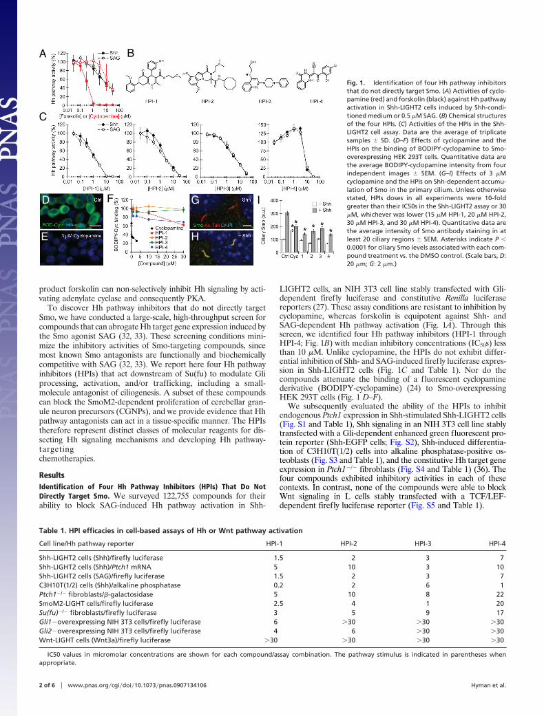
LIGHT2 cells, an NIH 3T3 cell line stably transfected with Gli-dependent firefly luciferase and constitutive Renilla luciferasereporters (27). These assay conditions are resistant to inhibition bycyclopamine, whereas forskolin is equipotent against Shh- andSAG-dependent Hh pathway activation (Fig. 1A). Through thisscreen, we identified four Hh pathway inhibitors (HPI-1 throughHPI-4; Fig. 1B) with median inhibitory concentrations (IC50s) lessthan 10 �M. Unlike cyclopamine, the HPIs do not exhibit differ-ential inhibition of Shh- and SAG-induced firefly luciferase expres-sion in Shh-LIGHT2 cells (Fig. 1C and Table 1). Nor do thecompounds attenuate the binding of a fluorescent cyclopaminederivative (BODIPY-cyclopamine) (24) to Smo-overexpressingHEK 293T cells (Fig. 1 D–F).
We subsequently evaluated the ability of the HPIs to inhibitendogenous Ptch1 expression in Shh-stimulated Shh-LIGHT2 cells(Fig. S1 and Table 1), Shh signaling in an NIH 3T3 cell line stablytransfected with a Gli-dependent enhanced green fluorescent pro-tein reporter (Shh-EGFP cells; Fig. S2), Shh-induced differentia-tion of C3H10T(1/2) cells into alkaline phosphatase-positive os-teoblasts (Fig. S3 and Table 1), and the constitutive Hh target geneexpression in Ptch1�/� fibroblasts (Fig. S4 and Table 1) (36). Thefour compounds exhibited inhibitory activities in each of thesecontexts. In contrast, none of the compounds were able to blockWnt signaling in L cells stably transfected with a TCF/LEF-dependent firefly luciferase reporter (Fig. S5 and Table 1).
A
D
E
G
H
I
B
F
C
Fig. 1. Identification of four Hh pathway inhibitorsthat do not directly target Smo. (A) Activities of cyclo-pamine (red) and forskolin (black) against Hh pathwayactivation in Shh-LIGHT2 cells induced by Shh-condi-tioned medium or 0.5 �M SAG. (B) Chemical structuresof the four HPIs. (C) Activities of the HPIs in the Shh-LIGHT2 cell assay. Data are the average of triplicatesamples � SD. (D–F) Effects of cyclopamine and theHPIs on the binding of BODIPY-cyclopamine to Smo-overexpressing HEK 293T cells. Quantitative data arethe average BODIPY-cyclopamine intensity from fourindependent images � SEM. (G–I) Effects of 3 �Mcyclopamine and the HPIs on Shh-dependent accumu-lation of Smo in the primary cilium. Unless otherwisestated, HPIs doses in all experiments were 10-foldgreater than their IC50s in the Shh-LIGHT2 assay or 30�M, whichever was lower (15 �M HPI-1, 20 �M HPI-2,30 �M HPI-3, and 30 �M HPI-4). Quantitative data arethe average intensity of Smo antibody staining in atleast 20 ciliary regions � SEM. Asterisks indicate P �0.0001 for ciliary Smo levels associated with each com-pound treatment vs. the DMSO control. (Scale bars, D:20 �m; G: 2 �m.)
Table 1. HPI efficacies in cell-based assays of Hh or Wnt pathway activation
Cell line/Hh pathway reporter HPI-1 HPI-2 HPI-3 HPI-4
Shh-LIGHT2 cells (Shh)/firefly luciferase 1.5 2 3 7Shh-LIGHT2 cells (Shh)/Ptch1 mRNA 5 10 3 10Shh-LIGHT2 cells (SAG)/firefly luciferase 1.5 2 3 7C3H10T(1/2) cells (Shh)/alkaline phosphatase 0.2 2 6 1Ptch1�/� fibroblasts/�-galactosidase 5 10 8 22SmoM2-LIGHT cells/firefly luciferase 2.5 4 1 20Su(fu)�/� fibroblasts/firefly luciferase 3 5 9 17Gli1�overexpressing NIH 3T3 cells/firefly luciferase 6 �30 �30 �30Gli2�overexpressing NIH 3T3 cells/firefly luciferase 4 6 �30 �30Wnt-LIGHT cells (Wnt3a)/firefly luciferase �30 �30 �30 �30
IC50 values in micromolar concentrations are shown for each compound/assay combination. The pathway stimulus is indicated in parentheses whenappropriate.
2 of 6 � www.pnas.org�cgi�doi�10.1073�pnas.0907134106 Hyman et al.

The HPIs Partially Inhibit Smo Multimerization and Trafficking. Theinability of the HPIs to block BODIPY-cyclopamine/Smo bindingand their non-competitive interactions with respect to SAG suggestthat the four inhibitors do not directly target Smo (24, 32). It ispossible, however, that they indirectly disrupt other aspects of Smofunction. For example, Smo activation is believed to involve phos-phorylation-dependent structural changes that alter its conforma-tion and aggregation state (24, 32, 37), and ciliary accumulation ofSmo is observed in cells treated with either Shh or SAG (13, 14).We therefore evaluated whether the compounds can perturb Shh-induced Smo multimerization, which can be monitored as anincrease in fluorescence resonance energy transfer (FRET) be-tween Smo proteins C-terminally tagged with cyan or yellowfluorescent proteins (Smo-CFP and Smo-YFP) (37). HPI-1 andHPI-2 attenuated the Shh-induced fold-change in Smo-CFP/Smo-YFP FRET in NIH 3T3 cells, while HPI-2, HPI-3, and HPI-4decreased basal FRET levels (Fig. S6). We next analyzed theShh-dependent trafficking of endogenous Smo to the primarycilium, which can be perturbed by Smo antagonists (13, 38). Whilenone of the HPIs completely blocked Smo trafficking to the cilium,all four compounds decreased the extent of ciliary Smo accumu-lation in response to Shh (Fig. 1 G–I).
The HPIs Are Epistatic to Su(fu). To determine whether these partialeffects on Smo multimerization and trafficking account for theinhibitory activities of the HPIs, we investigated their epistaticinteractions with Hh signaling proteins downstream of Smo. Forexample, Su(fu)�/� fibroblasts exhibit constitutive, Smo-independent Hh target gene expression that is insensitive to cyclo-pamine and partially inhibited by forskolin, as can be detected bytransiently transfecting these cells with a Gli-dependent fireflyluciferase reporter (39). Like GANT-61, all four HPIs were able torepress Hh pathway activity in Su(fu)�/� fibroblasts to near-basallevels (Fig. 2A, Table 1, and Fig. S7).
We then mapped the activities of the HPIs relative to Gli1 andGli2 by transiently overexpressing these transcription factors in NIH3T3 cells (Fig. 2B and Table 1). As measured by co-transfectedGli-dependent firefly luciferase and constitutive Renilla luciferasereporters, HPI-1 and HPI-2 were able to inhibit Gli-induced Hhpathway activation in a dose-dependent manner, with HPI-2 pref-erentially inhibiting Gli2 (Fig. 2B and Fig. S8). HPI-3 and HPI-4had no significant activity under these conditions, suggesting thesecompounds counteract the activities of endogenous Gli1 and Gli2through mechanisms that are circumvented by overexpressed Gliproteins. We also observed that GANT-61 was unable to antago-nize exogenous Gli1 or Gli2 in NIH 3T3 cells (Fig. S7), contrastingprevious findings in HEK 293 cells (35).
The HPIs Do Not Inhibit Gli Activity by Modulating PKA, PI3K/Akt, orMAPK Signaling. Since the HPIs act downstream of Su(fu) and likelyat the level of the Gli transcription factors, we investigated whetherthey target non-Hh pathway-specific signaling mechanisms previ-
ously shown to modulate Gli function. We first evaluated the abilityof the compounds to activate PKA in NIH 3T3 cells, as gauged bythe phosphorylation state of cAMP response element binding(CREB) protein (Fig. 3A). Forskolin strongly induced CREBphosphorylation that could be abrogated by the PKA inhibitor H89,but none of the HPIs produced comparable levels of H89-sensitivephospho-CREB. We also assessed the effects of the HPIs onplatelet-derived growth factor (PDGF)-induced PI3K and MAPKsignaling in NIH 3T3 cells, which results in the phosphorylation ofAkt and p44/p22 MAPK, respectively (Fig. 3B). The PI3K inhibitorLY294002 prevented Akt phosphorylation under these conditions,and the Mek1/Mek2 inhibitor U0126 blocked p44/p22 MAPKphosphorylation. In contrast, none of the HPIs inhibited thePDGF-induced phosphorylation of either downstream substrate.
The HPIs Differentially Perturb Gli Processing and Stability. To furthercharacterize the mechanisms by which HPIs inhibit Hh target geneexpression, we analyzed their effects on Gli processing and stability.We infected Shh-EGFP cells with a retroviral vector for FLAG-Gli2 expression and selected clones with low levels of the exogenousGli2 protein (Shh-EGFPFLAG�Gli2 cells). FLAG-Gli2 protein inthese cells exists in both full-length and N-terminal repressor forms,with the full-length/repressor ratio reflecting the level of Hhpathway activation. Shh stimulation of these cells significantlyincreases this ratio, and cyclopamine can suppress the effects of Shh(Fig. 4A and Fig. S9). HPI-1 and HPI-4 also prevented an increasein the FLAG-Gli2 full-length/repressor ratio upon Shh stimulation,but HPI-2 and HPI-3 had no significant effect (Fig. 4A and Fig. S9).
We similarly infected Shh-LIGHT2 cells with a retroviral vectorfor FLAG-Gli1 expression and selected clones with low levels of the
Fig. 2. Epistatic mapping of HPI activity relative toSu(fu), Gli1, and Gli2. (A) Effects of cyclopamine andthe HPIs on Hh pathway activation in Su(fu)�/� fibro-blasts. (B) Effects of the Hh pathway inhibitors on Hhpathway activation induced by Gli1 or Gli2 overexpres-sion, as measured using a co-transfected Gli-dependentfirefly luciferase reporter. Data are the average oftriplicate samples � SD.
Fig. 3. HPI activity is not due to modulation of PKA, PI3K/Akt, or MAPKsignaling. (A) Effects of 50 �M forskolin and the HPIs on PKA activity in NIH 3T3cells, as determined by the H89-sensitive phosphorylation state of CREB. (B)Effects of the HPIs on PDGF-induced activation of the PI3K/Akt and MAPKsignaling pathways in NIH 3T3 cells, as determined by the phosphorylationstates of Akt and p44/p42 MAPK. Fifty micromolar LY294002 and 10 �M U0126were used as positive controls.
Hyman et al. PNAS Early Edition � 3 of 6
PHA
RMA
COLO
GY
CHEM
ISTR
Y

exogenous Gli1 protein (Shh-LIGHT2FLAG-Gli1 cells). HPI-4 de-creased FLAG-Gli1 stability in these cells, revealing another mech-anism by which this small molecule can inhibit Hh target geneexpression, while neither HPI-2 or HPI-3 had any significant effecton FLAG-Gli1 levels (Fig. 4B and Fig. S9). HPI-1 actually increasedFLAG-Gli1 levels, indicating that this compound may inhibitFLAG-Gli1 activity through a mechanism that also decreases therate of Gli1 degradation.
The HPIs Differentially Perturb Gli Trafficking to the Primary Ciliumand Ciliogenesis. We next analyzed the effects of the HPIs onGli trafficking, using the Shh-EGFPFLAG-Gli2 and Shh-LIGHT2FLAG-Gli1 cells as model systems. In both cell lines, theFLAG-tagged Gli proteins are distributed throughout the cyto-plasm and nucleus in a punctate manner and localized to tip ofthe primary cilium (Fig. 4 C and H). HPI-2, HPI-3, and HPI-4increased ciliary levels of FLAG-Gli2 in a manner dispropor-tionate to their effects on total FLAG-Gli2 levels, while HPI-1had no obvious effect (Fig. 4 A, D–G, and M). In addition,Shh-EGFPFLAG-Gli2 cells cultured with HPI-4 had truncatedprimary cilia, and this cellular organelle was absent in a signif-icant fraction of HPI-4-treated cells (Fig. 4G and Fig. S10).HPI-4 also perturbed primary cilia formation in the Shh-LIGHT2FLAG-Gli1 cells and promoted accumulation of FLAG-Gli1 at the distal tip of this organelle, but ciliary FLAG-Gli1levels were not significantly changed by any of the other HPIs(Fig. 4 I–L and N and Fig. S10). These structural defects appearto be cilia-specific, as the non-ciliary microtubule cytoskeletonwas not grossly perturbed by any of the HPIs (Fig. S11).
The HPIs Have Divergent Activity Profiles Against Gli2 Mutants. Torefine our understanding of how small molecules can regulate Gli
activity, we studied the actions of HPI-1 and HPI-2 on Gli2 mutantslacking either PKA or GSK phosphorylation sites (Gli2 �PKA andGli2 �GSK) or the N-terminal repressor domain (Gli2 �N) (11, 31).The other HPIs were excluded from these studies since they areineffective against overexpressed Gli proteins. Hh pathway activa-tion in NIH 3T3 cells induced by the expression of wild-type Gli2or the Gli2 �GSK mutant was inhibited to a similar extent by HPI-1and HPI-2, whereas the Gli2 �PKA mutant was partially resistantto both compounds (Fig. 4O). In accordance with previous studies(31), Gli2 �PKA-induced Hh pathway activation in NIH 3T3 cellswas also resistant to forskolin and LY294002. HPI-1 and HPI-2,however, differentially antagonized Hh pathway activation inducedby the Gli2 �N mutant (Fig. 4O). HPI-1 activity was not dependenton this N-terminal repressor domain, consistent with its ability toinhibit both Gli1 and Gli2, while the Gli2 �N mutant was partiallyresistant to HPI-2, as well as LY294002.
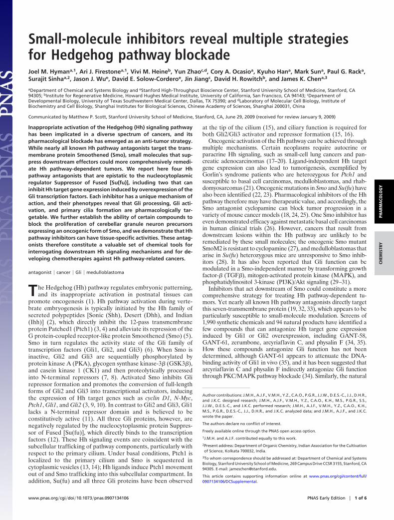
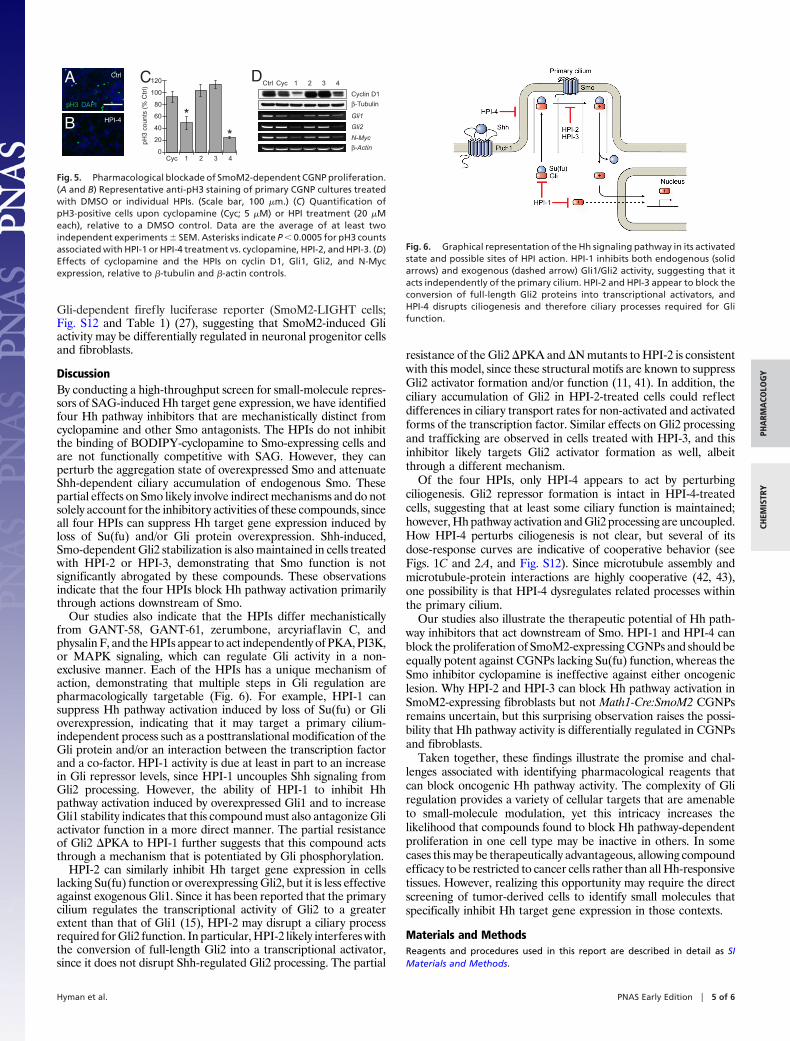
A Subset of the HPIs Can Block SmoM2-Dependent Proliferation ofCerebellar Granule Neuron Precursors. We concluded our studies byinvestigating the ability of the HPIs to block oncogenic Hh targetgene expression. We isolated CGNPs from Math1-Cre:SmoM2 miceat postnatal day seven (P7), which grow in a Hh ligand-independentand cyclopamine-resistant manner as primary cultures and give riseto medulloblastomas in vivo (40). HPI-1 and HPI-4 significantlyinhibited the proliferation of these neuronal progenitors, as mea-sured by histone H3 phosphorylation (pH3) levels (Fig. 5 A–C), andboth compounds reduced cellular levels of cyclin D1 protein andGli1, Gli2, and N-Myc transcripts in the CGNPs (Fig. 5D). Incontrast, HPI-2 and HPI-3 did not block CGNP proliferation orinhibit Hh target gene expression. These observations contrast theability of all four HPIs to block Hh pathway activation in NIH 3T3cells stably transfected with a SmoM2 expression vector and a
A B C D E F G
H I J K L
M N O
Fig. 4. The HPIs differentially perturb Gli processing, stability, localization, and function. (A) Effects of cyclopamine and the HPIs on full-length and repressorforms of FLAG-Gli2 in a clonal NIH 3T3 cell line, including representative immunoblotting results and full-length/repressor ratios (bar graph). Total FLAG-Gli2levels observed for each compound treatment are also indicated (red circles), normalized with respect to the DMSO control. Data are the average of fourindependent experiments � SEM. Asterisks and double asterisks respectively indicate P � 0.03 for full-length/repressor ratios and P � 0.05 for total FLAG-Gli2levels associated with compound treatment vs. the DMSO control. (B) Effects of the Hh pathway inhibitors on FLAG-Gli1 levels in a clonal NIH 3T3 cell line.Representative immunoblotting results are shown, and quantitative data are the average FLAG-Gli1 levels from three independent experiments � SEM,normalized with respect to DMSO control. Asterisks indicate P � 0.02 for total FLAG-Gli1 levels associated with compound treatment vs. the DMSO control. (C–G)Subcellular localization of FLAG-Gli2 (green) with respect to the primary cilium (red) and nucleus (blue) in cells treated with DMSO or individual HPIs. (H–L)Subcellular localization of FLAG-Gli1 (green) with respect to the primary cilium (red) and nucleus (blue) in cells under analogous conditions. (M and N)Quantification of ciliary FLAG-Gli2 and FLAG-Gli1 levels associated with HPI treatment. Data are the average intensity of anti-FLAG antibody staining in at least40 ciliary regions � SEM, and both absolute ciliary intensities and those normalized with respect to total FLAG-Gli2 or FLAG-Gli1 levels are shown. Asterisksindicate P � 0.003 for normalized ciliary FLAG-Gli levels associated with compound treatment vs. the DMSO control. (O) Differential inhibition of wildtype (black),�N (red), �GSK (green) and �PKA (blue) forms of Gli2 by 50 �M forskolin, 50 �M LY294002, HPI-1, or HPI-2. Wild-type and mutant forms of Gli2 are depictedschematically with the DNA-binding zinc finger region shown in gray and mutated phosphorylation sites shown in red. Immunoblotting data for each Gli2construct are also shown to confirm protein expression levels. Hh pathway activities are expressed relative to those observed for each Gli2 form in the absenceof compound and are the average of nine replicates � SEM. Asterisks indicate P � 0.002 and greater than a 1.5-fold change for Hh pathway activities associatedwith mutant vs. wildtype Gli2 expression in the presence of compound. (Scale bars, 2 �m.)
4 of 6 � www.pnas.org�cgi�doi�10.1073�pnas.0907134106 Hyman et al.
Gli-dependent firefly luciferase reporter (SmoM2-LIGHT cells;Fig. S12 and Table 1) (27), suggesting that SmoM2-induced Gliactivity may be differentially regulated in neuronal progenitor cellsand fibroblasts.
DiscussionBy conducting a high-throughput screen for small-molecule repres-sors of SAG-induced Hh target gene expression, we have identifiedfour Hh pathway inhibitors that are mechanistically distinct fromcyclopamine and other Smo antagonists. The HPIs do not inhibitthe binding of BODIPY-cyclopamine to Smo-expressing cells andare not functionally competitive with SAG. However, they canperturb the aggregation state of overexpressed Smo and attenuateShh-dependent ciliary accumulation of endogenous Smo. Thesepartial effects on Smo likely involve indirect mechanisms and do notsolely account for the inhibitory activities of these compounds, sinceall four HPIs can suppress Hh target gene expression induced byloss of Su(fu) and/or Gli protein overexpression. Shh-induced,Smo-dependent Gli2 stabilization is also maintained in cells treatedwith HPI-2 or HPI-3, demonstrating that Smo function is notsignificantly abrogated by these compounds. These observationsindicate that the four HPIs block Hh pathway activation primarilythrough actions downstream of Smo.
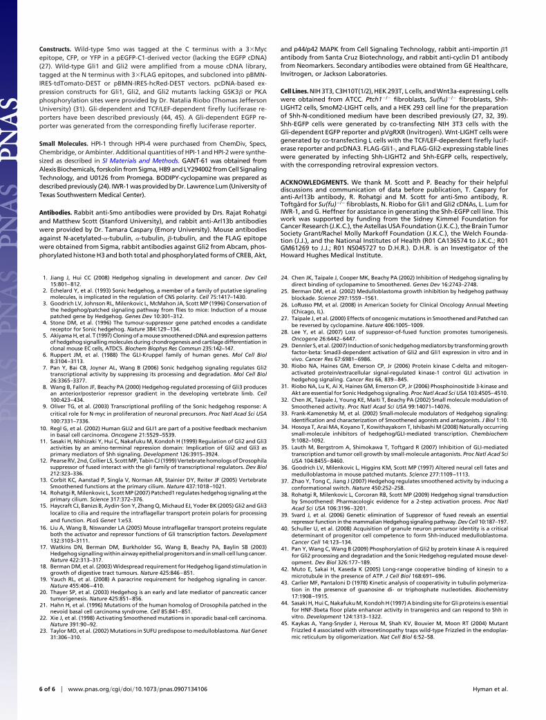
Our studies also indicate that the HPIs differ mechanisticallyfrom GANT-58, GANT-61, zerumbone, arcyriaflavin C, andphysalin F, and the HPIs appear to act independently of PKA, PI3K,or MAPK signaling, which can regulate Gli activity in a non-exclusive manner. Each of the HPIs has a unique mechanism ofaction, demonstrating that multiple steps in Gli regulation arepharmacologically targetable (Fig. 6). For example, HPI-1 cansuppress Hh pathway activation induced by loss of Su(fu) or Glioverexpression, indicating that it may target a primary cilium-independent process such as a posttranslational modification of theGli protein and/or an interaction between the transcription factorand a co-factor. HPI-1 activity is due at least in part to an increasein Gli repressor levels, since HPI-1 uncouples Shh signaling fromGli2 processing. However, the ability of HPI-1 to inhibit Hhpathway activation induced by overexpressed Gli1 and to increaseGli1 stability indicates that this compound must also antagonize Gliactivator function in a more direct manner. The partial resistanceof Gli2 �PKA to HPI-1 further suggests that this compound actsthrough a mechanism that is potentiated by Gli phosphorylation.
HPI-2 can similarly inhibit Hh target gene expression in cellslacking Su(fu) function or overexpressing Gli2, but it is less effectiveagainst exogenous Gli1. Since it has been reported that the primarycilium regulates the transcriptional activity of Gli2 to a greaterextent than that of Gli1 (15), HPI-2 may disrupt a ciliary processrequired for Gli2 function. In particular, HPI-2 likely interferes withthe conversion of full-length Gli2 into a transcriptional activator,since it does not disrupt Shh-regulated Gli2 processing. The partial
resistance of the Gli2 �PKA and �N mutants to HPI-2 is consistentwith this model, since these structural motifs are known to suppressGli2 activator formation and/or function (11, 41). In addition, theciliary accumulation of Gli2 in HPI-2-treated cells could reflectdifferences in ciliary transport rates for non-activated and activatedforms of the transcription factor. Similar effects on Gli2 processingand trafficking are observed in cells treated with HPI-3, and thisinhibitor likely targets Gli2 activator formation as well, albeitthrough a different mechanism.
Of the four HPIs, only HPI-4 appears to act by perturbingciliogenesis. Gli2 repressor formation is intact in HPI-4-treatedcells, suggesting that at least some ciliary function is maintained;however, Hh pathway activation and Gli2 processing are uncoupled.How HPI-4 perturbs ciliogenesis is not clear, but several of itsdose-response curves are indicative of cooperative behavior (seeFigs. 1C and 2A, and Fig. S12). Since microtubule assembly andmicrotubule-protein interactions are highly cooperative (42, 43),one possibility is that HPI-4 dysregulates related processes withinthe primary cilium.
Our studies also illustrate the therapeutic potential of Hh path-way inhibitors that act downstream of Smo. HPI-1 and HPI-4 canblock the proliferation of SmoM2-expressing CGNPs and should beequally potent against CGNPs lacking Su(fu) function, whereas theSmo inhibitor cyclopamine is ineffective against either oncogeniclesion. Why HPI-2 and HPI-3 can block Hh pathway activation inSmoM2-expressing fibroblasts but not Math1-Cre:SmoM2 CGNPsremains uncertain, but this surprising observation raises the possi-bility that Hh pathway activity is differentially regulated in CGNPsand fibroblasts.
Taken together, these findings illustrate the promise and chal-lenges associated with identifying pharmacological reagents thatcan block oncogenic Hh pathway activity. The complexity of Gliregulation provides a variety of cellular targets that are amenableto small-molecule modulation, yet this intricacy increases thelikelihood that compounds found to block Hh pathway-dependentproliferation in one cell type may be inactive in others. In somecases this may be therapeutically advantageous, allowing compoundefficacy to be restricted to cancer cells rather than all Hh-responsivetissues. However, realizing this opportunity may require the directscreening of tumor-derived cells to identify small molecules thatspecifically inhibit Hh target gene expression in those contexts.
Materials and MethodsReagents and procedures used in this report are described in detail as SIMaterials and Methods.
D
pH3
coun
ts (
% C
trl)
Cyc 1 2 3 4
120
100
80
60
40
20
0
Cyclin D1
β-Tubulin
β-Actin
Gli1
Gli2
N-Myc
CycCtrl 1 2 3 4CA
B
Ctrl
HPI-4
pH3 DAPI
*
*
Fig. 5. Pharmacological blockade of SmoM2-dependent CGNP proliferation.(A and B) Representative anti-pH3 staining of primary CGNP cultures treatedwith DMSO or individual HPIs. (Scale bar, 100 �m.) (C) Quantification ofpH3-positive cells upon cyclopamine (Cyc; 5 �M) or HPI treatment (20 �Meach), relative to a DMSO control. Data are the average of at least twoindependent experiments � SEM. Asterisks indicate P � 0.0005 for pH3 countsassociated with HPI-1 or HPI-4 treatment vs. cyclopamine, HPI-2, and HPI-3. (D)Effects of cyclopamine and the HPIs on cyclin D1, Gli1, Gli2, and N-Mycexpression, relative to �-tubulin and �-actin controls.
Fig. 6. Graphical representation of the Hh signaling pathway in its activatedstate and possible sites of HPI action. HPI-1 inhibits both endogenous (solidarrows) and exogenous (dashed arrow) Gli1/Gli2 activity, suggesting that itacts independently of the primary cilium. HPI-2 and HPI-3 appear to block theconversion of full-length Gli2 proteins into transcriptional activators, andHPI-4 disrupts ciliogenesis and therefore ciliary processes required for Glifunction.
Hyman et al. PNAS Early Edition � 5 of 6
PHA
RMA
COLO
GY
CHEM
ISTR
Y
Constructs. Wild-type Smo was tagged at the C terminus with a 3�Mycepitope, CFP, or YFP in a pEGFP-C1-derived vector (lacking the EGFP cDNA)(27). Wild-type Gli1 and Gli2 were amplified from a mouse cDNA library,tagged at the N terminus with 3�FLAG epitopes, and subcloned into pBMN-IRES-tdTomato-DEST or pBMN-IRES-hcRed-DEST vectors. pcDNA-based ex-pression constructs for Gli1, Gli2, and Gli2 mutants lacking GSK3� or PKAphosphorylation sites were provided by Dr. Natalia Riobo (Thomas JeffersonUniversity) (31). Gli-dependent and TCF/LEF-dependent firefly luciferase re-porters have been described previously (44, 45). A Gli-dependent EGFP re-porter was generated from the corresponding firefly luciferase reporter.
Small Molecules. HPI-1 through HPI-4 were purchased from ChemDiv, Specs,Chembridge, or Ambinter. Additional quantities of HPI-1 and HPI-2 were synthe-sized as described in SI Materials and Methods. GANT-61 was obtained fromAlexis Biochemicals, forskolin from Sigma, H89 and LY294002 from Cell SignalingTechnology, and U0126 from Promega. BODIPY-cyclopamine was prepared asdescribed previously (24). IWR-1 was provided by Dr. Lawrence Lum (University ofTexas Southwestern Medical Center).
Antibodies. Rabbit anti-Smo antibodies were provided by Drs. Rajat Rohatgiand Matthew Scott (Stanford University), and rabbit anti-Arl13b antibodieswere provided by Dr. Tamara Caspary (Emory University). Mouse antibodiesagainst N-acetylated-�-tubulin, �-tubulin, �-tubulin, and the FLAG epitopewere obtained from Sigma, rabbit antibodies against Gli2 from Abcam, phos-phorylated histone H3 and both total and phosphorylated forms of CREB, Akt,
and p44/p42 MAPK from Cell Signaling Technology, rabbit anti-importin �1antibody from Santa Cruz Biotechnology, and rabbit anti-cyclin D1 antibodyfrom Neomarkers. Secondary antibodies were obtained from GE Healthcare,Invitrogen, or Jackson Laboratories.
Cell Lines. NIH 3T3, C3H10T(1/2), HEK 293T, L cells, and Wnt3a-expressing L cellswere obtained from ATCC. Ptch1�/� fibroblasts, Su(fu)�/� fibroblasts, Shh-LIGHT2 cells, SmoM2-LIGHT cells, and a HEK 293 cell line for the preparationof Shh-N-conditioned medium have been described previously (27, 32, 39).Shh-EGFP cells were generated by co-transfecting NIH 3T3 cells with theGli-dependent EGFP reporter and pVgRXR (Invitrogen). Wnt-LIGHT cells weregenerated by co-transfecting L cells with the TCF/LEF-dependent firefly lucif-erase reporter and pcDNA3. FLAG-Gli1-, and FLAG-Gli2-expressing stable lineswere generated by infecting Shh-LIGHT2 and Shh-EGFP cells, respectively,with the corresponding retroviral expression vectors.
ACKNOWLEDGMENTS. We thank M. Scott and P. Beachy for their helpfuldiscussions and communication of data before publication, T. Caspary foranti-Arl13b antibody, R. Rohatgi and M. Scott for anti-Smo antibody, R.Toftgård for Su(fu)�/� fibroblasts, N. Riobo for Gli1 and Gli2 cDNAs, L. Lum forIWR-1, and G. Heffner for assistance in generating the Shh-EGFP cell line. Thiswork was supported by funding from the Sidney Kimmel Foundation forCancer Research (J.K.C.), the Astellas USA Foundation (J.K.C.), the Brain TumorSociety Grant/Rachel Molly Markoff Foundation (J.K.C.), the Welch Founda-tion (J.J.), and the National Institutes of Health (R01 CA136574 to J.K.C.; R01GM61269 to J.J.; R01 NS045727 to D.H.R.). D.H.R. is an Investigator of theHoward Hughes Medical Institute.
1. Jiang J, Hui CC (2008) Hedgehog signaling in development and cancer. Dev Cell15:801–812.
2. Echelard Y, et al. (1993) Sonic hedgehog, a member of a family of putative signalingmolecules, is implicated in the regulation of CNS polarity. Cell 75:1417–1430.
3. Goodrich LV, Johnson RL, Milenkovic L, McMahon JA, Scott MP (1996) Conservation ofthe hedgehog/patched signaling pathway from flies to mice: Induction of a mousepatched gene by Hedgehog. Genes Dev 10:301–312.
4. Stone DM, et al. (1996) The tumour-suppressor gene patched encodes a candidatereceptor for Sonic hedgehog. Nature 384:129–134.
5. Akiyama H, et al. T (1997) Cloning of a mouse smoothened cDNA and expression patternsof hedgehog signalling molecules during chondrogenesis and cartilage differentiation inclonal mouse EC cells, ATDC5. Biochem Biophys Res Commun 235:142–147.
6. Ruppert JM, et al. (1988) The GLI-Kruppel family of human genes. Mol Cell Biol8:3104–3113.
7. Pan Y, Bai CB, Joyner AL, Wang B (2006) Sonic hedgehog signaling regulates Gli2transcriptional activity by suppressing its processing and degradation. Mol Cell Biol26:3365–3377.
8. Wang B, Fallon JF, Beachy PA (2000) Hedgehog-regulated processing of Gli3 producesan anterior/posterior repressor gradient in the developing vertebrate limb. Cell100:423–434.
9. Oliver TG, et al. (2003) Transcriptional profiling of the Sonic hedgehog response: Acritical role for N-myc in proliferation of neuronal precursors. Proc Natl Acad Sci USA100:7331–7336.
10. Regl G, et al. (2002) Human GLI2 and GLI1 are part of a positive feedback mechanismin basal cell carcinoma. Oncogene 21:5529–5539.
11. Sasaki H, Nishizaki Y, Hui C, Nakafuku M, Kondoh H (1999) Regulation of Gli2 and Gli3activities by an amino-terminal repression domain: Implication of Gli2 and Gli3 asprimary mediators of Shh signaling. Development 126:3915–3924.
12. Pearse RV, 2nd, Collier LS, Scott MP, Tabin CJ (1999) Vertebrate homologs of Drosophilasuppressor of fused interact with the gli family of transcriptional regulators. Dev Biol212:323–336.
13. Corbit KC, Aanstad P, Singla V, Norman AR, Stainier DY, Reiter JF (2005) VertebrateSmoothened functions at the primary cilium. Nature 437:1018–1021.
14. Rohatgi R, Milenkovic L, Scott MP (2007) Patched1 regulates hedgehog signaling at theprimary cilium. Science 317:372–376.
15. Haycraft CJ, Banizs B, Aydin-Son Y, Zhang Q, Michaud EJ, Yoder BK (2005) Gli2 and Gli3localize to cilia and require the intraflagellar transport protein polaris for processingand function. PLoS Genet 1:e53.
16. Liu A, Wang B, Niswander LA (2005) Mouse intraflagellar transport proteins regulateboth the activator and repressor functions of Gli transcription factors. Development132:3103–3111.
17. Watkins DN, Berman DM, Burkholder SG, Wang B, Beachy PA, Baylin SB (2003)Hedgehog signalling within airway epithelial progenitors and in small-cell lung cancer.Nature 422:313–317.
18. Berman DM, et al. (2003) Widespread requirement for Hedgehog ligand stimulation ingrowth of digestive tract tumours. Nature 425:846–851.
19. Yauch RL, et al. (2008) A paracrine requirement for hedgehog signaling in cancer.Nature 455:406–410.
20. Thayer SP, et al. (2003) Hedgehog is an early and late mediator of pancreatic cancertumorigenesis. Nature 425:851–856.
21. Hahn H, et al. (1996) Mutations of the human homolog of Drosophila patched in thenevoid basal cell carcinoma syndrome. Cell 85:841–851.
22. Xie J, et al. (1998) Activating Smoothened mutations in sporadic basal-cell carcinoma.Nature 391:90–92.
23. Taylor MD, et al. (2002) Mutations in SUFU predispose to medulloblastoma. Nat Genet31:306–310.
24. Chen JK, Taipale J, Cooper MK, Beachy PA (2002) Inhibition of Hedgehog signaling bydirect binding of cyclopamine to Smoothened. Genes Dev 16:2743–2748.
25. Berman DM, et al. (2002) Medulloblastoma growth inhibition by hedgehog pathwayblockade. Science 297:1559–1561.
26. LoRusso PM, et al. (2008) in American Society for Clinical Oncology Annual Meeting(Chicago, IL).
27. Taipale J, et al. (2000) Effects of oncogenic mutations in Smoothened and Patched canbe reversed by cyclopamine. Nature 406:1005–1009.
28. Lee Y, et al. (2007) Loss of suppressor-of-fused function promotes tumorigenesis.Oncogene 26:6442–6447.
29. Dennler S, et al. (2007) Induction of sonic hedgehog mediators by transforming growthfactor-beta: Smad3-dependent activation of Gli2 and Gli1 expression in vitro and invivo. Cancer Res 67:6981–6986.
30. Riobo NA, Haines GM, Emerson CP, Jr (2006) Protein kinase C-delta and mitogen-activated protein/extracellular signal-regulated kinase-1 control GLI activation inhedgehog signaling. Cancer Res 66, 839–845.
31. Riobo NA, Lu K, Ai X, Haines GM, Emerson CP, Jr (2006) Phosphoinositide 3-kinase andAkt are essential for Sonic Hedgehog signaling. Proc Natl Acad Sci USA 103:4505–4510.
32. Chen JK, Taipale J, Young KE, Maiti T, Beachy PA (2002) Small molecule modulation ofSmoothened activity. Proc Natl Acad Sci USA 99:14071–14076.
33. Frank-Kamenetsky M, et al. (2002) Small-molecule modulators of Hedgehog signaling:Identification and characterization of Smoothened agonists and antagonists. J Biol 1:10.
34. Hosoya T, Arai MA, Koyano T, Kowithayakorn T, Ishibashi M (2008) Naturally occurringsmall-molecule inhibitors of hedgehog/GLI-mediated transcription. Chembiochem9:1082–1092.
35. Lauth M, Bergstrom A, Shimokawa T, Toftgard R (2007) Inhibition of GLI-mediatedtranscription and tumor cell growth by small-molecule antagonists. Proc Natl Acad SciUSA 104:8455–8460.
36. Goodrich LV, Milenkovic L, Higgins KM, Scott MP (1997) Altered neural cell fates andmedulloblastoma in mouse patched mutants. Science 277:1109–1113.
37. Zhao Y, Tong C, Jiang J (2007) Hedgehog regulates smoothened activity by inducing aconformational switch. Nature 450:252–258.
38. Rohatgi R, Milenkovic L, Corcoran RB, Scott MP (2009) Hedgehog signal transductionby Smoothened: Pharmacologic evidence for a 2-step activation process. Proc NatlAcad Sci USA 106:3196–3201.
39. Svard J, et al. (2006) Genetic elimination of Suppressor of fused reveals an essentialrepressor function in the mammalian Hedgehog signaling pathway. Dev Cell 10:187–197.
40. Schuller U, et al. (2008) Acquisition of granule neuron precursor identity is a criticaldeterminant of progenitor cell competence to form Shh-induced medulloblastoma.Cancer Cell 14:123–134.
41. Pan Y, Wang C, Wang B (2009) Phosphorylation of Gli2 by protein kinase A is requiredfor Gli2 processing and degradation and the Sonic Hedgehog-regulated mouse devel-opment. Dev Biol 326:177–189.
42. Muto E, Sakai H, Kaseda K (2005) Long-range cooperative binding of kinesin to amicrotubule in the presence of ATP. J Cell Biol 168:691–696.
43. Carlier MF, Pantaloni D (1978) Kinetic analysis of cooperativity in tubulin polymeriza-tion in the presence of guanosine di- or triphosphate nucleotides. Biochemistry17:1908–1915.
44. Sasaki H, Hui C, Nakafuku M, Kondoh H (1997) A binding site for Gli proteins is essentialfor HNF-3beta floor plate enhancer activity in transgenics and can respond to Shh invitro. Development 124:1313–1322.
45. Kaykas A, Yang-Snyder J, Heroux M, Shah KV, Bouvier M, Moon RT (2004) MutantFrizzled 4 associated with vitreoretinopathy traps wild-type Frizzled in the endoplas-mic reticulum by oligomerization. Nat Cell Biol 6:52–58.
6 of 6 � www.pnas.org�cgi�doi�10.1073�pnas.0907134106 Hyman et al.