Embed Size (px)
Citation preview
SLIT LAMP MICROSCOPYSKILL SESSION
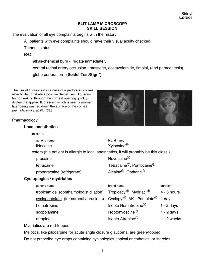
The evaluation of all eye complaints begins with the history.All patients with eye complaints should have their visual acuity checked.Tetanus statusR/O alkali/chemical burn - irrigate immediately central retinal artery occlusion - massage, acetazolamide, timolol, (and paracentesis) globe perforation (Seidel Test/Sign*)
The use of fluorescein in a case of a perforated corneal ulcer to demonstrate a positive Seidel Test. Aqueous humor leaking through the corneal opening quickly dilutes the applied fluorescein which is seen a moment later being washed down the surface of the cornea.(from Martonyi et al, Fig 103.)
PharmacologyLocal anesthetics amides
generic name brand name
lidocaine Xylocaine® esters (If a patient is allergic to local anesthetics, it will probably be this class.) procaine Novocaine®
tetracaine Tetracaine®, Pontocaine®
proparacaine (refrigerate) Alcaine®, Opthane®
Cycloplegics / mydriaticsgeneric name brand name duration
tropicamide (ophthalmologist dilation) Tropicacyl®, Mydriacil® 4 - 6 hourscyclopentolate (for corneal abrasions) Cyclogyl®, AK - Pentolate® 1 dayhomatropine Isopto Homatropine® 1 - 2 daysscopolamine Isoptohyoscine® 1 - 2 daysatropine Isopto Atropine® 1 - 2 weeks
Mydriatics are red-topped.Meiotics, like pilocarpine for acute angle closure glaucoma, are green-topped.Do not prescribe eye drops containing cycloplegics, topical anesthetics, or steroids.
Birinyi7/29/2004
1
Slit lamp technical points to remember.Align (roughly) the eyes with the black line on the microscope.The patient’s head must abut the headrest. This is the most likely source of error.
Fluorescein stainingUse the cobalt blue lamp to check for corneal defects.
The cobalt blue lamp is similar to the ‘black light’ from your undergraduate days.The red-free filter has a blue-green appearance.
A patient who complains of a FB sensation has a corneal abrasion. This is very common.The light should be obliquely oriented, and ‘wide open’, initially. If a stained defect is not
readily apparent, narrow the slit to detect a small defect.Blinking will dilute the dye.Focus on the cornea, not the iris. When the patient blinks, the fluorescein film layer is readily apparent.
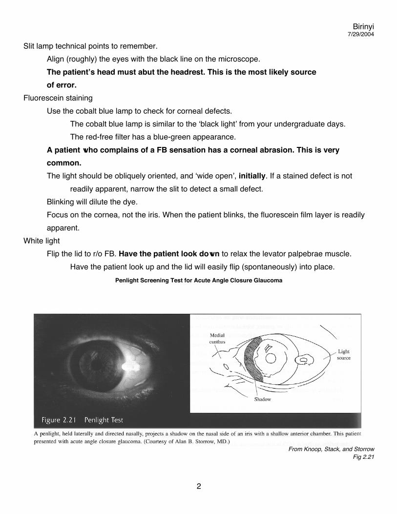
White lightFlip the lid to r/o FB. Have the patient look down to relax the levator palpebrae muscle.
Have the patient look up and the lid will easily flip (spontaneously) into place.Penlight Screening Test for Acute Angle Closure Glaucoma
From Knoop, Stack, and Storrow Fig 2.21
Birinyi7/29/2004
2
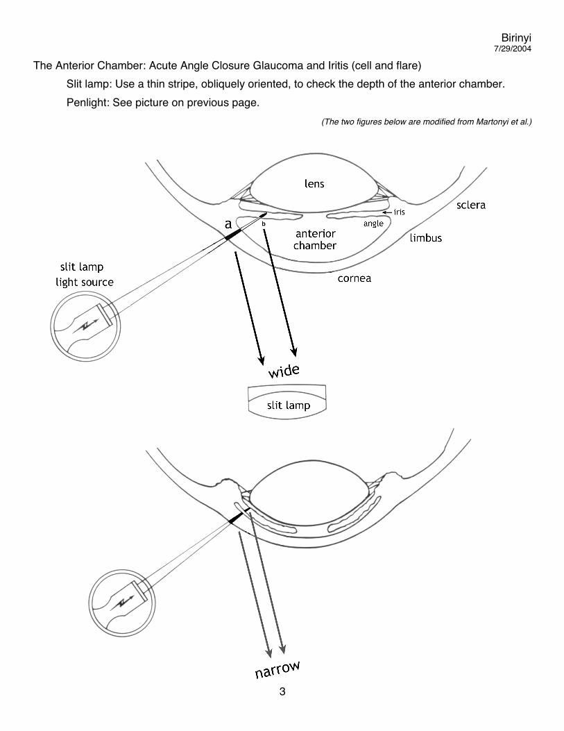
The Anterior Chamber: Acute Angle Closure Glaucoma and Iritis (cell and flare)Slit lamp: Use a thin stripe, obliquely oriented, to check the depth of the anterior chamber.Penlight: See picture on previous page.
(The two figures below are modified from Martonyi et al.)
Birinyi7/29/2004
3
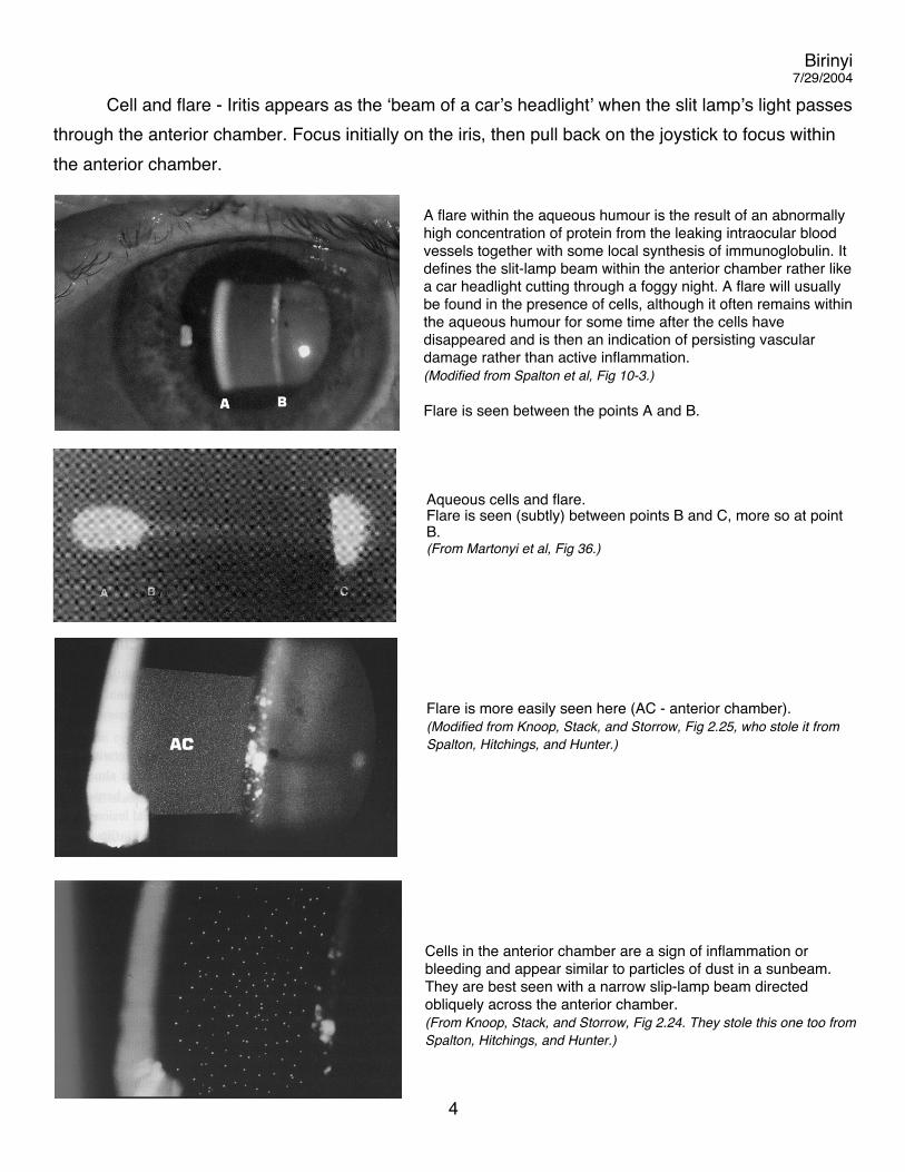
Cell and flare - Iritis appears as the ‘beam of a car’s headlight’ when the slit lamp’s light passes through the anterior chamber. Focus initially on the iris, then pull back on the joystick to focus within the anterior chamber.
A flare within the aqueous humour is the result of an abnormally high concentration of protein from the leaking intraocular blood vessels together with some local synthesis of immunoglobulin. It defines the slit-lamp beam within the anterior chamber rather like a car headlight cutting through a foggy night. A flare will usually be found in the presence of cells, although it often remains within the aqueous humour for some time after the cells have disappeared and is then an indication of persisting vascular damage rather than active inflammation. (Modified from Spalton et al, Fig 10-3.)
Flare is seen between the points A and B.
Aqueous cells and flare. Flare is seen (subtly) between points B and C, more so at point B.(From Martonyi et al, Fig 36.)
Flare is more easily seen here (AC - anterior chamber).(Modified from Knoop, Stack, and Storrow, Fig 2.25, who stole it from Spalton, Hitchings, and Hunter.)
Cells in the anterior chamber are a sign of inflammation or bleeding and appear similar to particles of dust in a sunbeam. They are best seen with a narrow slip-lamp beam directed obliquely across the anterior chamber. (From Knoop, Stack, and Storrow, Fig 2.24. They stole this one too from Spalton, Hitchings, and Hunter.)
Birinyi7/29/2004
4
References Berson FG. Basic Ophthalmology for Medical Students and Primary Care Residents
Sixth edition. American Academy of Ophthalmology. 1994Brandreth RH. Clinical Slit Lamp Biomicroscopy 1978
Cullom RD and Chang B. The Wills Eye Manual - Office and Emergency Room Diagnosis and Treatment of Eye Disease J.B. Lippincott Second edition 1994
Knoop KJ, Stack LB, and Storrow AB. Atlas of Emergency MedicineFirst edition McGraw-Hill 1997
Knoop KJ, Trott A. Ophthalmologic Procedures in the ED, AEM 1994-5Part I: Immediate Sight-saving Procedures. 1(4) p 408Part 2: Routine Evaluation Procedures. 2(2) p 144Part 3: Slit Lamp Use and Foreign Bodies. 2(3) p 224
Martonyi CL, Bahn CF, and Meyer RF. Clinical Slit Lamp BIomicroscopy and Photo Slit Lamp Biomicrography Time One Ink, Ltd. Second edition 1985
Spalton DJ, Hitchings RA, and Hunter PA. Atlas of Clinical OphthalmologyWolfe Publishing Second edition 1994
Appendix
Birinyi7/29/2004
5
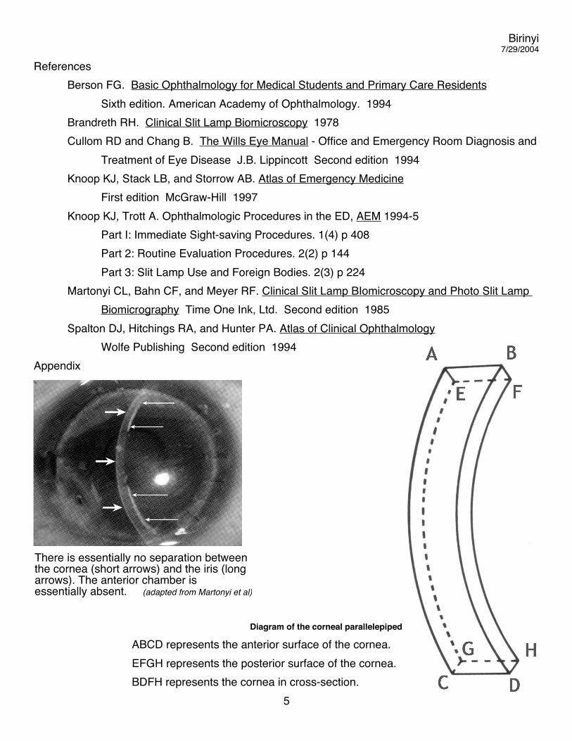
There is essentially no separation between the cornea (short arrows) and the iris (long arrows). The anterior chamber is essentially absent. (adapted from Martonyi et al)
Diagram of the corneal parallelepiped
ABCD represents the anterior surface of the cornea.EFGH represents the posterior surface of the cornea.BDFH represents the cornea in cross-section.