Embed Size (px)
Citation preview
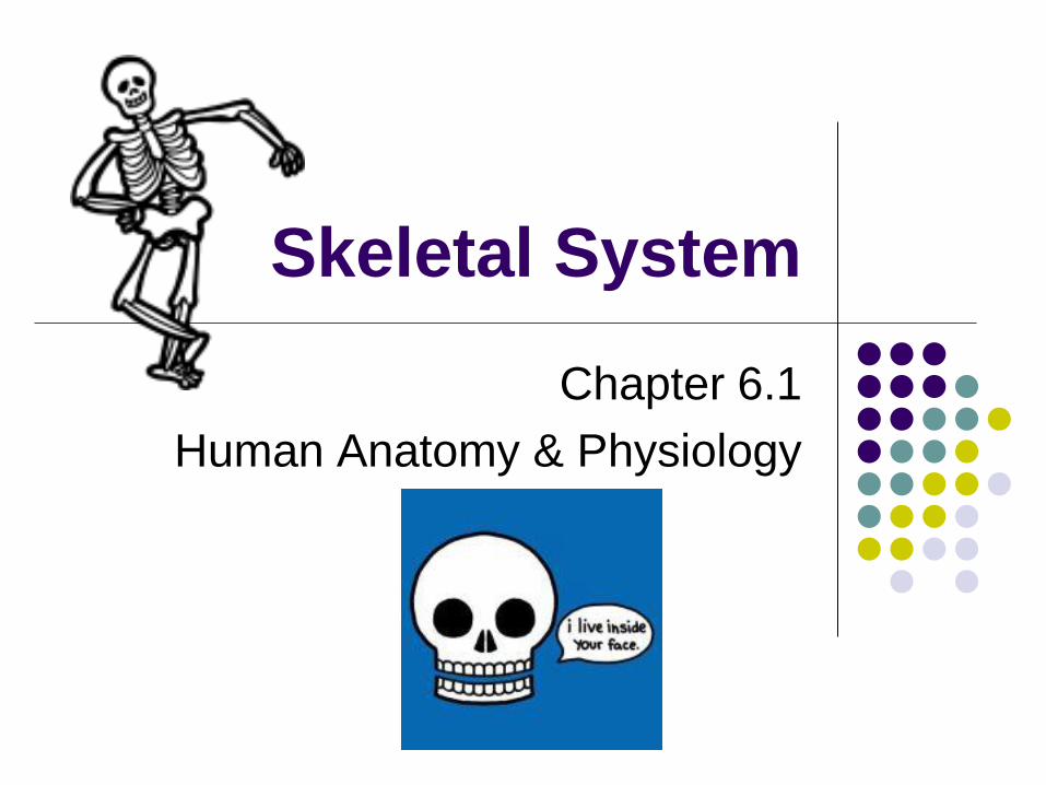
Skeletal System
Chapter 6.1
Human Anatomy & Physiology
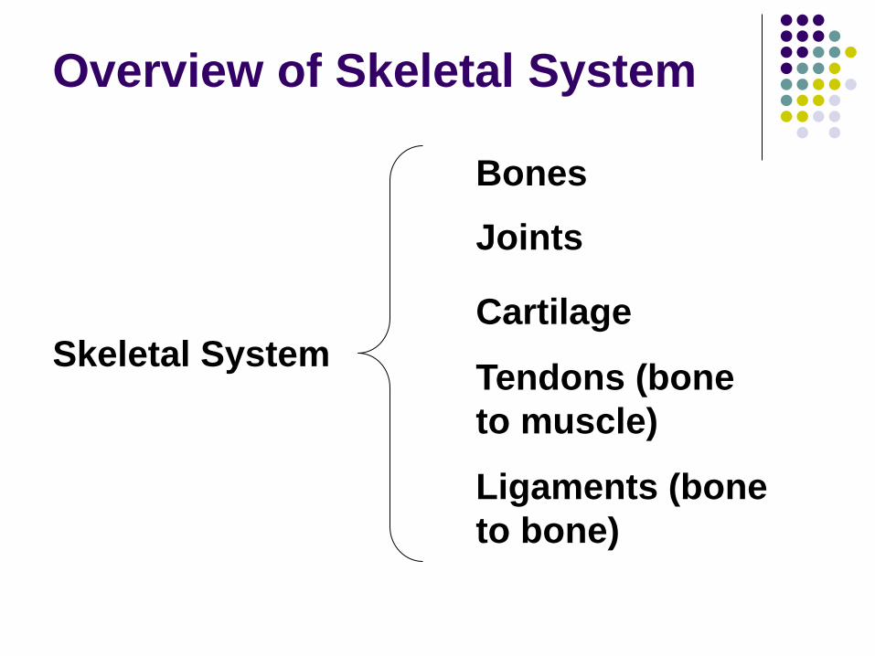
Overview of Skeletal System
Skeletal System
Bones
Joints
Cartilage
Ligaments (bone
to bone)
Tendons (bone
to muscle)
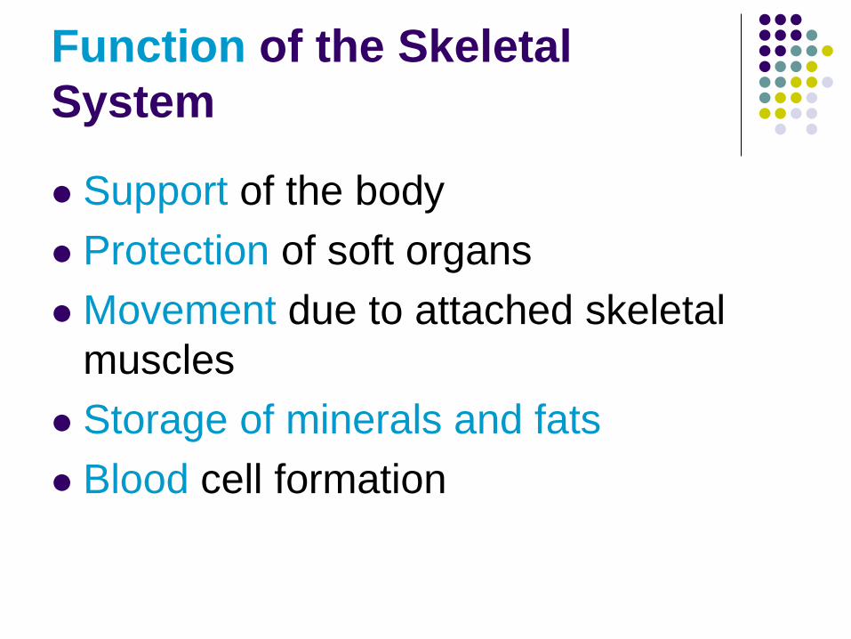
Function of the Skeletal
System
Support of the body
Protection of soft organs
Movement due to attached skeletal
muscles
Storage of minerals and fats
Blood cell formation
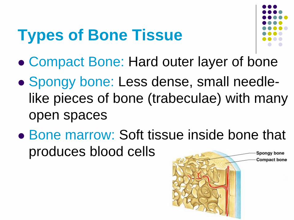
Types of Bone Tissue
Compact Bone: Hard outer layer of bone
Spongy bone: Less dense, small needle-
like pieces of bone (trabeculae) with many
open spaces
Bone marrow: Soft tissue inside bone that
produces blood cells
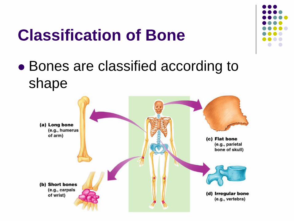
Classification of Bone
Bones are classified according to
shape
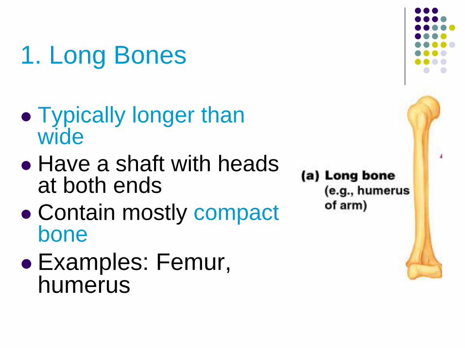
1. Long Bones
Typically longer than
wide
Have a shaft with heads at both ends
Contain mostly compact bone
Examples: Femur, humerus
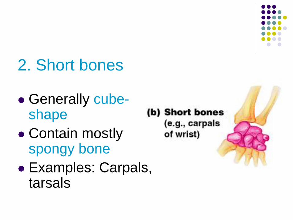
2. Short bones
Generally cube-shape
Contain mostly spongy bone
Examples: Carpals, tarsals
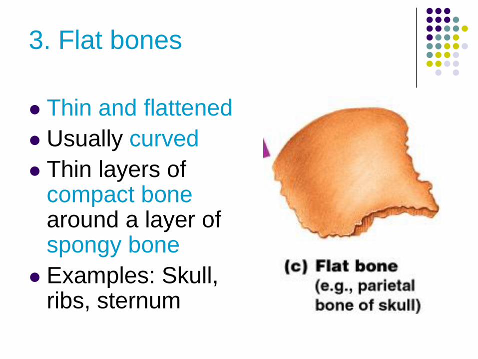
3. Flat bones
Thin and flattened
Usually curved
Thin layers of compact bone around a layer of spongy bone
Examples: Skull, ribs, sternum
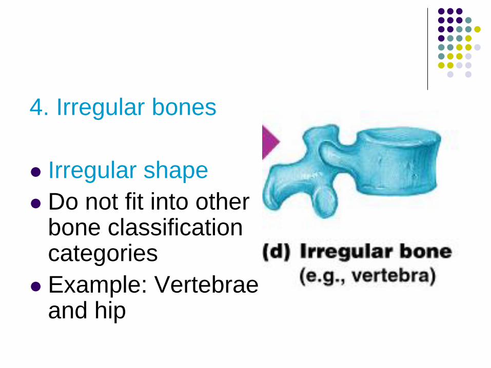
4. Irregular bones
Irregular shape
Do not fit into other bone classification categories
Example: Vertebrae and hip
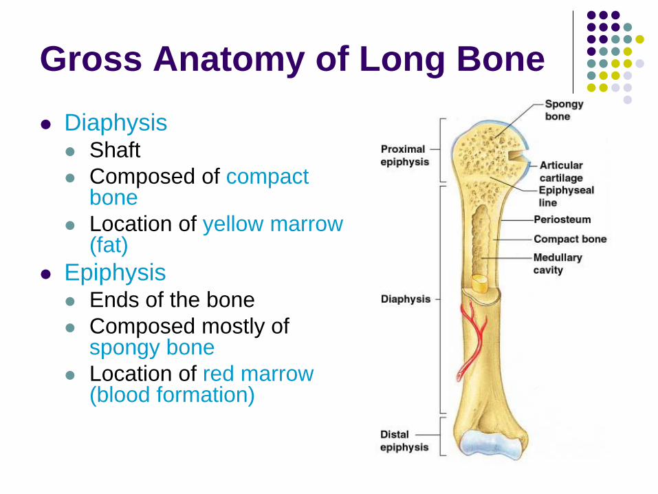
Gross Anatomy of Long Bone
Diaphysis Shaft
Composed of compact bone
Location of yellow marrow (fat)
Epiphysis Ends of the bone
Composed mostly of spongy bone
Location of red marrow (blood formation)
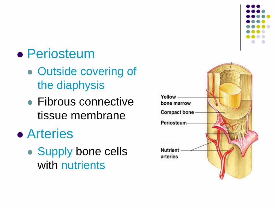
Periosteum
Outside covering of
the diaphysis
Fibrous connective
tissue membrane
Arteries
Supply bone cells
with nutrients
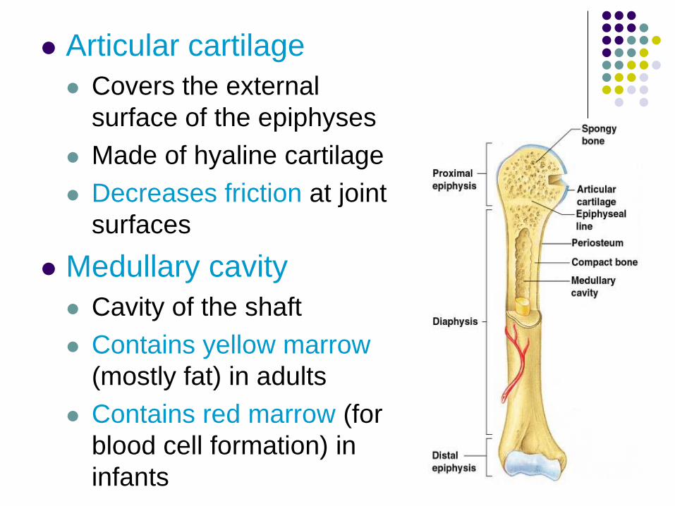
Articular cartilage
Covers the external
surface of the epiphyses
Made of hyaline cartilage
Decreases friction at joint
surfaces
Medullary cavity
Cavity of the shaft
Contains yellow marrow
(mostly fat) in adults
Contains red marrow (for
blood cell formation) in
infants
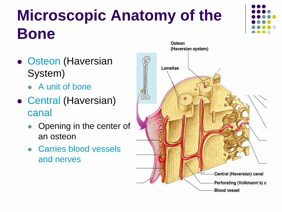
Microscopic Anatomy of the
Bone
Osteon (Haversian
System)
A unit of bone
Central (Haversian)
canal
Opening in the center of
an osteon
Carries blood vessels
and nerves
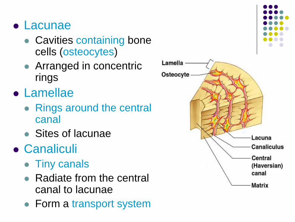
Lacunae
Cavities containing bone cells (osteocytes)
Arranged in concentric rings
Lamellae
Rings around the central canal
Sites of lacunae
Canaliculi
Tiny canals
Radiate from the central canal to lacunae
Form a transport system
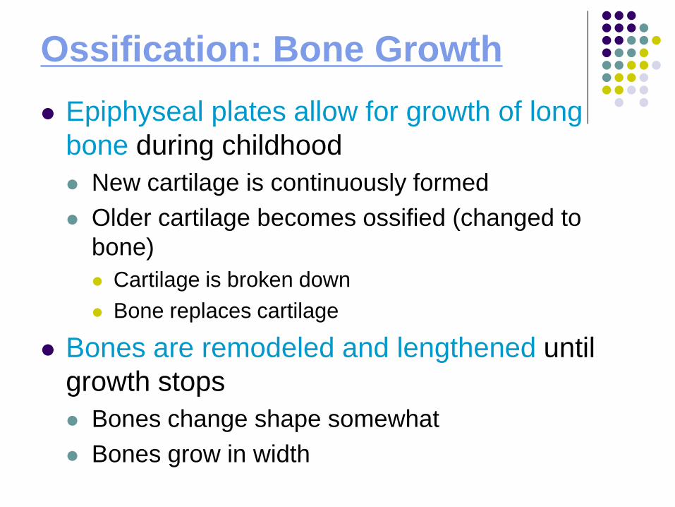
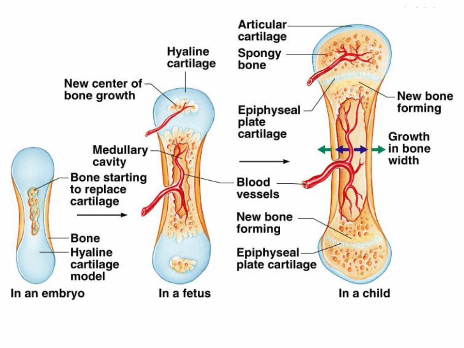
Ossification: Bone Growth
Epiphyseal plates allow for growth of long
bone during childhood
New cartilage is continuously formed
Older cartilage becomes ossified (changed to
bone)
Cartilage is broken down
Bone replaces cartilage
Bones are remodeled and lengthened until
growth stops
Bones change shape somewhat
Bones grow in width
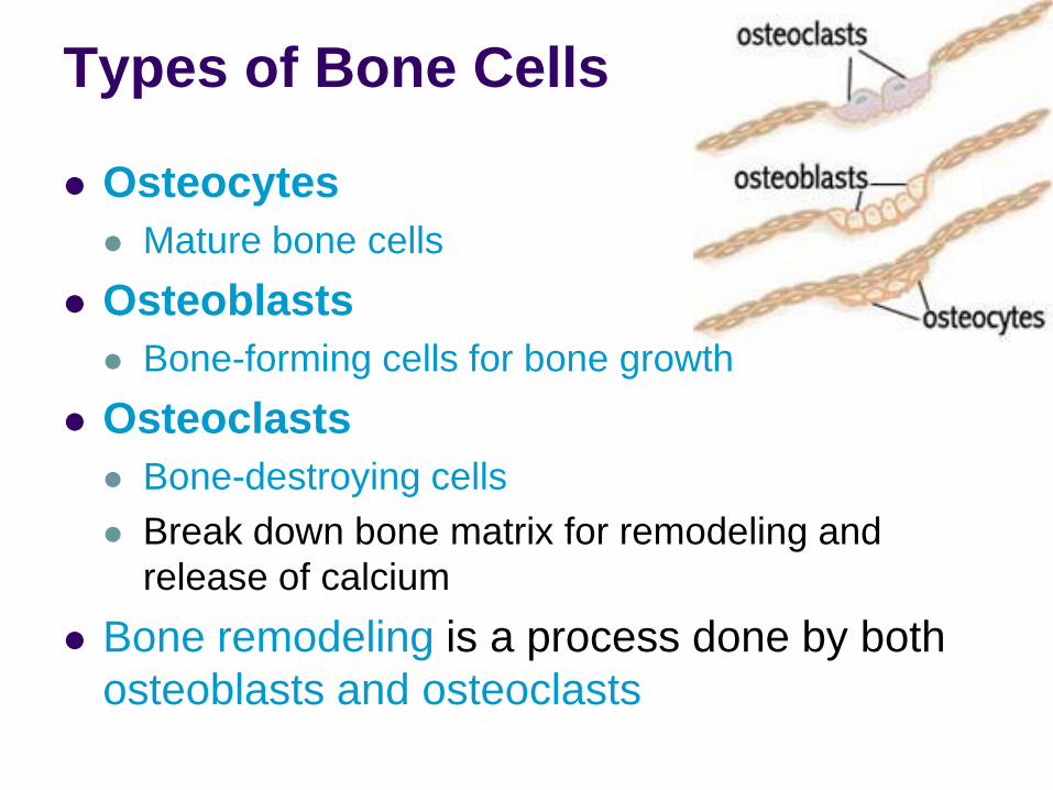
Types of Bone Cells
Osteocytes
Mature bone cells
Osteoblasts
Bone-forming cells for bone growth
Osteoclasts
Bone-destroying cells
Break down bone matrix for remodeling and
release of calcium
Bone remodeling is a process done by both
osteoblasts and osteoclasts
Ticket out the Door
Identify and Describe the 5 functions of the
skeletal system.
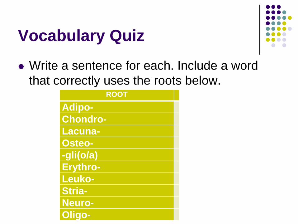
Vocabulary Quiz
Write a sentence for each. Include a word
that correctly uses the roots below.
ROOT
Adipo-
Chondro-
Lacuna-
Osteo-
-gli(o/a)
Erythro-
Leuko-
Stria-
Neuro-
Oligo-
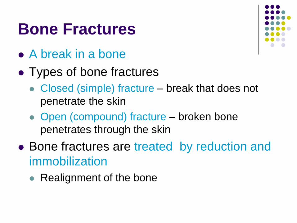
Bone Fractures
A break in a bone
Types of bone fractures
Closed (simple) fracture – break that does not
penetrate the skin
Open (compound) fracture – broken bone
penetrates through the skin
Bone fractures are treated by reduction and
immobilization
Realignment of the bone
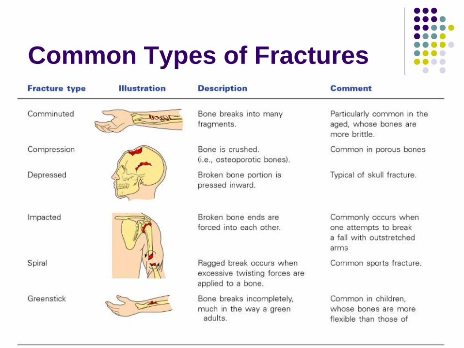
Common Types of Fractures
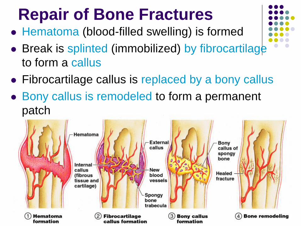
Repair of Bone Fractures Hematoma (blood-filled swelling) is formed
Break is splinted (immobilized) by fibrocartilage
to form a callus
Fibrocartilage callus is replaced by a bony callus
Bony callus is remodeled to form a permanent
patch
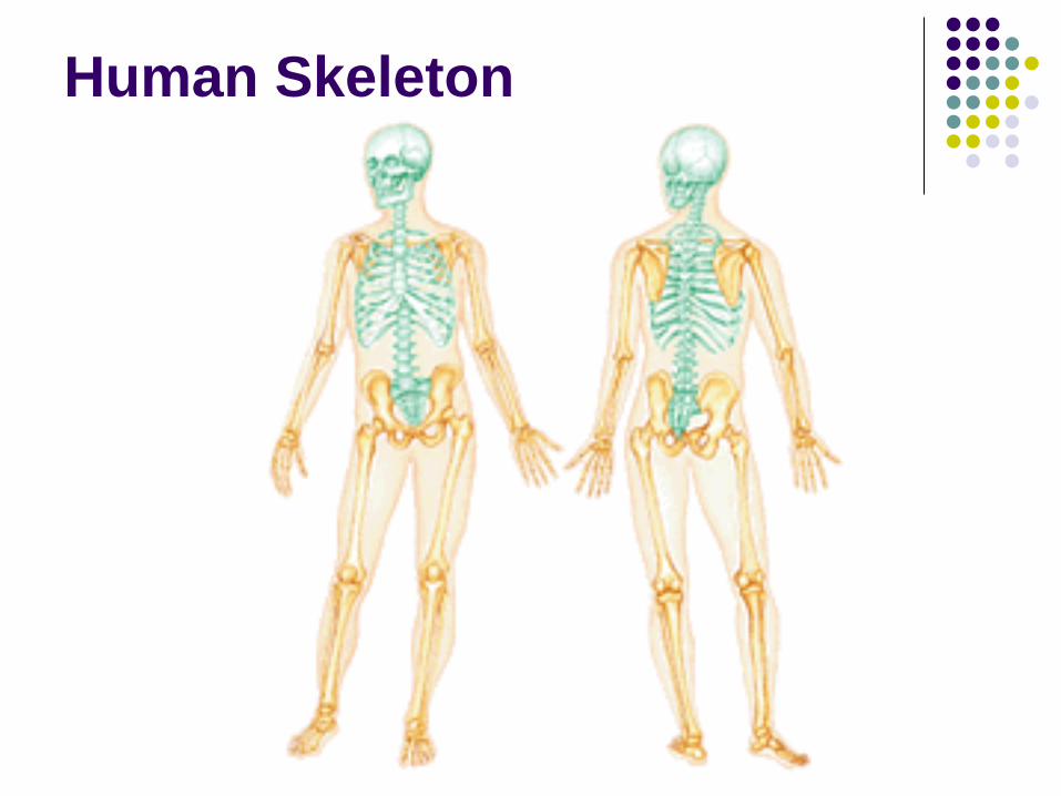
Human Skeleton
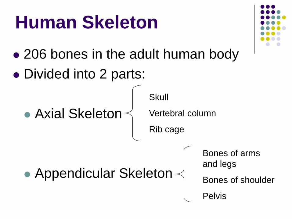
Human Skeleton
206 bones in the adult human body
Divided into 2 parts:
Axial Skeleton
Appendicular Skeleton
Skull
Vertebral column
Rib cage
Bones of arms
and legs
Bones of shoulder
Pelvis
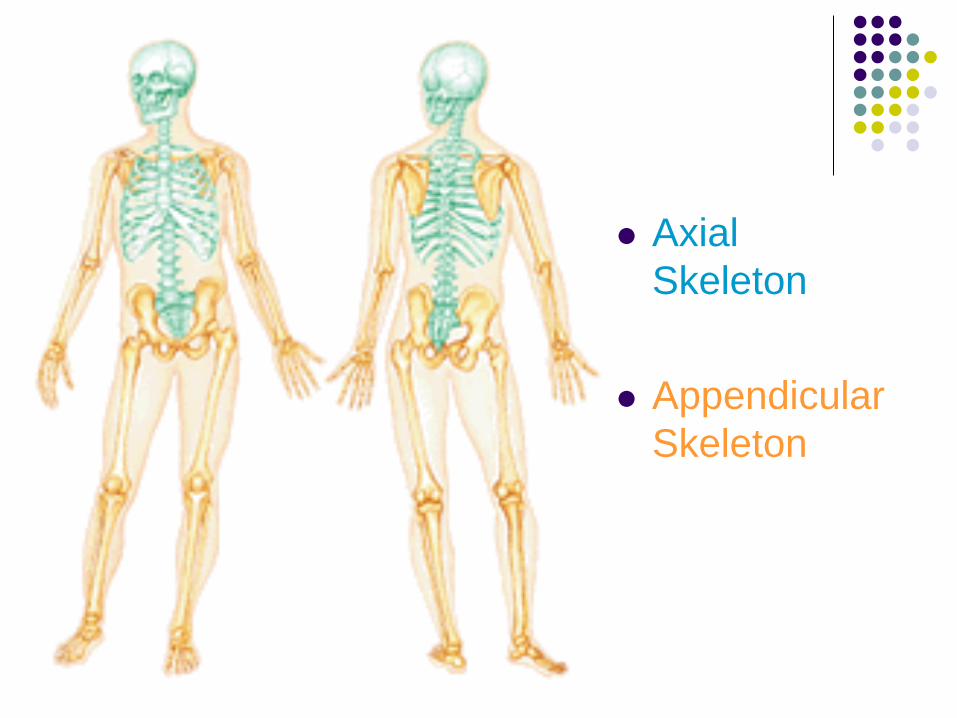
Axial
Skeleton
Appendicular
Skeleton
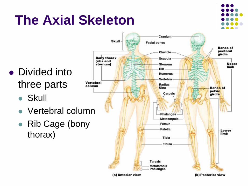
Divided into
three parts
Skull
Vertebral column
Rib Cage (bony
thorax)
The Axial Skeleton
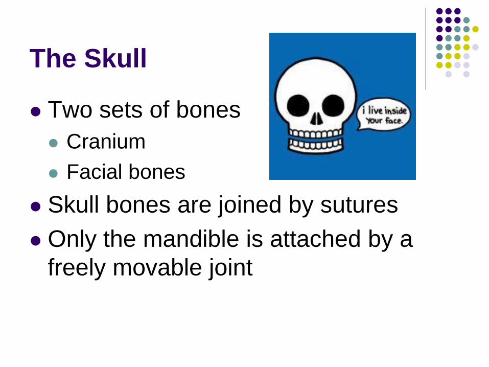
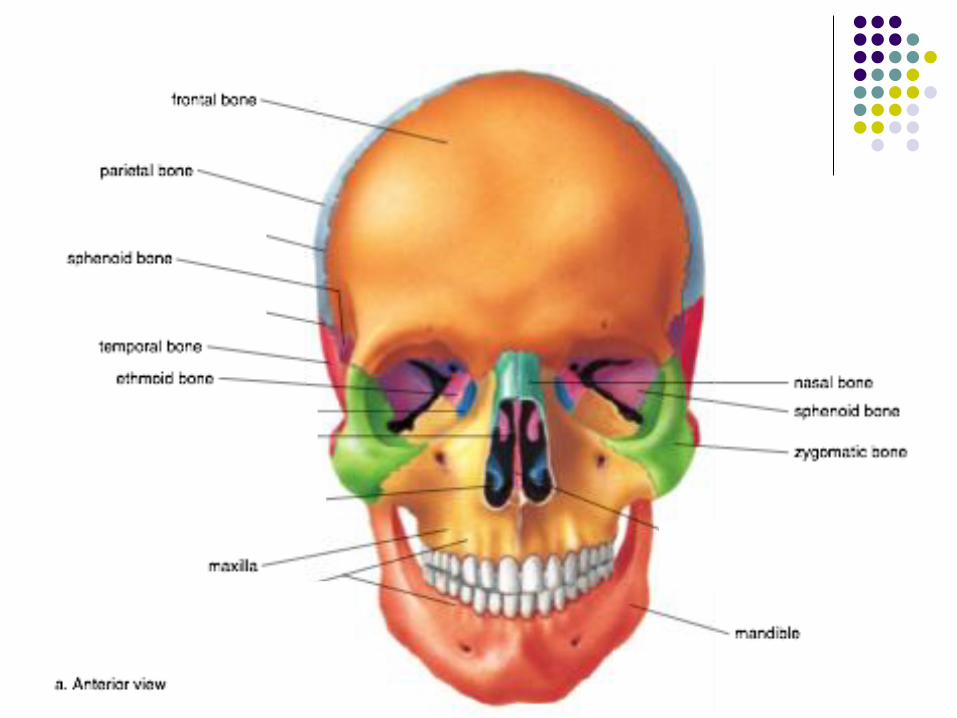
The Skull
Two sets of bones
Cranium
Facial bones
Skull bones are joined by sutures
Only the mandible is attached by a
freely movable joint
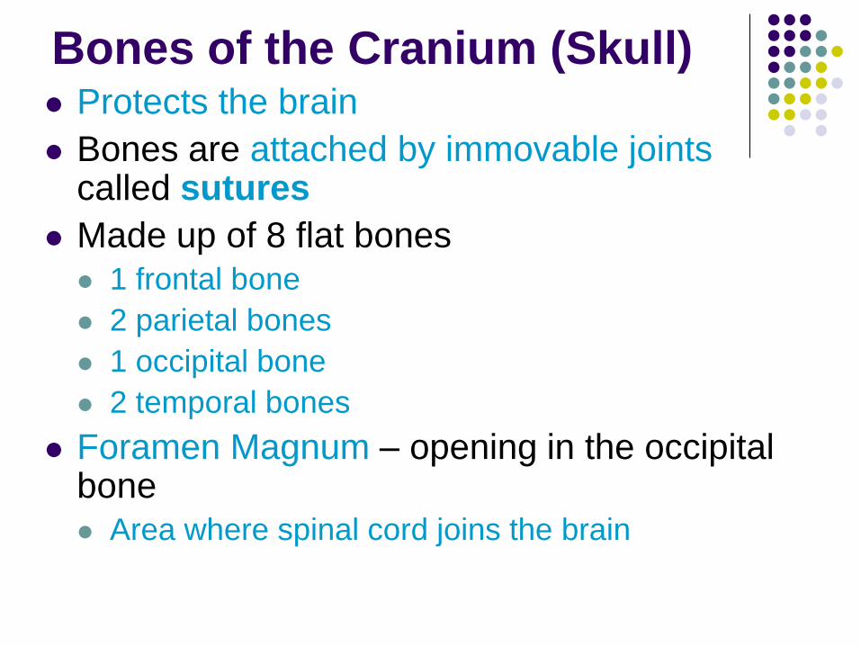
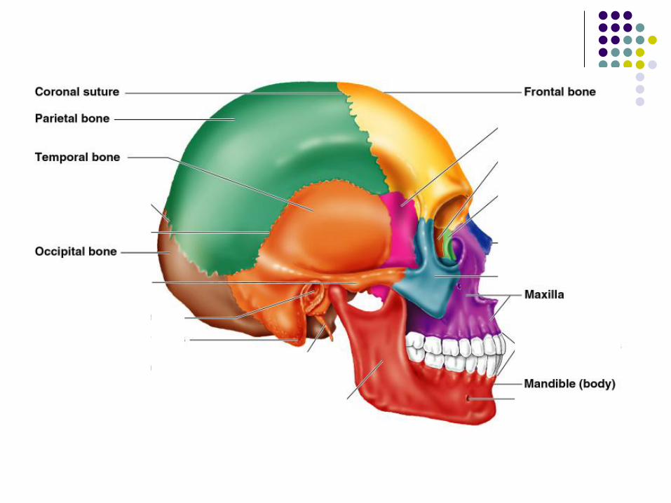
Bones of the Cranium (Skull) Protects the brain
Bones are attached by immovable joints called sutures
Made up of 8 flat bones
1 frontal bone
2 parietal bones
1 occipital bone
2 temporal bones
Foramen Magnum – opening in the occipital bone
Area where spinal cord joins the brain
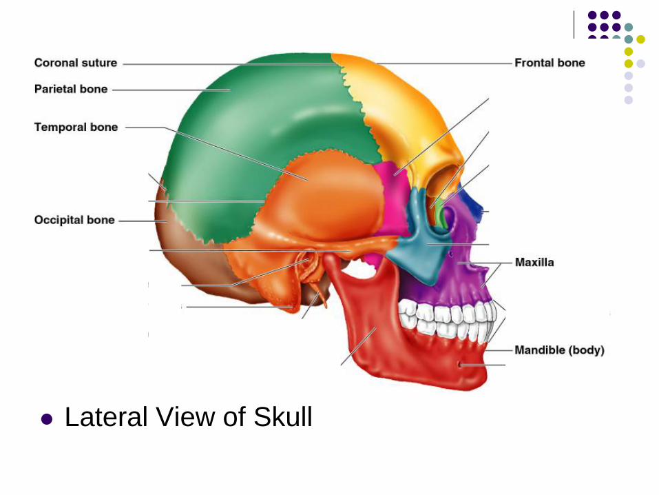
Lateral View of Skull
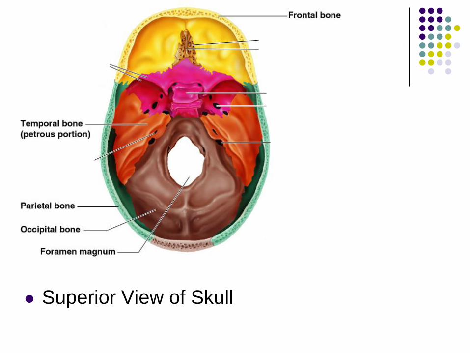
Superior View of Skull
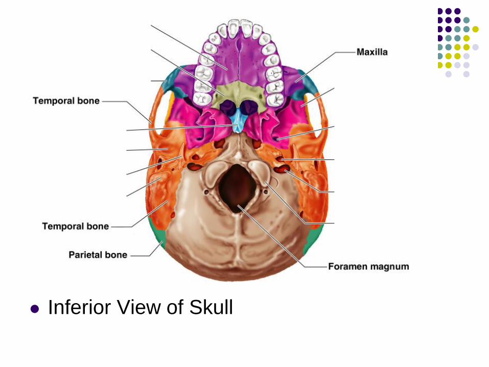
Inferior View of Skull
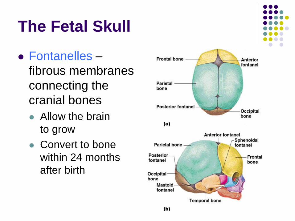
The Fetal Skull
Fontanelles –
fibrous membranes
connecting the
cranial bones
Allow the brain
to grow
Convert to bone
within 24 months
after birth
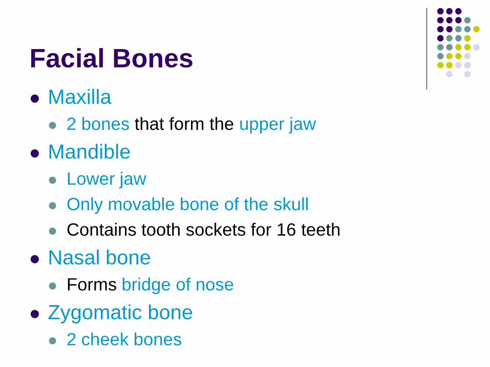
Facial Bones
Maxilla
2 bones that form the upper jaw
Mandible
Lower jaw
Only movable bone of the skull
Contains tooth sockets for 16 teeth
Nasal bone
Forms bridge of nose
Zygomatic bone
2 cheek bones
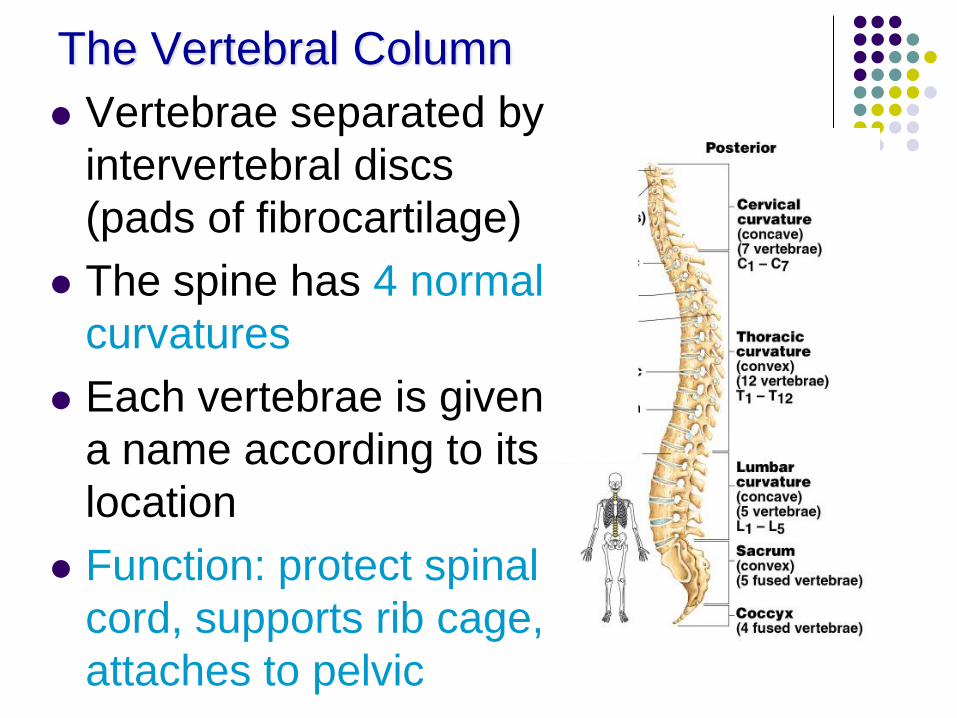
The Vertebral Column
Vertebrae separated by
intervertebral discs
(pads of fibrocartilage)
The spine has 4 normal
curvatures
Each vertebrae is given
a name according to its
location
Function: protect spinal
cord, supports rib cage,
attaches to pelvic
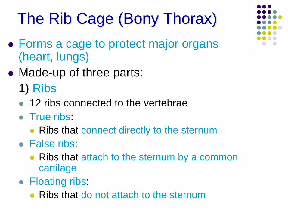
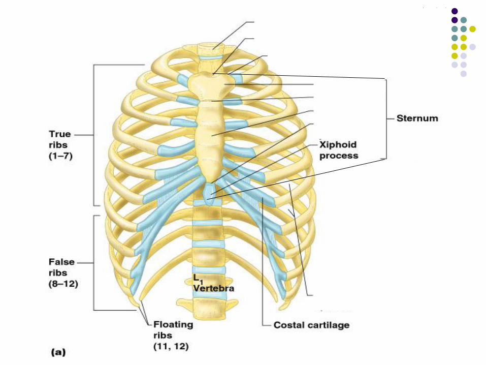
The Rib Cage (Bony Thorax)
Forms a cage to protect major organs (heart, lungs)
Made-up of three parts:
1) Ribs
12 ribs connected to the vertebrae
True ribs:
Ribs that connect directly to the sternum
False ribs:
Ribs that attach to the sternum by a common cartilage
Floating ribs:
Ribs that do not attach to the sternum
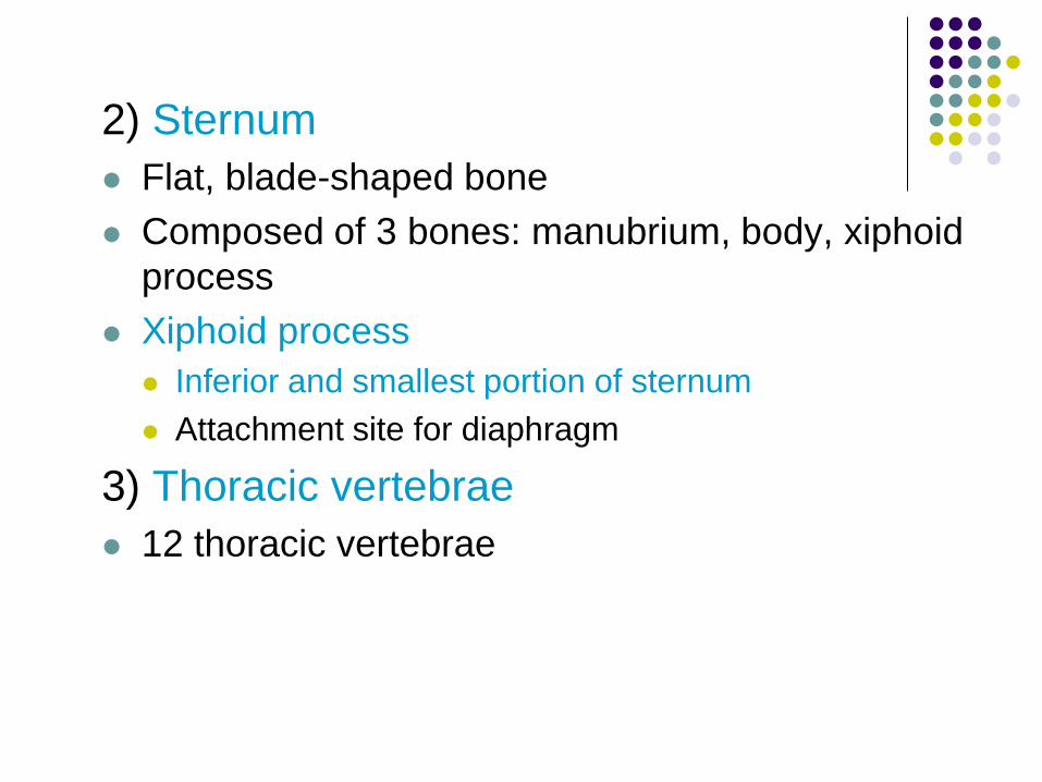
2) Sternum
Flat, blade-shaped bone
Composed of 3 bones: manubrium, body, xiphoid
process
Xiphoid process
Inferior and smallest portion of sternum
Attachment site for diaphragm
3) Thoracic vertebrae
12 thoracic vertebrae
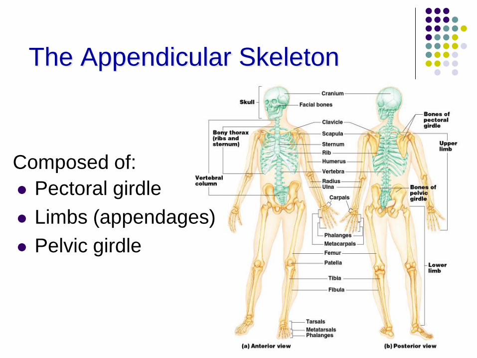
The Appendicular Skeleton
Pectoral girdle
Limbs (appendages)
Pelvic girdle
Composed of:
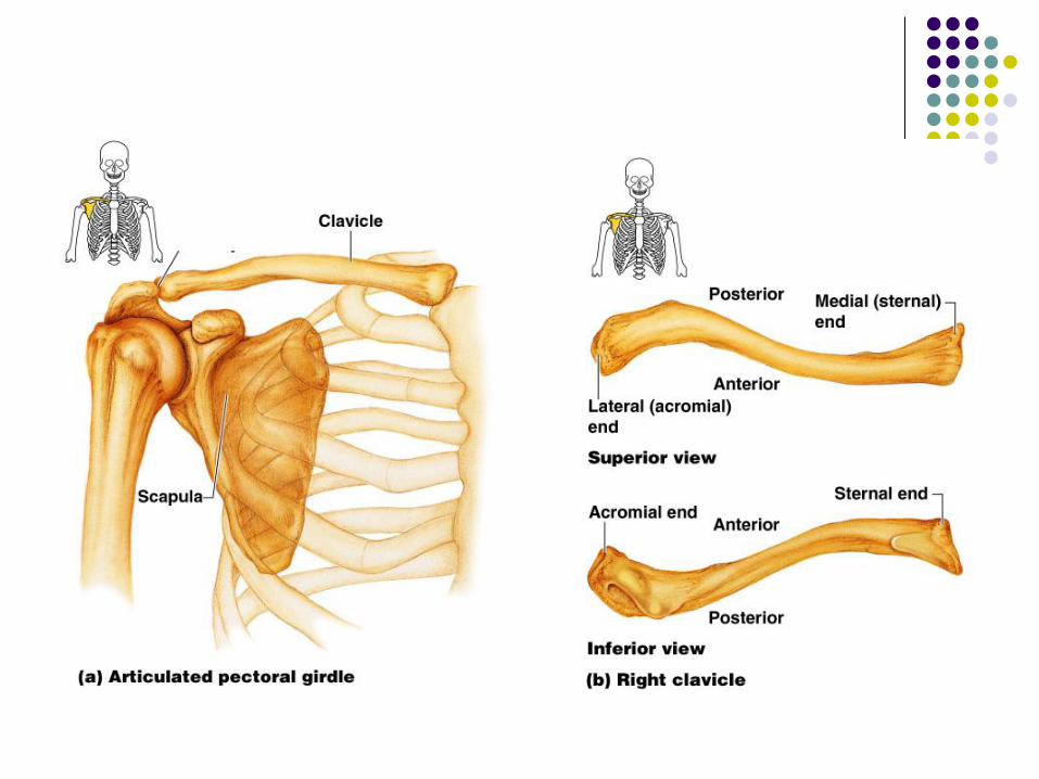
The Pectoral (Shoulder) Girdle
These bones allow the upper limbs to have exceptionally free movement
Composed of 4 bones
- 2 Clavicles – collarbone
Slender and s-shaped
Stabilizes shoulder but structurally weak (breaks easily)
- 2 Scapulas – shoulder blade
Triangular shape
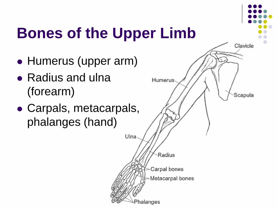
Humerus (upper arm)
Radius and ulna
(forearm)
Carpals, metacarpals,
phalanges (hand)
Bones of the Upper Limb
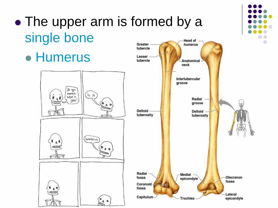
The upper arm is formed by a
single bone
Humerus
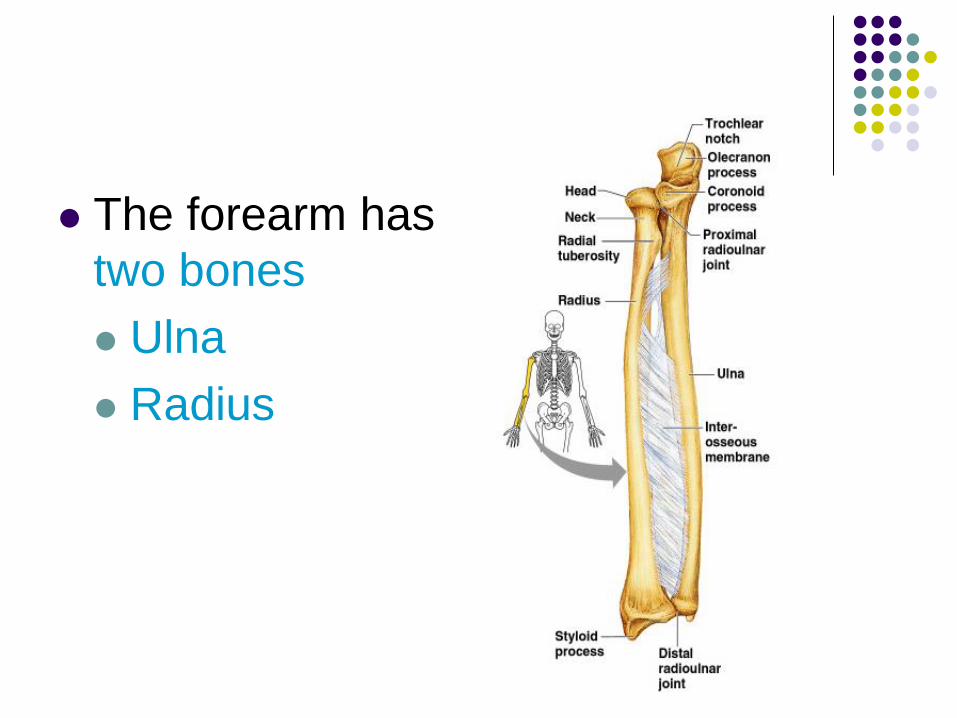
The forearm has
two bones
Ulna
Radius
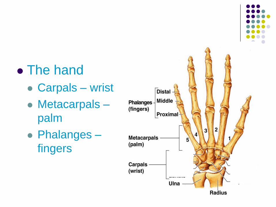
The hand
Carpals – wrist
Metacarpals –
palm
Phalanges –
fingers
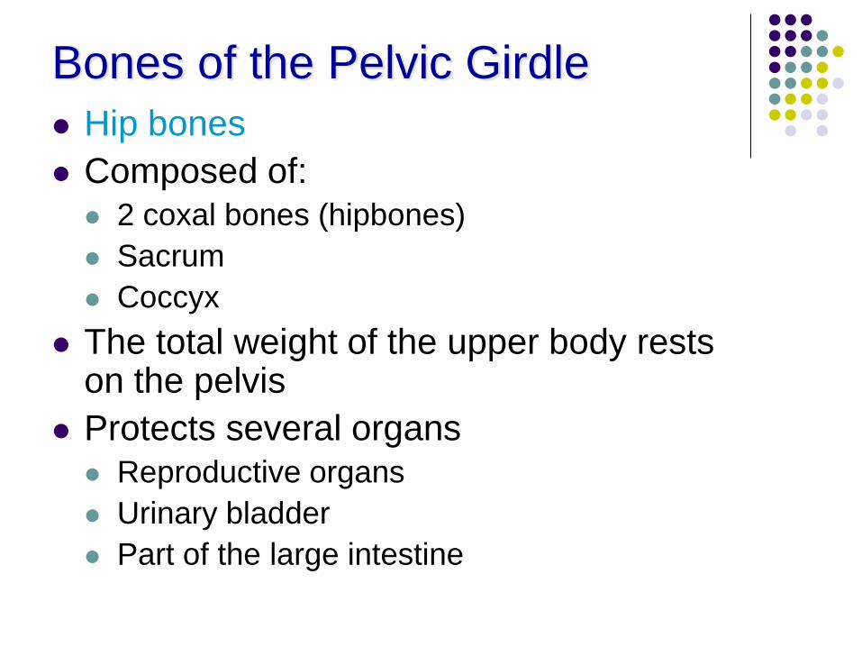
Bones of the Pelvic Girdle
Hip bones
Composed of:
2 coxal bones (hipbones)
Sacrum
Coccyx
The total weight of the upper body rests on the pelvis
Protects several organs
Reproductive organs
Urinary bladder
Part of the large intestine
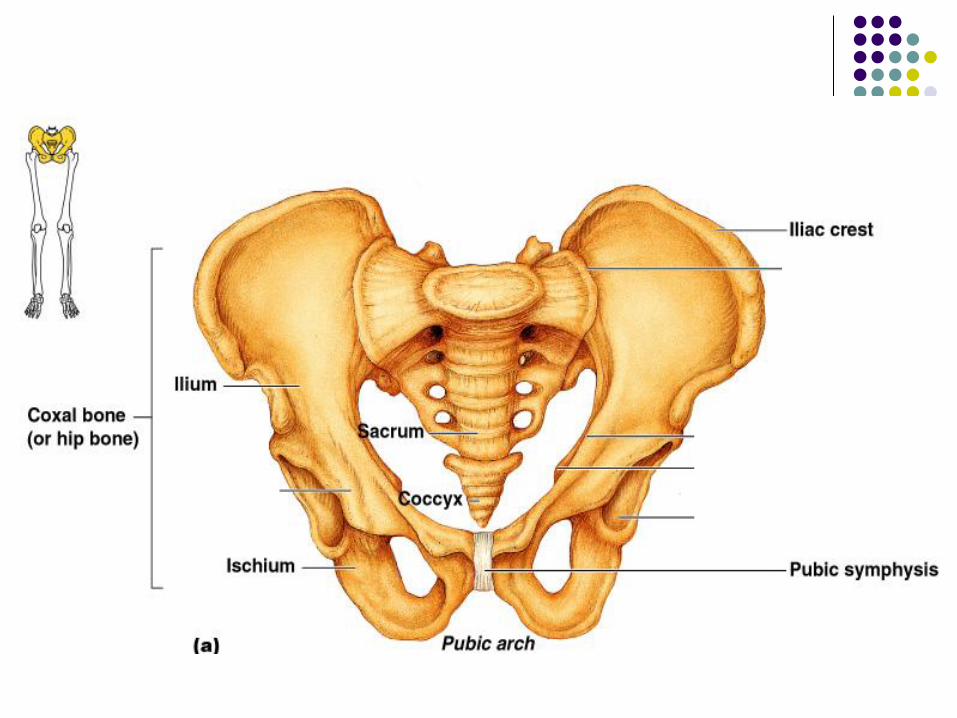
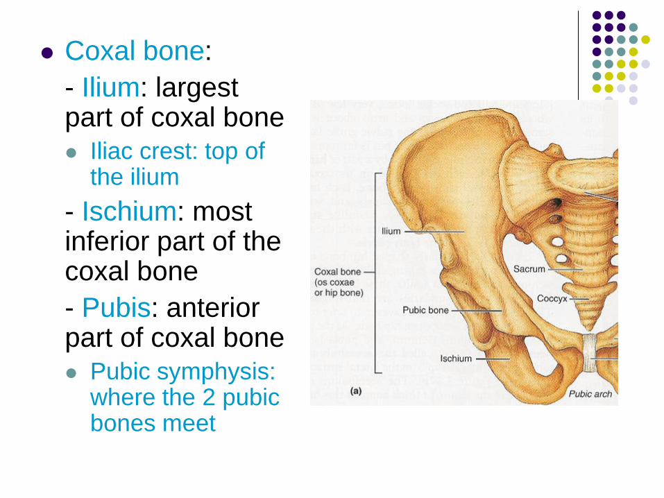
Coxal bone:
- Ilium: largest part of coxal bone
Iliac crest: top of the ilium
- Ischium: most inferior part of the coxal bone
- Pubis: anterior part of coxal bone
Pubic symphysis: where the 2 pubic bones meet
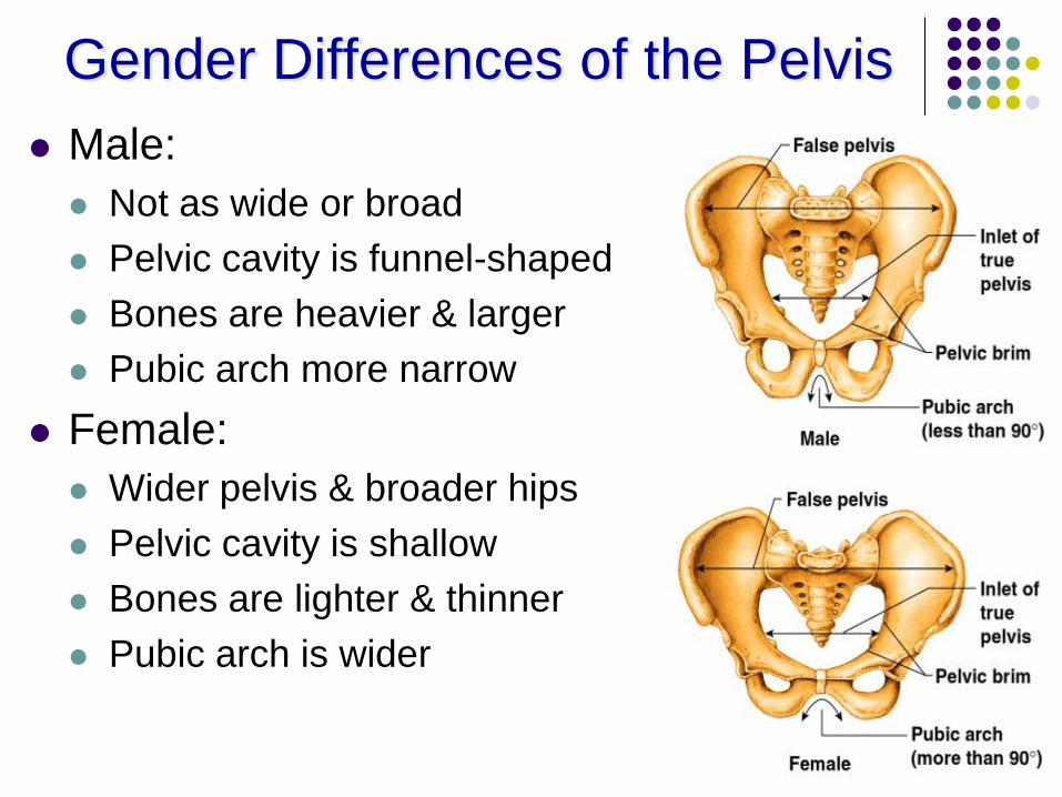
Gender Differences of the Pelvis
Male:
Not as wide or broad
Pelvic cavity is funnel-shaped
Bones are heavier & larger
Pubic arch more narrow
Female:
Wider pelvis & broader hips
Pelvic cavity is shallow
Bones are lighter & thinner
Pubic arch is wider
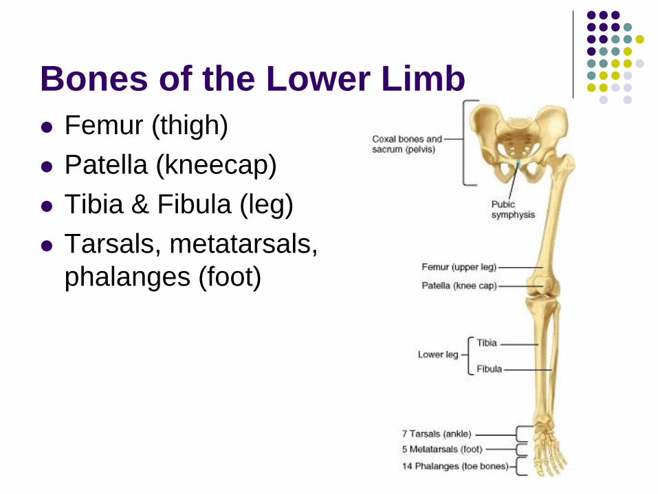
Bones of the Lower Limb
Femur (thigh)
Patella (kneecap)
Tibia & Fibula (leg)
Tarsals, metatarsals,
phalanges (foot)
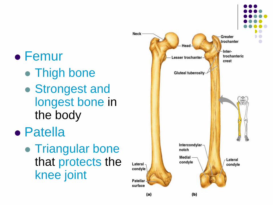
Femur
Thigh bone
Strongest and longest bone in the body
Patella
Triangular bone that protects the knee joint
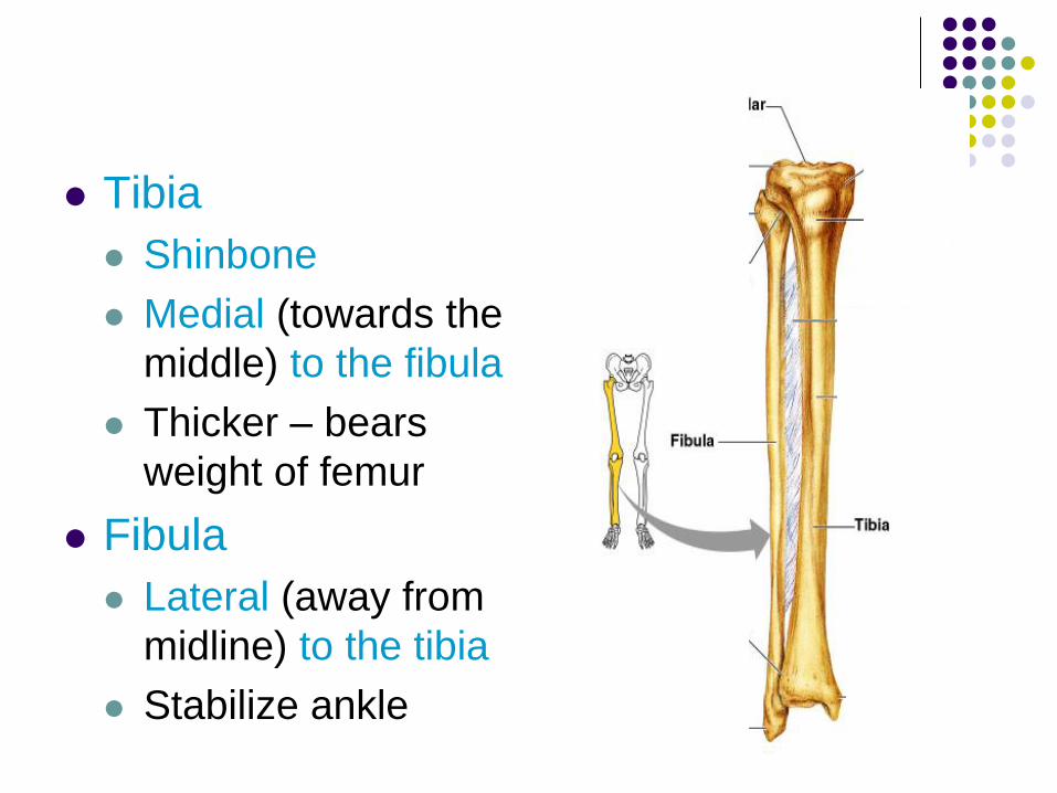
Tibia
Shinbone
Medial (towards the
middle) to the fibula
Thicker – bears
weight of femur
Fibula
Lateral (away from
midline) to the tibia
Stabilize ankle
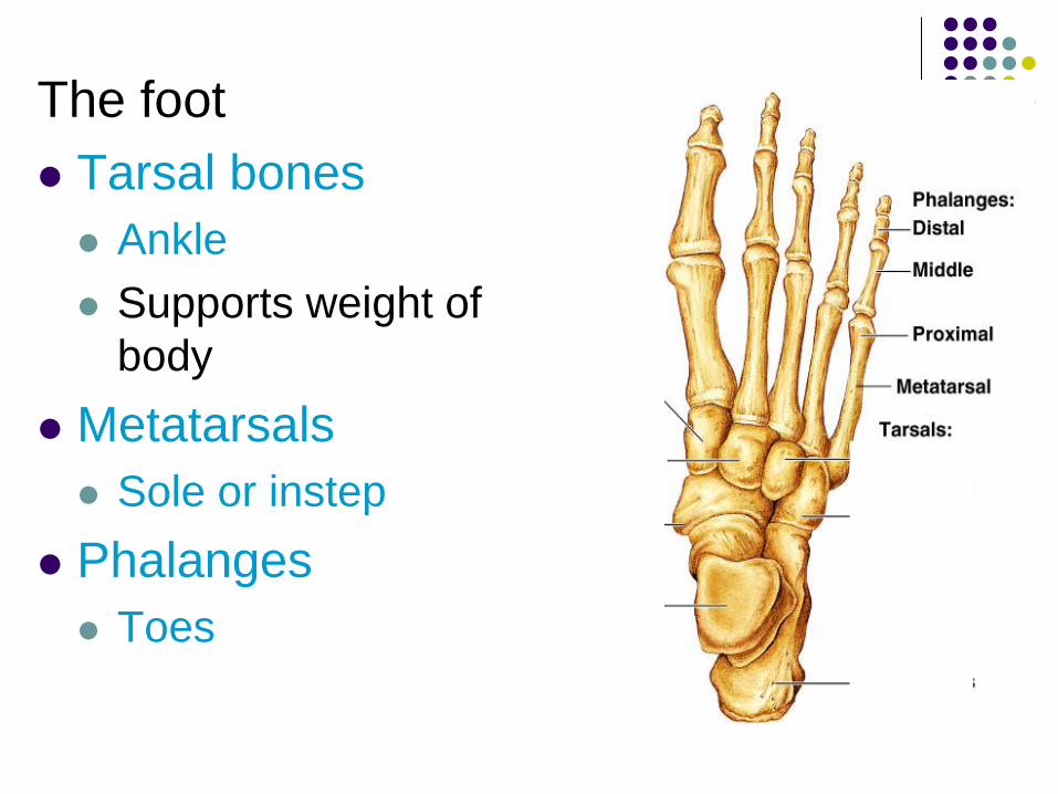
The foot
Tarsal bones
Ankle
Supports weight of
body
Metatarsals
Sole or instep
Phalanges
Toes
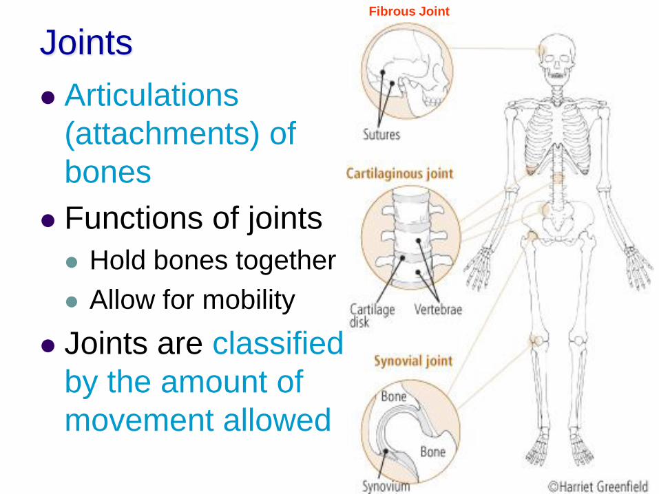
Joints
Articulations
(attachments) of
bones
Functions of joints
Hold bones together
Allow for mobility
Joints are classified
by the amount of
movement allowed
Fibrous Joint
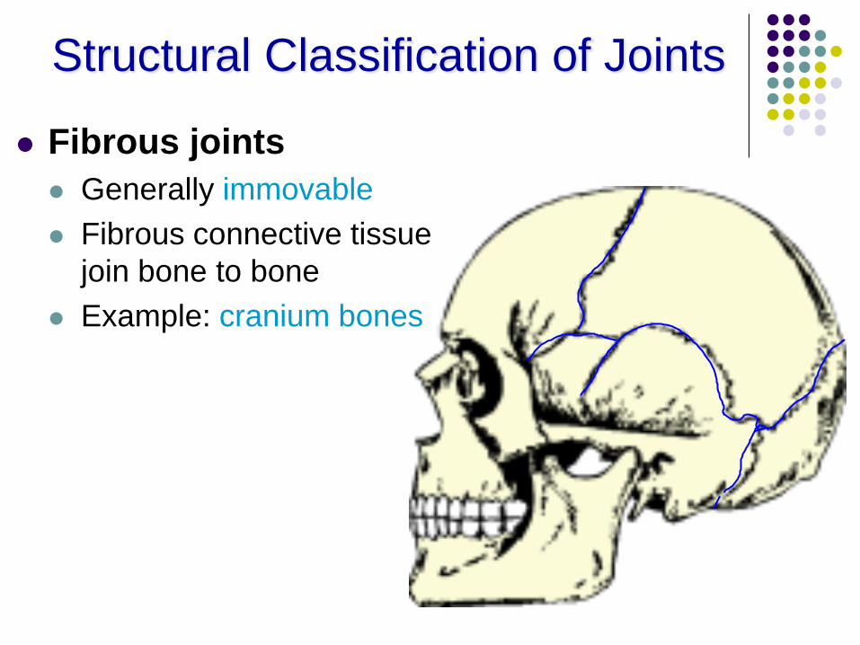
Structural Classification of Joints
Fibrous joints
Generally immovable
Fibrous connective tissue
join bone to bone
Example: cranium bones
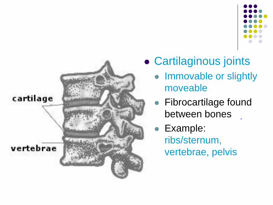
Cartilaginous joints
Immovable or slightly
moveable
Fibrocartilage found
between bones
Example:
ribs/sternum,
vertebrae, pelvis
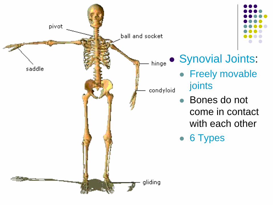
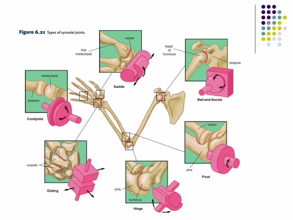
Synovial Joints:
Freely movable
joints
Bones do not
come in contact
with each other
6 Types
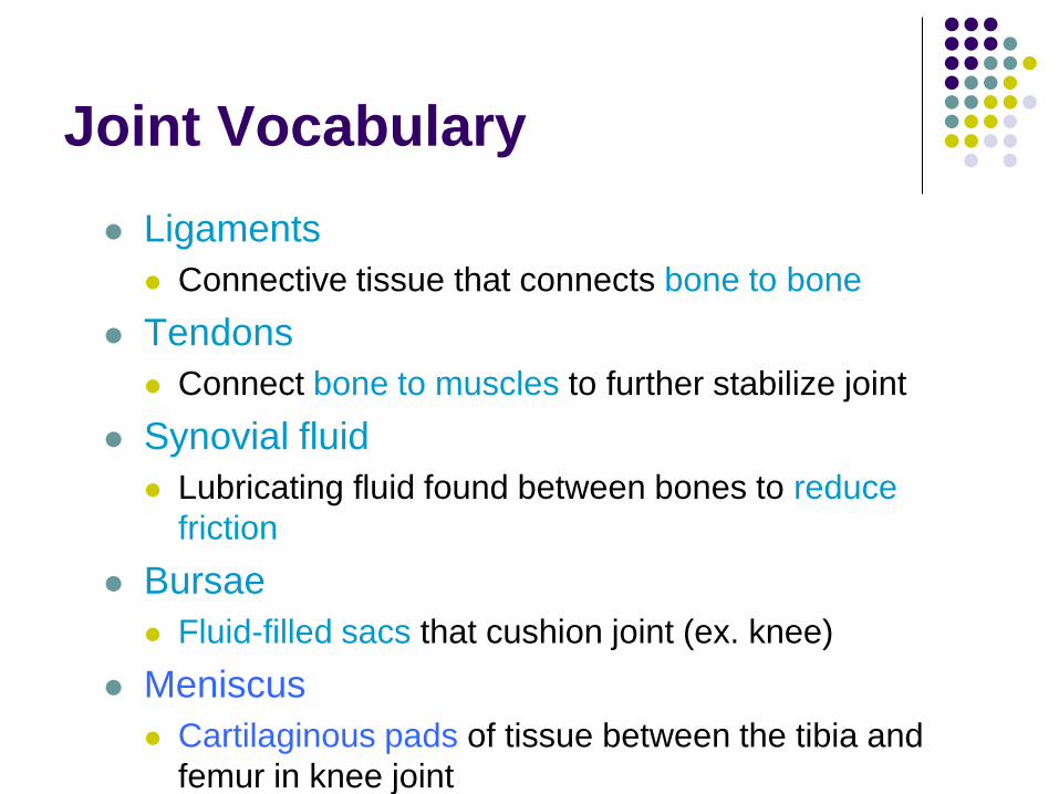
Joint Vocabulary
Ligaments
Connective tissue that connects bone to bone
Tendons
Connect bone to muscles to further stabilize joint
Synovial fluid
Lubricating fluid found between bones to reduce
friction
Bursae
Fluid-filled sacs that cushion joint (ex. knee)
Meniscus
Cartilaginous pads of tissue between the tibia and
femur in knee joint
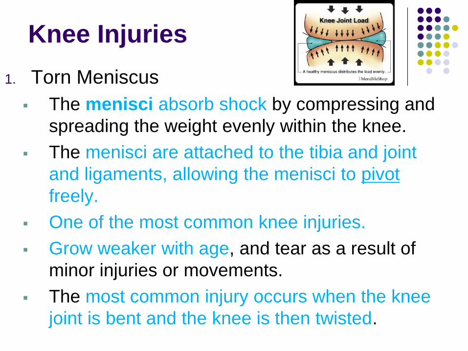
Knee Injuries
1. Torn Meniscus
The menisci absorb shock by compressing and
spreading the weight evenly within the knee.
The menisci are attached to the tibia and joint
and ligaments, allowing the menisci to pivot
freely.
One of the most common knee injuries.
Grow weaker with age, and tear as a result of
minor injuries or movements.
The most common injury occurs when the knee
joint is bent and the knee is then twisted.
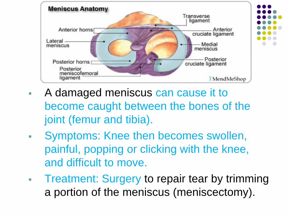
A damaged meniscus can cause it to
become caught between the bones of the
joint (femur and tibia).
Symptoms: Knee then becomes swollen,
painful, popping or clicking with the knee,
and difficult to move.
Treatment: Surgery to repair tear by trimming
a portion of the meniscus (meniscectomy).
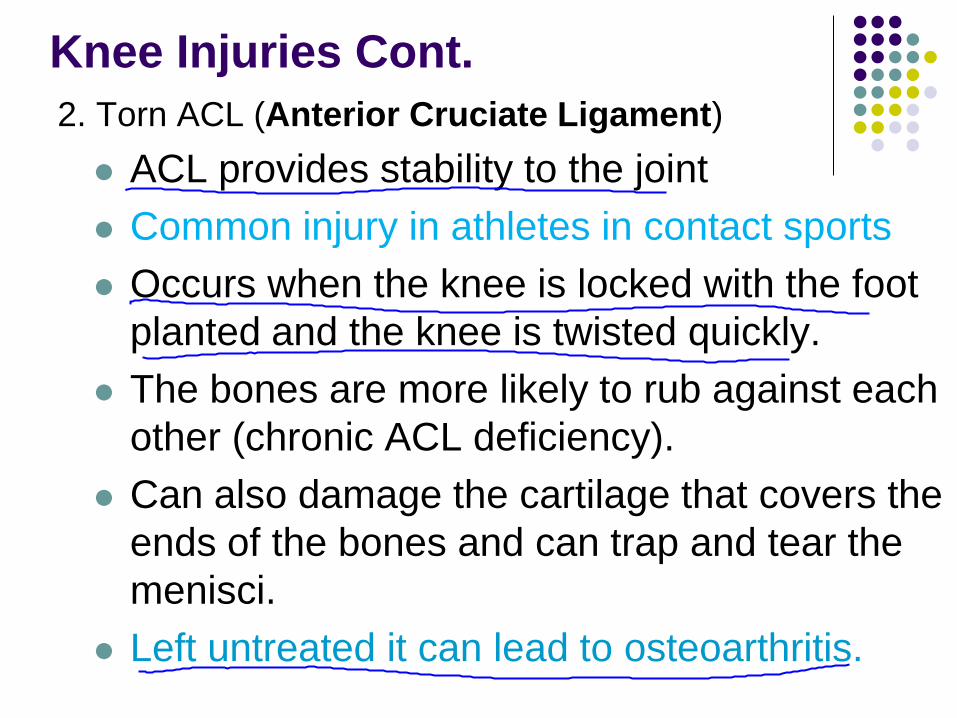
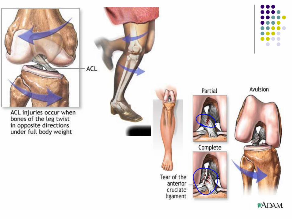
Knee Injuries Cont.
2. Torn ACL (Anterior Cruciate Ligament)
ACL provides stability to the joint
Common injury in athletes in contact sports
Occurs when the knee is locked with the foot
planted and the knee is twisted quickly.
The bones are more likely to rub against each
other (chronic ACL deficiency).
Can also damage the cartilage that covers the
ends of the bones and can trap and tear the
menisci.
Left untreated it can lead to osteoarthritis.
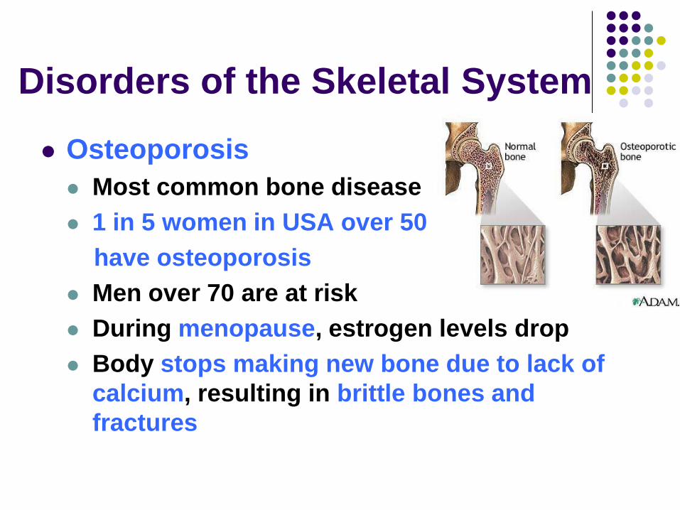
Disorders of the Skeletal System
Osteoporosis
Most common bone disease
1 in 5 women in USA over 50
have osteoporosis
Men over 70 are at risk
During menopause, estrogen levels drop
Body stops making new bone due to lack of
calcium, resulting in brittle bones and
fractures
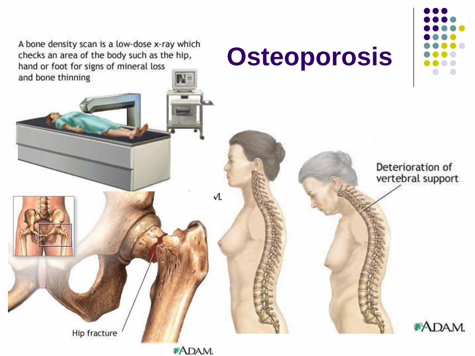
Osteoporosis
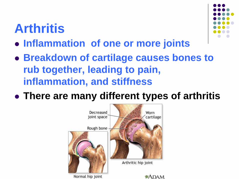
Arthritis Inflammation of one or more joints
Breakdown of cartilage causes bones to
rub together, leading to pain,
inflammation, and stiffness
There are many different types of arthritis
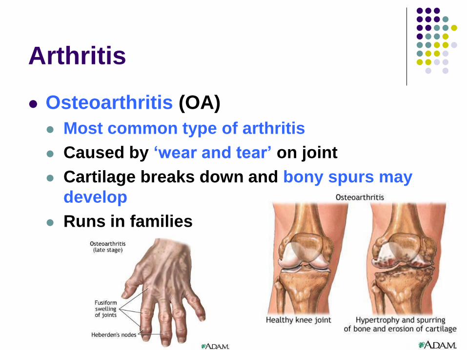
Arthritis
Osteoarthritis (OA)
Most common type of arthritis
Caused by ‘wear and tear’ on joint
Cartilage breaks down and bony spurs may
develop
Runs in families
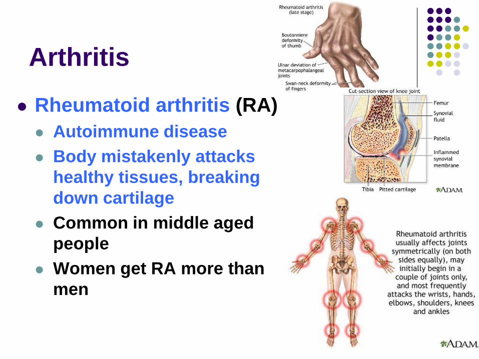
Arthritis
Rheumatoid arthritis (RA)
Autoimmune disease
Body mistakenly attacks
healthy tissues, breaking
down cartilage
Common in middle aged
people
Women get RA more than
men
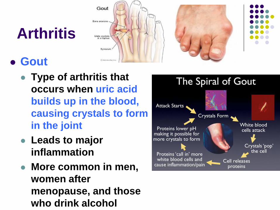
Arthritis
Gout
Type of arthritis that
occurs when uric acid
builds up in the blood,
causing crystals to form
in the joint
Leads to major
inflammation
More common in men,
women after
menopause, and those
who drink alcohol
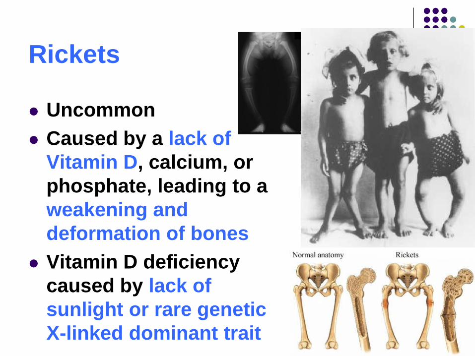
Rickets
Uncommon
Caused by a lack of
Vitamin D, calcium, or
phosphate, leading to a
weakening and
deformation of bones
Vitamin D deficiency
caused by lack of
sunlight or rare genetic
X-linked dominant trait