Embed Size (px)
Citation preview

International J. of Healthcare and Biomedical Research, Volume: 2, Issue: 4, July 2014, Pages 122- 126
122
www.ijhbr.com ISSN: 2319-7072
Case Report:
Skeletal Muscle Cysticercosis
Dr. Kandukuri Mahesh Kumar , Dr. V.Indira , Dr. Ravikanth Soni , Dr. Chintakindi ,
Dr. Divyagna. T , Dr. Konduru Omkareshwar
Department of Pathology, Malla Reddy Institute of Medical Sciences, Suraram, Hyderabad, India
Corresponding author: Dr. Kandukuri Mahesh Kumar
Abstract:
Cysticercosis is a disease caused by cysticercus cellulosae, the larval form of tapeworm, Taenia solium. Cystic-
ercosis is endemic with high prevalence in most of the developing countries because of the co-existence of poor
sanitary conditions and domestic pig raising. Human cysticercosis occurs when eggs are ingested via faecal-oral
transmission from an infected host. The infected human becomes an accidental intermediate host, with development
of cysticercosis within various organs. The parasite has a strong predilection to involve central nervous system
(CNS). Solitary muscular and soft tissue involvement without central nervous system involvement is rare and often
presents a diagnostic challenge. Imaging and histopathological examination plays an important role in establishing
the diagnosis by demonstrating a scolex on MRI and/or ultrasound. We report a rare case of an anterior abdominal
wall cysticercosis diagnosed by ultrasonography and confirmed by histopathology.
Keywords: cysticercosis, skeletal muscle, taenia solium
INTRODUCTION
Cysticercosis is a systemic manifestation caused by
dissemination of the larval form of the pork
tapeworm, Taenia solium. A high prevalence has
been reported from the developing countries because
of the co-existence of poor sanitary conditions and
domestic pig raising without proper veterinary
control or surveillance systems (1) .It occurs mainly in
pork eating nations due to consumption undercooked
pork or measly pork. Humans are the definitive hosts
and carry intestinal adult tapeworm. Intermittent
faecal shedding of proglottids or free eggs occurs,
and the intermediate host (normally pigs) ingests the
excreted eggs in contaminated food or water.
Embryos penetrate the gastrointestinal mucosa of the
pig and are haematogenously disseminated to
peripheral tissues with formation of larval cysts.
When undercooked pork is consumed, an intestinal
tapeworm is formed again, completing the life cycle
of the worm. Human cysticercosis occurs when eggs
are ingested via faecal-oral transmission from a
tapeworm host. The human then becomes an
accidental intermediate host, with development of
cysticercosis within organs. Cysticercosis can affect
various organs the brain, spinal cord, muscles, orbit,
subcutaneous tissues and heart. The clinical
manifestation of the patient varies depending upon
the site of larval encystment, number of cyst and the
extent of associated inflammatory responses. Isolated
involvement of the soft tissues is very rare and can
potentially mimic other soft tissue lesions including
infective, inflammatory and neoplastic lesions.
International J. of Healthcare and Biomedical Research, Volume: 2, Issue: 4, July 2014, Pages 122- 126
123
www.ijhbr.com ISSN: 2319-7072
CASE REPORT
A 16 year old male patient came to the surgical
outpatient department with complaints of swelling
the right side of the abdomen since one month.
Initially no pain was experienced by the patient, but
since one week moderate to severe pain was
experienced. Patient also complained of fever, nausea
and vomiting since 3 days which led to apprehension
in the patient and made the patient report to the
hospital. On general physical examination patient
was conscious, coherent with temperature of 99.8
Fahrenheit. A swelling of size 2 x 2 cm is noted in
the right anterior abdominal wall (lumbar region) in
subcutaneous /muscular plane firm in consistency,
tender, skin over the swelling appeared normal.
Routine work up of patient was done – which was
non contributory. Specific investigations requested
were Ultrasound abdomen, Fine needle aspiration
cytology (FNAC).
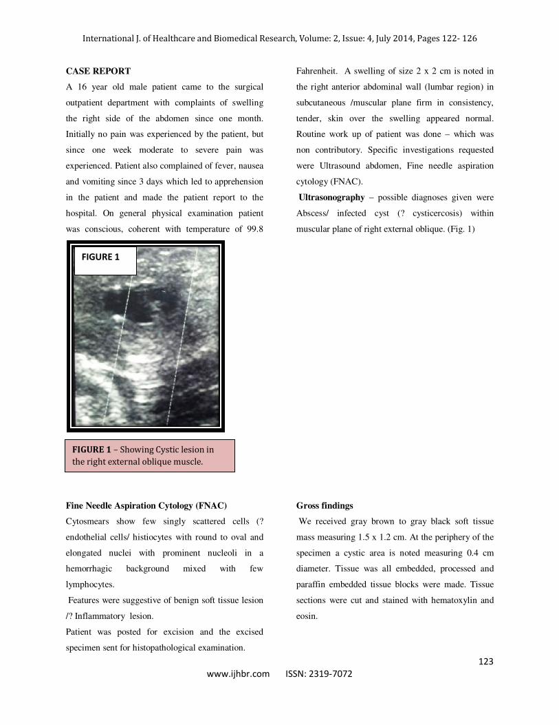
Ultrasonography – possible diagnoses given were
Abscess/ infected cyst (? cysticercosis) within
muscular plane of right external oblique. (Fig. 1)
Fine Needle Aspiration Cytology (FNAC)
Cytosmears show few singly scattered cells (?
endothelial cells/ histiocytes with round to oval and
elongated nuclei with prominent nucleoli in a
hemorrhagic background mixed with few
lymphocytes.
Features were suggestive of benign soft tissue lesion
/? Inflammatory lesion.
Patient was posted for excision and the excised
specimen sent for histopathological examination.
Gross findings
We received gray brown to gray black soft tissue
mass measuring 1.5 x 1.2 cm. At the periphery of the
specimen a cystic area is noted measuring 0.4 cm
diameter. Tissue was all embedded, processed and
paraffin embedded tissue blocks were made. Tissue
sections were cut and stained with hematoxylin and
eosin.
FIGURE 1 – Showing Cystic lesion in
the right external oblique muscle.
FIGURE 1
International J. of Healthcare and Biomedical Research, Volume: 2, Issue: 4, July 2014, Pages 122- 126
123
www.ijhbr.com ISSN: 2319-7072
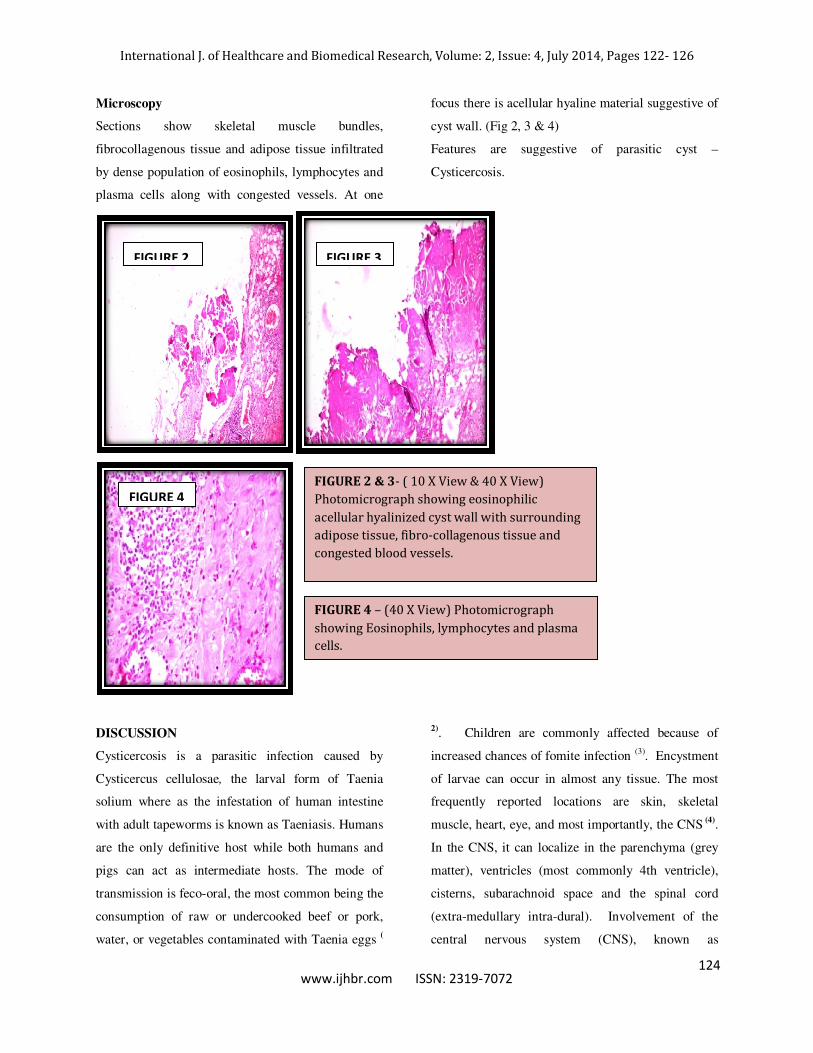
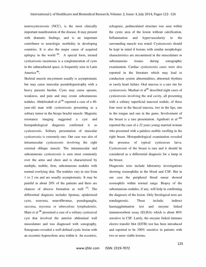
Microscopy
Sections show skeletal muscle bundles,
fibrocollagenous tissue and adipose tissue infiltrated
by dense population of eosinophils, lymphocytes and
plasma cells along with congested vessels. At one
focus there is acellular hyaline material suggestive of
cyst wall. (Fig 2, 3 & 4)
Features are suggestive of parasitic cyst –
Cysticercosis.
DISCUSSION
Cysticercosis is a parasitic infection caused by
Cysticercus cellulosae, the larval form of Taenia
solium where as the infestation of human intestine
with adult tapeworms is known as Taeniasis. Humans
are the only definitive host while both humans and
pigs can act as intermediate hosts. The mode of
transmission is feco-oral, the most common being the
consumption of raw or undercooked beef or pork,
water, or vegetables contaminated with Taenia eggs (
2). Children are commonly affected because of
increased chances of fomite infection (3). Encystment
of larvae can occur in almost any tissue. The most
frequently reported locations are skin, skeletal
muscle, heart, eye, and most importantly, the CNS (4).
In the CNS, it can localize in the parenchyma (grey
matter), ventricles (most commonly 4th ventricle),
cisterns, subarachnoid space and the spinal cord
(extra-medullary intra-dural). Involvement of the
central nervous system (CNS), known as
FIGURE 2 & 3- ( 10 X View & 40 X View)
Photomicrograph showing eosinophilic
acellular hyalinized cyst wall with surrounding
adipose tissue, fibro-collagenous tissue and
congested blood vessels.
FIGURE 4 – (40 X View) Photomicrograph
showing Eosinophils, lymphocytes and plasma
cells.
FIGURE 4
FIGURE 2 FIGURE 3
124
International J. of Healthcare and Biomedical Research, Volume: 2, Issue: 4, July 2014, Pages 122- 126
123
www.ijhbr.com ISSN: 2319-7072
neurocysticercosis (NCC), is the most clinically
important manifestation of the disease. It may present
with dramatic findings, and is an important
contributor to neurologic morbidity in developing
countries. It is also the major cause of acquired
epilepsy in the world (5). A special form, termed
cysticercosis racemosus is a conglomeration of cysts
in the subarachnoid space, is frequently seen in Latin
America (6).
Skeletal muscle encystment usually is asymptomatic
but may cause muscular pseudohypertrophy with a
heavy parasite burden. Cysts may cause spasms,
weakness, and pain and may create subcutaneous
nodules. Abdelwahab et al (6) reported a case of a 40-
year-old man with cysticercosis presenting as a
solitary tumor in the biceps brachii muscle. Magnetic
resonance imaging suggested a cyst and
histopathological diagnosis confirmed it as
cysticercosis. Solitary presentation of muscular
cysticercosis is extremely rare. Our case was also of
intramuscular cysticercosis involving the right
external oblique muscle. The intramuscular and
subcutaneous cysticercosis is seen most commonly
over the arms and chest and is characterized by
multiple, mobile, firm, subcutaneous nodules with
normal overlying skin. The nodules vary in size from
1 to 2 cm and are usually asymptomatic. It may be
painful in about 20% of the patients and there are
chances of abscess formation as well (7). The
differential diagnosis includes lipomas, epidermoid
cysts, neuroma, neurofibromas, pseudoganglia,
sarcoma, myxoma or tuberculous lymphadenitis.
Mani et al (8) presented a case of a solitary cysticercal
cyst that involved the anterior abdominal wall
musculature and was diagnosed with sonography.
Sonograms revealed a well-defined cystic lesion with
an eccentric hyperechoic area within it. An eccentric,
echogenic, pedunculated structure was seen within
the cystic area of the lesion without calcification.
Inflammation and hypervascularity in the
surrounding muscle was noted. Cysticercosis should
be kept in mind if lesions with similar morphologic
characteristics are encountered in the musculature or
subcutaneous tissues during sonographic
examination. Cardiac cysticercosis cases were also
reported in the literature which may lead to
conduction system abnormalities, abnormal rhythms
or rarely heart failure. Oral mucosa is a rare site for
cysticercosis. Mazhari et al(9) described eight cases of
cysticercosis involving the oral cavity, all presenting
with a solitary superficial mucosal nodule, of these
four were in the buccal mucosa, two in the lips, one
in the tongue and one in the gums. Involvement of
the breast is a rare presentation. Agnihotri et al (10)
reported the case of a 22 years young married woman
who presented with a painless mobile swelling in the
right breast. Histopathological examination revealed
the presence of typical cysticercus larva.
Cysticercosis of the breast is rare and it should be
considered as a differential diagnosis for a lump in
the breast.
Diagnostic tests include laboratory investigations
showing eosinophilia in the blood and CSF. But in
our case the peripheral blood smear showed
eosinophils within normal range. Biopsy of the
subcutaneous nodules, if any, will help in confirming
the diagnosis of the lesion. Only Serological tests are
nondiagnostic. These include, indirect
haemagglutination test and enzyme linked
immunosorbent assay (ELISA) which is about 80%
sensitive in CSF. Lately, the enzyme linked immuno
electro transfer blot (EITB) test has been introduced
and reported to be 100% sensitive in patients with
two or more viable lesions.
125
International J. of Healthcare and Biomedical Research, Volume: 2, Issue: 4, July 2014, Pages 122- 126
124
www.ijhbr.com ISSN: 2319-7072
Praziquantel and albendazole have been used
extensively in the treatment of cysticercosis and are
the accepted therapies. Cysticercosis is a preventable
faeco-oral transmitted infection. It is possible to
prevent infection by avoiding undercooked food and
pork, and water contamination with human faeces.
Care should be taken in places with poor hygiene or
meat inspection laws.
CONCLUSION
Cysticercosis of the abdominal wall is a rare entity
and a clinical diagnosis is challenging as it can mimic
other pathological entities which can be differentiated
by histopathology.
REFERENCES
1. García HH, Gilman RH, Gonzalez AE, Verastegui M, Rodriguez S, Garidia C, et al. Hyperendemic human
and porcine Taenia solium infection in perú.
2. Am J Trop Med Hyg 2003; 68: 268-75.
3. González AE, Lopez Urbina T, Tsang B et al. Transmission dynamics of Taenia solium and potential for
pig-to-pig transmission. Parasitol Int. 2006;55:131-5.
4. Jones TC. Cestodes (tapeworms). In: Mandell GL, Douglas RG Jr, Bennet JE, editors. Principles and
Practice of
5. Infectious Diseases. New York: Churchill Livinstone Inc. 1999; Pp:2183-5.
6. Garcia HH, Gilman R, Martinez M, Tsang VC, Pilcher JB, Herrera G et al. Cysticercosis as a major cause
of epilepsy in Peru. Lancet 1993; 341:197-200.
7. Kim JH, Suh SI, Kim JH, Kwon TH, Chung HS. Giant neurocysticercosis cyst in the cerebellar
hemisphere. Neurol Med Chir (Tokyo) 2006; 46:412-4.
8. Abdelwahab IF, Klein MJ, Hermann G, Abdul-Quader M.Solitary cysticercosis of the biceps brachii in a
vegetarian: a rare and unusual pseudotumor. Skeletal Radiol 2003; 32:424-8.
9. Sorvillo FJ, Portigal L, Degiorgio C, Smith L, Waterman SH, Berlin GW, et al. Cysticercosis-Related
Deaths, California. Emerg Infect Dis 2004; 10:465-9.
10. Mani NB, Kalra N, Jain M, Sidhu R. Sonographic diagnosis of a solitary intramuscular cysticercal cyst. J
Clin Ultrasound 2001; 29:472-5.
11. Mazhari NJ; Kumar N; Jain S. Cysticercosis of the oral mucosa: aspiration cytologic diagnosis. J Oral
Pathol Med 2001; 30:187-9.
12. Agnihotri S, Talwar OP, Pudasaini S, Baral R. Cysticercosis of breast - a case report. Pol J Pathol 2006;
57:53-4.
126