Embed Size (px)
Citation preview

doi:10.1016/j.jmb.2009.11.072 J. Mol. Biol. (2010) 395, 995–1006
Available online at www.sciencedirect.com
Single-Molecule Study of DNA Polymerization Activity ofHIV-1 Reverse Transcriptase on DNA Templates
Sangjin Kim1, Charles M. Schroeder1,2 and X. Sunney Xie1⁎
1Department of Chemistry andChemical Biology, HarvardUniversity, 12 Oxford Street,Cambridge, MA 02138, USA2Department of Chemical &Biomolecular Engineering,University of Illinois atUrbana–Champaign, 600 SouthMathews Avenue, Urbana,IL 61801, USAReceived 14 September 2009;received in revised form12 November 2009;accepted 30 November 2009Available online4 December 2009
*Corresponding author. E-mail [email protected] used: HIV-1 RT, hu
immunodeficiency virus type-1 revessDNA, single-stranded DNA; SSB,binding protein; dsDNA, double-strprimer extension rate; ksd, strand dinucleocapsid.
0022-2836/$ - see front matter. Publishe
HIV-1 RT (human immunodeficiency virus-1 reverse transcriptase) is amultifunctional polymerase responsible for reverse transcription of the HIVgenome, including DNA replication on both RNA and DNA templates.During reverse transcription in vivo, HIV-1 RT replicates through varioussecondary structures on RNA and single-stranded DNA (ssDNA) templateswithout the need for a nucleic acid unwinding protein, such as a helicase. Inorder to understand the mechanism of polymerization through secondarystructures, we investigated the DNA polymerization activity of HIV-1 RTon long ssDNA templates using a multiplexed single-molecule DNA flow-stretching assay.We observed that HIV-1 RT performs fast primer extensionDNA synthesis on single-stranded regions of DNA (18.7 nt/s) and switchesits activity to slow strand displacement synthesis at DNA hairpin locations(2.3 nt/s). Furthermore, we found that the rate of strand displacementsynthesis is dependent on the GC content in hairpin stems and templatestretching force. This indicates that the strand displacement synthesisoccurs through a mechanism that is neither completely active nor passive:that is, the opening of the DNA hairpin is driven by a combination of freeenergy released during dNTP (deoxyribonucleotide triphosphate) hydro-lysis and thermal fraying of base pairs. Our experimental observationsprovide new insight into the interchanging modes of DNA replication byHIV-1 RT on long ssDNA templates.
Published by Elsevier Ltd.
Keywords: HIV-1 reverse transcriptase; single molecule; DNA flow-stretching assay; DNA hairpin; strand displacement synthesis
Edited by J. KarnIntroduction
HIV-1 RT (human immunodeficiency virus-1reverse transcriptase) is a 66-/51-kDa heterodimericDNA polymerase that synthesizes a double-stranded proviral DNA from a viral RNA genome.The 66-kDa subunit is derived from the viral polgene and contains enzymatic active sites for DNApolymerization and RNase (ribonuclease) H activi-ty, whereas the 51-kDa subunit is derived fromproteolytic cleavage of the RNase H domain in the66-kDa subunit and lacks any catalytic role.1–3 In
ess:
manrse transcriptase;single-stranded DNAanded DNA; kpe,splacement rate; NC,
d by Elsevier Ltd.
vivo, HIV-1 RT possesses three distinct enzymaticactivities: (1) polymerization of cDNA on RNAtemplates yielding RNA/DNA duplexes; (2) enzy-matic degradation of RNA templates; and (3)synthesis of DNA using cDNA templates. HIV-1RT also executes strand displacement synthesis onan ∼634-base-long duplex DNA region (longterminal repeat sequences) in order to completereplication of its genome.4,5 Although multifunc-tional, HIV-1 RT is not an exceedingly efficient DNApolymerase. DNA polymerization by HIV-1 RT onboth RNA and DNA templates exhibits a slowenzymatic rate (maximum rate of single-nucleotideincorporation of ∼33 s−1 at 37 °C),6 low processivity(1–300 nt, depending on the template sequence),4,7and poor fidelity (error frequency of 1 in 5900 ntpolymerized on the DNA template).8 Because of itspivotal role in the HIV-1 life cycle and as a drugtarget for the clinical treatment of HIV infection andAIDS, there have been extensive biochemical studieson HIV-1 RT, including several kinetic studies forsingle-nucleotide addition.6,9

996 DNA Polymerization Activity of HIV-1 RT
An interesting open question for HIV-1 RT interms of viral life cycle is the effect of the largenumber of stable RNA or DNA secondary structuresin the HIV genome during reverse transcription.During DNA replication, HIV-1 RT uses longstretches of single-stranded RNA or DNAmoleculesas templates. However, unlike most DNA poly-merases that form a replisome, HIV-1 RT does nothave customary accessory proteins, such as a heli-case, sliding clamp, or single-stranded DNA bindingproteins (SSBs), which accelerate destabilization ofthe nucleic acid duplex on the template and increaseprocessivity of DNA polymerase. Hence, secondarystructures may serve as physical barriers for proces-sive enzymatic synthesis, and HIV-1 RT must have adifferent mechanism to cope with secondary struc-tures on the template. Based on the currentlyacceptedHIV infectionmechanism, stable secondarystructures induce pausing of DNA synthesis, andenzymatic pauses during DNA replication mayresult in DNA recombination through templateswitching or base misincorporation.10,11 In particu-lar, recombination hot spots were found wherestable stem–loop structures are present in the HIVgenome, such as in the trans-activating responseregion.12,13 At present, however, the molecularmechanism responsible for the ability of HIV-1 RTto replicate through successive secondary structureson a whole genome is not well understood becausebulk studies mainly used short nucleic acids with asingle hairpin for replication templates. The dynamicnature of HIV-1 RT can be easily obscured in bulkexperiments when a population of RT molecules isstudied because the asynchronous behavior of eachenzyme molecule is not well represented by anensemble average. Furthermore, it is experimentallychallenging to study the sequence dependence oftransient enzymatic pauses on templates comparablein length with the 9.7-kb HIV genome using bulkenzymology techniques.In this work, we investigated the DNA polymer-
ization activity of HIV-1 RT using a single-moleculetechnique based on the hydrodynamic manipula-tion of DNA molecules. Single-molecule techniquesoffer the ability to monitor multiple turnovers ofthe polymerase in real time without ensembleaveraging and therefore present advantageousmethods to study the unsynchronized, sequence-dependent dynamics during polymerization on along DNA template.14–17 Unlike short oligonucleo-tide DNA templates heretofore used for HIV-1 RTstudies,7,11,18–21 bacteriophage λ (48.5 kb) containsvariable DNA sequences and secondary structuresin a row as commonly encountered in HIV genomein vivo.22 We found that during polymerization onλ DNA, HIV-1 RT can switch between two distinctmodes of DNA synthesis activity—fast primerextension and slow strand displacement, depend-ing on whether downstream DNA is singlestranded or hairpin structured. The rate of stranddisplacement synthesis is further analyzed as afunction of the base sequence of hairpin stems andthe template stretching force. The dependence of
the strand displacement rate (ksd) on GC-richhairpins and high template force led us toconclude that the free energy contribution ofHIV-1 RT in base-pair destabilization is significant,such that HIV-1 RT falls in between two limitingmechanisms—either the free energy release fromdNTP (deoxyribonucleotide triphosphate) hydro-lysis drives opening of the DNA duplex or thepolymerase waits for thermal fraying of the DNAduplex. These new findings allow for a morequantitative understanding of strand displacementsynthesis catalyzed by HIV-1 RT and offer newinsights on hairpin-induced switching of HIV-1RT activity along viral RNA or cDNA templatesin vivo.
Results
Single-molecule trajectory of DNA synthesiscatalyzed by HIV-1 RT on single-strandedDNA template
In our single-molecule experiments, individualsingle-strandedDNA (ssDNA) templates are stretchedby hydrodynamic flow in amicrochannel (Fig. 1a). Thetermini of λ-phage DNA molecules are specificallyend-functionalized with biotin and digoxigeninseparately, such that the biotin-functionalized termi-nus of DNA is linked to a streptavidin-coated surfaceof a flow cell and the digoxigenin end is tetheredto 2.8-μm magnetic beads functionalized with anti-digoxigenin (Materials and Methods).23–25 Usingthe differential extension of ssDNA and double-stranded DNA (dsDNA) at a constant stretchingforce (Fig. 1b),wewere able tomonitor the enzymaticconversion of ssDNA to dsDNAon a single-moleculelevel based on the change in DNA extension overtime, which is directly measured by observationof the tethered bead position as a function of time(Fig. 1c).We studied the primer extension DNA replication
activity of HIV-1 RT on flow-stretched ssDNAtemplates at room temperature (21 °C). An enzymesolution containing 11 nM HIV-1 RT and asaturating amount of dNTP (200 μM) was infusedat a flow rate, resulting in a template force of 3.7 pN(Materials and Methods). In the primer extensionassay, we observed processive stretches of DNAsynthesis occasionally interrupted by a slow modeof DNA replication, which appeared as a plateauranging in time from a few seconds to 200 s (anexample trace is shown in Fig. 1d).When instantaneous rates of the DNA polymeri-
zation trajectories are calculated and drawn into ahistogram (Fig. 1e;Materials andMethods), there area dominant peak (Gaussian shape) near 1 nt/s due tothe contribution of the apparent plateaus andanother peak near ∼20 nt/s with a long tail. Incontrast, when instantaneous rates are calculatedfrom bead trajectories that do not show any DNAsynthesis events, the histogram is a simple Gaussian
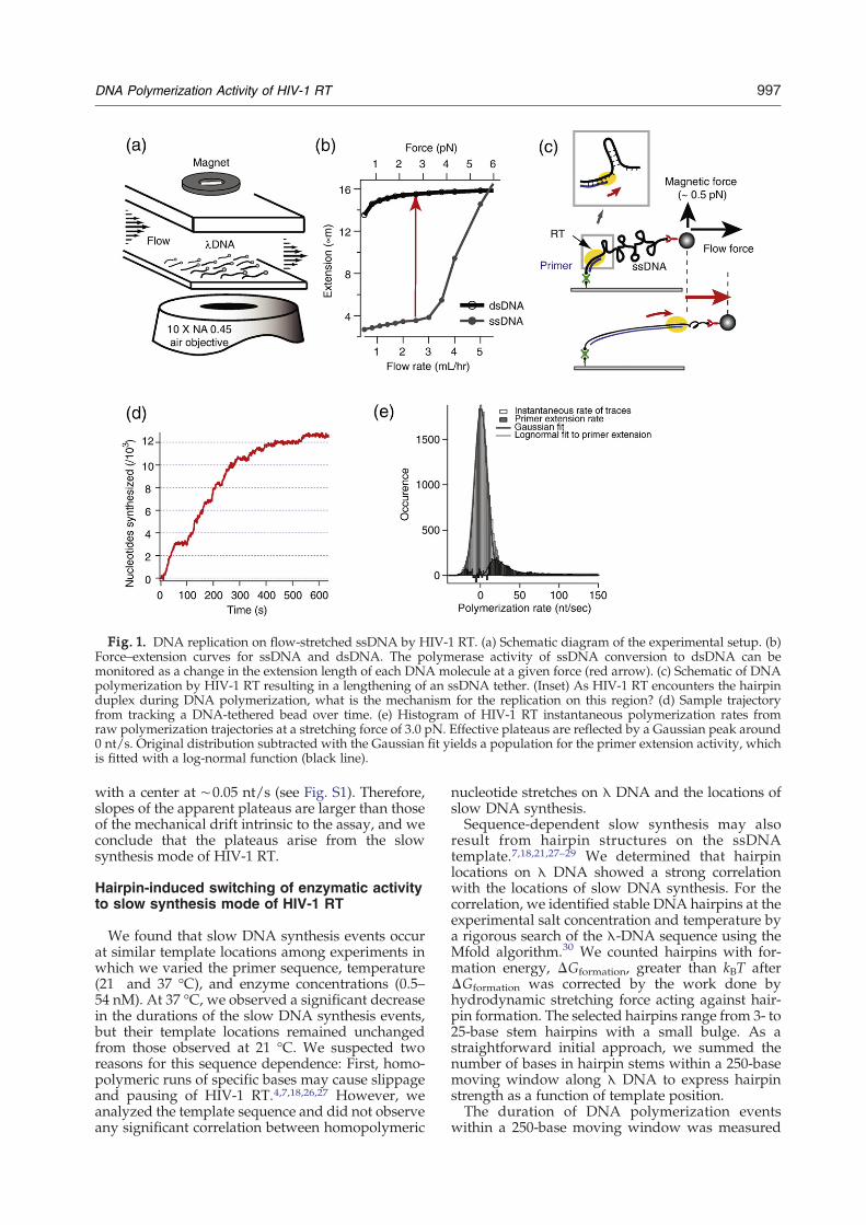
Fig. 1. DNA replication on flow-stretched ssDNA by HIV-1 RT. (a) Schematic diagram of the experimental setup. (b)Force–extension curves for ssDNA and dsDNA. The polymerase activity of ssDNA conversion to dsDNA can bemonitored as a change in the extension length of each DNA molecule at a given force (red arrow). (c) Schematic of DNApolymerization by HIV-1 RT resulting in a lengthening of an ssDNA tether. (Inset) As HIV-1 RT encounters the hairpinduplex during DNA polymerization, what is the mechanism for the replication on this region? (d) Sample trajectoryfrom tracking a DNA-tethered bead over time. (e) Histogram of HIV-1 RT instantaneous polymerization rates fromraw polymerization trajectories at a stretching force of 3.0 pN. Effective plateaus are reflected by a Gaussian peak around0 nt/s. Original distribution subtracted with the Gaussian fit yields a population for the primer extension activity, whichis fitted with a log-normal function (black line).
997DNA Polymerization Activity of HIV-1 RT
with a center at ∼0.05 nt/s (see Fig. S1). Therefore,slopes of the apparent plateaus are larger than thoseof the mechanical drift intrinsic to the assay, and weconclude that the plateaus arise from the slowsynthesis mode of HIV-1 RT.
Hairpin-induced switching of enzymatic activityto slow synthesis mode of HIV-1 RT
We found that slow DNA synthesis events occurat similar template locations among experiments inwhich we varied the primer sequence, temperature(21 and 37 °C), and enzyme concentrations (0.5–54 nM). At 37 °C, we observed a significant decreasein the durations of the slow DNA synthesis events,but their template locations remained unchangedfrom those observed at 21 °C. We suspected tworeasons for this sequence dependence: First, homo-polymeric runs of specific bases may cause slippageand pausing of HIV-1 RT.4,7,18,26,27 However, weanalyzed the template sequence and did not observeany significant correlation between homopolymeric
nucleotide stretches on λ DNA and the locations ofslow DNA synthesis.Sequence-dependent slow synthesis may also
result from hairpin structures on the ssDNAtemplate.7,18,21,27–29 We determined that hairpinlocations on λ DNA showed a strong correlationwith the locations of slow DNA synthesis. For thecorrelation, we identified stable DNA hairpins at theexperimental salt concentration and temperature bya rigorous search of the λ-DNA sequence using theMfold algorithm.30 We counted hairpins with for-mation energy, ΔGformation, greater than kBT afterΔGformation was corrected by the work done byhydrodynamic stretching force acting against hair-pin formation. The selected hairpins range from 3- to25-base stem hairpins with a small bulge. As astraightforward initial approach, we summed thenumber of bases in hairpin stems within a 250-basemoving window along λ DNA to express hairpinstrength as a function of template position.The duration of DNA polymerization events
within a 250-base moving window was measured

Fig. 2. Sequence-dependent strand displacement syn-thesis of HIV-1 RT. Average dwell time (blue dotted line)measured along a λ-DNA template (average of 64 traces)and the number of bases involved in hairpin stems in a250-base moving window (green continuous line). Corre-lation analysis between the average dwell time andhairpin stem number density yields a Pearson coefficientof 0.75 up to 10,000 nt.
Fig. 3. Enzyme concentration dependence of durationsof slowDNA synthesis. (a) Histogram of durations of slowDNA synthesis regions observed during DNA polymeri-zation of 11 nMHIV-1 RT. The histogram is well-fitted to asingle exponential function with a decay constant 0.026±0.001 s−1. (b) Decay constants for the single exponential fitto histograms of slow synthesis durations measured atenzyme concentrations between 1 and 54 nM.
998 DNA Polymerization Activity of HIV-1 RT
from 64 experimental trajectories (Fig. 2; also seeSupplementary Material). The amplitude of thedwell time function accounts for durations of slowsynthesis and the number of slow synthesis eventswithin the moving window; therefore, the functionstatistically reflects the relative “strength” of slowsynthesis along the template sequence. The Pearsoncorrelation coefficient between hairpin strength andthe duration of DNA polymerization was 0.75 up to10,000 nt. Hence, the observed slow synthesis byHIV-1 RT is interpreted as hairpin-induced switch-ing of HIV-1 RT activity to slow DNA synthesis,namely strand displacement synthesis.
Enzyme concentration-independent kinetics ofslow DNA synthesis
Given that hairpins induce HIV-1 RT switching toa much slower mode of synthesis, one easilyquestions whether HIV-1 RT dissociates at thestem of hairpins and, if so, whether the rebindingtime dominates the duration of the slow synthesis.Enzyme dissociation at hairpin stems is likely tooccur considering the low processivity of the HIV-1RT and the process of template switching in vivo,during which RT jumps between two copies of theviral genome. We investigated the contributions ofenzyme dissociation and rebinding during slowsynthesis steps by measuring the time duration ofslow synthesis regions at various RT concentrations(Materials and Methods). If enzyme rebinding is themajor rate-limiting process during slow DNAsynthesis events, then the time durations should bedependent on the enzyme concentration due to thewaiting time between enzyme dissociation andrebinding.For experiments with RT concentrations in the
range of 1–54 nM, the distribution of slow synthesisdurations is well-fitted with a single exponentialdecay with similar decay constants (see Fig. 3a andb; mean of the similar decay constants was 0.027±
0.0008 s−1). When the concentration of HIV-1 RTwas less than 1 nM, the distribution is better fit witha double exponential function, suggesting that thereis more than one major rate-limiting process andthat enzyme dissociation and rebinding processeslikely play a role in the slow synthesis eventsobserved in between fast stretches of DNA synthe-sis. Therefore, enzyme dissociation and rebindingare not rate-limiting processes during slow synthesisevents if the enzyme concentration is in excess of1 nM (our experiments were mainly conducted at anenzyme concentration of 11 nM). These observationsare consistent with the reported dissociation con-stant for interaction of HIV-1 RT and primer–template DNA between 0.65 and 5 nM6,31 andwith a bulk-level experiment demonstrating thatsecondary structures on DNA templates affectneither RT enzyme dissociation rate nor the enzymedissociation constant compared with those mea-sured on a hairpin-free template.21 Because crystalstructures of HIV-1 RT have shown that HIV-1 RThas a large binding cleft,1–3 it is possible that thedissociation constant of HIV-1 RT on a DNAtemplate may be tolerant to the hairpin structure.

Fig. 4. Active and passive mechanisms for stranddisplacement DNA synthesis by HIV-1 RT near hairpinlocations. (a) Active strand displacement. Yellow imprintdemonstrates the interaction between the enzyme and theDNA, which destabilizes the DNA junction (red base). (b)Passive strand displacement. The enzyme waits for thejunction opening by thermal fluctuations. In both cases, “nbases” is the number of bases open in front of the enzymein each turnover of strand displacement synthesis.
999DNA Polymerization Activity of HIV-1 RT
We further examined the role of hairpin thermalopening in the slow synthesis events. We found thatthe complete opening of a hairpin by thermalfluctuations has a much longer timescale than theduration of the slow synthesis (see SupplementaryMaterial). Considering these observations, we con-clude that the rate-limiting process underlying theobserved slow synthesis is based solely on enzymekinetics of strand displacement synthesis.
χ2 fitting of single-molecule trajectories todetermine the two synthesis rates
Assuming that primer extension and strand dis-placement synthesis are the rate-limiting processesduring fast synthesis at open, nonhybridized basesand slow synthesis at hairpin stem bases, respectively,we performed a weighted least-squares fitting of ourexperimental trajectories with two parameters: theprimer extension rate (kpe) and the ksd (Materials andMethods). We determined a kpe of 18.7±6.0 nt/s and aksd of 2.3±0.56 nt/s. Within the error range, the kpe issimilar to the 22.1±2.4 nt/s acquired from ahistogram of instantaneous slopes in raw trajectories.The ksd is higher than the 0.28 nt/s measured at 37 °Cin vitro5 or 0.05–0.10 nt/s measured using a single-molecule FRET technique32 and lower than the 2.98–4.5 nt/s measured with Moloney murine leukemiavirus RT.33,34 Clearly, the sequence of the templateand stretching force can affect the measured ksd, asdescribed in the next section.
Active or passive mechanism for stranddisplacement synthesis of HIV-1 RT
Active and passive mechanisms have been used todescribe the duplex unwinding activity of heli-cases.35,36 Briefly, the active mechanism describes ahelicase as a strong molecular motor such that itconverts chemical energy from NTP hydrolysis tounzip a duplexed template with high efficiency. Onthe contrary, the passive mechanism states that ahelicase translocates to the next base after theimmediately adjacent downstream base pair opensas a result of thermal fluctuations. Indeed, thesemechanisms are not inherently contradictory, as theyrepresent two extremes of an energetic continuumand describe helicases that exhibit a partial passivemechanism of unwinding, such as T7 helicase.37
While the speed of active unzipping can be as high asthe velocity of translocation on a single-strandedtemplate, the rate of passive unzipping is limited bythermal breathing of a base pair at the duplexjunction. Recent theoretical work predicted that theduplex unwinding rate of a strictly passive helicasewith a kinetic step size of 1 bp would be seven timesslower than the rate of translocation along a single-stranded nucleic acid template.38
Our correlation analysis shown in Fig. 2 consid-ered the number of bases in hairpin stems as aputative “strength” of each hairpin and is in reaso-nably good agreement with the active mechanism ofstrand displacement of HIV-1 RT. The active me-
chanism suggests that the enzyme may efficientlycouple the energy released from dNTP hydrolysis(∼20 kBT) to unzip the duplex junction, such that thesmall base-pair energy difference between AT andGC (∼1-kBT range) is not large enough to contributeto enzyme dynamics (Fig. 4a). Hence, the timerequired for RT to proceed through a hairpin isproportional to the number of bases in the hairpinstem and is negligibly affected by variation in basecontent in the hairpin stem.However, the rate of HIV-1 RT strand displace-
ment is nearly 10-fold slower than the rate of primerextension, which suggests that an additional duplexmelting step in the strand displacement processmay be rate limiting (Fig. 4b).38,39 If we considerthermal breathing of base pairs during stranddisplacement synthesis, we should weight GCover AT base pairs in hairpin stems to estimatehairpin strength in the correlation analysis. Weestimated base-pair opening energies using litera-ture values of 2.9 kBT for a GC pair (EGC) and 1.3 kBTfor an AT pair (EAT).
40 These values includecontributions from unpairing of hydrogen bonds,unstacking from the next base, and base rearrange-ments. Based on these free energies for baseopening, we estimated transition-state activationenergies for base opening (ΔG‡) using a propor-tionality factor β (i.e., Brønsted value).41 The ratio oftime required for thermal melting of a GC base pair
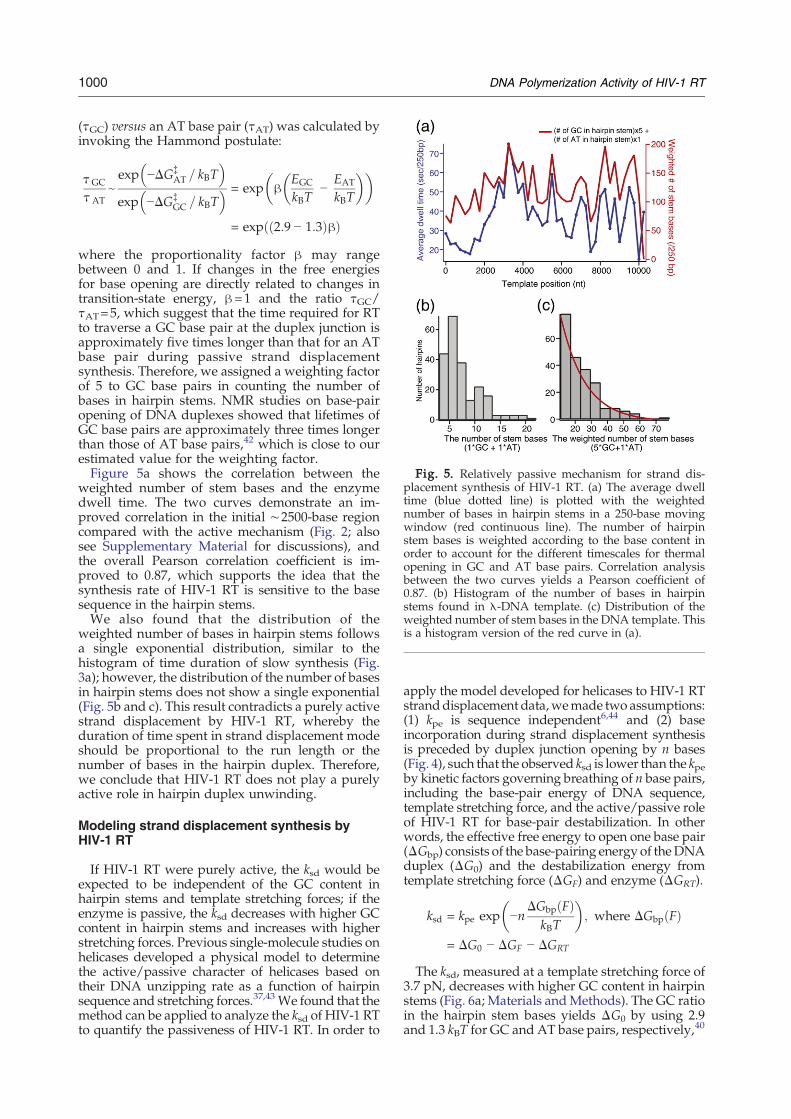
Fig. 5. Relatively passive mechanism for strand dis-placement synthesis of HIV-1 RT. (a) The average dwelltime (blue dotted line) is plotted with the weightednumber of bases in hairpin stems in a 250-base movingwindow (red continuous line). The number of hairpinstem bases is weighted according to the base content inorder to account for the different timescales for thermalopening in GC and AT base pairs. Correlation analysisbetween the two curves yields a Pearson coefficient of0.87. (b) Histogram of the number of bases in hairpinstems found in λ-DNA template. (c) Distribution of theweighted number of stem bases in the DNA template. Thisis a histogram version of the red curve in (a).
1000 DNA Polymerization Activity of HIV-1 RT
(τGC) versus an AT base pair (τAT) was calculated byinvoking the Hammond postulate:
H GC
H ATe exp −DG‡
AT = kBT� �
exp −DG‡GC = kBT
� � = exp hEGC
kBT−
EAT
kBT
� �� �
= exp 2:9 − 1:3ð Þhð Þwhere the proportionality factor β may rangebetween 0 and 1. If changes in the free energiesfor base opening are directly related to changes intransition-state energy, β=1 and the ratio τGC/τAT=5, which suggest that the time required for RTto traverse a GC base pair at the duplex junction isapproximately five times longer than that for an ATbase pair during passive strand displacementsynthesis. Therefore, we assigned a weighting factorof 5 to GC base pairs in counting the number ofbases in hairpin stems. NMR studies on base-pairopening of DNA duplexes showed that lifetimes ofGC base pairs are approximately three times longerthan those of AT base pairs,42 which is close to ourestimated value for the weighting factor.Figure 5a shows the correlation between the
weighted number of stem bases and the enzymedwell time. The two curves demonstrate an im-proved correlation in the initial ∼2500-base regioncompared with the active mechanism (Fig. 2; alsosee Supplementary Material for discussions), andthe overall Pearson correlation coefficient is im-proved to 0.87, which supports the idea that thesynthesis rate of HIV-1 RT is sensitive to the basesequence in the hairpin stems.We also found that the distribution of the
weighted number of bases in hairpin stems followsa single exponential distribution, similar to thehistogram of time duration of slow synthesis (Fig.3a); however, the distribution of the number of basesin hairpin stems does not show a single exponential(Fig. 5b and c). This result contradicts a purely activestrand displacement by HIV-1 RT, whereby theduration of time spent in strand displacement modeshould be proportional to the run length or thenumber of bases in the hairpin duplex. Therefore,we conclude that HIV-1 RT does not play a purelyactive role in hairpin duplex unwinding.
Modeling strand displacement synthesis byHIV-1 RT
If HIV-1 RT were purely active, the ksd would beexpected to be independent of the GC content inhairpin stems and template stretching forces; if theenzyme is passive, the ksd decreases with higher GCcontent in hairpin stems and increases with higherstretching forces. Previous single-molecule studies onhelicases developed a physical model to determinethe active/passive character of helicases based ontheir DNA unzipping rate as a function of hairpinsequence and stretching forces.37,43 We found that themethod can be applied to analyze the ksd of HIV-1 RTto quantify the passiveness of HIV-1 RT. In order to
apply the model developed for helicases to HIV-1 RTstranddisplacement data,wemade two assumptions:(1) kpe is sequence independent6,44 and (2) baseincorporation during strand displacement synthesisis preceded by duplex junction opening by n bases(Fig. 4), such that the observed ksd is lower than the kpeby kinetic factors governing breathing of n base pairs,including the base-pair energy of DNA sequence,template stretching force, and the active/passive roleof HIV-1 RT for base-pair destabilization. In otherwords, the effective free energy to open one base pair(ΔGbp) consists of the base-pairing energy of theDNAduplex (ΔG0) and the destabilization energy fromtemplate stretching force (ΔGF) and enzyme (ΔGRT).
ksd = kpe exp −nDGbp Fð Þ
kBT
� �; where DGbp Fð Þ
= DG0 − DGF − DGRT
The ksd, measured at a template stretching force of3.7 pN, decreases with higher GC content in hairpinstems (Fig. 6a; Materials andMethods). The GC ratioin the hairpin stem bases yields ΔG0 by using 2.9and 1.3 kBT for GC and AT base pairs, respectively,40
Fig. 6. Sequence and stretching force dependence of ksdand kpe. (a) ksd as a function of GC content in hairpin stems.The exponential fit based on the simple model for helicaseunzipping yields n=3 bases and ΔGRT=1.3 kBT. (b) Force-dependent kpe of HIV-1 RT. (c) The ratio between ksd andkpe is shown as a function of template stretching force. Thebest fit is done with n=4 bases and ΔGRT=1.4 kBT (seeSupplementary Material for analysis).
1001DNA Polymerization Activity of HIV-1 RT
andΔGF and kpe can be obtained experimentally (seeSupplementary Material). Fitting of data in Fig. 6ayields n=2.6±0.30 bases and ΔGRT=1.2±0.09 kBT(estimated standard deviation of the fit). For theclosest integer value of n=3 bases, ΔGRT=1.3 kBT.In template force-dependent measurements, we
observed kpe changes with template forces (Fig. 6b;Materials and Methods). In order to avoid sequence-dependent variation of ksd, we calculated ksd aroundthe template position of 3700 nt where the strongest
hairpin occurs in the first 10,000 nt of λ DNA(average GC ratio=0.44). We observed that ksdmoderately increases with template forces (Fig. 6c).The best fit for ksd(F)/kpe(F) was obtained withn=3.8±2.29 bases and ΔGRT=1.4±0.30 kBT. For theclosest integer value of n=4 bases, ΔGRT=1.4 kBT. IfHIV-1 RT were a purely passive enzyme, ΔGRTshould equal 0. Our results indicate that thecontribution of the HIV-1 RT enzyme in destabili-zation of the DNA duplex is comparable with 72% ofbase-pairing energy. Hence, strand displacement ofHIV-1 RT cannot be explained by either a complete-ly passive or a purely active mechanism; however,HIV-1 RT stands somewhere in between these twoextreme mechanisms.
Discussion
How do other DNA polymerases respond tohairpins on the template?
The hairpin-induced switch in DNA polymeriza-tion activity has been observed in other DNApolymerases in vitro (e.g., DNA polymerase α,45,46
Escherichia coliDNApolymerase II47 and polymeraseIII,48 and T7 or T4 bacteriophage DNA poly-merase21,49). However, addition of helicases or SSBoftentimes resolved the inhibitory effect of second-ary structures on DNA replication by these DNApolymerases. With our single-molecule assay, wealso observed frequent pausing of T7 DNA poly-merase spanning as long as a few tens of secondsduring primer extension replication on flow-stretched λ-DNA templates.25 On the contrary, Lee,et al. found no significant pause of T7 DNA poly-merase during leading-strand synthesis in thepresence of helicase (gp4).17 With phi29 DNApolymerase, we observed a small number of lesspronounced enzymatic pausing events duringprimer extension synthesis on ssDNA templatesand little or no evidence of enzymatic pausing eventsduring strand displacement replication on flow-stretched dsDNA templates (in all single-moleculeexperiments, the enzyme concentration significantlyexceeded the dissociation constant for the enzyme/primer–template pair).23 Enzymatic pauses of DNApolymerases at template secondary structures maybe a general phenomenon in many DNA poly-merases, but each DNA polymerase shows adifferent degree of sensitivity to the secondarystructures. Moreover, most DNA polymeraseswork with a helicase or an SSB in the context ofa large macromolecular protein superstructure.Efficient strand displacement activity in some DNApolymerases, such as phi29 DNA polymerase, couldalso facilitate DNA polymerization through second-ary structures. HIV-1 RT does not function in areplisome with customary accessory proteins thatdestabilize secondary structures and exhibits anintrinsically inefficient strand displacement activity;therefore, HIV-1 RT can be significantly affected bytemplate secondary structures.7,11,18,21,50

1002 DNA Polymerization Activity of HIV-1 RT
Comparison with previous kinetic studies
Our data suggest that HIV-1 RT follows arelatively passive mechanism during strand dis-placement synthesis on secondary structures. Kineticmeasurements of single-nucleotide incorporation byHIV-1 RT at pause or nonpause template sitesprovided a basis for understanding our data.20,21
Suo and Johnson determined that polymerization atthe template pause sites (generally occurring nearthe first base of a DNA hairpin stem) displaysbiphasic kinetics—small reaction amplitudes of fastphase (10–20 s−1) and large amplitudes of slowphase (0.02–0.07 s−1); only single-phase kinetics wasobserved at nonpause template DNA sites, com-posed solely of a large amplitude fast phase (33–37 s−1).21 To explain their data, the authors proposeda model whereby HIV-1 RT binds to DNA either in aproductive or in a nonproductive state at pause sites.A fast kinetic phase suggests that HIV-1 RTproductively turns over to add a single nucleotideto the extending DNA chain; however, a slow kineticphase results from the nonproductive enzyme state,which must be converted to the productive stateupon melting of the next stem base pair. We canrelate these two distinct kinetic schemes for pauseand nonpause sites to two distinct kinetic patterns inour single-molecule assay: replication trajectoryregions exhibiting positive slopes represent fastsingle kinetics, whereas slow synthesis regionsrepresent the dominant slow phase in biphasickinetics at pause sites. The timescale of the slow-phase kinetics was approximately 30 s,21 whichagrees with the range of our observed slowsynthesis durations. While a conformational changeof the polymerase is likely the rate-limiting step atnonpause sites,6,9,19 thermal melting of the nextstem base pair, or conversion of the nonproductivestate to the productive state, may serve as the rate-limiting step at pause sites, which supports arelatively passive mechanism of strand displace-ment synthesis for HIV-1 RT.
Force dependence of kpe
Template force dependence of HIV-1 RT activityof DNA polymerization allowed for a comparison ofHIV-1 RT with other DNA polymerases that werestudied at single-molecule level. Although molecu-lar dynamics simulations have generated an inter-esting discussion regarding polymerization rate as afunction of force,51 we focused our attention on therelatively low stall force of HIV-1 RT (Fig. 6b). Thestall force for HIV-1 RT was ∼7 pN, whereas T7DNA polymerase stalls at ∼34 pN,14 E. coli Klenowfragment stalls at ∼20 pN,15 and phi29 DNApolymerase stalls at ∼37 pN;52 in addition, thisstall force is lower than that measured by atomicforce microscopy where HIV-1 RT molecules wereadsorbed on the cantilever tip. The atomic forcemicroscopy experiment was conducted by applyinga stretching force to both the enzyme molecule andthe ssDNA template, as opposed to application of a
small stretching force to DNA templates in ourassay.53 We found that phi29 DNA polymeraseshows robust DNA polymerization activity attemplate forces far in excess of 8 pN in the sameexperimental conditions, so the strong dependenceof DNA synthesis on the template stretching force isan intrinsic property of HIV-1 RT. The strongsensitivity of HIV-1 RT replication to templatestretching force can be understood when consider-ing the low processivity of HIV-1 RT and its weakinteraction with DNA template. In the DNApolymerase active site, amino acid residues of theDNA polymerase form a fine network with thetemplate DNA base, incoming dNTP, two Mg2+
ions, and water molecules, and the correct coordi-nation or interaction among these elements isrequired for the phosphodiester bond formationstep.54,55 As higher stretching force is applied to thessDNA template, the probability of incoming dNTPpositioning itself correctly in the fine coordinationnetwork of the active site decreases. Compared withfast and highly processive T7 DNA polymerase,56
HIV-1 RT does not bind as strongly to the DNAtemplate, and the protein surface area around theDNA polymerization active site in HIV-1 RT is moreopen compared with that in T7 DNA polymerase.Previous mutational studies also showed that theflexible active site may explain the low fidelity ofHIV-1 RT.57 We conjecture that these loose struc-tural characteristics may allow HIV-1 RT to toleratesecondary structures on single-stranded templatesbut render HIV-1 RT unable to replicate ontemplates stretched with even moderate forces.
HIV-1 RT strand displacement mechanism
The sequence dependence in correlation analysis(Fig. 5a) and sequence-dependent ksd (Fig. 6a) ruleout a purely active mechanism for HIV-1 RT.However, the energetic contribution of the enzyme(ΔGRT) near 1.4 kBT opposes a purely passivemechanism for HIV-1 RT as well. Although theexact mechanism is not known, HIV-1 RT mayundergo a conformational change after dNTP hydro-lysis, which contributes nearly 72% of the averagebase-pairing energy and promotes template duplexunwinding. This suggests that HIV-1 RT cannot beexplained by either a purely active or an entirelypassive mechanism. HIV-1 RT is another examplewhere the extreme active or passive classificationcannot be applied for duplex unzipping processes.37
A previous in vitro study using KMnO4 oxidationrevealed that HIV-1 RT melts two bases ahead ofthe primer terminus in a strand displacementconstruct.58,59 The small difference between ourresults of n=3 or 4 and 2 may be attributed todifferences in experimental design and simpleassumptions in the fitting model.Note that fitting of the model to the sequence-
dependent ksd (Fig. 6a) yields similar values of n andΔGRT as from force-dependent rates (Fig. 6c).Analysis of the unzipping rate as a function of GCcontent in hairpin stems was not tried in previous
1003DNA Polymerization Activity of HIV-1 RT
studies where DNA templates with a single hairpinwere used. By using λ-DNA templates, we hadvarious naturally occurring hairpins in one templatemolecule, and we were able to study ksd as afunction of hairpin stem sequence as well.
Significance of hairpin-induced activity of HIV-1RT for viral survival
Why does the HIV-1 genome have severalhairpins,22 and why does HIV-1 RT remain aninefficient polymerase with high mutation rates?These are interesting and relevant questions. Fre-quent mutation of the HIV-1 genome is critical forviral proliferation and results in continued resistanceto antiretroviral drugs. In addition to lack of anintrinsic proofreadingmechanism and low fidelity ofHIV-1 RT,6,8 enzymatic pausing events at homopoly-meric sequences or at hairpin structures have beenproposed to enhance viral mutagenesis.10,11,26 Inparticular, stable secondary structures on RNA andnewly synthesized cDNA facilitate strand transferand template switching, leading to genetic recombi-nation in the retrovirus.7,10,11,13 Also, a recent studyon secondary structures in the HIV-1 RNA genomereported the role of stem–loop structures in ribo-somal pausing during translation, which may resultin frame shifting.22 Therefore, a large number ofstable secondary structures in the HIV-1 genomemay serve an important role for viral survival: HIV-1per semay favor secondary structures in its genomicRNA (and intermediate cDNA) in order to increaseoverall mutations in future generations of thevirus.60 In this view, a moderately passive mecha-nism for strand displacementmay allowHIV-1 RT toreplicate through various hairpins while maintain-ing the ability to introduce mutations during slowstrand displacement synthesis.We should note that nucleocapsid (NC) proteins
interact with HIV-1 RT in several steps duringreverse transcription in vivo. NC is known tofacilitate DNA duplex melting and reannealing invitro,60–63 and previous in vitro studies have shownthat complex formation between NC and templatehelps in the synthesis of a long DNA product byHIV-1 RT.29,64,65 It is interesting to consider amolecular mechanism whereby NC affects second-ary structure for the passage of HIV-1 RT duringDNA replication. Future single-molecule studies ofHIV-1 RT polymerization activity on the HIV-1 viralgenomic sequence in the presence of NC proteinshave the potential to reveal useful informationregarding the sequence-dependent DNA polymeri-zation dynamics of HIV-1 RT.
Materials and Methods
Hydrodynamic flow-stretching assay
In order to observe slow HIV-1 RT activity, we used apreviously described experimental single-molecule assaywith high mechanical stability and spatial resolution.23
High degrees of mechanical stability and spatial resolutionare crucial to observe the relatively slow DNA polymer-ization activity of HIV-1 RT over a long period; further-more, a stable assay enables direct identification of fast andslow modes of DNA synthesis from raw experimentaltrajectories without postprocessing of the experimentaldata.The hydrodynamic flow force exerted on the DNA-
tethered beads was calibrated using the equipartitiontheorem, F=kBTl/⟨δy
2⟩, where kB is Boltzmann's constant,T is absolute temperature, l is the length of DNAmolecule,and ⟨δy2⟩ is the transverse mean square displacement of adsDNA-tethered bead.66 We confirmed that ssDNA anddsDNA templates experience the same force under aconstant flow rate, regardless of the degree of ssDNA/dsDNA conversion.23 In addition to the hydrodynamicforce exerted on DNA-tethered beads, a small rare-earthmagnet positioned over the flow cell provides a weakvertical force (∼0.5 pN) to gently levitate beads above thesurface, thereby minimizing nonspecific bead adsorptionto the cover-slip surface. DNA-tethered beads are imagedusing through-objective dark-field microscopy, whichresults in a mechanically stable apparatus. Multiplexedimages of 20–40 ssDNA-tethered beads are simultaneouslyrecorded by a high-resolution charge-coupled devicecamera at a frame rate of 1 Hz. Trajectories of bead-tethered ssDNA molecules that exhibit DNA synthesisevents are obtained by Gaussian centroid determination ofbead positions in each frame by using Diatrack software(Semasopht, North Epping, Sydney, New South Wales,Australia).The number of bases synthesized by HIV-1 RT was
calculated from displacement of bead centroid position bythe following equation:
no: of nucleotides added
=change in bead centroid position ðAmÞ
jLdsDNA Fð Þ − LssDNA Fð Þ j × 48:5kb
where LdsDNA(F) and LssDNA(F) are the extension lengthsof λ dsDNA and ssDNA at stretching force F, respectively.
DNA templates and enzymes
Bacteriophage λ DNA (48.5 kb) was purchased fromNew England Biolabs (Ipswich, MA) and was used astemplate DNA in primer extension experiments. Biotin- ordigoxigenin-labeled oligonucleotides and the DNAprimerfor the primer extension assay were purchased fromIntegrated DNA Technologies (Coralville, IA). Furtherinformation on DNA template construction had beendescribed in detail by Kim et al.23 Recombinant HIV-1 RTwas purchased from Worthington Biochemical Corpora-tion (Lakewood, NJ), and phi29 DNA polymerase waspurchased from New England Biolabs. We confirmed thepurity of the HIV-1 RT stock by running SDS-PAGE.
Primer extension assay
Single-molecule experiments for primer extension DNAsynthesis were conducted with ssDNA tethers, whichwere generated either by denaturing dsDNA with highpH treatment or by digestion of the nontethered strand byλ exonuclease (experimental details may be foundelsewhere23,25). DNA synthesis was monitored whileHIV-1 RT reaction buffer (50 mM Tris–HCl, pH 8.3,40 mM KCl, 10 mM MgCl2, 1 mM DTT, 100 μg/ml of

1004 DNA Polymerization Activity of HIV-1 RT
bovine serum albumin) containing saturating amounts ofsubstrate dNTP (200 μM) and HIV-1 RT (11 nM) wasinfused into the microfluidic flow cell. DNA replicationinitiates at the 3′ end of a 21-base primer (5′-AGG TCGCCG CCC CGT AAC CTG-3′) annealed near the surfaceend of the ssDNA tether. Unless otherwise stated,experiments were conducted at room temperature(21 °C) with a solution flow rate of 2.9 ml/h, whichresults in an equivalent template force of 3.7 pN.In phi29 DNA polymerase experiments, we used 15 nM
enzyme solution in 50 mM Tris–HCl, pH 7.5, 10 mM(NH4)2SO4, 10 mM MgCl2, 4 mM DTT, and 200 μg/ml ofbovine serum albumin.
Data analysis
Instantaneous polymerization rates from raw trajectories
We defined instantaneous polymerization rates as thecurrent DNA synthesis rates at any moment in time alongan experimental replication trajectory (e.g., Fig. 1d) andcalculated them from raw trajectories by a least-squaresfitting approach with a fixed moving window. Thewindow size was chosen large enough to avoid experi-mental noise taken as enzymatic rate and small enough toavoid slow synthesis phase underestimating the rate ofprimer extension polymerization events. We used awindow size of 20 for data acquired at 1 Hz (also seeSupplementary Material).
Determining the duration of slow synthesis events
When the instantaneous rate is under a certain thresholdvalue, it is taken as a slow synthesis. The threshold fordetermining slow synthesis is critical as low thresholdvalues result in experimental noise underestimating thedurations, whereas large threshold values result in failureto detect events in a trajectory. The first and last slowsynthesis events recorded in each trajectory were dis-carded because they can include enzyme binding andcomplete dissociation, respectively. The calculations wereperformed with Matlab, and exponential curve fitting onthe duration histogram was performed using Igor Pro(WaveMetrics, Lake Oswego, OR).
χ2 fitting
Ourmodel predicts that enzyme dwell time is composedof primer extension (rate of a) and strand displacement
synthesis (rate of b): T xið Þ = 250 − stem xið Þa + stem xið Þ
b ; wherestem(xi) is the number of bases in hairpin stems withinmoving window xi along the template. We found para-
meters (a, b), which minimize v2 =Pni=1
t xið Þ−T xið Þji
� �2; where
t(xi) is enzyme dwell time in a 250-bpmovingwindow andσi is the standard error in t(xi).
67 We used the standarddeviation in t(xi) in 64 traces for the standard error, σi.Further details and a fitting plot are provided in Supple-mentary Material.
Estimation of ksd versus GC ratio
Based on the assumptions that enzyme dwell time iscomposed of primer extension and strand displacementand that kpe is not sequence dependent (18.7±6.0 nt/s),
6,44
we estimated ksd from the average dwell time with awindow of 250 bp. The error bars in Fig. 6a were estimatedfrom error in average dwell time and kpe. See Supplemen-tary Material for further details.
kpe as a function of template force
We calculated force-dependent kpe from a histogram ofinstantaneous polymerization rates from multiple tracesof DNA synthesis (N20; Fig. 1e). We extracted the primerextension activity population by fitting a Gaussianfunction to the peak around 1 nt/s and subtracting theGaussian fit from the original histogram. The remaininghistogram for primer extension activity was fitted with alog-normal function in order to fit a relatively long tail ofthe kpe values. kpe was calculated from the mean of the log-normal function (Fig. 6b). See Supplementary Material forfurther details.
Acknowledgements
This work was supported by a Jane Coffin ChildsMemorial Fund for Medical Research Fellowshipand a National Institutes of Health Pathway toIndependence Award to C.M.S. and a NationalInstitutes of Health Director's Pioneer Award toX.S.X. We thank Dr. Paul C. Blainey for discussions.
Supplementary Data
Supplementary data associated with this articlecan be found, in the online version, at doi:10.1016/j.jmb.2009.11.072
References
1. Kohlstaedt, L. A., Wang, J., Friedman, J. M., Rice, P. A.& Steitz, T. A. (1992). Crystal structure at 3.5 Åresolution of HIV-1 reverse transcriptase complexedwith an inhibitor. Science, 256, 1783–1790.
2. Huang, H., Chopra, R., Verdine, G. L. &Harrison, S. C.(1998). Structure of a covalently trapped catalyticcomplex of HIV-1 reverse transcriptase: implicationsfor drug resistance. Science, 282, 1669–1675.
3. Ding, J., Das, K., Hsio, Y., Sarafianos, S. G., Clark,A. D. J., Jacobo-Molina, A. et al. (1998). Structureand functional implications of the polymerase activesite region in a complex of HIV-1 RT with a double-stranded DNA template–primer and an antibodyFab fragment at 2.8 Å resolution. J. Mol. Biol. 284,1095–1111.
4. Huber, H. E., McCoy, J. M., Seehra, J. S. & Richardson,C. C. (1989). Human immunodeficiency virus 1reverse transcriptase. J. Biol. Chem. 264, 4669–4678.
5. Fuentes, G.M., Rodriguez-Rodriguez, L., Palaniappan,C., Fay, P. J. & Bambara, R. A. (1996). Stranddisplacement synthesis of the long terminal repeatsby HIV reverse transcriptase. J. Biol. Chem. 271,1966–1971.
6. Kati, W. M., Johnson, K. A., Jerva, L. F. & Anderson,K. S. (1992). Mechanism and fidelity of HIV reversetranscriptase. J. Biol. Chem. 267, 25988–25997.
7. Klarmann, G. J., Schauber, C. A. & Preston, B. D.(1993). Template-directed pausing of DNA synthesisby HIV-1 reverse transcriptase during polymeriza-tion of HIV-1 sequence in vitro. J. Biol. Chem. 268,9793–9802.

1005DNA Polymerization Activity of HIV-1 RT
8. Ji, J. & Loeb, L. A. (1992). Fidelity of HIV-1 reversetranscriptase copying RNA in vitro. Biochemistry, 31,954–958.
9. Hsieh, J., Zinnen, S. & Modrich, P. (1993). Kineticmechanism of the DNA-dependent DNA polymeraseactivity of HIV reverse transcriptase. J. Biol. Chem. 268,24607–24613.
10. Negroni, M. & Buc, H. (2001). Mechanisms ofretroviral recombination. Annu. Rev. Genet. 35,275–302.
11. Roda, R. H., Balakrishnan, M., Hanson, M. N., Wohrl,B. M., Le Grice, S. F. J., Roques, B. P. et al. (2003). Roleof the reverse transcriptase, nucleocapsid protein, andtemplate structure in the two-step transfer mecha-nism in retroviral recombination. J. Biol. Chem. 278,31536–31546.
12. Balakrishnan, M., Fay, P. J. & Bambara, R. A. (2001).The kissing hairpin sequence promotes recombinationwithin the HIV-I 5′ leader region. J. Biol. Chem. 276,36482–36492.
13. Moumen, A., Polomack, L., Roques, B., Buc, H. &Negroni, M. (2001). The HIV-1 repeated sequence R asa robust hot-spot for copy-choice recombination.Nucleic Acids Res. 29, 3814–3821.
14. Wuite, G. J. L., Smith, S. B., Young, M., Keller, D. &Bustamante, C. (2000). Single-molecule studies of theeffect of template tension on T7 DNA polymeraseactivity. Nature, 404, 103–106.
15. Maier, B., Bensimon, D. & Croquette, V. (2000).Replication by a single DNA polymerase of astretched single-stranded DNA. Proc. Natl Acad. Sci.USA, 97, 12002–12007.
16. Shundrovsky, A., Santangelo, T. J., Roberts, J. W. &Wang, M. D. (2004). A single-molecule technique tostudy sequence-dependent transcription pausing.Biophys. J. 87, 3945–3953.
17. Lee, J.-B., Hite, R. K., Hamdan, S. M., Xie, X. S.,Richardson, C. C. & van Oijen, A. M. (2006). DNAprimase acts as a molecular brake in DNA replication.Nature, 439, 621–624.
18. Harrison, G. P., Mayo, M. S., Hunter, E. & Lever,A. M. L. (1998). Pausing of reverse transcriptase onretroviral RNA templates is influenced by second-ary structures both 5′ and 3′ of the catalytic site.Nucleic Acids Res. 26, 3433–3442.
19. Pop, M. P. & Biebricher, C. K. (1996). Kinetic analysisof pausing and fidelity of human immunodeficiencyvirus type 1 reverse transcription. Biochemistry, 35,5054–5062.
20. Suo, Z. & Johnson, K. A. (1997). Effect of RNAsecondary structure on the kinetics of DNA synthesiscatalyzed byHIV-1 reverse transcriptase. Biochemistry,36, 12459–12467.
21. Suo, Z. & Johnson, K. A. (1998). DNA secondarystructure effects on DNA synthesis catalyzed by HIV-1 reverse transcriptase. J. Biol. Chem. 273, 27259–27267.
22. Watts, J.M.,Dang, K. K., Gorelick, R. J., Leonard, C.W.,Bess, J.W., Swanstrom, R. et al. (2009). Architecture andsecondary structure of an entire HIV-1 RNA genome.Nature, 460, 711–716.
23. Kim, S., Blainey, P. C., Schroeder, C. M. & Xie, X. S.(2007). Multiplexed single-molecule assay for enzy-matic activity on flow-stretched DNA.Nat. Methods, 4,397–399.
24. van Oijen, A. M., Blainey, P. C., Crampton, D. J.,Richardson, C. C., Ellenberger, T. & Xie, X. S. (2003).Single-molecule kinetics of lambda exonuclease revealbase dependence and dynamic disorder. Science, 301,1235–1238.
25. Schroeder, C. M., Blainey, P. C., Kim, S. & Xie, X. S.(2008). Hydrodynamic flow-stretching assay for sin-gle-molecule studies of nucleic acid–protein interac-tions. In Single-Molecule Techniques: A LaboratoryManual (Selvin, P. R. & Ha, T., eds), pp. 461–492,Cold Spring Harbor Laboratory Press, Cold SpringHarbor, NY.
26. Ji, J., Hoffmann, J.-S. & Loeb, L. (1994). Mutagenicityand pausing of HIV reverse transcriptase during HIVplus-strand DNA synthesis. Nucleic Acids Res. 22,47–52.
27. DeStefano, J. J., Buiser, R. G., Mallaber, L. M., Fay, P. J.& Bambara, R. A. (1992). Parameters that influenceprocessive synthesis and site-specific termination byhuman immunodeficiency virus reverse transcriptaseon RNA and DNA templates. Biochim. Biophys. Acta,1131, 270–280.
28. Olsen, D. B., Carroll, S. S., Culberson, J. C., Shafer, J. A.& Kuo, L. C. (1994). Effect of template secondarystructure on the inhibition of HIV-1 reverse transcrip-tase by a pyridinone non-nucleoside inhibitor. NucleicAcids Res. 22, 1437–1443.
29. Klasens, B. I. F.,Huthoff,H. T.,Das,A. T., Jeeninga,R. E.& Berkhout, B. (1999). The effect of template RNAstructure on elongation by HIV-1 reverse transcriptase.Biochim. Biophys. Acta, 1444, 355–370.
30. Zuker, M. (2003). Mfold Web server for nucleic acidfolding and hybridization prediction. Nucleic AcidsRes. 31, 3406–3415.
31. Muller, B., Restle, T., Reinstein, J. &Goody, R. S. (1991).Interaction of fluorescently labeled dideoxynucleo-tides with HIV-1 reverse transcriptase. Biochemistry,30, 3709–3715.
32. Liu, S., Abbondanzieri, E. A., Rausch, J. W., Le Grice,S. F. J. & Zhuang, X. (2008). Slide into action: dynamicshuttling of HIV reverse transcriptase on nucleic acidsubstrates. Science, 322, 1092–1097.
33. Kelleher, C. D. & Champoux, J. J. (1998). Character-ization of RNA strand displacement synthesis byMoloney murine leukemia virus reverse transcriptase.J. Biol. Chem. 273, 9976–9986.
34. Whiting, S. H. & Champoux, J. J. (1998). Properties ofstrand displacement synthesis by Moloney murineleukemia virus reverse transcriptase: mechanisticimplications. J. Mol. Biol. 278, 559–577.
35. Lohman, T. M., Tomko, E. J. & Wu, C. G. (2008).Non-hexameric DNA helicases and translocases:mechanisms and regulation. Nat. Rev. Mol. Cell Biol.9, 391–401.
36. Pyle, A. M. (2008). Translocation and unwindingmechanisms of RNA and DNA helicases. Annu. Rev.Biophys. 37, 317–336.
37. Johnson, D. S., Bai, L., Smith, B. Y., Patel, S. S. &Wang,M. D. (2007). Single-molecule studies reveal dynamicsof DNA unwinding by the ring-shaped T7 helicase.Cell, 129, 1299–1309.
38. Betterton, M. D. & Julicher, F. (2005). Opening ofnucleic-acid double strands by helicases: active versuspassive opening. Phys. Rev. E, 71, 011904.
39. Whiting, S. H. & Champoux, J. J. (1994). Stranddisplacement synthesis capability of Moloney murineleukemia virus reverse transcriptase. J. Virol. 68,4747–4758.
40. Bockelmann, U., Essevaz-Roulet, B. & Heslot, F.(1997). Molecular stick-slip motion revealed byopening DNA with piconewton forces. Phys. Rev.Lett. 79, 4489–4492.
41. Fersht, A. (1999). Structure and Mechanism in ProteinScience.W.H. Freeman and Company, New York, NY.
1006 DNA Polymerization Activity of HIV-1 RT
42. Leroy, J. L., Kochoyan, M., Huynh-Dinh, T. & Gueron,M. (1988). Characterization of base-pair opening indeoxynucleotide duplexes using catalyzed exchangeof the imino proton. J. Mol. Biol. 200, 223–238.
43. Lionnet, T., Spiering, M. M., Benkovic, S. J., Bensimon,D. & Croquette, V. (2007). Real-time observation ofbacteriophage T4 gp41 helicase reveals an unwindingmechanism. Proc. Natl Acad. Sci. USA, 104,19790–19795.
44. Thrall, S. H., Krebs, R., Wohrl, B. M., Cellai, L., Goody,R. S. & Restle, T. (1998). Pre-steady-state kineticcharacterization of RNA-primed initiation of tran-scription by HIV-1 reverse transcriptase and analysisof the transition to a processive DNA-primed poly-merization mode. Biochemistry, 37, 13349–13358.
45. Villani, G., Fay, P. J., Bambara, R. A. & Lehman, I. R.(1981). Elongation of RNA-primed DNA templates byDNA polymerase alpha from Drosophila melanogasterembryos. J. Biol. Chem. 256, 8202–8207.
46. Weaver, D. T. & DePamphilis, M. L. (1984). The role ofpalindromic and non-palindromic sequences in ar-resting DNA synthesis in vitro and in vivo. J. Mol. Biol.180, 961–986.
47. Sherman, L. A. & Gefter, M. L. (1976). Studies on themechanism of enzymatic DNA elongation by E. coliDNA polymerase II. J. Mol. Biol. 103, 61–76.
48. LaDuca, R. J., Fay, P. J., Chuang, C., McHenry, C. S. &Bambara, R. A. (1983). Site-specific pausing ofdeoxyribonucleic acid synthesis catalyzed by fourforms of E. coli DNA polymerase III. Biochemistry, 22,5177–5188.
49. Hacker, K. J. & Alberts, B. M. (1994). The rapiddissociation of the T4 DNA polymerase holoenzymewhen stopped by a DNA hairpin helix. A model forpolymerase release following the termination of eachOkazaki fragment. J. Biol. Chem. 269, 24221–24228.
50. Suo, Z. & Johnson, K. A. (1997). RNA secondarystructure switching during DNA synthesis catalyzedby HIV-1 reverse transcriptase. Biochemistry, 36,14778–14785.
51. Andricioaei, I., Goel, A., Herschbach, D. & Karplus,M. (2004). Dependence of DNA polymerase replica-tion rate on external forces: a model based onmolecular dynamics simulations. Biophys. J. 87,1478–1497.
52. Ibarra, B., Chemla, Y. R., Plyasunov, S., Smith, S. B.,Lazaro, J. M., Salas, M. & Bustamante, C. (2009).Proofreading dynamics of a processive DNA poly-merase. EMBO J. 28, 2794–2802.
53. Lu,H.,Macosko, J., Habel-Rodriguez, D., Keller, R.W.,Brozik, J. A. &Keller, D. J. (2004). Closing of the fingersdomain generates motor forces in the HIV reversetranscriptase. J. Biol. Chem. 279, 54529–54532.
54. Steitz, T. A. (1993). DNA- and RNA-dependent DNApolymerases. Curr. Opin. Struct. Biol. 3, 31–38.
55. Steitz, T. A. (1999). DNA polymerases: structuraldiversity and common mechanisms. J. Biol. Chem. 274,17395–17398.
56. Doublie, S., Tabor, S., Long, A. M., Richardson, C. C.& Ellenberger, T. (1998). Crystal structure of abacteriophage T7 DNA replication complex at 2.2 Åresolution. Nature, 391, 251–258.
57. Harris, D., Kaushik, N., Pandey, P. K., Yadav, P. N. S.& Pandey, V. N. (1998). Functional analysis of aminoacid residues constituting the dNTP binding pocket ofHIV-1 reverse transcriptase. J. Biol. Chem. 273,33624–33634.
58. Winshell, J. & Champouz, J. J. (2001). Structuralalterations in the DNA ahead of the primer terminusduring displacement synthesis by reverse transcrip-tases. J. Mol. Biol. 306, 931–943.
59. Winshell, J., Paulson, B. A., Buelow, B. D. &Champoux, J. J. (2004). Requirements for DNAunpairing during displacement synthesis by HIV-1reverse transcriptase. J. Biol. Chem. 279, 52924–52933.
60. Bampi, C., Jacquenet, S., Lener, D., Decimo, D. &Darlix, J. (2004). The chaperoning and assistance rolesof the HIV-1 nucleocapsid protein in proviral DNAsynthesis and maintenance. Curr. HIV Res. 2, 79–92.
61. Williams, M. C., Rouzina, I., Wenner, J. R., Gorelick,R. J., Musier-Forsyth, K. & Bloomfield, V. A. (2001).Mechanism for nucleic acid chaperone activity ofHIV-1nucleocapsid protein revealed by single moleculestretching. Proc. Natl Acad. Sci. USA, 98, 6121–6126.
62. Cosa, G., Harbron, E. J., Zeng, Y., Liu, H.-W.,O'Connor, D. B., Eta-Hosokawa, C. et al. (2004).Secondary structure and secondary structure dynam-ics of DNA hairpins complexed with HIV-1 NCprotein. Biophys. J. 87, 2759–2767.
63. Levin, J. G., Guo, J. H., Rouzina, I. & Musier-Forsyth,K. (2005). Nucleic acid chaperone activity of HIV-1nucleocapsid protein: critical role in reverse transcrip-tion and molecular mechanism. In Progress in NucleicAcid Research and Molecular Biology (Moldave, K. ed),vol. 80, pp. 217–286, Elsevier/Academic Press Inc.,San Diego, CA.
64. Ji, X., Klarmann, G. J. & Preston, B. D. (1996). Effect ofhuman immunodeficiency virus type 1 nucleocapsidprotein on HIV-1 reverse transcriptase activity in vitro.Biochemistry, 35, 132–143.
65. Anthony, R. M. & DeStefano, J. J. (2007). In vitrosynthesis of long DNA products in reactions withHIV-RT and nucleocapsid protein. J. Mol. Biol. 365,310–324.
66. Strick, T. R., Allemand, J. F., Bensimon, D. &Croquette, V. (1996). The elasticity of a single super-coiled DNA molecule. Science, 271, 1835–1837.
67. Press, W. H., Teukolsky, S. A., Vetterling, W. T. &Flannery, B. P. (1992). Numerical Recipes in C. Cam-bridge University Press, New York, NY.


![[49] Single-Molecule DNA Nanomanipulation: Detection of](https://img.dokumen.tips/doc/110x75/617358589073e71ea24d792e/49-single-molecule-dna-nanomanipulation-detection-of-.jpg)