Embed Size (px)
Citation preview

SURGICAL TECHNIQUE
Single-incision laparoscopic distal pancreatectomy with orwithout splenic preservation: How we do itTakeyuki Misawa, Ryusuke Ito, Yasuro Futagawa, Yuki Fujiwara, Hiroaki Kitamura, Nobuhiro Tsutsui,Hiroaki Shiba, Shigeki Wakiyama, Yuichi Ishida & Katsuhiko Yanaga
Department of Surgery, The Jikei University School of Medicine, Tokyo, Japan
KeywordsLaparoscopic distal pancreatectomy;
single-incision laparoscopic surgery (SILS);
splenic preservation
CorrespondenceTakeyuki Misawa, Department of Surgery,
The Jikei University School of Medicine,
3-25-8 Nishi-shinbashi, Minato-ku, Tokyo
105-8461, Japan.
Tel: +81 3 3433 1111 (ext. 3401/3402)
Fax: +81 3 5472 4240
Email: [email protected]
Received: 11 June 2012; accepted 30 July
2012
DOI:10.1111/j.1758-5910.2012.00155.x
Abstract
Introduction: Recent interest in improving cosmetic outcomes has led to single-incision laparoscopic surgery (SILS) being performed in a variety of organs.However, this innovative technique has rarely been introduced in pancreaticsurgery, as it is considered to be a challenging procedure. We report herein ourtechnique of single-incision laparoscopic distal pancreatectomy with orwithout splenic preservation.Materials and Surgical Technique: A 2.5-cm intraumbilical mini-laparotomy wasmade for the placement of a SILS Port as a single access site. The overallprocedures were similar to those performed in the standard laparoscopic distalpancreatectomy with multiple trocars. To obtain better exposure of the opera-tive field, we made technical refinements by employing gastric suspensionwith sutures, the tug-exposure technique, a balloon retractor, and gravity bychanging the patient’s position. The pancreas was transected with a linearstapler, and the specimen was extracted through the umbilical wound.Discussion: Patients were discharged without any complications. The umbilicalwounds were almost invisible 1 month after surgery. We believe that SILS,with some technical refinements, can be safely applied for distal pancreatec-tomy. Although the cosmetic benefits of single-incision laparoscopic distalpancreatectomy are obvious, several issues such as the extent of invasiveness,cost, indications, and learning curve need to be investigated.
Introduction
Since the initial reports of distal pancreatectomy (DP)using a laparoscopic approach (1,2), multi-trocar laparo-scopic distal pancreatectomy (LDP) has rapidly increasedpopularity, and there have been a growing number ofcase series and multi-institutional reports about LDP’ssafety and efficacy (3). Additionally, recent interest inbetter cosmetic outcomes has led to SILS being per-formed in a variety of target organs and drawn a greatdeal of attention (4–9). However, application of this tech-nique to DP is highly challenging, and there have beenonly a few case series in the literature (10–12). Therefore,we believe single-incision laparoscopic distal pancreatec-tomy (SILS-DP) is an emerging procedure with greatpotential for technical improvement to ensure its safeapplication. The aim of this study is to demonstrate tech-nical refinements in SILS-DP with or without splenicpreservation.
Materials and Surgical Technique
Surgical procedure
The operating room setup was the same as for standardlaparoscopic cases. The patient was positioned in a rightsemilateral position with the left arm fixed above thepatient’s head. The surgeon stands on the right side andthe assistant on the left side of the patient (Figure 1a). Inboth cases discussed herein, the access site was the umbi-licus (Figure 1b). With the patient under general anes-thesia, a 2-cm intraumbilical skin incision was made foran approximately 2.5-cm mini-laparotomy. A SILS Port(Covidien, Norwalk, USA) with three 5-mm trocars wasthen placed on the mini-laparotomy site as a single accesssite. Under the maximum of 10 mmHg CO2 pneumoperi-toneum, the patient was placed in a reversed Trendelen-burg position, with the right side rotated down. A flexible
bs_bs_banner
Asian J Endosc Surg ISSN 1758-5902
Asian J Endosc Surg 5 (2012) 195–199© 2012 Japan Society for Endoscopic Surgery, Asia Endosurgery Task Force and Wiley Publishing Asia Pty Ltd 195

5-mm laparoscope (Olympus LTF Type VH, OlympusSurgical, Orangeburg, USA) and an articulating grasper(Roticulator EndoGRASP, Covidien, Norwalk) were usedin addition to standard laparoscopic equipment. Once thelaparoscope, grasper, and dissector were in position, theoverall procedures were similar to those performed inthe standard laparoscopic distal pancreatectomy usingmultiple trocars (13). The technical refinements that weperformed in SILS-DP with or without splenic preserva-tion are described below. Patients were informed aboutthe details of the surgical procedures, including the pos-sibility of conversion from SILS-DP to laparoscopic oropen splenectomy, and informed consent was obtained inboth cases.
Case 1: SILS-DP without splenic preservation
The patient in Case 1 was a 53-year-old woman with a65-mm cystic lesion in the pancreatic tail that was firmly
adhered to the splenic vessels (Figure 2a). The first stepwas to dissect the ligamentous attachment around thespleen to mobilize the spleen. A 5-mm LigaSure V (Val-leylab, Boulder, USA) was used to dissect all ligaments,including small vessels such as the short gastric vessels.The gastrosplenic ligament was opened toward thesuperior pole of the spleen, and the splenic hilum wasexposed. Also, the phrenosplenic ligament was divided.The inferior pole of the spleen was then liberated bydissecting the splenocolic ligament. After the dissection ofboth the superior and inferior poles of the spleen, theretroperitoneum lateral to the spleen, the splenorenalligament, was dissected toward the superior pole of thespleen. After rough completion of the splenic hilar dis-section, one of three 5-mm trocars was temporarilyreplaced with a 12-mm trocar, through which a 3–5 mmwide cloth tape was introduced intraperitoneally toencircle and tug the splenic hilum. Both ends of the
Figure 1 (a) Operating room setting:
a, operator; b, assistant; c, nurse. (b) Access
site in the umbilicus: a, SILS Port; b: 5-mm
flexible scope.
Figure 2 Case 1. (a) Preoperative MRI (T2-
weighted image). A 65-mm cystic lesion is
located in the pancreatic tail (arrow). (b) The
tail to body of the pancreas is mobilized and
lifted from the retroperitoneum by a cloth
tape. a, pancreas; b, cystic tumor; c, spleen;
d, retroperitoneal space; e, cloth tape. (c) The
cyst contents were aspirated within a retrieval
bag. (d) Extraction of the pancreatic tail, the
cyst, and the spleen from the umbilical
wound.
Single-incision laparoscopic pancreatectomy T Misawa et al.
Asian J Endosc Surg 5 (2012) 195–199© 2012 Japan Society for Endoscopic Surgery, Asia Endosurgery Task Force and Wiley Publishing Asia Pty Ltd196
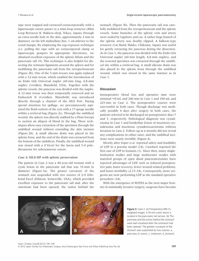
tape were trapped and extracted extracorporeally with alaparoscopic suture passer or a mini-loop retractor (MiniLoop Retractor II, Hakkou-shoji, Tokyo, Japan) throughan extra needle hole in the skin, approximately 2 mm indiameter, on the left midaxillary line, 1 cm inferior to thecostal margin. By employing the tug-exposure technique(i.e. pulling the tape with an extracorporeal clamp orlaparoscopic graspers in appropriate directions), weobtained excellent exposure of the splenic hilum and thepancreatic tail (9). This technique is also helpful for dis-secting the remnant ligaments around the spleen and formobilizing the pancreatic tail from the retroperitoneum(Figure 2b). One of the 5-mm trocars was again replacedwith a 12-mm trocar, which enabled the introduction ofan Endo GIA Universal stapler (60 mm long, 4.8-mmstaples; Covidien, Mansfield, USA). Together with thesplenic vessels, the pancreas was divided with the stapler.A 12-mm trocar was then temporarily removed and anEndocatch II (Covidien, Mansfield) was introduceddirectly through a channel of the SILS Port. Payingspecial attention for spillage, we percutaneously aspi-rated the fluid content of the cyst with a 17-gauge needlewithin a retrieval bag (Figure 2c). Through the umbilicalwound, the spleen was directly stabbed by a Péan forcepsto suction an aliquot of blood in the bag. These tech-niques allow easy extraction of the specimen through theumbilical wound without extending the skin incision(Figure 2d). A small silicone drain was placed in thesplenic fossa, and the end of the drain was extracted fromthe bottom of the umbilicus. Finally, the umbilical woundwas closed with a 0 Vicryl for the fascia and 5-0 poly-dioxanone for subcutaneous suture.
Case 2: SILS-DP with splenic preservation
The patient in Case 2 was a 40-year-old woman with acystic lesion in the pancreatic tail that was 35-mm indiameter (Figure 3a). The greater curvature of thestomach was suspended with two sutures of 2-0 Ethi-bond Excel (Ethicon, Somerville, USA), which providedexcellent exposure to the pancreatic tail and, after theomentum had been opened, the tumor behind the
stomach (Figure 3b). Then the pancreatic tail was care-fully mobilized from the retroperitoneum and the splenicvessels. Some branches of the splenic vein and arterywere sealed by LigaSure and cut. A rather large branch ofthe splenic artery was doubly clipped. A balloon-typeretractor (Cat Hand, Hakko, Chikuma, Japan) was usefulfor gently retracting the pancreas during the dissection.As in Case 1, the pancreas was divided with the Endo GIAUniversal stapler (60 mm length, 4.8-mm staples), andthe resected specimen was extracted through the umbili-cal site within a retrieval bag. A small silicone drain wasalso placed in the splenic fossa through the umbilicalwound, which was closed in the same manner as inCase 1.
Discussion
Intraoperative blood loss and operative time wereminimal �0 mL and 240 min in Case 1 and 100 mL and225 min in Case 2. The postoperative courses wereuneventful in both cases. Though discharge was medi-cally possible 4 days after surgery in both cases, thepatients selected to be discharged on postoperative days 7and 5, respectively. Pathological diagnosis was cystad-enoma in Case 1 and borderline lesion of mucinous cys-tadenoma and mucinous cystadenocarcinoma withoutinvasion in Case 2. Follow-up at 6 months did not revealany complications in either casre, and the umbilical inci-sions were nearly invisible (Figure 4).
Shortly after Soper et al. reported safety and feasibilityof LDP in a porcine model (14), Cuschieri reported thefirst case of LDP in humans (1). Since then, many single-institution studies and large multicenter studies withmatched groups of open distal pancreatectomies havereported advantages of LDP, such as reduced postopera-tive pain, faster recovery, fewer wound-related problems,and lower morbidity (2,15–18). Consequently, more sur-geons are now performing LDP as the standard operativeprocedure (18).
With the emergence of NOTES as the next major fron-tier in minimally invasive surgery, surgeons have become
Figure 3 Case 2. (a) Preoperative MRI (T2-
weighted image). A 35-mm cystic lesion is
located in the pancreatic tail (arrow). (b) The
pancreas and the tumor behind the stomach
were well visualized after the omentum had
been opened. The greater curvature of the
stomach was suspended by two sutures. a,
pancreas; b, tumor; c, omentum; d, stomach.
T Misawa et al. Single-incision laparoscopic pancreatectomy
Asian J Endosc Surg 5 (2012) 195–199© 2012 Japan Society for Endoscopic Surgery, Asia Endosurgery Task Force and Wiley Publishing Asia Pty Ltd 197

more conscious of cosmetic factors such as limiting thenumber of trocars. Terms such as “single-incision lapa-roscopic surgery” (SILS), “single-port access surgery”(SPA), or “laparoscopic single-site surgery” (LESS),have been used for a variety of procedures includingnephrectomy, colectomy, sleeve gastrectomy, herniarepair, appendectomy, cholecystectomy, and splenec-tomy (4–9,19). However, to our knowledge, reports onSILS-DP have been limited to sporadic case reports (10–12). Therefore, we believe SILS-DP remains a challeng-ing, emerging procedure with much room for technicalimprovements to ensure safety.
During SILS-DP procedures, one of the critical points isto obtain good exposure of the pancreas, which liesbehind the stomach. For this purpose in Case 2, as inprevious reports (12), we suspended the greater curva-ture of the stomach with two stay sutures to the abdomi-nal wall. This technique helped maintain a stableoperative field during the operation. Also, a balloonretractor was useful for retracting the stomach, spleen,and pancreas. To complement the limited number ofinstruments, use of gravity by rotating or positioning thepatient in the reversed Trendelenburg position wasessential. Gravity pulls the pancreas and the spleen closeto the umbilicus and keeps good exposure during opera-tion. Furthermore, the tug-exposure technique was ben-eficial for SILS-DP without splenic preservation (Case 1)as in SILS splenectomy (9). This technique allowedoptimal tension to the tissue as well as easy dissectionand stapling of the pancreas.
At our institution, we have performed laparoscopicpancreatectomy including LDP and enucleation as well asSILS-DP. Our indication for laparoscopic pancreatectomyat the present time is limited to benign or low malignant
lesions of the pancreas such as intraductal papillary muci-nous neoplasms, mucinous cystic neoplasms, pancreaticneuroendocrine tumors, serous cystic tumors, and solidand pseudopapillary tumors. When a lesion shows char-acteristics that indicate malignancy on the preoperativeimages, we select the patient for open pancreatectomywith systematic lymphadenectomy. In this pilot studywith SILS-DP, the lesions were located in the pancreatictail and did not present malignant features on preopera-tive images. Though recent reports suggest the feasibilityof LDP for pancreatic cancer (20), the induction ofSILS-DP for malignant tumors, such as invasive ductalcell carcinoma of the pancreas, should be investigated.
Although several other issues such as the extentof invasiveness, costs, and learning curve of SILS-DPremain to be investigated relative to traditional LDP, thecosmetic benefits of SILS-DP are obvious. In conclusion,with some technical refinements such as gastric suspen-sion or the tug-exposure technique, SILS-DP can besafely performed by experienced surgeons.
Acknowledgments
The authors have no conflicts of interest and received nofinancial support for this study.
References
1. Cuschieri A. Laparoscopic surgery of the pancreas. J R Coll
Surg Edinb 1994; 39: 178–184.
2. Gagner M, Pomp A, Herrera MF. Early experience with
laparoscopic resections of islet cell tumors. Surgery 1996;
120: 1051–1054.
3. Borja-Cacho D, Al-Refaie WB, Vickers SM et al. Laparo-
scopic distal pancreatectomy. J Am Coll Surg 2009; 209: 758–
765.
4. Esposito C. One-trocar appendectomy in pediatric surgery.
Surg Endosc 1998; 12: 177–178.
5. Piskun G & Rajpal S. Transumbilical laparoscopic
cholecystectomy utilizes no incisions outside the umbilicus.
J Laparoendosc Adv Surg Tech A 1999; 9: 361–364.
6. Remzi FH, Kirat HT, Kaouk JH et al. Single-port laparoscopy
in colorectal surgery. Colorectal Dis 2008; 10: 823–826.
7. Reavis KM, Hinojosa MW, Smith BR et al. Single-
laparoscopic incision transabdominal surgery sleeve gastrec-
tomy. Obes Surg 2008; 18: 1492–1494.
8. Ponsky LE, Cherullo EE, Sawyer M et al. Single access
site laparoscopic radical nephrectomy: Initial clinical expe-
rience. J Endourol 2008; 22: 663–666.
9. Misawa T, Sakamoto T, Ito R et al. Single-incision laparo-
scopic splenectomy using the “tug-exposure technique” in
adults: Results of ten initial cases. Surg Endosc 2011; 25:
3222–3227.
Figure 4 The umbilical wound is almost invisible 1 month after surgery
(Case 1).
Single-incision laparoscopic pancreatectomy T Misawa et al.
Asian J Endosc Surg 5 (2012) 195–199© 2012 Japan Society for Endoscopic Surgery, Asia Endosurgery Task Force and Wiley Publishing Asia Pty Ltd198
10. Barbaros U, Sümer A, Demirel T et al. Single incision lap-
aroscopic pancreas resection for pancreatic metastasis of
renal cell carcinoma. JSLS 2010; 14: 566–570.
11. Kuroki T, Adachi T, Okamoto T et al. Single-incision laparo-
scopic distalpancreatectomy. Hepatogastroenterology 2011; 58:
1022–1024.
12. Chang SK, Lomanto D, Mayasari M. Single-port laparo-
scopic spleen preserving distal pancreatectomy. Minim
Invasive Surg 2012; 2012: 197–429. Epub 2012 Feb 26.
13. Takaori K & Tanigawa N. Laparoscopic pancreatic resec-
tion: The past, present, and future. Surg Today 2007; 37:
535–545.
14. Soper NJ, Brunt LM, Dunnegan DL et al. Laparoscopic distal
pancreatectomy in the porcine model. Surg Endosc 1994; 8:
57–60.
15. Baker MS, Bentrem DJ, Ujiki MB et al. A prospective
single-institution comparison of perioperative outcomes for
laparoscopic and open distal pancreatectomy. Surgery 2009;
146: 635–643.
16. Melotti G, Butturini G, Piccoli M et al. Laparoscopic distal
pancreatectomy: Results on a consecutive series of 58
patients. Ann Surg 2007; 246: 77–82.
17. Mabrut J-Y, Fernandez-Cruz L, Azagra JS et al. Hepatobil-
iary and Pancreatic Section (HBPS) of the Royal Belgian
Society of Surgery; Belgian Group for Endoscopic Surgery
(BGES); Club Coelio. Laparoscopic pancreatic resection:
Results of a multicenter European study of 127 patients.
Surgery 2005; 137: 597–605.
18. Kooby DA, Gillespie T, Bentrem D et al. Left-sided pancre-
atectomy: A multicenter comparison of aparoscopic and
open approaches. Ann Surg 2008; 248: 438–446.
19. Cugura JF, Kirac I, Kulis T et al. First case of single incision
laparoscopic surgery for totally extraperitoneal inguinal
hernia repair. Acta Clin Croat 2008; 47: 249–252.
20. Marangos IP, Buanes T, Røsok BI et al. Laparoscopic resec-
tion of exocrine carcinoma in central and distal pancreas
results in a high rate of radical resections and long postop-
erative survival. Surgery 2012; 151: 717–723.
T Misawa et al. Single-incision laparoscopic pancreatectomy
Asian J Endosc Surg 5 (2012) 195–199© 2012 Japan Society for Endoscopic Surgery, Asia Endosurgery Task Force and Wiley Publishing Asia Pty Ltd 199