Embed Size (px)
Citation preview

1Review provided courtesy of Elevate Oral Care • West Palm Beach, FL
Silver Diamine Fluoride 38%
Scientific Literature Review
March 2016
Silver Diamine Fluoride (SDF) 38% has been receiving a great deal of attention by U.S. dental professionals since it was cleared for use by the Food and Drug Administration in August 2104 under the provisions of the Federal Food, Drug and Cosmetics Act. The Cleared Indications For Use are for the “Treatment of dentinal hypersensitivity. For use in adults over the age of 21.”
In the age of the Internet, access to information that can sometimes be credible and sometimes not, could cause confusion about the histroy, safety and efficacy of SDF. In addition, a number of local television news programs and social media postings around the U.S have recently begun commu-nicating information about the use of SDF by both general and pedictric dentists who have begun using it for the treatment of carious lesions in populations of all ages.
While SDF only recently received FDA Clearance it has been used by dental professionals outside the U.S for both the treatment of dentinal hypersensitivity and as a caries therapy for more than 45 years. This review is intended to provide U.S medical professionals with an understanding of the history of SDF around the world, including the most current information available regarding its use in the U.S.
Under federal law, the use of a drug or medical device by a licensed medical professional for an indication not Approved or Cleared by the FDA is allowable and not uncommon. This is termed “off-label” use.
As the organization permitted to market the only FDA Cleared SDF product in the United States, (Advantage Arrest™ Silver Diamine Fluoride 38%,) it is our intention to provide a review of all scientific literature available to us in order to help insure that medical professionals, and through them, their patients are as well informed as possible about this therapy.
This document is not assumed to contain all published information regarding SDF, as that would be virtually impossible, since SDF has been in use in many countries around the world for de-cades. It is however meant to provide a fair and balanced view of the benefits and risks of the use of SDF. If, after reading this document, you have any questions please send an email to the address below and we will get back to you promptly.
Please address any questions to:
Steve PardueElevate Oral [email protected]
Elevate Oral Care346 Pike Rd. Suite 5West Palm Beach, FL 33411

2Review provided courtesy of Elevate Oral Care • West Palm Beach, FL
Table of Contents
Advantage Arrest Package Insert 3
The Short Term Effects of Diammine Silver Fluoride 4on Tooth Sensitivity: a Randomized Controlled TrialJ Dent Res 90(2):203-208, 2011J.L. Castillo1, S. Rivera, T. Aparicio, R. Lazo1, T.-C. Aw, L.L. Mancl,and P. Milgrom
UCSF Protocol for Caries Arrest Using Silver Diamine 10 Fluoride: Rationale, Indications and ConsentCDA Journal , Vol 44, No.1Jeremy A. Horst, DDS, PhD; Hellene Ellenikiotis, DDS; and Peter L. Milgrom, DDS
Clinical Use of Silver Diamine Fluoride in Dental Treatment 22Compendium of Continuing Education Volume 37, Number 2May L. Mei, BDS, MDS, PhD; Edward Chin-Man Lo, BDS, MDS, PhD; and Chun-Hung Chu, BDS, MDS, PhD.
Fresh Approach to Caries Arrest in Adults 23Decisions in Dentistry Volume 1, Number 1Dr. John Featherstone, Dean of the University of California San Francisco School of Dentistry and Dr. Jeremy Horst, DDS, PhD
Silver Diamine Fluoride: A Caries “Silver-Fluoride Bullet” 24J Dent Res 88(2): 116-125, 2009 A. Rosenblatt. T.C.M. Stamford, and R. Niederman
Additional Information Published after February 2009 34
Frequently Asked Questions 42

3Review provided courtesy of Elevate Oral Care • West Palm Beach, FL
Advantage Arrest Package Insert
Professional Tooth Desensitizer
Rx Only
Desensitizing Ingredient: Aqueous Silver Diamine Fluoride, 38.3% to 43.2% w/v
Inactive Ingredients: Purified water
Clinical Pharmacology: Product forms insoluble precipitates with calcium or phosphate in the dentinal tubules to block nerve impulses.
Indication and Usage: Treatment of dentinal hypersensi-tivity. For use in adults over the age of 21.
Contraindications: This product is contraindicated in patients with ulcerative gingivitis or stomatitis, or known sensitivity to silver or other heavy-metal ions. Patients with more than six affected sites, patients having had full mouth gingivectomies and patients showing abnormal skin sensitization in daily circumstances are recom-mended for exclusion.
Warnings: This product is intended for local application only. Not for ingestion. Protect the patient’s eyes. Use caution to avoid contact with skin or clothing. In the event of exposure to eyes or skin, flush the area copiously with water and immediately seek medical consultation. This product yielded positive cytotoxicity in standard testing.
Precautions for Use:
1). Advantage Arrest does not normally stain enamel or burnished dentin. Advise patients that soft dentin or margins of composite restorations may be stained. Staining may be reversed by gentle polishing with tincture of iodine (weak iodine solution).
2) Advise patients that air-drying and product application can cause momentary transient pain to hypersensitive areas. Advantage Arrest has not been shown to cause pulpal necrosis even when soft dentin is treated.
3) Minimize product contact with gingiva and mucous membrane by using recommended amounts and care-ful application. Advantage Arrest may cause reversible short-term irritation. When applying Advantage Arrest to areas near the gingiva, apply petroleum jelly or co-coa butter and use cotton rolls to protect the gingival tissues. Alternatively, a rubber dam can be used to isolate the area.
4) If accidental contact occurs, thoroughly wash the area with water, saline solution or ~3% hydrogen peroxide. This includes contact with skin, clothes, floors and cabinets. Because Advantage Arrest is clear and thus
may be difficult to see, use caution to avoid transfer-ring the material from gloved hands to other surfaces.
Precautions for Handling:
1. Storage Precautions
1) Store in original packaging in a cool, dark place.
2) Replace cap immediately after use.
3) Use as soon as dispensed.
2. Advantage Arrest will stain skin, clothes, counter tops, floors and instruments brown or black. Refer to the following for stain removal:
1) Skin; wash immediately with water, soap, ammonia or iodine tincture and then rinse thoroughly with water. Do not use excessive methods in an attempt to remove difficult stains from skin as the stains will eventually fade.
2) Clothing/Countertops/Floors/Instruments; use the same procedures as with stained skin. Difficult stains may be treated with sodium hypochlorite.
3. If Advantage Arrest is dispensed into a separate container, be sure to wash or thoroughly wipe the container clean immediately after use.
Adverse Reactions: Transient irritation of the gingiva has rarely been reported.
Dosage and Administration:
1. Isolate the affected area of the tooth with cotton rolls or protect the gingival tissue of the affected tooth with petroleum jelly. Alternatively, a rubber dam can be used to isolate the area.
2. Clean and dry the affected tooth surface.
3. For up to 5 treated sites per patient, dispense 1-2 drops of solution into a disposable dappen dish. Transfer material directly to the tooth surface with an applicator.
4. Air-dry.
If needed, one or two reapplications may be adminis-tered at intervals of one week.
How Supplied: Single 10 mL dropper-bottle containing 8 mL of product. Not sterile.
Storage: Do not freeze or expose to extreme heat. Keep in an air-tight container in a dark place.
Caution: Federal law restricts this device to sale by or on the order of a dentist or physician.
Distributed by: Elevate Oral Care, LLC.West Palm Beach, FL 33411877-866-9113 © 2015 Elevate Oral Care
ELE137-0315

4Review provided courtesy of Elevate Oral Care • West Palm Beach, FL
203
RESEARCH REPORTSClinical
DOI: 10.1177/0022034510388516
Received August 13, 2010; Revised September 3, 2010; Accepted September 3, 2010
© International & American Associations for Dental Research
J.L. Castillo1, S. Rivera1, T. Aparicio2,R. Lazo1, T.-C. Aw3, L.L. Mancl4,and P. Milgrom4*
1School of Dentistry, Universidad Peruana Cayetano Heredia, Lima, Peru; 2Private Practice, Cusco, Peru; 3Department of Restorative Dentistry, University of Washington, Seattle, USA; and 4Department of Dental Public Health Sciences, Box 357475, University of Washington, Seattle, WA 98195, USA; *corresponding author, [email protected]
J Dent Res 90(2):203-208, 2011
ABSTRACTTooth sensitivity is a common clinical problem. This multi-center randomized clinical trial assessed the effectiveness and safety of topical diammine silver fluoride. From two sites (Lima and Cusco, Peru), 126 adults with at least one tooth sensitive to compressed air were randomly assigned to either the experimental treatment or sterile water, and pain was assessed by means of a 100-mm visual analogue scale at 24 hours and 7 days. The diammine silver fluoride reduced pain at 7 days at both sites. At the Lima site, the average change in pain scores between baseline and day 7 for the silver fluoride group was -35.8 (SD = 27.7) mm vs. 0.4 (SD = 16.2) mm for the control group (P < 0.001). In Cusco, the average change in pain scores for the silver fluoride group was –23.4 (SD = 21.0) mm and -5.5 (18.1) mm for the control group (P = 0.002). No tissue ulceration, white changes, or argyria was observed. A small number of participants in the silver fluoride group experi-enced a mild but transient increase in erythema in the gingiva near the tooth. No changes were observed in the Gingival Index. We concluded that diammine silver fluoride is a clinically effective and safe tooth desensitizer.
KEY WORDS: tooth sensitivity, silver diaminefluoride, diammine silver fluoride, silver diam-mine fluoride, fluorides, topical.
INTRODUCTION
Tooth sensitivity to various stimuli, including cold air, has been explained by hydrodynamic changes within the dentinal tubules that activate intradental
nerves (Markowitz and Pashley, 2008). Incidence is thought to be increasing. The etiology can be tooth wear, aggressive oral hygiene, and diet. Successful treatments physically block dentinal tubules (Arends et al., 1997).
Sodium fluoride varnish and fluoride solutions and gels have been shown to reduce sensitivity (Thrash et al., 1992; Ritter et al., 2006). However, there is continuing interest in finding effective treatments. Nevertheless, recent studies have designs that are weak or statistically underpowered (Erdemir et al., 2010; Jalali and Lindh, 2010).
The purpose of this study was to assess the clinical effectiveness and safety of topical diammine silver fluoride as a tooth desensitizer in adults.
METHODS
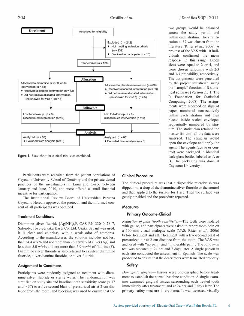
Design
This is a randomized clinical trial with two groups (Fig. 1). The study tested application of diammine silver fluoride in a single visit, because previous unpublished work had shown that a single application forms insoluble pre-cipitates with calcium and phosphate that physically block dentinal tubules. The International Clinical Trials Registry number is NCT01063530.
Study Sites
The study was conducted in two sites, Lima and Cusco, Peru.
Participants
To be included, a participant must have at least one vital cuspid or premolar with a buccal cervical defect and clinical hypersensitivity in response to com-pressed air with a score ≥ 15 on a visual analogue scale (VAS) for pain. The individual will have had generally healthy gum tissue surrounding this tooth and no ulceration and no leukoplakia in this gingival tissue.
Candidates were excluded if they were using any type of tooth desensitizer, had received a fluoride varnish treatment within the preceding month, or were taking prescription medications, aspirin, or non-steroidal anti-inflammatory drugs; women who were pregnant were also excluded. Individuals using smokeless tobacco or chewing coca leaves were excluded. Individuals with known sensitivity to silver or other heavy-metal ions were excluded.
The Short-term Effects of Diammine Silver Fluoride on Tooth Sensitivity: a Randomized Controlled Trial
5Review provided courtesy of Elevate Oral Care • West Palm Beach, FL
204 Castillo et al. J Dent Res 90(2) 2011
Participants were recruited from the patient populations of Cayetano University School of Dentistry and the private dental practices of the investigators in Lima and Cusco between January and June, 2010, and were offered a small financial incentive for participation.
The Institutional Review Board of Universidad Peruana Cayetano Heredia approved the protocol, and the informed con-sent of all participants was obtained.
Treatment Conditions
Diammine silver fluoride [Ag(NH3)2F, CAS RN 33040–28–7, Saforide, Toyo Seiyaku Kasei Co. Ltd. Osaka, Japan] was used. It is clear and colorless, with a weak odor of ammonia. According to the manufacturer, the solution includes not less than 24.4 w/v% and not more than 26.8 w/v% of silver (Ag), not less than 5.0 w/v% and not more than 5.9 w/v% of fluorine (F). Diammine silver fluoride is also referred to as silver diammine fluoride, silver diamine fluoride, or silver fluoride.
Assignment to Conditions
Participants were randomly assigned to treatment with diam-mine silver fluoride or sterile water. The randomization was stratified on study site and baseline tooth sensitivity score (< 37 and ≥ 37) to a five-second blast of pressurized air at 2 cm dis-tance from the tooth, and blocking was used to ensure that the
two groups would be balanced across the study period and within each stratum. The stratifi-cation at 37 was chosen from the literature (Ritter et al., 2006). A pre-test of the VAS with 10 indi-viduals confirmed the mean response in this range. Block sizes were equal to 2 or 4, and were chosen randomly with 2/3 and 1/3 probability, respectively. The assignments were generated by the project statistician, using the “sample” function of R statis-tical software (Version 2.7.1, The R Foundation for Statistical Computing, 2008). The assign-ments were recorded on slips of paper numbered consecutively within each stratum and then placed inside sealed envelopes sequentially numbered by stra-tum. The statistician retained the master list until all the data were analyzed. The clinician would open the envelope and apply the agent. The agents (active or con-trol) were packaged in identical dark glass bottles labeled as A or B. The packaging was done atCayetano University.
Clinical Procedure
The clinical procedure was that a disposable microbrush was dipped into a drop of the diammine silver fluoride or the control and then applied to the surface for 1 sec. Then the surface was gently air-dried and the procedure repeated.
Measures
Primary Outcome-Clinical
Reduction of pain (tooth sensitivity)—The teeth were isolated with gauze, and participants were asked to report tooth pain on a 100-mm visual analogue scale (VAS; Ritter et al., 2006) before treatment and after treatment with a five-second blast of pressurized air at 2 cm distance from the tooth. The VAS was anchored with “no pain” and “intolerable pain”. The follow-up test was repeated at 24 hrs and 7 days later. A single person in each site conducted the assessment in Spanish. The scale was pre-tested to ensure that the descriptors were translated properly.
Safety
Damage to gingiva—Tissues were photographed before treat-ment to establish the normal baseline condition. A single exam-iner examined gingival tissues surrounding each treated tooth immediately after treatment, and at 24 hrs and 7 days later. The primary safety measure is erythema. It was assessed visually
Figure 1. Flow chart for clinical trial sites combined.
6Review provided courtesy of Elevate Oral Care • West Palm Beach, FL
J Dent Res 90(2) 2011 Diammine Silver Fluoride for Sensitive Teeth 205
with the use of a standard dental light. Erythema (red changes) was rated on a 1 to 3 scale, where 1 is no redness, 2 is redness with bleeding on probing, and 3 is a severe change. The Gingival Index (Löe, 1967) was used to measure gingival inflammation in the mouth overall. White changes, ulceration, and staining were secondary measures. Changes were rated as present or absent. Examiners were trained to criteria using pho-tographs and clinical cases. Intra- and inter-examiner reliability was established in 15 cases, and intraclass correlation was used to assess reliability. All intraclass correlations exceeded 0.8.
Data Analysis Plan
The data from the two sites were analyzed. To confirm reduction in pain, we calculated average difference scores between pre- and post-treatment VAS scores for each individual for each time-point (24 hrs and 7 days after treatment), and t tests were used to com-pare changes. The primary end point was at 7 days. Generalized estimating equations (GEE) linear regression was used in a sec-ondary analysis to compare the reduction in pain across the 3 time-points, where the outcome is pain at the 3 time-points, the baseline pain is a covariate, and robust standard errors are used to account for multiple observations per participant and heteroscedasticity (Hardin and Hilbe, 2002). In addition, separate analyses of covari-ance were done at each time-point to compare the reduction in pain due to the active treatment between the two study sites, where the outcome is the pain at a particular time-point, baseline pain was entered as a covariate, and treatment and site, as well as a treat-ment-group-by-site interaction, were entered as factors.
We used Fisher’s Exact Test to assess whether there were more participants with erythema score > 1 in the silver fluoride group vs. the control group at 24 hrs and 7 days post-treatment. The primary end-point was assessed at 24 hrs. A t test assessed any differences in Gingival Index. Any white changes, ulcer-ation, and staining (argyria) were reported.
Power Analysis
The data from the two sites were analyzed separately, and power is described below for the separate site analyses.
Reduction in tooth sensitivity—The primary end-point was assessed at 7 days post-treatment. In a similar desensiti-zation study comparing fluoride varnishes (Ritter et al., 2006), pain in response to air dropped from 36.9 (SD = 26.2) at baseline to 20.8 (SD = 4.3) at 2 wks post-treatment. We expected a similar or larger drop after 7 days with diammine silver fluoride, based on unpublished work from the University of Hong Kong, and little or no drop from the water. Thus, having 31 individuals in a group will allow for detection of effect size from 0.64 upwards, with an alpha of 0.05 and power of 0.8.
RESULTS
Participants
One hundred twenty-six adults (71 in Lima and 55 in Cusco) participated. About 378 candidates were screened between January and June 2010. The main reason (95%) for exclusion was lack of tooth sensitivity. The remainder were excluded because of the use of medications. No individuals were excluded because of tobacco use or coca. All of those eligible agreed to participate, but 10 were excluded because they failed to appear for the first visit. The proportion of women enrolled was 86% in Lima and 80% in Cusco. The average age of participants was 44 yrs and 43 yrs, respectively. There were no dropouts.
Participants and clinicians were blind to treatment assign-ment. Odor was not a threat to blinding, because the smell is not detectable clinically when such small quantities are used. Taste was not a threat in this study, because only minute amounts of material were applied and the tooth was air-dried after application.
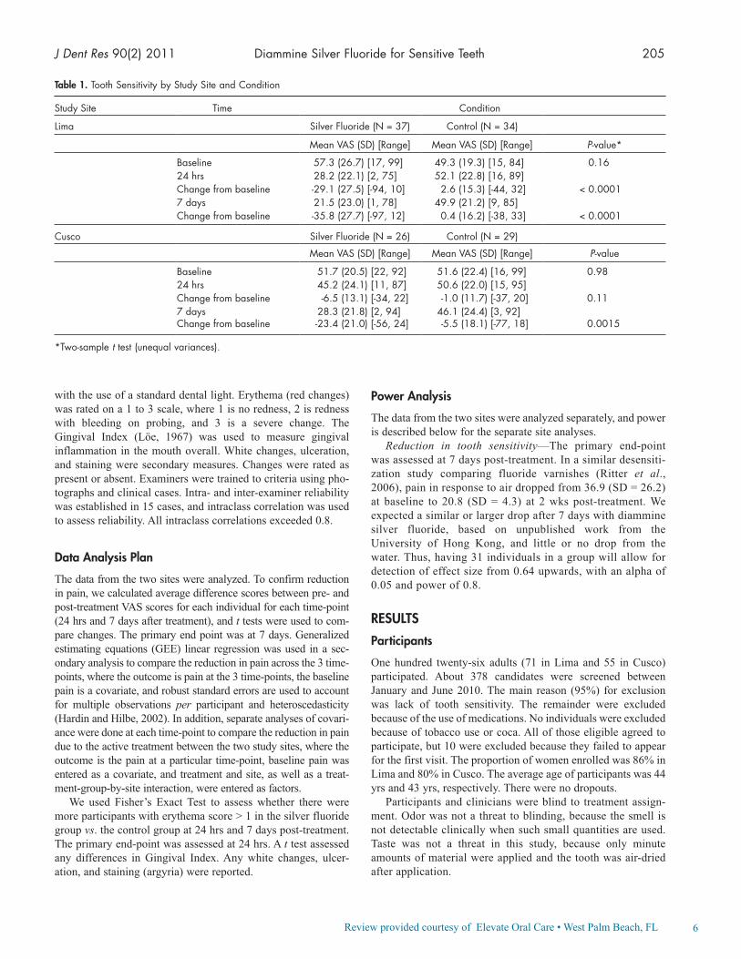
Table 1. Tooth Sensitivity by Study Site and Condition
Study Site Time Condition
Lima Silver Fluoride (N = 37) Control (N = 34)
Mean VAS (SD) [Range] Mean VAS (SD) [Range] P-value*
Baseline 57.3 (26.7) [17, 99] 49.3 (19.3) [15, 84] 0.1624 hrs 28.2 (22.1) [2, 75] 52.1 (22.8) [16, 89]Change from baseline -29.1 (27.5) [-94, 10] 2.6 (15.3) [-44, 32] < 0.00017 days 21.5 (23.0) [1, 78] 49.9 (21.2) [9, 85]Change from baseline -35.8 (27.7) [-97, 12] 0.4 (16.2) [-38, 33] < 0.0001
Cusco Silver Fluoride (N = 26) Control (N = 29)
Mean VAS (SD) [Range] Mean VAS (SD) [Range] P-value
Baseline 51.7 (20.5) [22, 92] 51.6 (22.4) [16, 99] 0.9824 hrs 45.2 (24.1) [11, 87] 50.6 (22.0) [15, 95]Change from baseline -6.5 (13.1) [-34, 22] -1.0 (11.7) [-37, 20] 0.117 days 28.3 (21.8) [2, 94] 46.1 (24.4) [3, 92]Change from baseline -23.4 (21.0) [-56, 24] -5.5 (18.1) [-77, 18] 0.0015
*Two-sample t test (unequal variances).

7Review provided courtesy of Elevate Oral Care • West Palm Beach, FL
206 Castillo et al. J Dent Res 90(2) 2011
Clinical Effectiveness
The average pain scores before and after treatment, by site, are given in Table 1. At the Lima site, the silver fluoride group had slightly higher baseline scores (average = 57.3) than the control (average = 49.3; P = 0.16). At the Cusco site, the baseline scores were similar between the silver fluoride group (average = 51.7) and control (average = 51.6; P = 0.98). The primary study end-point was the change from baseline to 7 days. In Lima, the aver-age change in pain score between baseline and day 7 for the silver fluoride group was -35.8 (SD = 27.7) mm vs. 0.4 (SD = 16.2) for the controls (P < 0.0001). In Cusco, the average change in pain score between baseline and day 7 for the silver fluoride group was -23.4 (SD = 21.0) mm vs. -5.5 (SD = 18.1) mm (P = 0.0015) for water.
Comparison of tooth sensitivity at 24 hrs and 7 days between study groups by analysis covariance, adjusted for the baseline sensitivity level, gave similar results.
There was no significant three-way interaction among study site, time, and study group (GEE linear regression; P = 0.20), but all two-way interactions were significant: study site by time (P = 0.043), study site by study group (P = 0.0006), and study group by time (P = 0.0076). Hence, an analysis of time effect was done separately by study site. In Lima, there was no sig-nificant time-by-study-group interaction (P = 0.21). The overall study group difference in tooth sensitivity (over both time-points), adjusted for baseline sensitivity, was 29.9 (P < 0.001). The overall difference in sensitivity between 24 hrs and 7 days was 4.5 (P = 0.014). In Cusco, there was a significant study-group-by-time interaction (P = 0.015), so the overall study group difference is not reported. The differences in sensitivity between 24 hrs and 7 days were 16.9 (P = 0.005) for silver fluo-ride and 4.5 (P = 0.097) in the control group, respectively.
Safety
The number and percent of participants with a erythema score of 2 for the gingival tissue of the tooth treated for each treatment condition by site and time are given in Table 2. Scores were low; no individual had score 3, severe erythema, either before or after the application of silver fluoride. There was no difference in the proportion of participants with erythema score > 1 between the silver fluoride group and the placebo (Fisher’s Exact Test, P = 1.0) at any time-point in the Lima population. There was a small but significant increase in the proportion of participants at the Cusco site who experienced an erythema score > 1 at 24 hrs (P = 0.0076). There was no difference in the proportion of par-ticipants with an erythema score > 1 between the groups in Cusco after 7 days (P = 1.0). No white or dark changes were noted in gingiva in any participant at any time in any condition at either site. An independent examiner, who was blind to treat-ment condition and time, examined the photographs and con-firmed this lack of change.
The Gingival Index scores for each treatment condition and site are listed in Table 3. The mean (SD) Gingival Index scores for the mouth for treatment and control groups at base-line were: (Lima) silver fluoride, 0.29 (0.24), control 0.33 (0.35) (P = 0.59); and (Cusco) silver fluoride, 0.47 (0.24), control 0.38 (0.27) (P = 0.19). At 7 days, the mean (SD) changes in GI scores were: (Lima) silver fluoride, -0.02 (0.09), control 0.03 (0.13) (P = 0.076); and (Cusco) silver fluoride, -0.16 (0.27), control -0.03 (0.09) (P = 0.023). Similar resultswere observed after 24 hrs.
Photographs of the teeth suggest that the silver fluoride did not stain most exposed root surfaces (see Fig. 2 for an example). This result was found only when surfaces had untreated decay.
Table 2. Numbers and Percentages of Participants with Erythema Score of 2 by Study Site and Condition
Study Site Time Condition
Lima Silver Fluoride (N = 37) Control (N = 34)
n (%) n (%) P-value*
Baseline 3 (8.1) 2 (5.9) 1.024 hrs 3 (8.1) 2 (5.9) 1.07 days 3 (8.1) 1 (2.9) 0.61
Cusco Silver Fluoride (N = 26) Control (N = 29)
n (%) n (%) P-value*
Baseline 6 (23.1) 7 (24.1) 1.024 hrs 10 (38.5) 2 (6.9) 0.00767 days 3 (11.5) 3 (10.3) 1.0
Sites combined Silver Fluoride (N = 63) Control (N = 63)
n (%) n (%) P-value*
Baseline 9 (14.3) 9 (14.3) 1.024 hrs 13 (20.6) 4 (6.3) 0.0357 days 6 (9.5) 4 (6.3) 0.74
*Fisher’s exact test

8Review provided courtesy of Elevate Oral Care • West Palm Beach, FL
J Dent Res 90(2) 2011 Diammine Silver Fluoride for Sensitive Teeth 207
DISCUSSION
In a population with teeth sensitive to air, this trial demonstrated that a topical solution of diammine silver fluoride was more effective than a placebo in reducing tooth pain. Reductions grew larger between 24 hrs and 7 days post-treatment. The study was con-ducted in two sites by different investigators to increase generalizability and had sufficient statistical power to detect clinically mean-ingful differences in pain. The study involved many more individuals than the typical study (Ritter et al., 2006).
The results, however, are consistent with those from similar studies of other desensitizers, such as self-administered 0.717% fluoride solution (Thrash et al., 1992) or fluoride varnish (Ritter et al., 2006). In the fluoride solution study, the authors con-cluded that two one-minute applications reduced sensitivity to cold. Participants in the varnish study experienced a pain reduc-tion in response to ice, but not to air, at 2 wks. The current study reported significant pain reductions in response to air in 24 hrs that were maintained at 7 days. The magnitude of reduction was considerably greater than in the other studies. The current study did not use ice as a stimulus.
There were no unintended effects on the gingiva, and any inflammation resulting from the treatment was minor and transient. No staining of the gingival tissues was observed.
Staining of teeth was found only when surfaces had untreated decay. The staining of carious dentin can be minimized by the application of potassium iodide solution after treatment without reducing the effect (Knight et al., 2006).
Diammine silver fluoride has been shown to arrest caries in animal models (Tanzer et al., 2010) and to be more effective than sodium fluoride varnish in human trials (Chu et al., 2002; Llodra et al., 2005; Rosenblatt et al., 2009; Tan et al., 2010). It did not cause abscesses in teeth with open cavities that were treated. The mechanism of action for caries arrest may be anti-microbial (Knight et al., 2009). Studies have also shown that diammine silver fluoride is free of adverse effects (Chu et al., 2002; Llodra et al., 2005; Tan et al., 2010). This suggests that diammine fluoride may be particularly effective in individuals in whom sensitivity is associated with demineralization and caries.
Figure 2. Root caries at baseline (left panel), 24 hrs after treatment (middle panel), and 7 days after treatment with diammine silver fluoride (right panel).
Table 3. Overall Gingival Index Score by Study Site and Condition
Study Site Time Condition
Lima Silver Fluoride (N = 37) Control (N = 34)
Mean (SD) [Range] Mean (SD) [Range] P-value*
Baseline 0.29 (0.24) [0.0, 1.2] 0.33 (0.35) [0.0, 1.5] 0.5924 hrs 0.28 (0.24) [0.0, 1.2] 0.35 (0.36) [0.0, 1.7]Change from baseline -0.01 (0.05) [-0.2, 0.1] 0.02 (0.07) [-0.2, 0.2] 0.0767 days 0.27 (0.23) [0.0, 1.2] 0.36 (0.39) [0.1, 1.8]Change from baseline -0.02 (0.09) [0.2, 0] 0.03 (0.13) [-0.5, 0.3] 0.076
Cusco Silver Fluoride (N = 26) Control (N = 29)
Mean (SD) [Range] Mean (SD) [Range] P-value*
Baseline 0.47 (0.24) [0.1, 0.9] 0.38 (0.27) [0.0, 1.2] 0.1924 hrs 0.36 (0.21) [0.1, 0.8] 0.36 (0.24) [0.0, 1.2]Change from baseline -0.11 (0.16) [-0.6, 0.1] -0.02 (0.12) [-0.3, 0.3] 0.0207 days 0.31 (0.19) [0.0, 0.8] 0.35 (0.26) [0.1, 1.2]Change from baseline -0.16 (0.27) [-0.8, 0.7] -0.03 (0.09) [-0.3, 0.2] 0.023
Sites Combined Silver Fluoride (N = 63) Control (N = 63)
Mean (SD) [Range] Mean (SD) [Range] P-value**
Baseline 0.36 (0.26) [0.0, 1.2] 0.35 (0.32) [0.0, 1.5] 0.7224 hrs 0.31 (0.23) [0.0, 1.2] 0.35 (0.31) [0.0, 1.7]Change from baseline -0.05 (0.12) [-0.6, 0.1] 0.00 (0.10) [-0.3, 0.3] 0.00237 days 0.28 (0.22) [0.0, 1.2] 0.35 (0.33) [0.1, 1.8]Change from baseline -0.08 (0.20) [-0.8, 0.7] 0.00 (0.12) [-0.5, 0.3] 0.0028
*Two-sample test (unequal variances).**Analysis of covariance, adjusted for study site, with heteroscedasticity-consistent standard errors.

9Review provided courtesy of Elevate Oral Care • West Palm Beach, FL
208 Castillo et al. J Dent Res 90(2) 2011
Diammine silver fluoride has been demonstrated to be a clini-cally effective and safe tooth desensitizer after 24 hrs and 7 days. Clinical trials are warranted to examine effectiveness over a lon-ger period of time and in comparison with other agents.
ACKNOWLEDGMENTS
The authors acknowledge the contributions of Silvia Navarro in recruitment of participants. ADP Silver Dental Arrest, LLC, Redmond, OR, USA, was the study sponsor.
REFERENCESArends J, Düschner H, Ruben JL (1997). Penetration of varnishes into
demineralized root dentin in vitro. Caries Res 31:201-205.Chu CH, Lo EC, Lin HC (2002). Effectiveness of silver diamine fluoride
and sodium fluoride varnish in arresting dentin caries in Chinese pre-school children. J Dent Res 81:767-770.
Erdemir U, Yildiz E, Kilic I, Yucel T, Ozel S (2010). The efficacy of three desensitizing agents used to treat dentin hypersensitivity. J Am Dent Assoc 141:285-296.
Hardin JW, Hilbe JM (2002). Generalized estimating equations. New York, NY: Chapman & Hall/CRC Press.
Jalali Y, Lindh L (2010). A randomized prospective clinical evaluation of two desensitizing agents on cervical dentine sensitivity. A pilot study. Swed Dent J 34:79-86.
Knight GM, McIntyre JM, Craig GG, Mulyani (2006). Ion uptake into demineralized dentine from glass ionomer cement following
pretreatment with silver fluoride and potassium iodide. Aust Dent J 51: 237-241.
Knight GM, McIntyre JM, Craig GG, Mulyani, Zilm PS, Gully NJ (2009). Inability to form a biofilm of Streptococcus mutans on silver fluoride- and potassium iodide-treated demineralized dentin. Quintessence Int 40:155-161.
Llodra JC, Rodriguez A, Ferrer B, Menardia V, Ramos T, Morato M (2005). Efficacy of silver diamine fluoride for caries reduction in primary teeth and permanent first molars of schoolchildren: 36-month clinical trial. J Dent Res 84:721-724.
Löe H (1967). The Gingival Index, the Plaque Index, and the Retention Index. J Periodontol 38(Suppl):610-616.
Markowitz K, Pashley DH (2008). Discovering new treatments for sensitive teeth: the long path from biology to therapy. J Oral Rehabil 35:300-315.
Ritter AV, Dias WdeL, Miguez P, Caplan DJ, Swift EJ Jr (2006). Treating cervical dentin hypersensitivity with fluoride varnish: a randomized clinical study. J Am Dent Assoc 137:1013-1020.
Rosenblatt A, Stamford TC, Niederman R (2009). Silver diamine fluoride: a caries “silver bullet”. J Dent Res 88:116-125.
Tan HP, Lo EC, Dyson JE, Luo Y, Corbet EF (2010). A randomized trial on root caries prevention in elders. J Dent Res 89(10):1086-1090. Epub 2010 Jul 29.
Tanzer J, Thompson A, Milgrom P, Shirtcliff M (2010). Diammino silver fluoride arrestment of caries associated with anti-microbial action (abstract). J Dent Res 89(Spec Iss B):2082. URL: http://iadr.confex.com/iadr/2010barce/webprogram/Paper131877.html (URL accessed 09/07/2010).
Thrash WJ, Jones DL, Dodds WJ (1992). Effect of a fluoride solution on dentinal hypersensitivity. Am J Dent 5:299-302.

10Review provided courtesy of Elevate Oral Care • West Palm Beach, FL
C DA J O U R N A L , V O L 4 4 , Nº 1
J A N UA R Y 2 0 1 6 17
s i l v e r d i a m i n e
Since its approval in Japan more than 80 years ago,2 more than 2 million containers have been sold. The silver acts as an antimicrobial, the fl uoride promotes remineralization and the ammonia stabilizes high concentrations in solution.3
Because silver diamine fl uoride is new to American dentistry and dental education, there is a need for a standardized guideline, protocol and consent. The University of California, San Francisco, School of Dentistry paradigm shift committee assembled a subcommittee with the following goals:
■ Use available evidence to develop a list of clinical indications.
■ Defi ne a protocol that maximized safety and effi cacy and minimized inadvertent staining of clinical facilities.
Until now, no option for the treatment of dental caries in the U.S. besides restorative dentistry has shown substantial effi cacy.1 Silver
diamine fl uoride is an inexpensive topical medicament used extensively in other countries to treat dental caries across the age spectrum. No other intervention approaches the ease of application and effi cacy. Multiple randomized clinical trials — with hundreds of patients each — support its use for caries treatment, thus substantiating an intervention that addresses an unmet need in American dentistry. In August 2014, the Food and Drug Administration (FDA) cleared the fi rst silver diamine fl uoride product for market, and as of April 2015, that product is available.
AUTHORS
Jeremy A. Horst, DDS, PhD, is a fellow at the University of California, San Francisco, School of Dentistry studying the bacteria that cause cavities, a pediatric dentist at Alameda Pediatric Dentistry and co-founder and CSO at OraViz.Confl ict of Interest Disclosure: Dr. Horst is co-founder and CSO at OraViz.
UCSF Protocol for Caries Arrest Using Silver Diamine Fluoride: Rationale, Indications and ConsentJeremy A. Horst, DDS, PhD; Hellene Ellenikiotis, DDS; and Peter L. Milgrom, DDS
A B S T R AC T The Food and Drug Administration recently cleared silver diamine fl uoride for reducing tooth sensitivity. Clinical trials document arrest and prevention of dental caries by silver diamine fl uoride. This off-label use is now permissible and appropriate under U.S. law. A CDT code was approved for caries arresting medicaments for 2016 to facilitate documentation and billing. We present a systematic review, clinical indications, clinical protocol and consent procedure to guide application for caries arrest treatment.
Hellene Ellenikiotis, DDS, is a resident in the University of California, San Francisco, general practice residency and a recent graduate of the University of California, San Francisco School of Dentistry.Confl ict of Interest Disclosure: None reported.
Peter M. Milgrom, DDS, is a professor of dental public health sciences and pediatric dentistry and director of the Northwest Center to Reduce Oral Health Disparities at the University of Washington in Seattle.Confl ict of Interest Disclosure: Dr. Milgrom is a principal in ADP Silver Dental Arrest LLC, which licenses permission to market Advantage Arrest to Elevate Oral Care LLC.

11Review provided courtesy of Elevate Oral Care • West Palm Beach, FL
C DA J O U R N A L , V O L 4 4 , Nº 1
18 J A N UA RY 2 01 6
■ Build an informed consent document at the eighth-grade reading level.
We conducted a systematic review, inquired of authors of published clinical and in vitro studies about details and considerations in their protocols and consulted experts in cariology and materials chemistry where evidence was lacking. The work of this committee resulted in the adoption of silver diamine fl uoride use in the UCSF student clinics.
MethodsA literature review was designed by
a medical librarian to search PubMed and the International Association of Dental Research abstract archive with the following search terms: “33040-28-7” OR “1Z00ZK3E66” OR “silver diamine fl uoride” OR “silver fl uoride” OR “silver diamine fl uoride” OR “diammine silver fl uoride” OR “ammonical silver fl uoride” OR “ammoniacal silver fl uoride”. Differences in nomenclature have led to confusion around this material. Another review was completed with the terms “dental” OR “caries” AND “silver nitrate” AND “clinical.”
MaterialSilver diamine fl uoride (38% w/v
Ag(NH3)2F, 30% w/w) is a colorless topical agent comprised of 24.4-28.8% (w/v) silver and 5.0-5.9% fl uoride at pH 10,4 and marketed as Advantage Arrest by Elevate Oral Care LLC (West Palm Beach, Fla.). Other companies may market silver diamine fl uoride in the future following determination of substantial equivalence and FDA clearance.
MechanismsSilver diamine fl uoride is used for
caries arrest and treatment of dentin hypersensitivity. In the treatment of exposed sensitive dentin surfaces,
s i l v e r d i a m i n e
topical application results in the development of a squamous layer on the exposed dentin, partially plugging the dentinal tubules.5 High concentration aqueous silver has been long known to form this protective layer.6 Decreased sensitivity in treated patients7,8 is consistent with the hydrodynamic theory of dentin hypersensitivity.9
Dental caries is a complex progression involving dietary sugars, bacterial metabolism, demineralization and organic degradation. The collagenous organic matrix is
exposed once a dentin surface is demineralized and destroyed by native and bacterial proteases to enable a lesion to enlarge.10 Upon application of silver diamine fl uoride to a decayed surface, the squamous layer of silver protein conjugates forms, increasing resistance to acid dissolution and enzymatic digestion.11 Hydroxyapatite and fl uorapatite form on the exposed organic matrix, along with the presence of silver chloride and metallic silver.5 The treated lesion increases in mineral density and hardness while the lesion depth decreases.5 Meanwhile, silver diamine fl uoride specifi cally inhibits the proteins that break down the exposed dentin organic matrix: matrix metalloproteinases,11 cathepsins12 and bacterial collagenases.5 Silver
ions act directly against bacteria in lesions by breaking membranes, denaturing proteins and inhibiting DNA replication.13,14 Ionic silver deactivates nearly any macromolecule. Silver diamine fl uoride outperforms other anticaries medicaments in killing cariogenic bacteria in dentinal tubules.15
Silver and fl uoride ions penetrate ~25 microns into enamel16 and 50-200 microns into dentin.17 Fluoride promotes remineralization, and silver is available for antimicrobial action upon release by re-acidifi cation.18 Silver diamine fl uoride arrested lesions are 150 microns thick.19
Artifi cial lesions treated with silver diamine fl uoride are resistant to biofi lm formation and further cavity formation, presumably due to remnant ionic silver.20,21 More silver and fl uoride is deposited in demineralized than nondemineralized dentin. Correspondingly, treated demineralized dentin is more resistant to caries bacteria than treated sound dentin.22 When bacteria killed by silver ions are added to living bacteria, the silver is re-activated so that effectively the dead bacteria kill the living bacteria in a “zombie effect.”23 This reservoir effect helps explain why silver deposited on bacteria and dentin proteins within a cavity has sustained antimicrobial effects.
Clinical Evidence
Silver Nitrate Plus Fluoride VarnishBefore the FDA cleared silver
diamine fl uoride, some U.S. dentists sequentially applied silver nitrate then fl uoride varnish to dentinal decay as the only available noninvasive option for caries treatment. Duffi n rediscovered silver nitrate from the early literature,24 which had been lost
Silver diamine fl uoride outperforms other anticaries medicaments in killing cariogenic bacteria in dentinal tubules.

12Review provided courtesy of Elevate Oral Care • West Palm Beach, FL
C DA J O U R N A L , V O L 4 4 , Nº 1
J A N UA RY 2 0 1 6 19
to modern cariology. Surprisingly, there is no mention of silver nitrate in either of the American Dental Association Council on Scientifi c Affairs reports on Nonfl uoride Caries-Preventive Agents25 or Managing Xerostomia and Salivary Gland Hypofunction,26 and it is not part of standard dental school curricula. Case series of carious lesions arrested by silver nitrate date to the 1800s. For example, in 1891, 87 of 142 treated lesions were arrested.27 Percy Howe, DDS, then director of the Forsyth Institute in Boston, added ammonia to silver nitrate, making it more stable and effective as an antimicrobial for application to any infected tooth structure from early cavitated lesions to infected root canals.28 Duffi n added the application of fl uoride varnish following silver nitrate, simulating silver diamine fl uoride. While his clinic doubled in patients, cases needing general anesthesia disappeared. His review of randomly selected charts showed only seven of 578 treated lesions progressed within two and a half years to the point that extractions were needed.24 Thus, with the exception of Duffi n’s and one other report, attention to silver nitrate largely disappeared by the 1950s. The lore is that use and teaching of this intervention were lost with the introduction of effective local anesthetic to enable painless restorations and fl uoride for caries prevention. Because no high-quality clinical trials have been performed, we did not include the silver nitrate plus fl uoride varnish regimen in our recommendation.
Silver Diamine FluorideWe found nine published randomized
clinical trials evaluating silver diamine fl uoride for caries arrest and/or prevention of at least one year in duration. These
studies each involved hundreds of children aged 3 to 9 or adults aged 60 to 89 (FIGURES 1 and 2). Most participants had low (< 0.3 ppm) fl uoride in the environmental water and reported using fl uoride toothpaste (e.g., 73 percent).29 Silver diamine fl uoride was applied with cotton isolation. Lesions were detected with mirror and explorer only. All studies were registered and met the Consolidated Standards of Reporting Trials requirements. Clinical cases and studies not meeting these criteria can be found elsewhere.30
Caries arrest increased dramatically after reapplication from one year posttreatment31-33 to one and a half years,31,34 and increasingly to two to three years (FIGURE 1).29,31,35 Single application without repeat lost effect over time in the elderly.32 Twice per year application resulted in more arrest than once per year.31,35 Twelve percent silver diamine fl uoride was markedly less effective.32
Darkening of the entire lesion indicated success at follow-up and is suggested to facilitate diagnosis of caries arrest status by nondentists. A longitudinal study reported that color activation of silver diamine fl uoride with 10% stannous fl uoride resulted in less fi rst molar caries.36 Tea extract was used in one group to activate color change for improved follow-up diagnosis; no differences
in arrest were seen.32 Indeed, when stannous fl uoride was used to activate color change, a break in the black color within a lesion at six months was highly sensitive and specifi c for active caries.37
Silver diamine fl uoride greatly outperformed fl uoride varnish for caries arrest29 and was equivalent or better than glass ionomer cement (GIC) (FIGURE 1).31,33 The addition of semiannual intensive oral health education with the application of silver diamine fl uoride in the elderly increased the arrest of root caries (FIGURE 1).38
Caries PreventionWhen silver diamine fl uoride
was applied only to carious lesions, impressive prevention was seen for other tooth surfaces.29,35 Fluoride-releasing GIC can have this effect but it is limited to surfaces adjacent to the treated surface and of short duration. Direct application to healthy surfaces in children also helps prevent caries (F IGURE 2).29,35,39 Two studies show great difference in the level of prevention in the elderly;38,40 the difference is hard to reconcile. As seen for arrest, prevention is less after one year without repeat application.41
Annual application of silver diamine fl uoride prevented many more carious lesions than four-times-per-year fl uoride varnish in both children29 and the elderly.40 Prevention was roughly equivalent to twice-per-year varnish in one study (FIGURE 2).39 The addition of semiannual intensive oral health education in a study of the elderly increased prevention.38 Although many fell out, GIC or resin sealants outperformed silver diamine fl uoride in preventing caries in the fi rst molars of children,39,41 though the cost was ~20 times more.
When stannous fl uoride was used to activate color change, a break in the black color within a lesion at six months was highly sensitive and specifi c for active caries.

13Review provided courtesy of Elevate Oral Care • West Palm Beach, FL
C DA J O U R N A L , V O L 4 4 , Nº 1
20 J A N UA RY 2 01 6
Ongoing TrialsUnpublished reports of clinical
studies unanimously confi rm better caries arrest and/or prevention by silver diamine fl uoride over control or other materials. A one-year report of a study of the elderly demonstrated that the addition of a saturated solution of potassium iodide (SSKI) to decrease discoloration did not signifi cantly alter caries arrest or prevention.42 This was confi rmed in the two-year examinations (personal communication, Edward Lo). A one-year report of a study in children showed that the application once per week for three consecutive weeks, once per year, was more effective than that of single annual application.43 Other studies have recently begun to evaluate the ability of silver diamine fl uoride to arrest interproximal carious lesions, to compare the relative effi cacy of silver diamine fl uoride to the combination of silver nitrate plus fl uoride varnish and to compare the effects on populations with or without access to fl uoridated water. Final reports from these studies will follow in the coming years.
Recommendations From the Literature on Clinical Effi cacy
These studies show that 38% silver diamine fl uoride is effective and effi cient in arresting and preventing carious lesions. Application only to lesions appears to be similarly effective in preventing cavities in other teeth and surfaces as applying directly. Single application appears insuffi cient for sustained effects, while annual re-application results in remarkable success, and even greater effects with semi-annual application. From these data, we recommend twice-per-year application, only to carious lesions without excavation, for at least the fi rst two years.
s i l v e r d i a m i n e
FIGURE 1. Graphic summary of randomized controlled trials demonstrating caries arrest after topical treatment with 38% silver diamine fl uoride (SDF). Studies are arranged vertically by frequency of silver diamine fl uoride application. Caries arrest is defi ned as the fraction of initially active carious lesions that became inactive and fi rm to a dental explorer. SDF (38% unless noted otherwise); q6mon, every six months; q1year, every year; q3mon, every three months; GIC, glass ionomer cement; NaF, 5% sodium fl uoride varnish; + OHI q6mon, SDF every year and oral hygiene instructions every six months.
For any patient with active caries, we recommend considering replacement of fl uoride varnish as the primary means to prevent new lesions, with application of silver diamine fl uoride to the active lesions only. For patients without access
to both sealants and monitoring, silver diamine fl uoride is the agent of choice for prevention of caries in permanent molars — particularly as there is no margin to leak and thereby facilitate deep caries and it does not stain sound enamel.
■ SDF q6mon ■ control
■ SDF q6mon ■ SDF q1year■ GIC q1year
■ SDF q1year■ exc SDF q1year■ exc NaF q3mon■ NaF q3mon■ control
■ SDF q1year■ + OHI q6mon■ control
■ SDF once■ SDF, tannate■ 12% SDF once■ control
■ 30% SDF once■ GIC once
Llodra et al., 2005373 6-year-olds3.2 lesions at start
Zhi et al., 2012181 3- to 4-year-olds3.4 surfaces at start
Chu et al., 2002308 3- to 5-year-olds6 lesions at start
Zhang et al., 2013227 60- to 89-year-olds0.91 lesions at start
Yee et al., 2009624 3- to 9- year olds6.8 lesions at start
Santos et al., 2014322 5- to 6-year-olds3.8 lesions at start3
0%
Time (in years)
Arre
sted
carie
s
.5 21.51 2.5
50%
50%
50%
50%
50%
50%
100%
100%
100%
100%
100%
100%

14Review provided courtesy of Elevate Oral Care • West Palm Beach, FL
C DA J O U R N A L , V O L 4 4 , Nº 1
J A N UA RY 2 0 1 6 21
Safety
Maximum Dose and Safety MarginThe margin of safety for dosing is of
paramount concern. In gaining clearance from the FDA, female and male rat and mouse studies were conducted to determine the lethal dose (LD50) of silver diamine fl uoride by oral and subcutaneous administration. Average LD50 by oral administration was 520 mg/kg and by subcutaneous administration was 380 mg/kg. The subcutaneous route is taken here as a worst-case scenario. One drop (25 μL) is ample material to treat fi ve teeth and contains 9.5 mg silver diamine fl uoride. Assuming the smallest child with caries would be in the range of 10 kg, the dose would be 0.95 mg/kg child. Thus, the relative safety margin of using an entire drop on a 10 kg child is 380 mg/kg LD50/0.95 mg/kg dose = four-hundredfold safety margin. The actual dose is likely to be much smaller, for example 2.37 mg total for three teeth was the largest dose measured in six patients.46 The most frequent application monitored in a clinical trial was weekly for three weeks, annually.43 Thus, we set our recommended limit as one drop (25 μL) per 10 kg per treatment visit, with weekly intervals at most. This dose is commensurate with the Environmental Protection Agency’s (EPA) allowable short-term exposure of 1.142 mg silver per liter of drinking water for one to 10 days (Agency for Toxic Substances and Disease Registry, ATSDR, 1990).
Cumulative exposure from lower-level acute or chronic silver intake has no real physiologic disease importance, but the bluing of skin in argyria should obviously be avoided. The EPA set the lifetime exposure conservatively at 1 gm to safely avoid argyria. The highest applied dose for three teeth measured in the pharmacokinetic study, 2.37 mg, would enable > 400 applications.46 Silver
FIGURE 2. Graphic summary of randomized controlled trials demonstrating caries prevention after topical treatment of carious lesions with 38% silver diamine fl uoride. Prevented caries is defi ned as the fraction of new carious lesions in treatment groups as compared to those in the placebo or no treatment control group. Chlorhex, 1% chlorhexidine varnish.
Longer studies are needed to determine whether caries arrest and prevention can be maintained with decreased application after two to three years, and whether more frequent use would enhance effi cacy. Traditional or
nontraditional restorative approaches, such as the atraumatic restorative technique (ART)44 and Hall crowns,45 should be performed as dictated by the response of the patient, disease progression and the nature of individual lesions.
■ SDF q6mon
■ SDF q1year ■ Sealant once■ NaF q6mon
■ SDF q1year■ exc SDF q1year■ exc NaF q3mon■ NaF q3mon
■ SDF q1year■ NaF q3mon■ Chlorhex q3mon
■ SDF q1year■ + OHI q6mon
■ SDF once■ GIC sealant once
Llodra et al., 2005373 6-year-oldscontrol: 2.5 new lesions (only applied to lesions)
Liu et al., 2012482 9.1-year-oldscontrol: 4.6 new lesions
Chu et al., 2002308 3- to 5-year-oldscontrol: 1.6 new lesions (only applied to lesions)
Tan et al., 2010203 79-year-oldscontrol: 2.5 new lesions
Zhang et al., 2013227 60- to 89-year-oldscontrol: 1.3 new lesions
Monse et al., 2012708 6- to 8-year-oldscontrol: 0.44 new lesions0%
Time (in years)
Prev
ente
d ca
ries
.5
50%
50%
50%
50%
50%
50%
100%
100%
100%
100%
100%
100%
321.51 2.5

15Review provided courtesy of Elevate Oral Care • West Palm Beach, FL
C DA J O U R N A L , V O L 4 4 , Nº 1
22 J A N UA RY 2 01 6
nitrate (typically a 25% solution) has been used for more than 100 years in the U.S. without incident, including acceptance by the ADA, and in other countries for arresting dental caries.3
Adverse EffectsNot a single adverse event has been
reported to the Japanese authorities since they approved silver diamine fl uoride (Saforide, Toyo Seiyaku Kasei Co. Ltd., Osaka, Japan) more than 80 years ago.47 The manufacturer estimates that more than 2 million multi-use containers have been sold, including > 41,000 units in each of the last three reporting years.
In the nine randomized clinical trials in which silver diamine fl uoride was applied to multiple teeth to arrest or prevent dental caries, the only side effect noted was for three of 1,493 children or elderly patients monitored for one to three years who experienced “a small, mildly painful white lesion in the mucosa, which disappeared at 48 [hours] without treatment.”29,31-33,35,38,40,41,48 The occurrence of reversible localized changes to the oral mucosa was predicted in the fi rst reports of longitudinal studies.49 No adverse pulpal response was observed.
Gingival responses have been minimal. In a pharmacokinetic study of silver diamine fl uoride application to three teeth in each of six 48- to 82-year-olds, no erythema, bleeding, white changes, ulceration or pigmentation was found after 24 hours. Serum fl uoride hardly went up from baseline, while serum silver increased about tenfold and stayed high past the four hours of measurement.46 In a two-site hypersensitivity trial of 126 patients in Peru, at baseline 9 percent of patients presented redness scores of 2 (1 being normal, 2 being mild to moderate redness and 3 being severe); and after one day, 13 percent in silver diamine fl uoride treated patients versus 4 percent
in controls. All redness was gone at seven days. Meanwhile, gingival index improved slightly in silver diamine fl uoride treated patients.7 Nonetheless, gingival contact should be minimized. In our experience, it has been adequate to coat the nearby gingiva with petroleum jelly, use the smallest available microsponge and dab the side of the dappen dish to remove excess liquid before application.
Concerns for fl uoride safety are most relevant to chronic exposure,50 whereas this is an acute exposure. Chronically high systemic fl uoride results in dental
fl uorosis. The ubiquitous use of fl uoride-based gas in general anesthetics has shown that the fi rst acute response is transient renal holding, and is rare.51 Concerns have been raised about poorly controlled silver diamine fl uoride concentrations52 and fl uorosis appearing in treated rats.53 However, silver and fl uoride levels are closely monitored for the U.S. product, and the Health Department of Western Australia conducted a study that found no evidence of fl uorosis resulting from long-term proper use of silver diamine fl uoride.54 Therefore, we have concluded that the development of fl uorosis after application of the U.S.-approved product is not a clinically signifi cant risk.
Silver allergy is a contraindication. Relative contraindications include any signifi cant desquamative gingivitis or
mucositis that disrupts the protective barrier formed by stratifi ed squamous epithelium. Increased absorption and pain would be expected with contact. Heightened caution and use of a protective gingival coating may suffi ce.
A saturated solution of potassium iodide (SSKI) is contraindicated in pregnant women and during the fi rst six months of breastfeeding because of the concern of overloading the developing thyroid with iodide; thyroid specialists suggested a pregnancy test prior to use in women of childbearing age uncertain of their status.
Nonmedical Side EffectsSilver diamine fl uoride darkens carious
lesions. At least for children, many parents have seen the color changes as a positive indication that the treatment was effective.29 Application of an SSKI immediately following silver diamine fl uoride treatment is thought to decrease staining (patent US6461161). This is an off-label use; potassium iodide is approved as an over-the-counter drug to facilitate mucus release to breathe more easily with chronic lung problems and to protect the thyroid from radioactive iodine in radiation emergencies. In our clinical experience, SSKI helps but does not dramatically effect stain; arrested lesions normally darken. Most stain remains at the dentin-enamel or cementum-enamel junction. However, SSKI maintains resistance to biofi lm formation or activity in laboratory studies.20 Also, SSKI maintained caries arrest effi cacy in the early results of an ongoing clinical trial.42 Meanwhile, silver diamine fl uoride-treated lesions can also be covered with GIC or composite (see below for discussion on bonding).
Patients note a transient metallic or bitter taste. In our experience, with judicious use, the taste and texture
s i l v e r d i a m i n e
At least for children, many parents have seen the color changes as a positive indication that the treatment was eff ective.
16Review provided courtesy of Elevate Oral Care • West Palm Beach, FL
C DA J O U R N A L , V O L 4 4 , Nº 1
J A N UA RY 2 0 1 6 23
response is more favorable than the response to fl uoride varnish.
Even a small amount of silver diamine fl uoride can cause a “temporary tattoo” to the skin (on the patient or provider), like a silver nitrate stain or henna tattoo, and does no harm. Stain on the skin resolves with the natural exfoliation of skin in two to 14 days. Universal precautions prevent most exposures. Long-term mucosal stain, local argyria akin to an amalgam tattoo, has been observed when applying silver nitrate to intraoral wounds; we anticipate similar stains with submucosal exposure to silver diamine fl uoride.
Silver diamine fl uoride stains clinic surfaces and clothes. The stain does not come out once it sets. Spills should be cleaned up immediately with copious water, ethanol or bleach. High pH solvents such as ammonia may be more successful. Secondary containers and plastic liners for surfaces are adequate preventives.
Effects on BondingUsing a contemporary bonding system,
silver diamine fl uoride had no effect on composite bonding to noncarious dentin using either self-etch or full-etch systems.55 In one study, simply rinsing after silver diamine fl uoride application avoided a 50 percent decrease in bond strength for GIC.56 In another study, increased dentin bond strength to GIC was observed.57 Silver diamine fl uoride decreased dentin bonding strength of resin-based crown cement by approximately one-third.58 Thus, rinsing will suffi ce for direct restorations, while excavation of the silver diamine fl uoride-treated superfi cial dentin is appropriate for cementing crowns.
IndicationsCountless patients would benefi t
from conservative treatment of nonsymptomatic active carious lesions. We discuss the following indications.
First, extreme caries risk is defi ned as patients with salivary dysfunction, usually secondary to cancer treatment, Sjogren’s syndrome, polypharmacy, aging or methamphetamine abuse. For these patients, frequent prevention visits and traditional restorations fail to stop disease progression. Similar disease recurrence occurs in severe early childhood caries.
Second, some patients cannot tolerate standard treatment for medical or psychological reasons. These include the precooperative child, the frail elderly, those with severe cognitive or physical
disabilities and those with dental phobias. Various forms of immunocompromise mean that these same patients have a much higher risk of systemic infection arising from untreated dental caries. Many only receive restorative care with general anesthesia or sedation and others are not good candidates for general anesthesia due to frailty or another medical complexity. The Centers for Disease Control and Prevention (CDC) estimates 1.4 million people in the U.S. live in nursing homes and 1.2 million live in hospice.59 These individuals tend to have medical, behavioral, physical and fi nancial limitations that beg a reasonable option.
Third, some patients have more lesions than can be treated in one visit, such that new lesions arise or existing lesions become symptomatic while
awaiting completion of treatment. This is particularly relevant to the dental school setting where treatment is slow. American dentistry has been desperately lacking an effi cient instrument to be used at the diagnostic visit to provide a step toward controlling the disease.
Fourth, some lesions are just diffi cult to treat. Recurrent caries at a crown margin, root caries in a furcation or the occlusal of a partially erupted wisdom tooth pose a challenge to access, isolation and cleansability necessary for restorative success.
Following the above considerations, we developed four indications for treatment of dental caries with silver diamine fl uoride:
1. Extreme caries risk (xerostomia or severe early childhood caries).
2. Treatment challenged by behavioral or medical management.
3. Patients with carious lesions that may not all be treated in one visit.
4. Diffi cult to treat dental carious lesions.
Finally, these indications are for our school clinics. They do not address access to care. The U.S. Department of Health and Human Services estimates 108 million Americans are without dental insurance, and there are 4,230 shortage areas with 49 million people without access to a dental health professional.60 Unlike fi llings, failure of silver diamine fl uoride treatment does not appear to create an environment that promotes caries, and thus needs to be monitored. Thus, a fi nal important indication is:
5. Patients without access to dental care.
Clinical ApplicationWe considered practical strategies
to maximize safety and effectiveness in the design of a clinical protocol for the UCSF dental clinics (FIGURE 3).
The key factor is repeat application
Countless patients would benefi t from conservative treatment of nonsymptomatic active carious lesions.
17Review provided courtesy of Elevate Oral Care • West Palm Beach, FL
C DA J O U R N A L , V O L 4 4 , Nº 1
24 J A N UA RY 2 01 6
over multiple years. We believe that dryness of the lesion during application is also important. Isolation with gauze and/or cotton rolls is suffi cient, while air drying prior to application is thought to improve effectiveness. Allowing one to three minutes for the silver diamine fl uoride to soak into and react with a lesion is thought to effect success.
Allowing only a few seconds to soak in due to the cooperation limits of very young patients commonly results in arrest. Application time in clinical studies does not correlate to outcome. However, our committee decided to be cautious in our recommendations for initial use. Longer absorption time also decreases concerns about removing silver
diamine fl uoride with a posttreatment rinse. Removing any excess material with the same cotton used to isolate is routine to minimize systemic absorption.
Many clinicians place silver diamine fl uoride at the diagnostic visit, then at one and/or three-month follow ups, then at semiannual recall visits (six, 12, 18, 24 months). Whether application needs
s i l v e r d i a m i n e
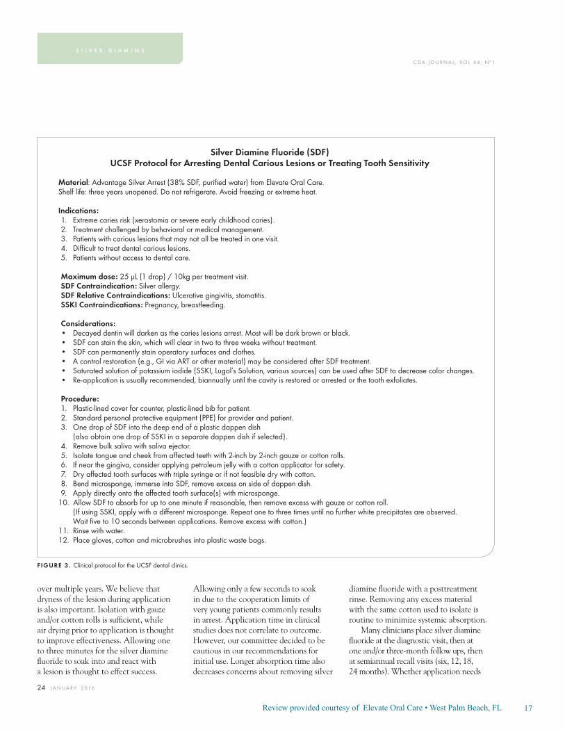
Silver Diamine Fluoride (SDF)UCSF Protocol for Arresting Dental Carious Lesions or Treating Tooth Sensitivity
Material: Advantage Silver Arrest (38% SDF, purifi ed water) from Elevate Oral Care.Shelf life: three years unopened. Do not refrigerate. Avoid freezing or extreme heat. Indications:1. Extreme caries risk (xerostomia or severe early childhood caries).2. Treatment challenged by behavioral or medical management.3. Patients with carious lesions that may not all be treated in one visit.4. Diffi cult to treat dental carious lesions.5. Patients without access to dental care.
Maximum dose: 25 μL (1 drop) / 10kg per treatment visit.SDF Contraindication: Silver allergy.SDF Relative Contraindications: Ulcerative gingivitis, stomatitis.SSKI Contraindications: Pregnancy, breastfeeding.
Considerations: • Decayed dentin will darken as the caries lesions arrest. Most will be dark brown or black. • SDF can stain the skin, which will clear in two to three weeks without treatment.• SDF can permanently stain operatory surfaces and clothes. • A control restoration (e.g., GI via ART or other material) may be considered after SDF treatment.• Saturated solution of potassium iodide (SSKI, Lugol’s Solution, various sources) can be used after SDF to decrease color changes.• Re-application is usually recommended, biannually until the cavity is restored or arrested or the tooth exfoliates.
Procedure:1. Plastic-lined cover for counter, plastic-lined bib for patient.2. Standard personal protective equipment (PPE) for provider and patient. 3. One drop of SDF into the deep end of a plastic dappen dish (also obtain one drop of SSKI in a separate dappen dish if selected).4. Remove bulk saliva with saliva ejector.5. Isolate tongue and cheek from aff ected teeth with 2-inch by 2-inch gauze or cotton rolls.6. If near the gingiva, consider applying petroleum jelly with a cotton applicator for safety.7. Dry aff ected tooth surfaces with triple syringe or if not feasible dry with cotton.8. Bend microsponge, immerse into SDF, remove excess on side of dappen dish.9. Apply directly onto the aff ected tooth surface(s) with microsponge.
10. Allow SDF to absorb for up to one minute if reasonable, then remove excess with gauze or cotton roll. (If using SSKI, apply with a diff erent microsponge. Repeat one to three times until no further white precipitates are observed. Wait fi ve to 10 seconds between applications. Remove excess with cotton.)
11. Rinse with water.12. Place gloves, cotton and microbrushes into plastic waste bags.
FIGURE 3 . Clinical protocol for the UCSF dental clinics.

18Review provided courtesy of Elevate Oral Care • West Palm Beach, FL
C DA J O U R N A L , V O L 4 4 , Nº 1
J A N UA RY 2 0 1 6 25
to continue after two or three years to maintain caries arrest is not known. Another approach is simply to substitute silver diamine fl uoride for any application of fl uoride varnish to a patient with untreated carious lesions. Increased frequency with higher disease burden follows the caries management by risk assessment (CAMBRA) principles.61 It is relevant to take photographs to track lesions over time.
Efforts to improve the penetration of silver diamine fl uoride into affected dentin by chemical cavity preparation have not been studied but are being explored clinically. Pretreatment with ethylenediaminetetraacetic acid (EDTA) to remove superfi cial hydroxyapatite in affected dentin may open the dentinal tubules to further silver diamine fl uoride penetration. Pretreatment with hypochlorite (bleach) may help breakdown bacteria and exposed dentin proteins, but this may be redundant to the action of the silver. Hypochlorite to decrease discoloration after silver diamine fl uoride treatment is not recommended, as the color comes from silver that cannot be broken down like organic chromophores and might break down dentin proteins stabilized against the effects of bacteria and acid by interactions with silver.
Experience with the combination of silver nitrate plus fl uoride varnish (see above) has many practitioners asking about a topical varnish after silver diamine fl uoride placement to prevent silver diamine fl uoride taste and keep the silver diamine fl uoride in the lesion. We see no evidence that varnish would help achieve either goal. Varnish does not seal. Rather, allowing more time for residence and diffusion of silver diamine fl uoride to react with and dry into the lesion is more likely to improve effectiveness. Also, in our experience, silver diamine fl uoride results in less aversive taste and texture responses than to fl uoride varnish.
Decreased darkening of lesions in the esthetic zone improves acceptance. SSKI is an option if the patient is not pregnant, though signifi cant darkening should still be expected. SSKI and silver diamine fl uoride are not to be combined prior to application — SSKI can be placed after drying the silver diamine fl uoride-treated tooth. Silver diamine fl uoride does not prevent restoration of a lesion, thus it does not prevent esthetic options. While silver diamine fl uoride has been shown to be more effective than ART or interim restorative treatment
(IRT),33 the two are compatible and can be combined across one or more visits.
The California Business and Professions Code permits dental hygienists and assistants to apply silver diamine fl uoride for the control of caries because they are topical fl uorides (Section 1910.(b)). Physicians, nurses and their assistants are permitted to apply fl uorides in California and in many other states and federal programs. The recent decision of the Oregon Dental Board to allow dental hygienists and assistants to place silver diamine fl uoride under existing rules for topical fl uoride medicaments sets a precedent. Dental hygienists and assistants in Oregon were barred from providing silver nitrate in a previous decision. All providers need to be trained. Applications should be tracked if applied to the same patient by multiple clinics.
Documentation and BillingA new code, D1354, for “interim
caries arresting medication application” was approved by the Code on Dental Procedures and Nomenclature (CDT) Code Maintenance Commission for 2016. The code defi nition is “Conservative treatment of an active, nonsymptomatic carious lesion by topical application of a caries arresting or inhibiting medicament and without mechanical removal of sound tooth structure.” The CDT Code is the U.S. HIPAA standard code set and is required for billing. The Commission includes representatives from the major insurers, Medicaid, ADA, AGD and specialty organizations. Insurers are in the process of evaluating coverage for this treatment.
Legal ConsiderationsSilver diamine fl uoride is cleared
by the FDA for marketing as a Class II medical device to treat tooth sensitivity. We are discussing off-label use as a drug to treat and prevent dental caries. This is a parallel situation to fl uoride varnish, which has the same device clearance but is ubiquitously used off label by dentists and physicians as a drug to prevent caries. The same public health dentists who achieved the FDA device clearance are now applying for a dental caries indication. However, this is a more complicated process, normally only carried out by large pharmaceutical companies, and is likely to take longer.
ConsentBecause silver diamine fl uoride
is new in the U.S., it is important to communicate effectively. In the UCSF clinics, we are using a special consent form (FIGURE 4) as a way to inform patients, parents and caregivers, and
In our experience, silver diamine fl uoride results in less aversive taste and texture responses than to fl uoride varnish.

19Review provided courtesy of Elevate Oral Care • West Palm Beach, FL
C DA J O U R N A L , V O L 4 4 , Nº 1
26 J A N UA RY 2 01 6
to standardize procedures because we have so many inexperienced student clinicians. All practices have established procedures for consent and an extra form may not be needed in the community. The normal elements of informed consent
apply. We sought to ensure awareness of the expected change in color of the dentin as the decay arrests, likelihood of reapplication and contraindications in the presence of silver allergy and stomatitis. Note the importance of distinguishing
between allergy to nickel and other trace metals rather than silver allergy, which is rare. We used readability measurements to guide intelligibility and included a progressively discoloring lesion to show stain of a lesion but not healthy enamel.
UCSF Dental Center Informed Consent for Silver Diamine Fluoride
Facts for consideration:• Silver diamine fl uoride (SDF) is an antibiotic liquid. We use SDF on cavities to help stop tooth decay. We also use it to treat tooth sensitivity. SDF application every six to 12 months is necessary.• The procedure: 1. Dry the aff ected area. 2. Place a small amount of SDF on the aff ected area. 3. Allow SDF to dry for one minute. 4. Rinse.• Treatment with SDF does not eliminate the need for dental fi llings or crowns to repair function or esthetics. Additional procedures will incur a separate fee.• I should not be treated with SDF if: 1. I am allergic to silver. 2. There are painful sores or raw areas on my gums (i.e., ulcerative gingivitis) or anywhere in my mouth (i.e., stomatitis).
Benefi ts of receiving SDF:• SDF can help stop tooth decay. • SDF can help relieve sensitivity.
Risks related to SDF include, but are not limited to:• The aff ected area will stain black permanently. Healthy tooth structure will not stain. Stained tooth structure can be replaced with a fi lling or a crown.• Tooth-colored fi llings and crowns may discolor if SDF is applied to them. Color changes on the surface can normally be polishe d off . The edge between a tooth and fi lling may keep the color.• If accidentally applied to the skin or gums, a brown or white stain may appear that causes no harm, cannot be washed off and will disappear in one to three weeks.• You may notice a metallic taste. This will go away rapidly.• If tooth decay is not arrested, the decay will progress. In that case the tooth will require further treatment, such as repeat SDF, a fi lling or crown, root canal treatment or extraction.• These side eff ects may not include all of the possible situations reported by the manufacturer. If you notice other eff ects, please contact your dental provider.• Every reasonable eff ort will be made to ensure the success of SDF treatment. There is a risk that the procedure will not stop the decay and no guarantee of success is granted or implied.
Alternatives to SDF, not limited to the following:• No treatment, which may lead to continued deterioration of tooth structures and cosmetic appearance. Symptoms may increase in severity.• Depending on the location and extent of the tooth decay, other treatment may include placement of fl uoride varnish, a fi lling or crown, extraction or referral for advanced treatment modalities.
I CERTIFY THAT I HAVE READ AND FULLY UNDERSTAND THIS DOCUMENTAND ALL MY QUESTIONS WERE ANSWERED:
________________________________(signature of patient) ___________________(date)
________________________________(signature of witness) ___________________(date)
FIGURE 4 . UCSF special consent form.
s i l v e r d i a m i n e

20Review provided courtesy of Elevate Oral Care • West Palm Beach, FL
C DA J O U R N A L , V O L 4 4 , Nº 1
J A N UA RY 2 0 1 6 27
ConclusionSilver diamine fl uoride is a safe,
effective treatment for dental caries across the age spectrum. At UCSF, it is indicated for patients with extreme caries risk, those who cannot tolerate conventional care, patients who must be stabilized so they can be restored over time, patients who are medically compromised or too frail to be treated conventionally and those in disparity populations with little access to care.
Application twice per year outperforms all minimally invasive options including the atraumatic restorative technique — with which it is compatible but 20 times less expensive. It approaches the success of dental fi llings after two or more years, and again, prevents future caries — while fi llings do not. Silver diamine fl uoride is more effective as a primary preventive than any other available material, with the exception of dental sealants, which are > 10 times more expensive and need to be monitored.
Saliva may play a role in caries arrest by silver diamine fl uoride. Lower rates of arrest are seen in geriatric patients.38 The elderly tend to have less abundant and less functional saliva, which generally explains their higher caries rate. In pediatric patients, higher rates of arrest are noted for buccal or lingual smooth surfaces and anterior teeth.31 These surfaces bathe more directly in saliva than others. It is surprising that silver chloride is the main precipitant in treated dentin, as chloride is not a common component of dentin or silver diamine fl uoride, so may come from the saliva.
Traditional approaches often provide only temporary benefi t, given the highest rates of recurrent caries are in patients with the worst disease burden. The advent of a treatment for nonsymptomatic caries not requiring general anesthesia or sedation addresses long-standing
concerns about the expense, danger and practical complexity of these services.
Experience suggests that dryness prior to application enhances effectiveness. Good patient management is still profoundly relevant to the very young and otherwise challenged patients, though this one-minute intervention is more tolerable than other options. Silver diamine fl uoride can readily replace fl uoride varnish for the prevention of caries in patients who have active caries. This as a powerful new tool in the fi ght against dental caries, particularly suited for those who suffer most from this disease.
Clinical evidence supports continued application one to two times per year until the tooth is restored or exfoliates, and otherwise perhaps indefi nitely. Some treated lesions keep growing, particularly those in the inner third of the dentin. It is unclear what will happen if treatment is stopped after two to three years and research is needed. ■
ACKNOWLEDGMENT
The UCSF paradigm shift committee subcommittee on silver caries arrest included Sean Mong, DDS, EdD; Spomenka Djordjevic, DDS, MS; Paul Atkinson, DDS, PhD; George Taylor, DMD, MPH, DrPh; Natalie Heaivilin, DDS; Ling Zhan, DDS, PhD; John Featherstone, PhD; Hellene Ellenikiotis, DDS; and Jeremy Horst, DDS, PhD. Thanks to Linda Milgrom for designing the PubMed search. Thanks to Chad Zillich, DDS, for help with the literature review. Thanks to study authors, particularly Edward Lo, BDS, MDS, PhD; Chun Hung Chu, BDS, MDS, PhD; and Geoff Knight, BDSc, MSc, MBA, for helpful discussions.
REFERENCES
1. Clarkson BH, Exterkate RAM. Noninvasive dentistry: A dream or reality? Caries Res 2015;49 suppl 1(1):11-17. doi:10.1159/000380887.2. Yamaga R, Yokomizo I. Arrestment of caries of deciduous teeth with diamine silver fl uoride. Dent Outlook 1969;33:1007-1013.3. Rosenblatt A, Stamford TCM, Niederman R. Silver diamine fl uoride: A caries “silver-fl uoride bullet.” J Dent Res 2009;88(2):116-125. doi:10.1177/0022034508329406.4. Mei ML, Chu CH, Lo ECM, Samaranayake LP. Fluoride and silver concentrations of silver diamine fl uoride solutions for dental use. Int J Paediatr Dent 2013;23(4):279-285. doi:10.1111/ipd.12005.5. Mei ML, Ito L, Cao Y, Li QL, Lo ECM, Chu CH. Inhibitory eff ect of silver diamine fl uoride on dentine demineralisation and collagen degradation. J Dent 2013;41(9):809-817.
doi:10.1016/j.jdent.2013.06.009.6. Hill TJ, Arnold FA. The eff ect of silver nitrate in the prevention of dental caries. I. The eff ect of silver nitrate upon the decalcifi cation of enamel. J Dent Res 1937;16:23-28.7. Castillo JL, Rivera S, Aparicio T, et al. The short-term eff ects of diamine silver fl uoride on tooth sensitivity: A randomized controlled trial. J Dent Res 2011;90(2):203-208. doi:10.1177/0022034510388516.8. Craig GG, Knight GM, McIntyre JM. Clinical evaluation of diamine silver fl uoride/potassium iodide as a dentine desensitizing agent. A pilot study. Aust Dent J 2012;57(3):308-311. doi:10.1111/j.1834-7819.2012.01700.x.9. Markowitz K, Pashley DH. Discovering new treatments for sensitive teeth: The long path from biology to therapy. J Oral Rehabil 2008;35(4):300-315. doi:10.1111/j.1365-2842.2007.01798.x.10. Featherstone JDB. The continuum of dental caries — evidence for a dynamic disease process. J Dent Res 2004;83 Spec No C:C39-C42.11. Mei ML, Li QL, Chu CH, Yiu CKY, Lo ECM. The inhibitory eff ects of silver diamine fl uoride at diff erent concentrations on matrix metalloproteinases. Dent Mater 2012;28(8):903-908. doi:10.1016/j.dental.2012.04.011.12. Mei ML, Ito L, Cao Y, Li QL, Chu CH, Lo ECM. The inhibitory eff ects of silver diamine fl uorides on cysteine cathepsins. J Dent 2014;42(3):329-335. doi:10.1016/j.jdent.2013.11.018.13. Klasen HJ. A Historical Review of the Use of Silver in the Treatment of Burns. II. Renewed Interest for Silver. 2000:131-138.14. Youravong N, Carlen A, Teanpaisan R, Dahlén G. Metal-ion susceptibility of oral bacterial species. Lett Appl Microbiol 2011;53(3):324-328. doi:10.1111/j.1472-765X.2011.03110.x.15. Hamama HH, Yiu CK, Burrow MF. Eff ect of silver diamine fl uoride and potassium iodide on residual bacteria in dentinal tubules. Aust Dent J 2015;60(1):80-87. doi:10.1111/adj.12276.16. Suzuki T, Nishida M, Sobue S, Moriwaki Y. Eff ects of diamine silver fl uoride on tooth enamel. J Osaka Univ Dent Sch 1974;14:61-72.17. Chu CH, Lo ECM. Micro-hardness of dentin in primary teeth after topical fl uoride applications. J Dent 2008;36(6):387-391. doi:10.1016/j.jdent.2008.02.013.18. Englander HR, James VE, Massler M. Histologic eff ects of silver nitrate of human dentin and pulp. J Am Dent Assoc 1958;57(5):621-630.19. Mei ML, Ito L, Cao Y, Lo ECM, Li QL, Chu CH. An ex vivo study of arrested primary teeth caries with silver diamine fl uoride therapy. J Dent 2014;42(4):395-402. doi:10.1016/j.jdent.2013.12.007.20. Knight GM, McIntyre JM, Craig GG, Mulyani, Zilm PS, Gully NJ. Inability to form a biofi lm of Streptococcus mutans on silver fl uoride- and potassium iodide-treated demineralized dentin. Quintessence Int 2009;40(2):155-161.21. Knight GM, McIntyre JM, Craig GG, Mulyani, Zilm PS, Gully NJ. An in vitro model to measure the eff ect of a silver fl uoride and potassium iodide treatment on the permeability of demineralized dentine to Streptococcus mutans. Aust Dent J 2005;50(4):242-245.22. Knight GM, McIntyre JM, Craig GG, Mulyani, Zilm PS, Gully NJ. Diff erences between normal and demineralized
21Review provided courtesy of Elevate Oral Care • West Palm Beach, FL
C DA J O U R N A L , V O L 4 4 , Nº 1
28 J A N UA RY 2 01 6
dentine pretreated with silver fl uoride and potassium iodide after an in vitro challenge by Streptococcus mutans. Aust Dent J 2007;52(1):16-21.23. Wakshlak RB-K, Pedahzur R, Avnir D. Antibacterial activity of silver-killed bacteria: the “zombies” eff ect. Sci Rep 2015;5:9555. doi:10.1038/srep09555.24. Duffi n S. Back to the future: The medical management of caries introduction. J Calif Dent Assoc 2012;40(11):852-858.25. Rethman MP, Beltrán-Aguilar ED, Billings RJ, et al. Nonfl uoride caries-preventive agents: Executive summary of evidence-based clinical recommendations. J Am Dent Assoc 2011;142(9):1065-1071.26. Plemons JM, Al-Hashimi I, Marek CL. Managing xerostomia and salivary gland hypofunction. J Am Dent Assoc 2014;145(8):867-873. doi:10.14219/jada.2014.44.27. Stebbins EA. What value has argenti nitras as a therapeutic agent in dentistry? Int Dent J 1891;12:661-670.28. Howe PR. A method of sterilizing and at the same time impregnating with a metal aff ected dentinal tissue. Dent Cosmos 1917;59:891-904.29. Chu CH, Lo ECM, Lin HC. Eff ectiveness of silver diamine fl uoride and sodium fl uoride varnish in arresting dentin caries in Chinese preschool children. J Dent Res 2002;81(11):767-770.30. Shah S, Bhaskar V, Venkataraghavan K, Choudhary P, Ganesh M, Trivedi K. Effi cacy of silver diamine fl uoride as an antibacterial as well as antiplaque agent compared to fl uoride varnish and acidulated phosphate fl uoride gel: An in vivo study. Indian J Dent Res 2013;24(5):575-581. doi:10.4103/0970-9290.123374.31. Zhi QH, Lo ECM, Lin HC. Randomized clinical trial on eff ectiveness of silver diamine fl uoride and glass ionomer in arresting dentine caries in preschool children. J Dent 2012;40(11):962-967. doi:10.1016/j.jdent.2012.08.002.32. Yee R, Holmgren C, Mulder J, Lama D, Walker D, van Palenstein Helderman W. Effi cacy of silver diamine fl uoride for arresting caries treatment. J Dent Res 2009;88(7):644-647. doi:10.1177/0022034509338671.33. Santos Dos VE, de Vasconcelos FMN, Ribeiro AG, Rosenblatt A. Paradigm shift in the eff ective treatment of caries in schoolchildren at risk. Int Dent J 2012;62(1):47-51. doi:10.1111/j.1875-595X.2011.00088.x.34. Lo EC, Chu CH, Lin HC. A community-based caries control program for preschool children using topical fl uorides: 18-month results. J Dent Res 2001;80(12):2071-2074.35. Llodra JC, Rodriguez A, Ferrer B, Menardia V, Ramos T, Morato M. Effi cacy of silver diamine fl uoride for caries reduction in primary teeth and fi rst permanent molars of schoolchildren: 36-month clinical trial. J Dent Res 2005;84(8):721-724.36. Green E. A clinical evaluation of two methods of caries prevention in newly erupted fi rst permanent molars. Aust Dent J 1989;34(5):407-409.37. Craig GG, Powell KR, Price CA. Clinical evaluation of a modifi ed silver fl uoride application technique designed to facilitate lesion assessment in outreach programs. BMC Oral Health 2013;13(1):73. doi:10.1186/1472-6831-13-73.38. Zhang W, McGrath C, Lo ECM, Li JY. Silver diamine fl uoride and education to prevent and arrest root caries among community-dwelling elders. Caries Res 2013;47(4):284-290. doi:10.1159/000346620.39. Liu BY, Lo ECM, Chu CH, Lin HC. Randomized trial on fl uorides and sealants for fi ssure caries prevention J Dent Res 2012;91(8):753-758. doi:10.1177/0022034512452278.
40. Tan HP, Lo ECM, Dyson JE, Luo Y, Corbet EF. A randomized trial on root caries prevention in elders. J Dent Res 2010;89(10):1086-1090. doi:10.1177/0022034510375825.41. Monse B, Heinrich-Weltzien R, Mulder J, Holmgren C, van Palenstein Helderman WH. Caries preventive effi cacy of silver diamine fl uoride (SDF) and ART sealants in a school-based daily fl uoride toothbrushing program in the Philippines. BMC Oral Health 2012;12(1):52. doi:10.1186/1472-6831-12-52.42. Li R, Lo ECM, Chu CH, Liu BY. Preventing and arresting root caries through silver diamine fl uoride applications. In: 2014. iadr.confex.com/iadr/14iags/webprogram/Paper189080.html.43. Duangthip D, Lo ECM, Chu CH. Arrest of dentin caries in preschool children by topical fl uorides. In: 2014. iadr.confex.com/iadr/14iags/webprogram/Paper189031.html.44. Frencken JE, Pilot T, Songpaisan Y, Phantumvanit P. Atraumatic Restorative Treatment (ART): Rationale, Technique and Development. J Public Health Dent 1996;56(3):135-140. doi:10.1111/j.1752-7325.1996.tb02423.x.45. Innes NP, Evans DJ, Stirrups DR. The Hall Technique: A randomized controlled clinical trial of a novel method of managing carious primary molars in general dental practice: Acceptability of the technique and outcomes at 23 months. BMC Oral Health 2007;7(1):18. doi:10.1186/1472-6831-7-18.46. Vasquez E, Zegarra G, Chirinos E, et al. Short-term serum pharmacokinetics of diamine silver fl uoride after oral application. BMC Oral Health 2012;12(1):60. doi:10.1186/1472-6831-12-60.47. Chu CH, Lo ECM. Promoting caries arrest in children with silver diamine fl uoride: A review. Oral Health Prev Dent 2008;6(4):315-321.48. Liu Y, Zhai W, Du F. [Clinical observation of treatment of tooth hypersensitiveness with silver ammonia fl uoride and potassium nitrate solution]. Zhonghua Kou Qiang Yi Xue Za Zhi. 1995;30(6):352-354.49. Yamaga R, Nishino M, Yoshida S, Yokomizo I. Diamine silver fl uoride and its clinical application. J Osaka Univ Dent Sch 1972;(12):1-20.50. Milgrom P, Taves DM, Kim AS, Watson GE, Horst JA. Pharmacokinetics of fl uoride in toddlers after application of 5% sodium fl uoride dental varnish. Pediatrics 2014;134(3):e870-e874. doi:10.1542/peds.2013-3501.51. Goldberg ME, Cantillo J, Larijani GE, Torjman M, Vekeman D, Schieren H. Sevofl urane versus isofl urane for maintenance of anesthesia: Are serum inorganic fl uoride ion concentrations of concern? Anesth Analg 1996;82(6):1268-1272.52. Gotjamanos T, Afonso F. Unacceptably high levels of fl uoride in commercial preparations of silver fl uoride. Aust Dent J 1997;42(1):52-53.53. Gotjamanos T, Ma P. Potential of 4 percent silver fl uoride to induce fl uorosis in rats: Clinical implications. Aust Dent J 2000;45(3):187-192.54. Neesham DC. Fluoride concentration in AgF and dental fl uorosis. Aust Dent J 1997;42(4):268-269.55. Quock RL, Barros JA, Yang SW, Patel SA. Eff ect of Silver Diamine Fluoride on Microtensile Bond Strength to Dentin. Oper Dent 2012;37(6):610-616. doi:10.2341/11-344-L.56. Knight GM, McIntyre JM, Mulyani. The eff ect of silver fl uoride and potassium iodide on the bond strength of auto cure glass ionomer cement to dentine. Aust Dent J 2006;51(1):42-45.
57. Yamaga M, Koide T, Hieda T. Adhesiveness of glass ionomer cement containing tannin-fl uoride preparation (HY agent) to dentin — an evaluation of adding various ratios of HY agent and combination with application diamine silver fl uoride. Dent Mater J 1993;12(1):36-44.58. Soeno K, Taira Y, Matsumura H, Atsuta M. Eff ect of desensitizers on bond strength of adhesive luting agents to dentin. J Oral Rehabil 2001;28(12):1122-1128.59. Centers for Disease Control and Prevention. Long-Term Care Services in the United States: 2013 Overview, table 4. www.cdc.gov/nchs/data/nsltcp/long_term_care_services_2013.pdf.60. U.S. Department of Health and Human Services. Integration of Oral Health and Primary Care Practice. www.hrsa.gov/publichealth/clinical/oralhealth. Accessed June 5, 2015.61. Featherstone JDB, Adair SM, Anderson MH, et al. Caries management by risk assessment: Consensus statement, April 2002. J Calif Dent Assoc 2003 Mar;31(3):257-69.
THE CORRESPONDING AUTHOR, Jeremy A. Horst, DDS, PhD, can be reached at [email protected].
s i l v e r d i a m i n e

22Review provided courtesy of Elevate Oral Care • West Palm Beach, FL
Compendium of Continuing Education article provides an overview on the use of Silver Diamine Fluoride in dental treatment. May L. Mei, BDS, MDS, PhD; Edward Chin-Man Lo, BDS, MDS, PhD; and Chun-Hung Chu, BDS, MDS, PhD.
ABSTRACTThe use of a topical fluoride solution, namely silver diamine fluoride (SDF), in dental treatment has been drawing increasing attention. SDF has been used in some countries in Asia, including Japan and China, as a caries-arresting and anti-hypersensitivity agent. It was recently cleared by the Food and Drug Admin-istration in the United States as a fluoride to manage hypersensitive teeth. Topical application of SDF is a noninvasive procedure that is quick and simple to use. Promising results of laboratory studies and clinical trials have suggested that SDF is more effective than other fluoride agents to halt the caries process. A review concluded that SDF is a safe, effective, efficient, and equitable caries control agent that has a potentially broad application in dentistry and may meet the criteria of both the WHO Millennium Development Goals and the US Institute of Medicine’s criteria for 21st century medical care. This article provides an overview of the clinical use of SDF in dental treatment.
Please use the link below to access the full article.
https://cced.cdeworld.com/courses/4990#sthash.d0aJoy9Y.dpuf
CLINICAL USE OF SILVER DIAMINE FLUORIDE IN DENTAL TREATMENT
FEBRUARY 2016

23Review provided courtesy of Elevate Oral Care • West Palm Beach, FL
Decisions in Dentistry article on the use of Silver Diamine Fluoride in adult patients. Dr. John Featherstone, Dean of the University of California San Francisco School of Dentistry and Dr Jeremy Horst, DDS, PhD.
KEY TAKEAWAYS• Cleared by the U.S. Food and Drug Administration for treating dentinal hyper-
sensitivity, in off-label use silver diamine fluoride can be used to prevent and arrest caries.
• The agent acts as an antimicrobial that remains active well after application. It also promotes remineralization and resistance to demineralization in enamel and dentin.
• In order to effectively implement treatment, clinicians should know the indica-tions and contraindications, and gain informed consent for use.
• Dentists and (if allowed by state practice acts) dental auxiliaries who apply this agent must understand precautions for handling silver diamine fluoride.
• Repeat application completely stops many, but not all lesions. Research is needed to determine why some caries are not arrested.
Please use the link below to access the full article.
http://decisionsindentistry.com/article/fresh-approach-to-caries-arrest-in-adults/
FRESH APPROACH TO CARIES ARREST IN ADULTS
SEPTEMBER/OCTOBER 2015

24Review provided courtesy of Elevate Oral Care • West Palm Beach, FL

25Review provided courtesy of Elevate Oral Care • West Palm Beach, FL

26Review provided courtesy of Elevate Oral Care • West Palm Beach, FL

27Review provided courtesy of Elevate Oral Care • West Palm Beach, FL

28Review provided courtesy of Elevate Oral Care • West Palm Beach, FL

29Review provided courtesy of Elevate Oral Care • West Palm Beach, FL

30Review provided courtesy of Elevate Oral Care • West Palm Beach, FL

31Review provided courtesy of Elevate Oral Care • West Palm Beach, FL

32Review provided courtesy of Elevate Oral Care • West Palm Beach, FL

33Review provided courtesy of Elevate Oral Care • West Palm Beach, FL

34Review provided courtesy of Elevate Oral Care • West Palm Beach, FL
Articles Published after Rosenblatt, et. al 2009 Systematic Review
1 Aust Dent J. 2015 Mar;60Suppl 1:95-105. doi: 10.1111/adj.12288. Improving the oral health of frail and functionally dependent elderly. Lewis A(1), Wal-lace J, Deutsch A, King P.
2. Int J Antimicrob Agents. 2015 Feb;45(2):183-7. doi: 10.1016/j.ijantimi-cag.2014.09.007. Epub 2014 Oct 14.Action of silver nanoparticles towards biological systems: cytotoxicity evaluation using hen’s egg test and inhibition of Streptococcus mutans biofilm formation. FreirePL(1), Stamford TC(2), Albuquerque AJ(3), Sampaio FC(3), Cavalcante HM(3), Macedo RO(3), Galembeck A(4), Flores MA(5), Rosenblatt A(6).
3. Trials. 2014 Nov 19;15:448. doi: 10.1186/1745-6215-15-448. New proposal of silver diamine fluoride use in arresting approximal caries: study protocol for a randomized controlled trial. Mattos-Silveira J, Floriano I, Ferreira FR, Viganó ME, Frizzo MA, Reyes A, Novaes TF, Moriyama CM, Raggio DP, Imparato JC, Mendes FM, Braga MM(1).
4. Int J Pediatric Dent. 2014 Sep 17.doi: 10.1111/ipd.12134. [Epub ahead of print] Children’s discomfort may vary among different treatments for initial approximal caries lesions: preliminary findings of a randomized controlled clinical trial. Mattos-SilveiraJ(1), Floriano I, Ferreira FR, Viganó ME, Mendes FM, Braga MM. Author information:
5. J Dent. 2014 Aug;42(8):945-51. doi: 10.1016/j.jdent.2014.05.017. Epub 2014 Jun 12. A new “silver-bullet” to treat caries in children--nano silver fluoride: a randomized clinical trial. Santos VE Jr(1), VasconcelosFilho A(2), Targino AG(3), Flores MA(4), Galembeck A(5), Caldas AF Jr(6), Rosenblatt A(7).
6. J Mater Sci Mater Med. 2014 Aug;25(8):2041-7. doi: 10.1007/ s10856-014-5221-5. Epub 2014 May 13.An innovative approach to treating dental decay in children. A new anti-caries agent. TarginoAG(1), Flores MA, dos San-tos Junior VE, de Godoy BenéBezerra F, de Luna Freire H, Galembeck A, Rosenblatt A.
7. Lasers Med Sci. 2015 Apr;30(3):985-91. doi: 10.1007/ s10103-014-1521-8. Epub 2014 Jan 26.Effect of laser irradiation on the fluoride uptake of silver diamine fluoride treated dentine. Mei ML(1), Ito L, Zhang CF, Lo EC, Chu CH.
8. BMC Res Notes. 2014 Jan 3;7:3. doi: 10.1186/1756-0500-7-3. Arresting ram-pant dental caries with silver diamine fluoride in a young teenager suffering from chronic oral graft versus host disease post-bone marrow transplantation: a case report. Chu CH(1), Lee AH, Zheng L, Mei ML, Chan GC.
9. Org Lett. 2014 Jan 3;16(1):102-5. doi: 10.1021/ol403083e. Epub 2013 Dec 2.AgF-mediated fluorinativehomocoupling of gem-difluoroalkenes. GaoB(1),

35Review provided courtesy of Elevate Oral Care • West Palm Beach, FL
Zhao Y, Ni C, Hu J.
10. Science. 2013 Nov 22;342(6161):956-60. doi: 10.1126/science. 1243759. Selective C-H fluorination of pyridines and diazines inspired by a classic amination reaction. FierPS(1), Hartwig JF.
11. Nat Protoc. 2013 Dec;8(12):2348-54. doi: 10.1038/nprot.2013.144. Epub 2013 Oct 31. Oxidative aliphatic C-H fluorination with manganese catalysts and fluoride ion.Liu W(1), Huang X, Groves JT.
12. J Dent. 2013 Sep;41(9):809-17. doi: 10.1016/j.jdent.2013.06.009. Epub 2013 Jun 27. Inhibitory effect of silver diamine fluoride on dentine demineralisa-tion and collagen degradation. Mei ML(1), Ito L, Cao Y, Li QL, Lo EC, Chu CH.
13. Lasers Med Sci. 2014 Nov;29(6):1785-91. doi: 10.1007/ s10103-013-1329-y. Epub 2013 Apr 26.Prevention of dentine caries using silver diamine fluo-ride application followed by Er:YAG laser irradiation: an in vitro study. Mei ML(1), Ito L, Chu CH, Lo EC, Zhang CF.
14. Spec Care Dentist. 2013 May-Jun;33(3):133-40. doi: 10.1111/j.1754-4505.2012.00318.x. Epub 2012 Dec 10.Prevention of root caries: a literature review of primary and secondary preventive agents. GluzmanR(1), Katz RV, Frey BJ, McGowan R.
15. Ann ClinMicrobiolAntimicrob. 2013 Feb 26;12:4. doi: 10.1186/1476-0711-12-4. Antibacterial effects of silver diamine fluoride on multi-species cario-genic biofilm on caries.Mei ML(1), Li QL, Chu CH, Lo EC, Samaranayake LP.
16. Aust Dent J. 2013 Mar;58(1):50-6. doi: 10.1111/adj.12033. Epub 2013 Jan 30.An in vitro study of silver and fluoride ions on remineralization of demin-eralized enamel and dentine. ZhiQH(1), Lo EC, Kwok AC.
17. Contemp Clin Dent. 2012 Jul;3(3):262-4. doi: 10.4103/0976-237X. 103615. Anti-microbial efficiency of silver diamine fluoride as an endodontic medica-ment -An ex vivo study. Mathew VB(1), Madhusudhana K, Sivakumar N, Venugopal T, Reddy RK.
18. Am J Dent. 2012 Oct;25(5):299-302. Changes in the crystallinity of hydroxy-apatite powder and structure of enamel treated with several concentrations of ammonium hexafluorosilicate. SugeT(1), Ishikawa K, Matsuo T.
19. J Oral Sci. 2012 Sep;54(3):267-72. Effects of ammonium hexafluorosili-cate application on demineralized enamel and dentin of primary teeth. Ho-soyaY(1), Watanabe E, Tadokoro K, Inoue T, Miyazaki M, Tay FR.
20. Int J Paediatr Dent. 2013 Jul;23(4):279-85. doi: 10.1111/ipd. 12005. Epub

36Review provided courtesy of Elevate Oral Care • West Palm Beach, FL
2012 Oct 3. Fluoride and silver concentrations of silver diammine fluoride solutions for dental use. Mei ML(1), Chu CH, Lo EC, Samaranayake LP.
21. In Vivo. 2012 Jul-Aug;26(4):657-64. Effect of three fluoride compounds on the growth of oral normal and tumor cells. AcraAM(1), Sakagami H, Matsuta T, Adachi K, Otsuki S, Nakajima H, Koh T, Machino M, Ogihara T, Wata-nabe K, Watanabe S, Salgado AV, Bastida NM.
22. J Dent Res. 2012 Aug;91(8):753-8. doi: 10.1177/0022034512452278. Epub 2012 Jun 26. Randomized trial on fluorides and sealants for fissure caries prevention. Liu BY(1), Lo EC, Chu CH, Lin HC.
23. J Evid Based Dent Pract. 2012 Jun;12(2):95-6. doi: 10.1016/ j.jeb-dp.2012.03.011. Chlorhexidine varnish, sodium fluoride varnish, and silver diamine fluoride solution can prevent the development of new root caries in elders living in senior homes in Hong Kong. NiessenLC(1).
24. J Am Chem Soc. 2012 Jul 4;134(26):10795-8. doi: 10.1021/ja304410x. Epub 2012 Jun 22. Copper-mediated fluorination of aryl iodides. Fier PS(1), Hartwig JF.
25. Oper Dent. 2012 Nov-Dec;37(6):610-6. doi: 10.2341/11-344-L. Epub 2012 May 22. Effect of silver diamine fluoride on microtensile bond strength to dentin. Quock RL(1), Barros JA, Yang SW, Patel SA.
26. Am J Dent. 2012 Feb;25(1):31-4. Antibacterial activity of ammonium hexa-fluorosilicate solution with antimicrobial agents for the prevention of dentin caries. Shibata S(1), Suge T, Kimura T, Ishikawa K, Matsuo T.
27. J Dent. 2012 Jul;40(7):531-41. doi: 10.1016/j.jdent.2012.03.009. Epub 2012 Apr 3. Silver compounds used in dentistry for caries management: a review. PengJJ(1), Botelho MG, Matinlinna JP.
28. J Nanosci Nanotechnol. 2011 Nov;11(11):10063-8. Fabrication and enhanced visible light photocatalytic activity of fluorine doped TiO2 by loaded with Ag. Lin X(1), Rong F, Ji X, Fu D, Yuan C.
29. Aust Dent J. 2012 Mar;57(1):65-70. doi: 10.1111/j. 1834-7819.2011.01641.x.
Effect of silver and fluoride ions on enamel demineralization: a quantitative study using micro-computed tomography. Liu BY(1), Lo EC, Li CM.
30. Acta Odontol Latinoam. 2011;24(2):127-31. In vitro antibacterial activity of silver diamine fluoride in different concentrations.de Almeida LdeF(1), Cav-alcanti YW, Valença AM.
31. J Calif Dent Assoc. 2011 Oct;39(10):735-41. Prevention-centered caries man-agement strategies during critical periods in early childhood. MilgromP(1), Chi DL.

37Review provided courtesy of Elevate Oral Care • West Palm Beach, FL
32. J Conserv Dent. 2011 Jul;14(3):233-6. doi: 10.4103/0972-0707.85796. Remineralizing efficacy of silver diamine fluoride and glass ionomer type VII for their proposed use as indirect pulp capping materials - Part II (A clinical study). SinhaN(1), Gupta A, Logani A, Shah N.
33. J Org Chem. 2011 Oct 21;76(20):8543-8. doi: 10.1021/jo2016168. Epub 2011 Sep 26.Palladium-catalyzed direct ortho C-H arylation of 2-arylpyridine derivatives with aryltrimethoxysilane. Li W(1), Yin Z, Jiang X, Sun P.
34. J Conserv Dent. 2011 Apr;14(2):113-6. doi: 10.4103/0972-0707.82603. An ex vivo study to evaluate the remineralizing and antimicrobial efficacy of silver diamine fluoride and glass ionomer cement type VII for their proposed use as indirect pulp capping materials - Part I. Gupta A(1), Sinha N, Logani A, Shah N.
35. J Dent. 2011 Sep;39(9):612-8. doi: 10.1016/j.jdent.2011.06.008. Epub 2011 Jul 1.Reaction of silver diamine [corrected] fluoride with hydroxyapatite and protein. Lou YL(1), Botelho MG, Darvell BW.
36. Int J Paediatr Dent. 2012 Jan;22(1):2-10. doi: 10.1111/j. 1365-263X.2011.01149.x. Epub 2011 Jun 27. Effects of silver diamine fluo-ride on dentine carious lesions induced by Streptococcus mutans and Actino-mycesnaeslundii biofilms. Chu CH(1), Mei L, Seneviratne CJ, Lo EC.
37. J Org Chem. 2011 Jul 15;76(14):5793-802. doi: 10.1021/jo200966k. Epub 2011 Jun 21. Synthesis of triazafluoranthenones via silver(I)-mediated non-oxidative and oxidative intramolecular palladium-catalyzed cyclizations. Koutentis PA(1), Loizou G, Lo Re D.
38. Med Hypotheses. 2011 Sep;77(3):315-7. doi: 10.1016/j.mehy. 2011.05.002. Epub 2011 May 19.Is a drill-less dental filling possible? QuockRL(1), Patel SA, Falcao FA, Barros JA.
39. J Evid Based Dent Pract. 2011 Mar;11(1):54-5. doi: 10.1016/ j.jeb-
dp.2010.11.011. Topical application of silver diamine fluoride may arrest dental caries. HouptM(1).
40. Org Lett. 2011 Mar 18;13(6):1510-3. doi: 10.1021/ol200196m. Epub 2011 Feb 11.Silver-mediated palladium-catalyzed direct C-H arylation of 3-bro-moisothiazole-4-carbonitrile.IoannidouHA(1), Koutentis PA.
41. J Mol Model. 2011 Sep;17(9):2237-48. doi: 10.1007/ s00894-010-0949-4. Epub 2011 Jan 22.On possible existence of pseudobinary mixed valence fluo-rides of Ag(I)/Ag(II): a DFT study. Grochala W(1).
42. Anal Chem. 2011 Feb 15;83(4):1315-20. doi: 10.1021/ac1024683. Epub 2011 Jan 14.Quantification of VX vapor in ambient air by liquid chromatography isotope dilution tandem mass spectrometric analysis of glass bead filled sam-
38Review provided courtesy of Elevate Oral Care • West Palm Beach, FL
pling tubes. Evans RA(1), Smith WL, Nguyen NP, Crouse KL, Crouse CL, Norman SD, Jakubowski EM.
43. Stat Med. 2011 Feb 10;30(3):250-9. doi: 10.1002/sim.4094. Epub 2010 Nov 5.Analysis of multilevel grouped survival data with time-varying regression coefficients. Wong MC(1), Lam KF, Lo EC.
44. MolPharmacol. 2010 Nov;78(5):952-60. doi: 10.1124/mol.110.066407. Epub 2010 Aug 20.Inhibition of large-conductance Ca2+-activated K+ channels by nanomolar concentrations of Ag+. Zhou Y(1), Xia X, Lingle CJ.
45. Zhongguo Yi XueKeXue Yuan XueBao. 2010 Jun;32(3):265-8. doi: 10.3881/j.issn.1000-503X.2010.03.005. [Effect of combination of pulsed CO2 laser irradiation and diammine silver fluoride treatment on ultrastructure of dentine].[Article in Chinese] Wang Q(1), Zhao JZ, Wu XM.
46. J Endod. 2010 Jun;36(6):1026-9. doi: 10.1016/j.joen.2010.02.029. Epub 2010 Apr 24. Antimicrobial efficacy of 3.8% silver diamine fluoride and its effect on root dentin. HiraishiN(1), Yiu CK, King NM, Tagami J, Tay FR.
47. J Evid Based Dent Pract. 2010 Jun;10(2):122-4. doi: 10.1016/ j.jeb-dp.2010.02.014. Silver diamine fluoride (SDF) may be better than fluoride varnish and no treatment in arresting and preventing cavitated carious lesions. Beltrán-Aguilar ED(1).
48. Dent Today. 2010 Feb;29(2):130, 132-3. Leave decay in my cavity? You must be kidding! Knight GM(1), McIntyre JM, Craig GG, Mulyani.
49. J Am Chem Soc. 2010 Feb 10;132(5):1476-7. doi: 10.1021/ja909806t. Silver dendrites from galvanic displacement on commercial aluminum foil as an effective SERS substrate. GutésA(1), Carraro C, Maboudian R.
50. Braz Oral Res. 2009 Jul-Sep;23(3):296-301. In vitro evaluation of fluoride products in the development of carious lesions in deciduous teeth. San-tosLdeM(1), Reis JI, Medeiros MP, Ramos SM, Araújo JM.
51. AcadPediatr. 2009 Nov-Dec;9(6):404-9. doi: 10.1016/j.acap. 2009.09.001. An examination of the advances in science and technology of prevention of tooth decay in young children since the Surgeon General’s Report on Oral Health. MilgromP(1), Zero DT, Tanzer JM.
52. Evid Based Dent. 2009;10(3):68. doi: 10.1038/sj.ebd.6400661. Silver lining for caries cloud? DeeryC(1).
53. J Org Chem. 2009 Nov 6;74(21):8232-42. doi: 10.1021/jo901725k. 5(6)-an-ti-Substituted-2-azabicyclo[2.1.1]hexanes: a nucleophilic displacement route. KrowGR(1), Edupuganti R, Gandla D, Choudhary A, Lin G, Sonnet PE, DeBrosse C, Ross CW 3rd, Cannon KC, Raines RT.

39Review provided courtesy of Elevate Oral Care • West Palm Beach, FL
54. Dent Mater. 2010 Jan;26(1):29-34. doi: 10.1016/j.dental. 2009.08.011. Ef-fects of ammonium hexafluorosilicate concentration on dentin tubule occlu-sion and composition of the precipitate. SugeT(1), Kawasaki A, Ishikawa K, Matsuo T, Ebisu S.
55. J Dent Child (Chic). 2009 Jan-Apr;76(1):28-33. Effect of silver diammine fluoride on incipient caries lesions in erupting permanent first molars: a pilot study. Braga MM(1), Mendes FM, De Benedetto MS, Imparato JC.
56. Journal of Advanced Oral Research / Jan-Apr 2014 / Vol. 5 No.1. Silver Diamine Fluoride: A Review and Current Applications. Shalin Shah1, Vijay Bhaskar2, Karthik Venkatraghavan3, Prashant Choudhary4, Ganesh M.5, Krishna Trivedi6
57. International Dental Journal 2012; 62: 47–51. doi: 10.1111/j.1875-595X.2011.00088.x. Paradigm shift in the effective treatment of caries in schoolchildren at risk. Valdeci E. dos Santos Jr, Fla´ via M. N. de Vasconce-los1, Andre´a G. Ribeiro and Aronita Rosenblatt
58. J Dent. 2015 May 30. pii: S0300-5712(15)00124-4. doi: 10.1016/j.jdent.2015.05.006. A randomized clinical trial on arresting dentine caries in preschool children by topical fluorides -18 month results. Duangthip D, Chu CH, Lo CM
59. Parental Acceptance of the Use of Diamine Silver Fluoride in Children Aged 0 to 3 Years in the City of Cascavel, PR, Brazil Thaisa Cezária TRICHES, Mabel Mariela Rodríguez CORDEIRO, Juliana Garcia Mugnai Vieira SOU-ZA, Eduardo Karam SALTORI, Beatriz Helena Sottile FRANÇA
60. Pulp response to high fluoride releasing glass ionomer, silver diamine fluo-ride,and calcium hydroxide used for indirect pulp treatment: An in-vivo com-parative study. AtishKorwar, Sidhartha Sharma, Ajay Logani, and Naseem-ShahContempClin Dent. 2015 Jul-Sep; 6(3): 288–292.
61. The evidence base for professional and self-care prevention - caries, erosion and sensitivity. Twetman S. BMC Oral Health. 2015;15 Suppl 1:S4. doi: 10.1186/1472-6831-15-S1-S4. Epub 2015 Sep 15.
62. Root Caries in Older Adults. Gregory D, Hyde S. J Calif Dent Assoc. 2015 Aug;43(8):439-45.
63. Silver diamine fluoride and glass ionomer differentially remineralize early caries lesions, in situ. Nantanee R, Santiwong B, Trairatvorakul C, Hamba H, Tagami J. Clin Oral Investig. 2015 Sep 23.
64. The effectiveness of the biannual application of silver nitrate solution fol-lowed by sodium fluoride varnish in arresting early childhood caries in preschool children: study protocol for a randomised controlled trial. Chu

40Review provided courtesy of Elevate Oral Care • West Palm Beach, FL
CH, Gao SS, Li SK, Wong MC, Lo EC. Trials. 2015 Sep 25;16(1):426. doi: 10.1186/s13063-015-0960-2
65. Prevention of secondary Caries by silver diamine fluoride. Mei ML, Zhao IS, Ito L, Lo EC, Chu CH. Int Dent J. 2015 Dec 22. doi: 10.1111/idj.12207
66. An alternate technique of care using silver fluoride followed by stannous fluoride in the management of root caries in aged care. Deutsch A. Spec Care Dentist. 2015 Dec 21. doi: 10.1111/scd.12153.
67. Caries remineralisation and arresting effect in children by professionally ap-plied fluoride treatment - a systematic review. Gao SS, Zhang S, Mei ML Lo EC, Chu CH. BMC Oral Health. 2016 Feb 1; 16(1), 12.

41Review provided courtesy of Elevate Oral Care • West Palm Beach, FL
Frequently Asked Questions
1. Should SDF be used as a caries preventive therapy in a similar manner to how fluoride varnish is used “off-label” as a caries prevention therapy?
No. SDF is indicated for site-specific application only. Fluoride varnish can be applied to specific sites, but most fluoride varnish applications are full mouth. SDF is never a full mouth application.
SDF treatment should be limited to up to five sites per appointment. Appli-cation to more than five sites should be spaced by 7 days. For patients with active caries, SDF can be considered as a replacement of fluoride varnish as the primary means to prevent new lesions, with application of SDF to the active lesions only.
2. Is SDF safe for use in children? Regarding the margin of safety for dosing, a study was conducted for FDA
review for market clearance in rats and mice to determine the lethal dose by oral and subcutaneous administration. The worst case scenario is subcutane-ous administration and that lethal dose was found to be 380 mg/kg. One drop (25uL) of 38% silver diamine fluoride (SDF) contains 9.5 mg silver diamine fluoride. Thus, one drop of 38% SDF applied to 10 kg (22 lb.) child would equal 0.95 mg/kg, equal to a four-hundredfold safety margin.
In setting up protocols for undergraduate application of 38% SDF the Univer-sity of California San Francisco set a recommended limit of one drop per 10 kg per treatment visit, with weekly intervals at most.
3. Does SDF discolor skin or oral tissue?
Contact to skin and oral tissue is not harmful but is likely to cause temporary “tattooing”. On skin and oral soft tissue the effect is not immediate, rather it will be noticed within hours. The speed of discoloration is accelerated with light contact. The staining will be limited to direct areas of contact and will fade over a period of 24-72 hours. Patients should be protected with bibs and safety glasses as in any clinical procedure. If you believe you have touched the applicator to the skin of a patient it is good to advise them of possible tattooing.
4. Does the application technique differ between the label indication of relief of dentinal hypersensitivity and the off label indication of caries control?
In countries where SDF is used for the control of caries the application tech-nique is identical to the instructions we provide in the Advantage Arrest Pack-age Insert for the relief of dentinal hypersensitivity. No excavation, decay removal or anesthesia is required. The area to be treated should be “dry tooth brush” clean, free of plaque and debris.The area should be free of saliva, so

42Review provided courtesy of Elevate Oral Care • West Palm Beach, FL
as not to dilute the material.Transfer the material from a disposable plastic dappen dish to the surface to be treated and allow time to dry, which gener-ally occurs in 30-60 seconds. If accelerated drying is required due to patient compliance use a low/weak air stream to dry the material.
The chemical action of the SDF occurs almost immediately in the outer layers of the softened dentin and can be confirmed by changes in the hardness and density of the dentin surface, similar to caries that arrests naturally because of positive changes in oral hygiene, diet, or daily application of fluoride in cus-tom trays. The darkening of the lesion occurs over 24 hours and may increase over a week. Reexamination of the lesion at the next regular recall is appro-priate and reapplication of SDF may be warranted. Repeat until the lesion has arrested
5. Is there a recommended frequency of application of SDF for caries con-trol?
Caries arrest studies were conducted with SDF applications of once and twice annually. Arrested lesions were retreated every six-months.
Clinicians have reported that they will recall their first cohort of SDF patients within 3-6 weeks to evaluate the application and action of the treatment. Once they have a feel for the predictability of the material with their application technique they will set recall appointments based on the risk level and caries activity of the patient with higher risk patients at 3-month intervals. Moderate to high-risk patients, where it appears that home care and diet counseling has had positive impact, are recalled at 6-months.
6. Does the application of SDF to a lesion cause discoloration?
Yes, darkening of decayed, demineralized sites occurs as the lesion arrests. This is similar to what is seen when caries arrests from changes in diet or increased use of other fluorides. A recent study showed that patients see the discoloration as a clear indication that the treatment is working. Similar to the treatment of eroded and hypersensitive dentin, the treated area can be restored using glass ionomer or with a sandwich restoration of both glass ionomer and composite. In lab studies, bond strength of composite by itself to treated surfaces is reduced but the clinical importance is unknown.
38% silver diamine fluoride should not be diluted in an attempt to reduce discoloration. Studies have shown that diluted solutions may not be effective for caries arrest.
Ionic silver adsorbs onto almost any protein surface and is especially te-naciously bound to denatured proteins. This accounts for the specificity to carious collagen over normal collagen, but both will stain. The differentiator between these stains is that with SDF use, intrinsic pigmentation of a carious lesion occurs and surface protein staining occurs primarily on healthy tissue. These oxides are bound to the tissue and don’t wash or polish away. This is why the blackened lesion retains its dark color for so long, and is most likely

43Review provided courtesy of Elevate Oral Care • West Palm Beach, FL
the reason the antimicrobial effect is long lasting.
The functional indicator of effectiveness is when the silver oxide is bound to the diseased collagen. If the surface doesn’t turn black, the silver didn’t bind and the antimicrobial effect will only be short lived.
7. Can SDF be used on a prepared tooth just prior to restoration cementa-tion?
Desensitizing agents, have been shown to be protective of the pulp when
placed on crown preparations to reduce dentin permeability. Advantage Arrest, a desensitizer, has been shown safe to the pulp when placed on ex-posed dentin. In addition, studies have shown desensitization and efficacy in treating softened dentin before placing direct restorations. Usually the tooth is first treated with silver diamine fluoride 38%. This provides the benefit of sealing tubules plus the antimicrobial benefits of both silver and fluoride
8. Are there any contraindications for the use of SDF for the control of car-ies?
SDF should not be placed on exposed pulps. Other topical fluorides (e.g.
fluoride varnish) should not be used in the same appointment. Studies have shown that 38% silver diamine fluoride conveys more effective protection against decay in other teeth than fluoride varnish with reduced overall fluo-ride exposure.
9. Is there evidence of caries prevention benefit to non-application sites fol-lowing SDF use for a patient?
Treating carious areas with silver diamine fluoride 38% acts as a whole
mouth fluoride treatment. It can also be used in place of sealants in grooves. A protective effect has been shown to non-treated teeth and surfaces. These findings come from high quality randomized clinical trials.
10. Are there any post appointment instructions for the patient or the care-givers/guardians?
There are no postoperative limitations. Patients may eat or drink immediate-
ly. Patients may brush their teeth with fluoridated toothpaste on their regular schedule.
11. Does SDF stain countertops, instruments etc.?
Yes. When dispensing SDF it is a good idea to use an absorbent material that has a coated bottom, like a patient bib, under the dappen dish and applicator to avoid contact with metal trays and office countertops. If SDF comes in contact with instruments or countertops wash immediately with water, soap, ammonia or iodine tincture and then rinse thoroughly with water. Sodium

44Review provided courtesy of Elevate Oral Care • West Palm Beach, FL
hypochlorite (household bleach) can also be used for difficult stains.
12. What are the safety implications for application of SDF for a patient that has more than six sites to be treated?
The Margin of Safety for the volume of product needed to treat six sites is
within 130 times the NOAEL (no-observed-adverse-effect-level). Treating more sites in one visit will likely have little practical impact on patient safety. Like protocols for fluoride varnish application, the suspension for several days of fluoride supplements is advised.
13. How does an arrested lesion treated with SDF look like on radiographs? Arrested lesions look like a scar on radiographs. You will observe radio-opac-
ity as the mineralization of the previously softened dentin increases. Ulti-mately the best test of arrest is still the color change and tactile hardness of the dentin surface.
It is advised that you educate your referring dentist about your use of Advan-tage Arrest since the appearance of a treated lesion might be new and confus-ing for many practitioners.
14. How can Advantage Arrest be coded using CDT? There is a new CDT code for 2016 specifically for the use of caries arresting
medicaments; the off-label use of Advantage Arrest. Code D1354
The nomenclature reads: “Interim caries arresting medicament application,” with the descriptor; “Conservative treatment of an active, non-symptomatic carious lesion by topical application of a caries arresting or inhibiting me-dicament and without mechanical removal of sound tooth structure.”
It is common for insurance providers to initially not reimburse for new codes as they are developing usual and customary rates for the procedure. Howev-er, it’s important the new code is used so the providers can see the volume of use and determine future coverage. There are several providers that have announced coverage in various states.
For a current list of providers visit: https://en.wikipedia.org/wiki/Silver_di-
amine_fluoride
There are three other options to code the use of Advantage Arrest Silver Di-amine Fluoride 38%. These codes are:
D1208 - Topical application of fluoride

45Review provided courtesy of Elevate Oral Care • West Palm Beach, FL
SDF is categorized as a fluoride and can be used to treat site-specific loca-tions under this code if there is no active lesion present. It’s application and effect is very different than most fluorides.
D 9910 - Application of a desensitizing medicament, per visit
SDF is indicated for dentinal hypersensitivity treatment and can be used to treat site-specific locations.
D1999 - Unspecified preventive procedure by report
From a third party payer perspective, this is the preferred code so providers can track the frequency of a procedure and develop usual and customary rates for future coverage.
It is also helpful to identify caries risk to justify the reimbursement with a recognized caries risk tool. Codes: D0601 (low), D0602 (moderate) and D0603 (high) codes are especially helpful in adult claims.
15. Can SDF be used as a cavity Liner?
SDF is cleared in the same FDA category as cavity liners. Although there are no head to head clinical trials comparing SDF as a cavity liner, it has been used successfully in this way.
SDF will not discolor intact enamel or dentin. SDF can discolor demineral-ized tooth structure brown/black. Some of this discoloration may shadow a restoration and can create less than optimal esthetic restorations.
16. Who is allowed to apply SDF in clinical practice in my State?
Each State dental practice act is different. Since SDF is a fluoride containing product indicated for the control of dentinal hypersensitivity it should fit into the same rules as fluoride varnishes. Please confirm that within your own State’s dental practice acts.
17 . How far into enamel and dentin does SDF penetrate?
Approximately 25 microns into enamel and 300 microns into dentin. This will seal off the surface of any lesions and cause the remainder of the lesion to arrest. In a 2002 study by Dr. Chu, 100% of lesions stained black to the outer edge of the lesion were arrested.