Embed Size (px)
Citation preview

Signal processing in neurons
• Metabotropic neurotransmission
• Electrical signals in dendrites
• Active properties of dendrites
• Signal processing at the– Synapse (post)– Dendrite– Soma– Synapse (pre)

Neuronal Anatomy
• Dendrites– Input-spine
• Cell Body/Soma
• Axon– Output-bouton

Neural circuits, cartoon version
• Spindle afferents excite the homonymous motor neuron and inhibit antagonists
MNa
Agonist
Ia
Antagonist
MNb
IN
Excitatory synapse
Inhibitory synapse

Synaptic structure
Garner 2002

Modulation of Input E/I PSPs
• Synaptic strength (fast)– Efficiency of neurotransmitter release– Area/receptor number– Channel conductance/sensitivity
• Dendrite morphology (slow)– Input resistance– Membrane capacitance– Electrical propagation

NMDA receptor mediated plasticity• Glutamineric synapses have both AMPA and NMDA
receptors– Long term potentiation: Tetanus increases subsequent
EPSPs– Tetanic depolarization relieves Mg2+ block (NMDA)– Calcium induced channel phosphorylation increases
conductance– Long term potentiation
• Ca2+ influx via NMDA receptors
• DepolNMDACa2+CaMKIIAMPA
• Ca2+(PKA)-|I1->PP1-|AMPA
High frequency stimulationHigh CalciumI1 is inhibitedReduces PP1
Increases AMPA

Metabotropic neurotransmission
• GPCRs– Gs Adenyl Cyclase
• AC->PKA->channel phos (NaV)
– Gq phospholipase C• PLC->DAG->PKC->channel phos (AMPA)
– Gbg GirK G-coupled inward rectifying potassium channel
– Gbg CaV N, P, Q type voltage gated calcium channel
• Slow – seconds to minutes

Girk
• Hippocampal neurons
• GABAA channel
– 1300 pA Cl- current
• GABAB GPCR
– 50 pA K+ current– Slow kinetics– Different GABA
sensitivity
• Cooperative currentsdifferent time
Sodickson & Bean 1996
Picrotoxin blocks GABAA
Distinct I-V curvesDifferent reversal potentials
Ba2+ blocks K+
GABAA
GABAB

Metabotropic Neuromodulation
• DSI stimulation triggers fast and slow depolarization– Slow depolarization is GTP dependent– Blocked by non-exchangeable GDP--S
Stimulation
Recording
Slow metabotropic depolarization
Fast Ionotropic depolarization
Blocks metabotropic process

mGluR1 suppression of m-current
• M-Current: potassium current, near threshold, helps set excitability
• After-hyperpolarization
Young S R et al. J Neurophysiol 2008;99:1105-1118
DHPG is an mGluR agonistBrief exposure Long exposure Prolonged exposure to
DHPG results in sustained inactivation of m-current
Sustained, but not immediate suppression requires p38 MAP kinase
EPSPs recorded in CA3 neurons of guinnea pig

Dendrite Morphology
• Multiple synapses (10k+)
• Multiple morphologies
• Post-synaptic density
VI Popov et al., 2004 Neuroscience

Electrical interaction in dendrites
• Local depolarization propagates– Internal resistance– Membrane capacitance– Time constants RC
• Signal attenuation– Leak current
Extracellular
CmCmRm Rm
RiIntracellular

Active properties of dendrites
• NaV
– Low density prevents AP– PSP regeneration, amplification
• CaV
– T-type, low threshold– “Window current” bistatility– Additional calcium-mediated magic
• Ih
– Slow depolarization– Pacemaker

Multiple inputs
• Consider Unitary PSP 5 mV– Input current ~ 750pA = GV = G(0.060-(-0.060)– G=6250 pS (multiple channels at one synapse)
• Simultaneous PSP– G=12,100pS– Input current 1500 pA
• Second PSP during coincident wave:– G=6250; V=(0.06-(-0.055))= 115 mV– Input current = 720 pA
• Dendritic branches isolate circuits

Coincidence reinforcement
• “Hebbian” plasticity– Neurons that fire together, wire together– Reinforcement of synapse consequent to AP– Back-propagation of AP, faster than PSP
Stuart & Hauser, 2001

Current interactions
• Multiple ions, multiple gatings• Local to synapse or distributed• Experimental models are incomplete:
– Intact, decerebrate, isolated spine, slice, culture– Unique populations of neurons
• See Grillner (2003); construct potential in a CPG or motor neuron w/nifedipine, stychnine, etc

Axon hillock
• Integrates signals across dendritic tree
• Dense NaV, highest probability of AP
• Rheobase
• Chronaxie
0 0.2 0.4 0.6 0.8 1.0 1.2 1.4 1.6 1.8 20
1
2
3
4
5
Stim Duration (ms)
Stim
Am
pl (
nA)
ActionPotential
No ActionPotential
Rheobase
2x Rheobase
Chronaxie
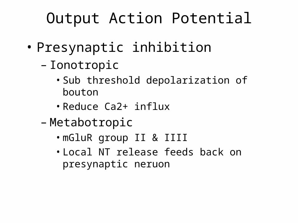
Output Action Potential
• Presynaptic inhibition– Ionotropic
• Sub threshold depolarization of bouton• Reduce Ca2+ influx
– Metabotropic• mGluR group II & IIII• Local NT release feeds back on presynaptic
neruon

Sea slug (tritonia) locomotion
• Characteristic escape response
• Alternate, vigorous body flexion
• Simple neural circuit
Lawrence & Watson 2002

Tritonia CPG
• Escape is a programmed response– Katz, et al., 2004
Dorsal Swim Interneuron
Ventral Swim Interneuron
Ventral Flexion Neuron
Dorsal Flexion Neuron
Flex
ExtendIn
tracellular p
oten
tialo
f neu
ron
s
Stimulate sensory neurons to elicit escape

Tritonia Metabotropic Neuromodulation
• DSI stimulation triggers fast and slow depolarization– Slow depolarization is GTP dependent– Blocked by non-exchangeable GDP--S
Stimulation
Recording
Slow metabotropic depolarization
Fast Ionotropic depolarization
Blocks metabotropic process


![ECE-V-DIGITAL SIGNAL PROCESSING [10EC52] …vtusolution.in/.../digital-signal-processing-10ec52.pdfDigital vtusolution.in Signal Processing 10EC52 TEXT BOOK: 1. DIGITAL SIGNAL PROCESSING](https://img.dokumen.tips/doc/110x75/5afe42bb7f8b9a256b8ccd2e/ece-v-digital-signal-processing-10ec52-signal-processing-10ec52-text-book.jpg)