Embed Size (px)
Citation preview
Severe Hyperglycemia, Diabetic Ketoacidosis, and Hyperglycemic Hyperosmolar State -
Ketan Dhatariya, Mark Savage, Mike Sampson, Glenn Matfin, and Adrian Scott
Dr Ketan Dhatariya MBBS MSc MD MS FRCP
Consultant in diabetes, endocrinology and general medicine
Elsie Bertram Diabetes Centre
Norfolk and Norwich University Hospitals NHS Foundation Trust
Colney Lane
Norwich, NR4 7UY, UK
+44(0) 1603 288170
Dr Mark W Savage MB ChB MD FRCP
Director of Medicine and Clinical Dean, Medical Services, Consultant Physician (Endocrinology),
Bendigo Health
PO Box 126 Bendigo Victoria 3552, Australia
Phone +61 (0)3 5454 7564
Fax: +61 (0)3 5454 7504
Professor Mike Sampson
Elsie Bertram Diabetes Centre,
Norfolk and Norwich University Hospital National Health Service Trust,
Colney Lane
Norwich, NR4 7UY, UK
+44(0) 1603 288170
Glenn Matfin,
Medical Director,
International Diabetes Center
3800 Park Nicollet Blvd
Minneapolis, MN 55416, USA
Phone: 952 993 3048
Fax: 952 993 1302
Adrian Scott
Clinical director, diabetes and endocrinology
Sheffield Teaching Hospitals NHS Foundation Trust,
Diabetes Centre,
Northern General Hospital,
Sheffield S5 7AU, UK
Abstract
Emergency admissions due to hyperglycemia remain some of the most common and challenging
metabolic conditions to deal with. Diabetic ketoacidosis (DKA) and hyperglycemic hyperosmolar
state (HHS) are biochemically different conditions that require different approaches to treatment.
They often occur in different age groups, and there is a need for co-ordinated care from the
multidisciplinary team to ensure the timely delivery of the correct treatments. Over the last few
years, the management of these conditions has changed. With DKA, the use of bedside
monitoring of plasma ketone levels now drives treatment. With HHS, the treatment now focusses
on the use of fluid rehydration rather than insulin treatment as the means by which glucose
lowering should be achieved, with insulin only being introduced when the rate of glucose lowering
levels off.
This chapter, which is based largely on published guidelines from the US and UK on the
management of these conditions highlights the recent changes in management, the differences
between the guidelines, and the rationale for these changes.
Introduction
The hyperglycemic urgency, severe hyperglycemia, and emergencies, DKA and HHS are acute
severe metabolic complications of uncontrolled diabetes mellitus 1. Severe hyperglycemia can
escalate into the potentially fatal complications of DKA and HHS. Subsequently, these conditions
demand immediate recognition and treatment.
Severe hypergycemia is characterized by significant hyperglycemia (i.e. glycosylated hemoglobin
[HbA1c] ≥10%); or fasting plasma glucose [FPG] >250 mg/dl [~14 mmol/L]; or random plasma
glucose >300 mg/dl [16.7mmol/L]); or when symptomatic (e.g. sudden persistent weight-loss,
polyuria, and polydipsia) 2.
DKA is a complex disordered metabolic state characterized by severe hyperglycemia (i.e. plasma
glucose levels >250 mg/dl [~14 mmol/L]), ketonemia (ketosis), and metabolic acidosis (pH ≤7.3,
serum bicarbonate <18 mEq/L [18 mmol/L]). The 2009 American Diabetes Association (ADA)
Hyperglycemic Crises consensus guidelines further subdivides DKA into mild DKA (serum
bicarbonate of 15 to 18 mEq/L [15 to 18 mmol/L], pH 7.25 to 7.30); moderate DKA (serum
bicarbonate 10 to <15 mEq/L [10 to <15 mmol/L], pH 7.00 to 7.24): and severe DKA (serum
bicarbonate <10 mEq/L [10 mmol/L], pH <7.00). More recently, the UK 2013 Joint British Diabetes
Societies Inpatient (JBDS IP) group DKA guidelines have incorporated serum ketone levels (3-
beta-hydroxybutyrate [βHBA]) in the definition of DKA 3. The measurement of βHBA for diagnosis
and monitoring of DKA was also recommended in the 2011 ADA Diabetes Laboratory guidelines 4.
The 2013 JBDS IP group DKA definition requires the combined presence of 3 biochemical
abnormalities: ketonemia ≥3 mmol/L or significant ketonuria (≥2+ urine ketones on standard urine
sticks); blood glucose >200 mg/dl (11.1 mmol/L) or known diabetes; and venous (or arterial) blood
bicarbonate <15 mEq/L (15 mmol/L) and/or pH <7.3. DKA primarily affects persons with type 1
diabetes and may be an initial manifestation of previously undiagnosed type 1 diabetes (~25%) or
may result from increased insulin requirements in existing patients during situations that increase
the release of counterregulatory hormones (i.e. glucagon, cortisol, epinephrine, and growth
hormone). When hyperglycemia initially presents in the presence of ketones or other signs of
metabolic decompensation, the diagnosis of type 1 diabetes is generally straightforward
(especially in children and adolescents). However, ketonemia can also be found in individuals with
type 2 diabetes (i.e. ketosis-prone hyperglycemia, especially in persons of African descent), with 5
to 25% having DKA 5.
HHS is characterized by marked hyperglycemia (blood glucose levels >600 mg/dl [33.3 mmol/L]);
hyperosmolarity (plasma osmolarity >320 mOsm/kg [320 mmol/kg]) and dehydration; the absence
of ketoacidosis; and depression of the sensorium. The 2012 JBDS IP group HHS definition
includes: marked hyperglycemia (>540 mg/dl [30 mmol/L]); no significant ketonemia (<3 mmol/L);
no acidosis (pH>7.3, bicarbonate >15 mEq/L (15 mmol/L); hypovolemia; and osmolality usually
>320 mosmol/kg [320 mmol/kg]. These guidelines also highlight that a mixed picture of HHS and
DKA may occur. HHS is seen most frequently in persons with type 2 diabetes, however,
approximately 20% of cases have no history of this diagnosis.
In the US, the prevalence of DKA has risen whilst mortality has decreased. This is related to an
improved understanding of the pathophysiology of DKA together with close monitoring and
correction of electrolytes. More than one-third of DKA hospitalizations are due to re-admissions.
Mortality rates have fallen significantly in the last 20 years from 7.96% to 0.67% 6;7. The mortality
rate is still high in low-income countries and among non-hospitalized patients 8. This high mortality
rate illustrates the necessity of early diagnosis and the implementation of effective prevention
programmes. The ready availability of insulin to all parts of the world should remain a priority.
Cerebral edema remains the most common cause of mortality, particularly in young children and
adolescents. The main causes of mortality in the adult population include severe hypokalemia
(and related cardiac dysrhythmias), adult respiratory distress syndrome (ARDS), and comorbid
states such as pneumonia, ACS and sepsis 9. In comparison, HHS is rare but mortality attributed
to HHS is considerably higher than that attributed to DKA, with recent mortality rates of 5–20%.
The prognosis of both conditions is substantially worsened at the extremes of age in the presence
of coma, hypotension, and severe comorbidities.
Pathophysiology
DKA usually occurs as a consequence of absolute or relative insulin deficiency that is
accompanied by an increase in counterregulatory hormones 10. This type of hormonal imbalance
enhances hepatic gluconeogenesis and glycogenolysis resulting in severe hyperglycemia.
Enhanced lipolysis increases serum free fatty acids that are then metabolised as an alternative
energy source in the process of ketogenesis 11. This results in accumulation of large quantities of
ketone bodies and subsequent metabolic acidosis. Ketones include acetone, βHBA, and
acetoacetate. The predominant ketone in DKA is βHBA.
There are several mechanisms responsible for fluid depletion in DKA. These include osmotic
diuresis due to hyperglycemia, vomiting (commonly associated with DKA) and eventually, inability
to take in fluid due to a diminished level of consciousness. Electrolyte shifts and depletion are in
part related to the osmotic diuresis. Hyperkalemia and hypokalemia need particular attention.
Unlike DKA, which is a condition most frequently associated with absolute insulin deficiency, in
HHS there is sufficient insulin to prevent ketogenesis, but insufficient insulin to either prevent
hepatic gluconeogenesis and cellular glucose uptake 12. If counterregulatory hormone excess is
also present (e.g. concomitant illness), then this leads to a rise in blood glucose, and a
subsequent osmotic diuresis. If sufficient water is not available, this leads to dehydration and
resultant impaired renal function. The high plasma glucose causes a raised serum osmolality. The
impaired renal function then leads to a further inability to excrete glucose thus perpetuating the
hyperglycaemia, osmotic diuresis, volume depletion, and dehydration 11. Alterations in mental
status are common with serum osmolalities over 330 mosmol/kg (330 mmol/kg).
Etiology
DKA and HHS can be precipitated by various conditions (which can be easily remembered by the
letter I), including Insulin deficiency (i.e. diabetes presentation or failure to take enough insulin);
Iatrogenic (e.g. glucocorticoids, thiazides, and atypical antipsychotic drugs); Infection (the most
common precipitating factor for both DKA and HHS); Inflammation (e.g. acute pancreatitis,
cholecystitis); Ischemia or Infarction (e.g. acute coronary syndromes [ACS], stroke, bowel); and
Intoxication (e.g. alcohol, cocaine).
Few studies have assessed factors associated with DKA in adults with type 1 diabetes. However,
the T1D Exchange clinic registry at 70 US endocrinology centers recently performed a cross-
sectional analysis including 7012 participants with type 1 diabetes 13. Higher frequencies of DKA
were associated with lower socioeconomic status (P < 0.001); and higher HbA1c levels
(P < 0.001), with 21.0% of those with HbA1c ≥10.0% having an event in the past 12 months.
Notably, the frequency was no higher in insulin pump users than injection users, an important
finding because DKA is a potential risk with pump infusion site failure.
Diagnostic considerations
The diagnostic criteria of severe hyperglycemia, DKA, and HHS are outlined in the introduction.
The initial laboratory evaluation of patients includes determination of plasma glucose, blood urea
nitrogen (BUN, or urea), creatinine, electrolytes (with calculated anion gap), serum osmolality,
serum and urinary ketones, urinalysis, baseline venous (or arterial) blood gases, complete (full)
blood count with differential, and ESR or CRP (if indicated). An electrocardiogram (ECG), chest X-
ray, and urine, sputum, and blood cultures should also be obtained. Other investigations are
performed as warranted by the clinical situation (e.g. cardiac troponins, serum lactate, appropriate
imaging, toxicology, and drug screen).
Point of care ketone testing should be used to measure the plasma ketone concentrations (in
particular, βHBA), because this is the direct marker of disease severity. The anion gap is
calculated by subtracting the sum of chloride (Cl) and bicarbonate (HCO3) concentration from the
uncorrected (see below) sodium (Na) concentration: [Na - (Cl + HCO3)]. A normal anion gap is
between 7 and 9 mEq/L (7-9 mmol/L) and an anion gap >10-12 mEq/L (10-12 mmol/L) indicates
the presence of increased anion gap metabolic acidosis. Whilst arterial blood gas (ABG) is the
most accurate method of assessing ventilation and acid-base status, venous blood gas (VBG) is
preferred to ABG for bicarbonate and pH measurements because the differences in arterial and
venous pH (e.g. venous pH only 0.03 lower than arterial), bicarbonate and potassium
measurements are not great enough (in either direction) to alter management.
The admission serum sodium is usually low or normal despite water loss (renal and gut) because
of an intracellular–extracellular fluid shift. To assess the severity of sodium and water deficit,
serum sodium may be corrected by adding 1.6 mg/L (1.6 mmol/L) to the measured serum sodium
for each 100 mg/dl (5.6 mmol/L) of glucose above 100 mg/dl (5.6 mmol/L) up to 440 mg/dl (~24
mmol/L) 14. For glucose levels >440 mg/dl (~24 mmol/L), serum sodium may be corrected by
adding 4 mg/L (4 mmol/L) to the measured serum sodium for each 100 mg/dl glucose above this
threshold. Serum potassium levels may also be low, normal, or elevated, despite total body
potassium depletion resulting from protracted polyuria and vomiting.
On admission, leukocytosis with cell counts in the 10,000 –15,000 mm3 range is the rule in DKA
and is generally attributed to stress and may not be indicative of an infection. However,
leukocytosis with cell counts >25,000 mm3 may designate infection and appropriate evaluation is
indicated.
Hyperamylasemia has been reported in 21–79% of patients with DKA; however, there is little
correlation between the presence, degree, or isoenzyme type of hyperamylasemia (e.g. salivary
amylase can also be increased in DKA) and the presence of gastrointestinal symptoms (nausea,
vomiting, and abdominal pain) or pancreatic imaging studies. A serum lipase determination may
be beneficial in the differential diagnosis of pancreatitis; however, lipase can also be elevated in
DKA in the absence of pancreatitis.
Not all patients with ketoacidosis have DKA. Starvation ketosis and alcoholic ketoacidosis are
distinguished by clinical history and by plasma glucose concentrations that range from mildly
elevated (rarely >200 mg/dl [11.1 mmol/L]) to hypoglycemia. A clinical history of previous drug
abuse and intoxication should be sought.
Clinical signs and features
The process of HHS usually evolves over several days to weeks, whereas the evolution of the
acute DKA episode tends to be much shorter (typically <24 h). For both DKA and HHS, the
classical clinical picture includes a history of polyuria, polydipsia, weight loss, vomiting,
dehydration, weakness, and mental status change. Physical findings may include increased rate
and depth of respiration in DKA (i.e. Kussmaul’s breathing) with odor of acetone, tachycardia,
and hypotension. Assessment of fluid status encompasses subjective observations (skin turgor,
mucous membranes, and cerebral dysfunction), objective measurements (blood pressure, pulse,
postural measurements, body weight) and laboratory measurements (serum sodium, serum
osmolality, BUN, hematocrit, and urine osmolality). Severe hypovolemia may manifest as
tachycardia (pulse >100 bpm) and/or hypotension (systolic blood pressure <100 mmHg). Each
has its limitations particularly on initial assessment where previous comparator data may not be
available.
Mental status can vary from full alertness to profound lethargy or coma, with the latter more
frequent in HHS. Acute impairment in cognitive function may be associated with dehydration but
is not specific to the condition and is not necessarily present. Alterations in mental status are
common with serum osmolalities over 330 mosmol/kg (330 mmol/kg). Focal neurologic signs
(hemianopia and hemiparesis), positive Babinski reflexes, aphasia, visual hallucinations, seizures
(focal or generalized), and coma may also be features of HHS. As HHS more frequently occurs in
older people, the neurological findings may be mistaken for a stroke.
Although infection is a common precipitating factor for both DKA and HHS, patients can be
normothermic or even hypothermic primarily because of peripheral vasodilation. Nausea,
vomiting, diffuse abdominal pain are frequent in patients with DKA (>50%) but are uncommon in
HHS. Further evaluation is necessary if this complaint does not resolve with resolution of
dehydration and metabolic acidosis.
Management of Severe Hyperglycemia, DKA, and HHS
Successful treatment of severe hyperglycemia, DKA and HHS requires correction of
hyperglycemia, dehydration, and electrolyte imbalances; identification of comorbid precipitating
events; and above all, frequent patient monitoring.
Acute intervention
Once severe hyperglycemia, DKA, or HHS is recognized the patient should be managed in an
appropriate location. This could be in the outpatient setting (e.g. rapid access clinic) for selected
cases of severe hyperglycemia and mild DKA. However, the majority of patients will be
hospitalized and managed in an emergency assessment unit (EAU), acute medical unit (AMU),
high dependency unit (HDU), or intensive care unit (ICU). As with all acute medical patients,
prompt assessment and management of the ABCs should occur (i.e. Airway; Breathing;
Circulation). Other markers of DKA and HHS severity should be assessed and recorded (Table 1).
General Supportive Care
Supportive care includes inserting large bore IV cannula and starting appropriate IV fluid
resuscitation, electrolyte replacement, nutritional support, continuous cardiac monitoring, and
pulse oximetry.
Hyperglycemia Bundle: Due to the increased risk of arterial and venous thromboembolism
(VTE), all patients with DKA or HHS should receive low molecular weight heparin (LMWH) for
the full duration of admission unless contraindicated. HHS (and some DKA) patients are also
at high risk of pressure ulceration. An initial foot assessment should be undertaken and heel
protectors applied in those with neuropathy, peripheral arterial disease (PAD) or lower limb
deformity. The feet should be re-examined daily. Consider NG tube with airway protection to
prevent aspiration if Glasgow Coma Scale (GCS) is <12 or excessive vomiting. Consider
urinary catheterization if the patient is incontinent, difficulty monitoring urine output (minimum
urine output should be no less than 0.5ml/kg/hr), or anuric (i.e. not passed urine by 60
minutes).
Hyperglycemia Specific Therapy
Severe Hyperglycemia
Healthcare providers are concerned by the risk of severe hyperglycemia during acute illness in
persons with diabetes, especially those treated with insulin therapy. Prevention of hyperglycemia,
and their management with a step-by-step procedure (generally referred to as “sick-day” rules)
when they eventually occur, are integral to diabetes management. Blood glucose levels normally
increase during illness because of the release of stress hormones. Thus, sick-day rules should be
initiated. The instructions include maintaining usual food plan, non-insulin therapies, and/or
insulin regimen whenever possible. Low-caloric fluid intake should be increased as appropriate.
Persons with diabetes, who experience nausea or vomiting, should initiate the sick-day food plan.
In patients treated with insulin, blood glucose and ketone (if available) monitoring should be
carried out as recommended (e.g. every 2 to 4 hours). If blood glucose is >250 mg/dl (~14
mmol/L) on two consecutive tests, persons with diabetes are recommended to contact their
clinician due to the possible need to supplement their current insulin regimen with short- or
rapid-acting insulin as necessary. The individual must be instructed to contact their healthcare
provider when the blood glucose is persistently raised (e.g. >300 mg/dl [16.6 mmol/L]) and/or
they develop moderate to high ketonuria (i.e. ≥2+ urine ketones) and/or increased serum
ketones if available. DKA or HHS can occur in this setting, thus frequent contact is necessary
between clinician and the individual to prevent the situation from deteriorating further.
The most important consideration in the initial assessment of acute illness in persons with
diabetes is whether the individual needs inpatient admission or can be safely managed in the
community or outpatient setting (including review in a rapid-access clinic). The majority of these
situations will be managed in the community or outpatient setting. However, this decision has to
take into account various factors including medical (e.g. severity of metabolic decompensation,
associated comorbidities, presence of confusion or impaired consciousness) and social issues
(e.g. whether the patient lives alone or has adequate family support, whether there are reliable
communication channels between healthcare provider and person with diabetes and/or
caregivers). Inpatient admission can be considered when (1) severe and prolonged
hyperglycemia, (2) presence of high ketones for more than 6 hours, (3) vomiting, diarrhea, and/or
abdominal pain (4) inadequate phone contact between healthcare provider and person with
diabetes and/or caregiver, or (5) at the discretion of the clinician and/or according to local practice
guidelines.
DKA and HHS
The 2009 ADA Hyperglycemic Crises consensus guidelines protocols for the management of
patients with DKA and HHS are summarized in Fig. 1. The overall goals in treating DKA are to
improve circulatory volume and tissue perfusion, decrease blood glucose, and correct the acidosis
and electrolyte imbalances. These objectives usually are accomplished through the administration
of low-dose insulin and IV fluid and electrolyte replacement solutions. An initial loading dose of
short-acting or rapid-acting insulin is often given IV, followed by IV continuous insulin infusion
(CII). Frequent laboratory tests are used to monitor blood glucose, venous pH, creatinine, and
serum electrolyte levels and to guide fluid and electrolyte replacement. It is important to replace
fluid and electrolytes and correct pH while bringing the blood glucose concentration to a normal
level. Too rapid a drop in blood glucose may cause hypoglycemia. A sudden change in the
osmolality of extracellular fluid can also occur when blood glucose levels are lowered too rapidly,
and this can cause cerebral edema. Serum potassium levels often fall as acidosis is corrected and
potassium moves from the extracellular into the intracellular compartment. Thus, it may be
necessary to add potassium to the IV infusion. Identification and treatment of the underlying
cause, such as infection, also are important.
During treatment of DKA, hyperglycemia is corrected faster than ketoacidosis. The mean duration
of treatment until blood glucose is <250 mg/dl (~14 mmol/L) and ketoacidosis (pH >7.30;
bicarbonate >18 mEq/L [18 mmol/L) is corrected is 6 and 12 h, respectively. Once the plasma
glucose is <200 mg/dl (11.1 mmol/L), 5% dextrose should be added to replacement fluids to allow
continued insulin administration until ketonemia is controlled while at the same time avoiding
hypoglycemia. For DKA occurring in individuals with type 2 diabetes (i.e. ketosis-prone
hyperglycemia), the initial management is similar to standard DKA 5.
In comparison, 2013 JBDS IP group DKA guidelines reflect two major recent developments a) a
change in the way patients with DKA present clinically and b) there has been development of
rapid near-patient testing technology. Until recently there was no easily available assay for
ketone bodies, hence capillary glucose, venous pH and bicarbonate were used to diagnose and
monitor response to treatment in DKA. Near patient testing for βHBA is now readily available for
the monitoring of the abnormal metabolite allowing for a shift away from using glucose levels to
drive treatment decisions in the management of DKA.
Some of the major recommendations of the 2013 JBDS IP group DKA guidelines includes:
a) Aim to treat the cause of the acidosis (i.e. ketonemia). Subsequently, bedside ketone
monitors should be used to measure βHBA, because this is the direct marker of
disease severity. The resolution of DKA depends upon the suppression of ketonemia,
and measurement of blood ketones now represents best practice in monitoring the
response to treatment 15-18.
b) Insulin is given as a standard dose per kg until the ketones are cleared. A weight-
based, fixed rate IV insulin infusion (FRIII) via an infusion pump should be used. 50
units short-acting insulin or rapid-acting insulin made up to 50ml with 0.9% sodium
chloride solution (resulting concentration of insulin is 1 unit per ml). The initial starting
dose of a fixed dose per kg body weight (0.1 units per kg per hour [i.e. 7 units per hour
for a 70 Kg individual]) enables rapid blood ketone clearance. The fixed rate may be
adjusted if the metabolic targets are not met (i.e. reduction of the blood ketone
concentration by at least 0.5 mmol/L/hour; increase in venous bicarbonate
concentrations by at least 3 mEq/L/hour [3 mmol/L/hour]); or reduction capillary blood
glucose by at least 50 mg/dl/hour [3 mmol/L/hour]). The insulin infusion rate is
increased by 1.0 unit/hr increments hourly until the ketones are falling at target rates
(also check infusion set for leaks and connection problems).There is no need to give a
bolus dose of insulin as long as the CII is set up promptly. Only use a variable-rate IV
insulin infusion (VRIII) with 10% dextrose when the blood glucose is <250 mg/dl (~14
mmol/L).
c) Subcutaneous injections of long-acting insulin should be continued if the patient is
already using these agents. They provide background insulin when the CII is
discontinued, and should avoid excess length of stay. This does not obviate the need
for giving short-acting or rapid-acting insulin before discontinuing the CII. Most units
experienced in managing DKA now also continue intermediate-acting insulin (NPH) if
that is what the patient normally uses. The CII is thus a “top up” on the (inadequate)
background insulin already circulating. Patients presenting with newly diagnosed type
1 diabetes should be given long-acting insulin (or NPH insulin, depending on local
policy) at a dose of 0.25 units/Kg subcutaneously once daily to mitigate against
rebound ketosis when they are taken off the FRIII 19.
d) Use 0.9% sodium chloride solution for resuscitation, not colloid. If the systolic blood
pressure (BP) is <90mmHg, consider causes other than fluid depletion, such as heart
failure, sepsis, etc. Give 500 ml of 0.9% sodium chloride solution over 10-15 minutes
and repeat if necessary (i.e. fluid challenge). If there has been no improvement in BP,
call for urgent senior help. If the systolic BP is >90 mmHg use the typical
recommendations outlined in Table 2. More cautious fluid replacement should be
considered in young people aged 18-25 years, elderly, pregnant, heart or renal failure
(also consider HDU and/or central line). Reduce the rate of fluid replacement in the
elderly/cardiac disease/mild-moderate DKA (e.g. bicarbonate >10 mEq/L [10 mmol/l]).
More rapid infusion increases risk of ARDS and cerebral edema.
e) Measure venous blood gas for pH, bicarbonate and potassium at 60 minutes, 2 hours
and 2 hourly thereafter.
f) Keep potassium between 4.5 and 5.5 mEq/L (4.5-5.5 mmol/L) (Table 3). Hypokalemia
and hyperkalemia are life threatening conditions and are common in DKA.
g) Avoid hypoglycemia. If the glucose falls below 250 mg/dl (~14.0 mmol/L), commence
10% dextrose given at 125mls/hour alongside the 0.9% sodium chloride solution. This
is to avoid hypoglycemia if the FRIII is still required to drive down the ketones and
acidosis.
h) Bicarbonate should not generally be given because it may worsen intracellular
acidosis, and it may precipitate cerebral edema, particularly in children and
adolescents 20;21.
i) Hypophosphatemia and hypomagnesemia are common in DKA and HHS, however
routine replacement is not recommended, unless associated with significant
malnutrition.
j) It is expected that by 24 hours the ketonemia (<0.6 mmol/L) and acidosis (venous
bicarbonate >15 mEq/L [15 mmol/L]; venous pH >7.3) should have resolved. Continue
IV fluids if the patient is not eating and drinking. If the patient is not eating and drinking
and there is no ketonemia move to a VRIII. Transfer to subcutaneous insulin if the
patient is eating and drinking normally. Ensure that the subcutaneous insulin is started
before the IV insulin is discontinued. Ideally give the subcutaneous short-acting or
rapid-acting insulin at a meal and discontinue IV insulin one hour later.
k) Where available, the diabetes inpatient team should ideally be involved as early as is
practical after admission.
Unlike DKA, guidelines on the management of the HHS in adults are uncommon and often there
is little to differentiate them from the management of DKA. However, HHS is different from DKA
and treatment requires a different approach. The person with HHS is often elderly, frequently with
multiple comorbidities but always very sick. Even when specific hospital guidelines are available,
adherence to and use of these is variable amongst inpatient teams. The major goals of treatment
of HHS are to gradually and safely normalize the osmolality; replace fluid and electrolyte losses;
and normalize blood glucose. Other goals includes identifying and treating the underlying cause;
prevent arterial or venous thrombosis; prevent other potential complications (e.g. cerebral
edema); and prevent foot ulceration.
Some of the major recommendations of the 2012 JBDS IP group HHS guidelines includes:
(a) Measure or calculate osmolality (2x Na [mEq/L] + glucose [mg/dl)]/18 + BUN
[mg/dl]/2.8; or (2x Na [mmol/L] + glucose [mmol/L] + urea [mmol/L]) frequently to
monitor the response to treatment.
(b) The goal of the initial therapy is expansion of the intra and extravascular volume
and to restore peripheral perfusion. The fluid replacement of choice is 0.9%
sodium chloride. Measurement or calculation of osmolality should be undertaken
every hour initially and the rate of fluid replacement adjusted to ensure a
positive fluid balance sufficient to promote a gradual decline in osmolality.
Urinary fluid losses may be considerable due to osmotic diuresis which may
persist for hours as glucose concentrations slowly decrease. The fall in
osmolality with lowering of blood glucose and shift of water into the intracellular
space inevitably results in a rise in serum sodium. This is not necessarily an
indication to give hypotonic solutions (so-called ‘isotonic’ 0.9% sodium chloride
is relatively hypotonic compared to the serum) especially if the person remains
clinically hypovolemic. A rise in serum sodium concentration must be interpreted
in the context of what is happening to tonicity (effective osmolality). Provided
plasma glucose is declining at a safe rate – for example, no-more than 90
mg/dl/hr (5 mmol/L/hr) this will be accompanied by a rise in serum sodium, but a
fall in osmolality. Serum sodium concentrations should be frequently monitored,
and the concentration of sodium in fluids adjusted to promote a gradual decline
in corrected serum sodium. An optimal rate of decline in serum sodium is 0.5
mEq/L (0.5 mmol/L) per hour has been recommended for hypernatremic
dehydration. The rate of fall of plasma sodium should not exceed 10-12 mEq/L
(10-12 mmol/L) per day. The aim of treatment should be to replace
approximately 50% of estimated fluid loss within the first 12 hr and the
remainder in the following 12 hours although this will, in part, be determined by
the initial severity, degree of renal impairment and associated comorbidities,
which may limit the speed of correction.
(c) If significant ketonemia is present (βHBA >1 mmol/L) this indicates relative
hypoinsulinemia and insulin should be started at time zero. If significant
ketonemia is not present (βHBA <1 mmol/L) insulin should not be started. Fluid
replacement alone with 0.9% sodium chloride will result in a drop in blood
glucose and because most patients with HHS are insulin sensitive, there is a
risk of lowering the osmolality precipitously. Insulin treatment prior to adequate
fluid replacement may result in cardiovascular collapse as water moves out of
the intravascular space, with a resulting decline in intravascular volume. Lack of
appropriate decline in serum glucose with rehydration should prompt
reassessment and evaluation of renal function. Insulin may be started at this
point, or if already in place the infusion rate increased (increased by 1 unit/hr).
The recommended insulin dose is an FRIII given at 0.05 units per kg per hour
(e.g. 4 units/hour in an 80 kg person) is used. A fall of glucose at a rate of up to
90 mg/dl/hr (5 mmol/L/hr) is ideal.
(d) Avoid hypoglycemia. A blood glucose target of between 180 and ~270 mg/dl (10
and 15mmol/L) is a reasonable goal in the first 24 hours. If the blood glucose
falls below 180 mg/dl (14 mmol/L), commence 5% or 10% dextrose at 125 ml/h
and continue the 0.9% sodium chloride solution.
(e) Potassium replacement. This is the same as DKA and the same principles can
be applied using Table 3.
(f) Complete normalization of electrolytes and osmolality may take up to 72 hours.
(g) Assess for any complications of treatment (e.g. fluid overload, cerebral edema,
osmotic demyelination syndrome [e.g. a deteriorating conscious level])
(h) Because of the increased risk of arterial and venous thromboembolism, all
patients should receive prophylactic LMWH for the full duration of admission
unless contraindicated. Consideration should be given to extending prophylaxis
beyond the duration of admission in HHS patients deemed to be at high risk.
(i) Discharge planning: because many of these patients have multiple
comorbidities, recovery will largely be determined by their previous functional
level and the underlying precipitant of HHS. IV insulin can usually be
discontinued once they are eating and drinking but their fluids may be required
for longer if intake remains inadequate. Many patients may require conversion to
subcutaneous insulin treatment. For patients with previously undiagnosed
diabetes or who were well controlled on oral agents, switching from insulin to the
appropriate non-insulin therapy should be considered after a period of stability.
(j) Where available, the diabetes inpatient team should ideally be involved as early
as is practical after admission.
Treatment of Precipitating Illness
For both DKA and HHS, consider any precipitating causes (especially sepsis) and treat
appropriately.
Conclusions
Severe hyperglycemia, DKA, and HHS demand immediate recognition and treatment. However,
prevention is of these states is always preferred and this requires appropriate education of
patients, carers, and healthcare practitioners on an ongoing basis.
Legends
Figure 1
Protocol for management of adult patients with DKA or HHS. DKA diagnostic criteria: blood glucose 250 mg/dl (13.8 mmol/L), arterial pH 7.3, bicarbonate <15 mEq/l (15 mmol/L), and moderate ketonuria or ketonemia. HHS diagnostic criteria: serum glucose >600 mg/dl, arterial pH >7.3, serum bicarbonate >15 mEq/l, and minimal ketonuria and ketonemia. †15–20 ml/kg/h; ‡serum Na should be corrected for hyperglycemia (for each 100 mg/dl [5.6 mmol/L) > glucose 100 mg/dl (5.6 mmol/L), add 1.6 mEq/l (1.6 mmol/L) to sodium value for corrected serum value). Bwt,body weight; IV, intravenous; SC, subcutaneous.
Table 1
Markers of severity in DKA (taken from Reference 3) and HHS (Taken from Reference 22).
After a diagnosis of DKA or HHS has been made, the presence of any of the following during the admission should prompt a swift senior review and/or indicate admission to a High Dependence Unit (HDU) environment.
GCS, Glasgow Coma Scale; AVPU (Alert, Voice, Pain, Unresponsive) scale
Table 2
Recommended rate of fluid replacement in DKA assuming the individual has normal baseline
cardiovascular reserve. (Taken from Reference 3)
Table 3
Recommended rate of potassium replacement in DKA and HHS assuming the individual has
normal baseline renal function. (Taken from Reference 3)
Reference List
(1) Kitabchi AE, Umpierrez GE, Miles JM, Fisher JN. Hyperglycemic crises in adult patients with diabetes. Diabetes Care 2009; 32(7):1335-1343.
(2) Nathan DM, Buse JB, Davidson MB, Ferrannini E, Holman RR, Sherwin R et al. Medical management of hyperglycemia in type 2 diabetes: A consensus algorithm for the initiation and adjustment of therapy: A consensus statement of the American Diabetes Association and the European Association for the Study of Diabetes. Diabetes Care 2009; 32(1):193-203.
(3) Dhatariya K, Savage M, Claydon A, Dyer P, Evans P, Khan A et al. Joint British Diabetes Societies Inpatient Care Group. The management of diabetic ketoacidosis in adults. Second Edition. Update: September 2013. http://www.diabetologists-abcd.org.uk/JBDS/JBDS_IP_DKA_Adults_Revised.pdf. 2013. Last accessed 12th November 2013.
(4) Sacks DB, Arnold M, Bakris GL, Bruns DE, Horvath AR, Kirkman MS et al. Guidelines and recommendations for laboratory analysis in the diagnosis and management of diabetes mellitus. Diabetes Care 2011; 34(6):e61-e99.
(5) Smiley D, Chandra P, Umpierrez GE. Update on diagnosis, pathogenesis and management of ketosis-prone Type 2 diabetes mellitus. Diabetes Manag (Lond) 2011; 1(6):589-600.
(6) Lin SF, Lin JD, Huang YY. Diabetic ketoacidosis: comparisons of patient characteristics, clinical presentations and outcomes today and 20 years ago. Chang Gung Med J 2005; 28(1):24-30.
(7) Wang J, Williams DE, Narayan KM, Geiss LS. Declining death rates from hyperglycemic crisis among adults with diabetes, U.S., 1985 - 2002. Diabetes Care 2006; 29(9):2018-2022.
(8) Otieno CF, Kayima JK, Omonge EO, Oyoo GO. Diabetic ketoacidosis: risk factors, mechanisms and management strategies in sub-Saharan Africa: a review. E Afr Med J 2005; 82(12 (Suppl)):S197-203.
(9) Hamblin PS, Topliss DJ, Chosich N, Lording DW, Stockigt JR. Deaths associated with diabetic ketoacidosis and hyperosmolar coma. 1973-1988. Med J Aust 1989; 151(8):441-442.
(10) Matfin G. Diabetes mellitus and the metabolic syndrome. In: Porth CM, Matfin G, editors. Pathophysiology - Concepts of Altered Health States. 8th Edition ed. Lippincott Williams & Wilkins; 2008. 1047-1077.
(11) English P, Williams G. Hyperglycaemic crises and lactic acidosis in diabetes mellitus. Postgrad Med J 2004; 80(943):253-261.
(12) Barwell ND, McKay GA, Fisher M. Drugs for diabetes: part 7 insulin. Br J Cardiol 2011; 18:224-228.
(13) Weinstock RS, Xing D, Maahs DM, Michels A, Rickels MR, Peters AL et al. Severe hypoglycemia and diabetic ketoacidosis in adults with type 1 diabetes: Results from the T1D Exchange Clinic Registry. J Clin Endocrinol Metab 2013; 98(8):3411-3419.
(14) Verbalis JG, Goldsmith SR, Greenberg A, Korzelius C, Schrier RW, Sterns RH et al. Diagnosis, evaluation, and treatment of hyponatremia: Expert panel recommendations. Am J Med 2013; 126(Supplement):S1-S42.
(15) Sheikh-Ali M, Karon BS, Basu A, Kudva YC, Muller LA, Xu J et al. Can serum beta-hydroxybutyrate be used to diagnose diabetic ketoacidosis? Diabetes Care 2008; 31(4):643-647.
(16) Bektas F, Eray O, Sari R, Akbas H. Point of care blood ketone testing of diabetic patients in the emergency department. Endocr Res 2004; 30(3):395-402.
(17) Khan AS, Talbot JA, Tieszen KL, Gardener EA, Gibson JM, New JP. Evaluation of a bedside blood ketone sensor: the effects of acidosis, hyperglycaemia and acetoacetate on sensor performance. Diabetic Med 2004; 21(7):782-785.
(18) Wallace TM, Matthews DR. Recent advances in the monitoring and management of diabetic ketoacidosis. QJM 2004; 97(12):773-780.
(19) Hsia E, Seggelke S, Gibbs J, Hawkins RM, Cohlmia E, Rasouli N et al. Subcutaneous administration of glargine to diabetic patients receiving insulin infusion prevents rebound hyperglycemia. J Clin Endocrinol Metab 2012; 97(9):3132-3137.
(20) Hale PJ, Crase JE, Nattrass M. Metabolic effects of bicarbonate in the treatment of diabetic ketoacidosis. Br Med J 1984; 290(6451):1035-1038.
(21) Chua HR, Schneider A, Bellomo R. Bicarbonate in diabetic ketoacidosis - a systematic review. Ann Intensive Care 2013; 1(23).
(22) Scott A, Claydon A, Brennan G, Carey P, Dhatariya K, Hammersley M et al. The management of the hyperosmolar hyperglycaemic state (HHS) in adults with diabetes. The Joint British Diabetes Societies Inpatient Care Group. http://www.diabetologists-abcd.org.uk/JBDS/JBDS.htm. 2012. Last accessed 12th November 2013.
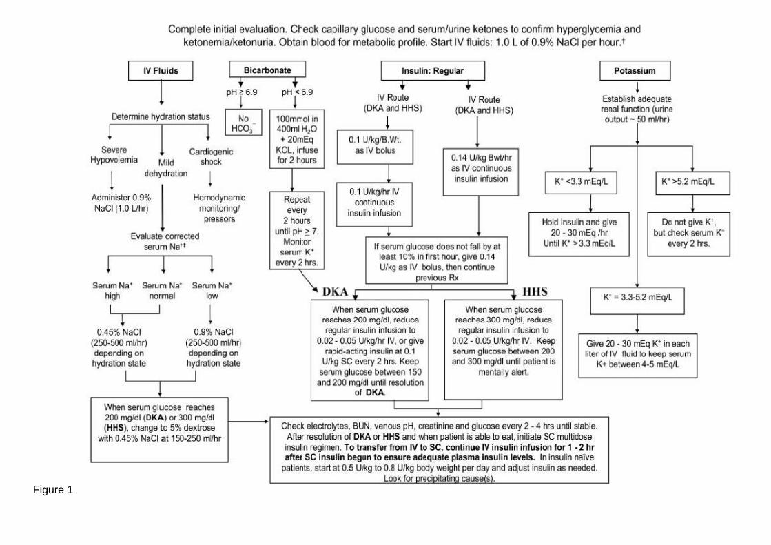
Figure 1
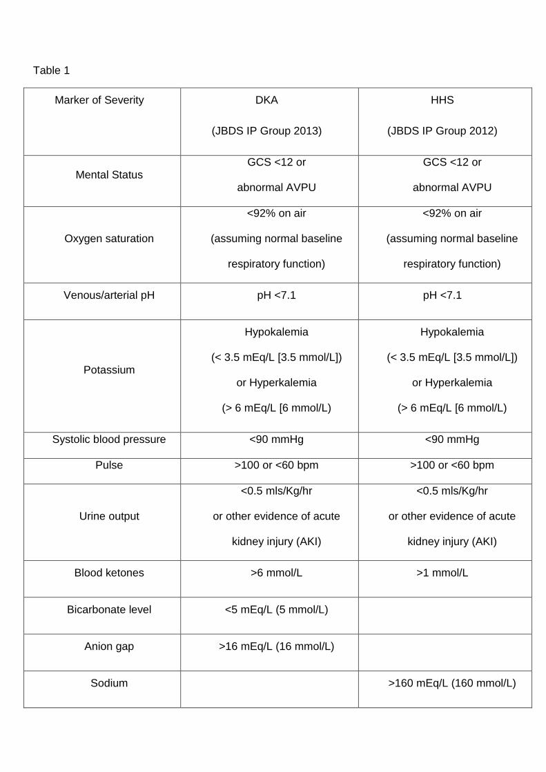
Table 1
Marker of Severity
DKA
(JBDS IP Group 2013)
HHS
(JBDS IP Group 2012)
Mental Status GCS <12 or
abnormal AVPU
GCS <12 or
abnormal AVPU
Oxygen saturation
<92% on air
(assuming normal baseline
respiratory function)
<92% on air
(assuming normal baseline
respiratory function)
Venous/arterial pH pH <7.1 pH <7.1
Potassium
Hypokalemia
(< 3.5 mEq/L [3.5 mmol/L])
or Hyperkalemia
(> 6 mEq/L [6 mmol/L)
Hypokalemia
(< 3.5 mEq/L [3.5 mmol/L])
or Hyperkalemia
(> 6 mEq/L [6 mmol/L)
Systolic blood pressure <90 mmHg <90 mmHg
Pulse >100 or <60 bpm >100 or <60 bpm
Urine output
<0.5 mls/Kg/hr
or other evidence of acute
kidney injury (AKI)
<0.5 mls/Kg/hr
or other evidence of acute
kidney injury (AKI)
Blood ketones >6 mmol/L >1 mmol/L
Bicarbonate level <5 mEq/L (5 mmol/L)
Anion gap >16 mEq/L (16 mmol/L)
Sodium >160 mEq/L (160 mmol/L)
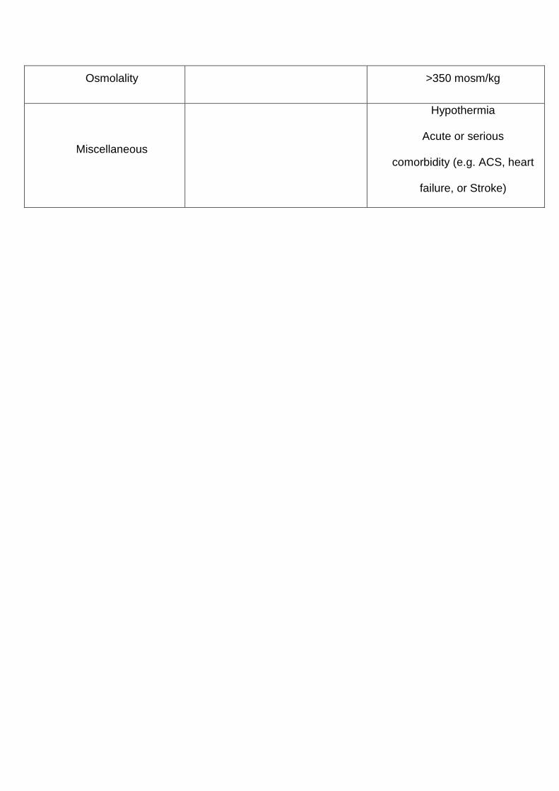
Osmolality >350 mosm/kg
Miscellaneous
Hypothermia
Acute or serious
comorbidity (e.g. ACS, heart
failure, or Stroke)
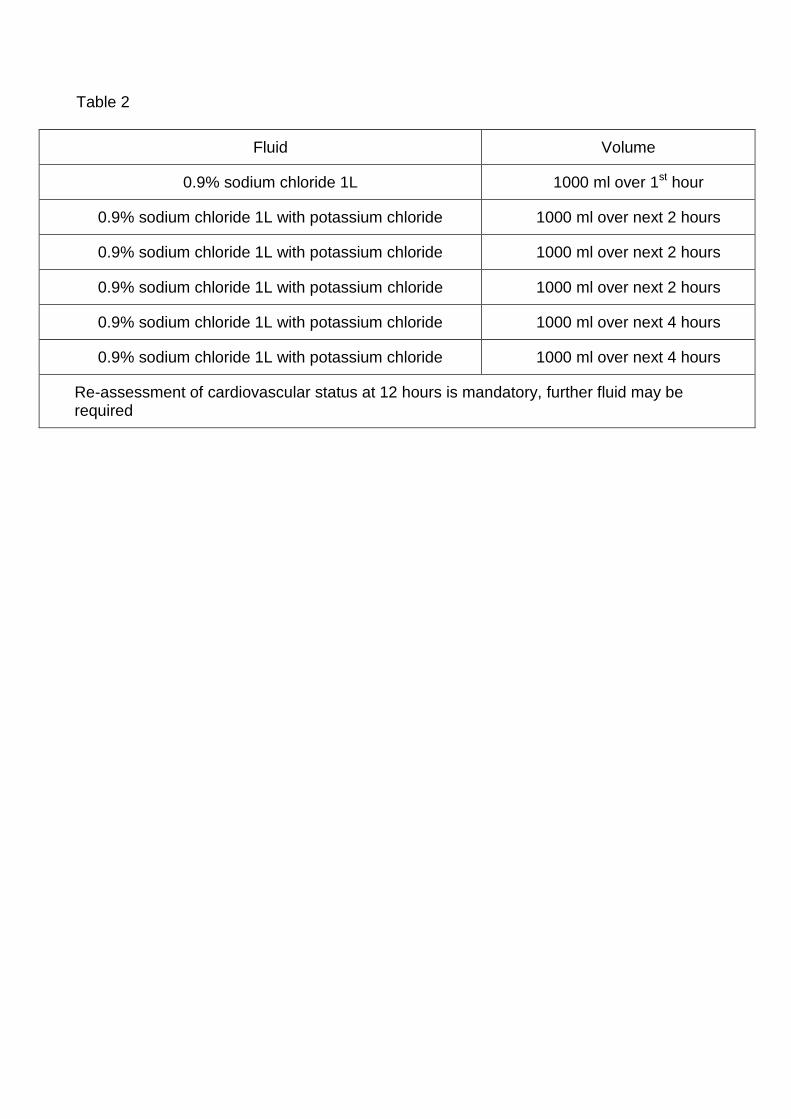
Table 2
Fluid Volume
0.9% sodium chloride 1L 1000 ml over 1st hour
0.9% sodium chloride 1L with potassium chloride 1000 ml over next 2 hours
0.9% sodium chloride 1L with potassium chloride 1000 ml over next 2 hours
0.9% sodium chloride 1L with potassium chloride 1000 ml over next 2 hours
0.9% sodium chloride 1L with potassium chloride 1000 ml over next 4 hours
0.9% sodium chloride 1L with potassium chloride 1000 ml over next 4 hours
Re-assessment of cardiovascular status at 12 hours is mandatory, further fluid may be required
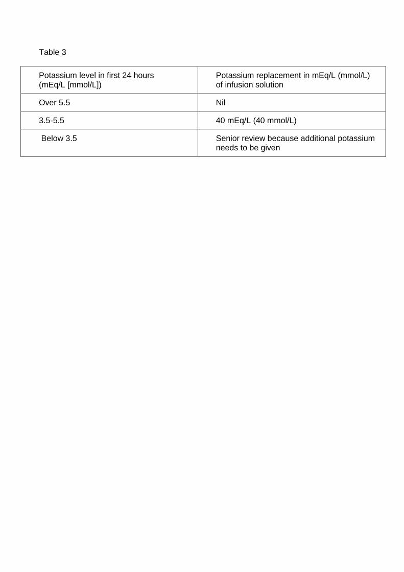
Table 3
Potassium level in first 24 hours (mEq/L [mmol/L])
Potassium replacement in mEq/L (mmol/L) of infusion solution
Over 5.5 Nil
3.5-5.5 40 mEq/L (40 mmol/L)
Below 3.5 Senior review because additional potassium needs to be given